Article

Significance of Mucosa-Associated Microbiota and Its Impacts on Intestinal Health of Pigs Challenged with F18+ E. coli

Department of Animal Science, North Carolina State University, Raleigh, NC 27695, USA
*
Author to whom correspondence should be addressed.
Pathogens 2022, 11(5), 589; https://doi.org/10.3390/pathogens11050589
Submission received: 7 April 2022 / Revised: 5 May 2022 / Accepted: 14 May 2022 / Published: 17 May 2022
(This article belongs to the Special Issue Modulation of Gut Microbiota & Microbiome in Pigs)

Abstract

:
The objective of this study was to evaluate the significance of jejunal mucosa-associated microbiota and its impacts on the intestinal health of pigs challenged with F18+ Escherichia coli. Forty-four newly-weaned pigs were allotted to two treatments in a randomized complete block design with sex as blocks. Pigs were fed common diets for 28 d. At d 7 post-weaning, pigs were orally inoculated with saline solution or F18+ E. coli. At d 21 post-challenge, feces and blood were collected and pigs were euthanized to collect jejunal tissue to evaluate microbiota and intestinal health parameters. The relative abundance of Firmicutes and Bacteroidetes was lower (p < 0.05) in jejunal mucosa than in feces, whereas Proteobacteria was greater (p < 0.05) in jejunal mucosa. F18+ E. coli increased (p < 0.05) protein carbonyl, Helicobacteraceae, Pseudomonadaceae, Xanthomonadaceae, and Peptostreptococcaceae and reduced (p < 0.05) villus height, Enterobacteriaceae, Campylobacteraceae, Brachyspiraceae, and Caulobacteraceae in jejunal mucosa, whereas it reduced (p < 0.05) Spirochaetaceae and Oscillospiraceae in feces. Collectively, jejunal mucosa-associated microbiota differed from those in feces. Compared with fecal microbiota, the change of mucosa-associated microbiota by F18+ E. coli was more prominent, and it was mainly correlated with increased protein carbonyl and reduced villus height in jejunal mucosa impairing the intestinal health of nursery pigs.

1. Introduction

The microbiota plays important roles on the maturation of the intestine and immune system and consequently affects the health of the host [1,2]. The physicochemical properties and the direct interaction with intestinal cells lead to a distinguishable composition of the microbiota along the gastrointestinal tract [3,4,5]. The microbiota in the intestinal lumen is more related to dietary compounds, whereas the mucosa-associated microbiota directly interacts with the mucus layer and the intestinal cells [6,7,8]. From the mucosa to the lumen the oxygen gradient is reduced which in turn makes the mucosal environment more propitious to aerobic and oxygen tolerant bacteria [3,9], whereas the mucus layer provides structures and substrates for bacteria with attachment characteristics and protein-degrading bacteria [10]. In addition, Zhao et al. [11] reported that the fecal microbiota is more similar to the microbiota in the lumen of the large intestine compared with the microbiota in the lumen of the small intestine.
The mucosa-associated microbiota provides the first line of defense preventing the colonization of opportunistic pathogens [12,13]. Enterotoxigenic Escherichia coli can colonize the small intestinal mucosa facilitated by fimbrial or no-fimbrial adhesins and produce enterotoxins inducing luminal fluid secretion, and thus contributing to diarrhea [14,15]. The F18+ E. coli is highly prevalent in pigs causing post-weaning diarrhea (PWD) [14,16]. The PWD caused by enterotoxigenic E. coli infection is well known to induce microbiota dysbiosis [17,18,19]. During infection, the increased nitrate and oxygen concentration may increase the proliferation of aerobic and facultative anaerobic bacteria, and reduce anaerobic bacteria [18,20,21]. The F18+ E. coli challenge has been associated to increased inflammatory response and oxidative stress, consequently affecting intestinal morphology and enterocyte proliferation [18,22,23,24]. According to Duarte at al. [18], F18+ E. coli challenge markedly affects the jejunal mucosa-associated microbiota in pigs 14 days post-challenge, although the symptoms of PWD caused by F18+ E. coli generally last up to 11 days post-challenge [14,18,22,25]. It has been demonstrated that dietary intervention has a longer-lasting effect on mucosa-associated microbiota rather than on fecal microbiota in nursery pigs [4,26]. Furthermore, Liu et al. [27] reported that understanding the modulation of mucosa-associated can be a key factor to provide a more precise nutritional intervention to enhance intestinal health.
Therefore, it was hypothesized that the jejunal mucosa-associated microbiota is directly related to intestinal health of nursery pigs and the impacts of F18+ E. coli challenged to nursery pigs are greater to jejunal mucosa-associated microbiota than in fecal microbiota. To test the hypothesis, the objective of this study was to evaluate the significance of jejunal mucosa-associated microbiota and its impacts on the intestinal health of pigs challenged with F18+ E. coli.

2. Results

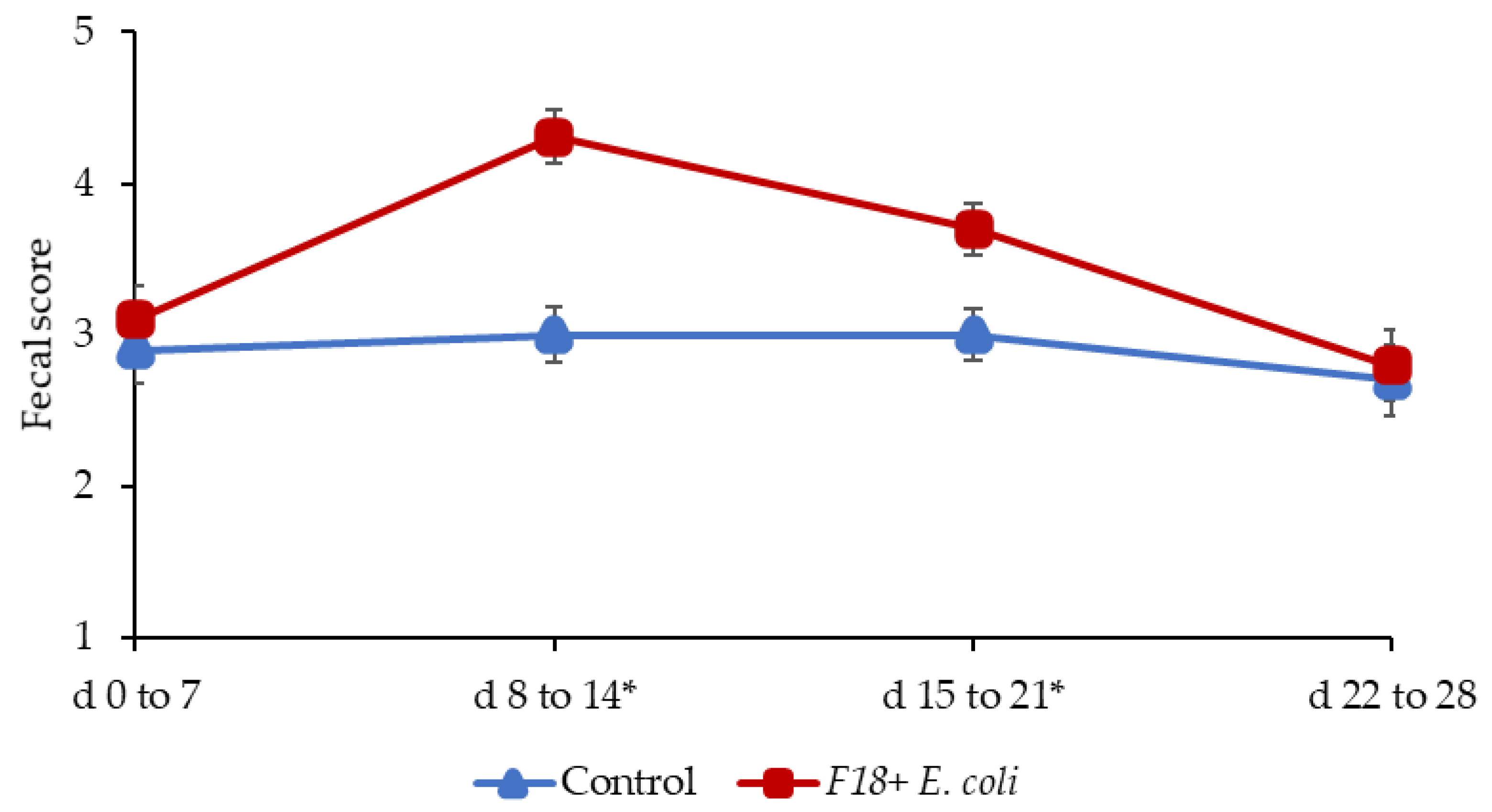
2.1. Growth Performance and Fecal Score

Pigs challenged with F18+ E. coli showed a reduced growth performance at the end of experiments. During the pre-challenge period (d 0 to 7) the fecal scores of pigs was not different (Figure 1), confirming that pigs were in normal fecal score before the F18+ E. coli challenge. The F18+ E. coli challenge increased (p < 0.05) the fecal score of pigs from d 8 to 14 and from d 15 to 21 post-weaning. The F18+ E. coli challenge did not affect the fecal score of pigs from d 22 to 28 post-weaning.

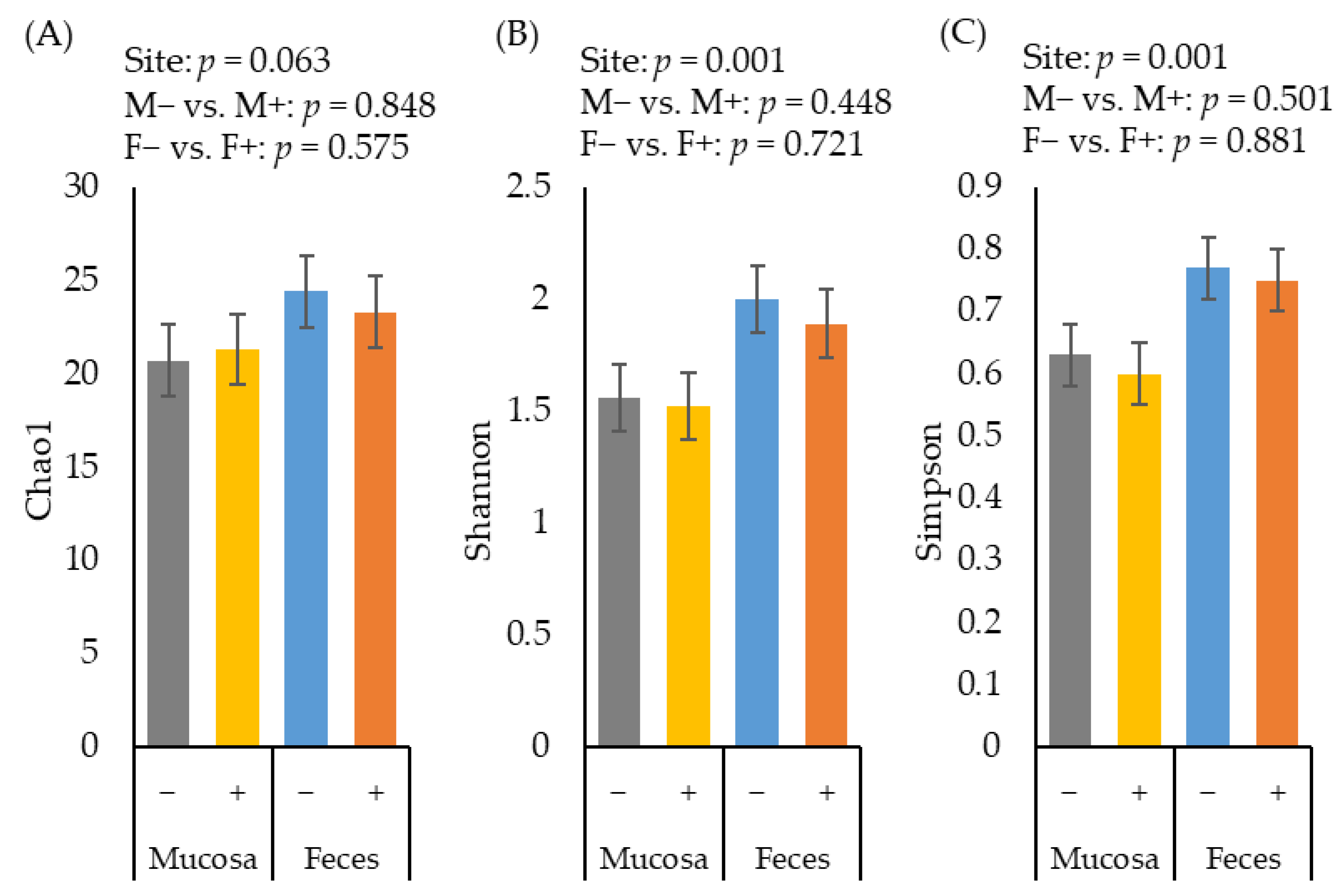
2.2. Alpha Diversity of Microbiota

The alpha diversity of fecal microbiota estimated with Chao1 tended to be greater (p = 0.063) than the diversity of jejunal mucosa-associated microbiota (Figure 2A). The alpha diversity of fecal microbiota estimated with Shannon (Figure 2B) and Simpson (Figure 2C) were greater (p < 0.05) than the diversity of jejunal mucosa-associated microbiota. The F18+ E. coli challenge did not affect the alpha diversity of fecal or jejunal mucosa-associated microbiota at d 21 post-challenge.

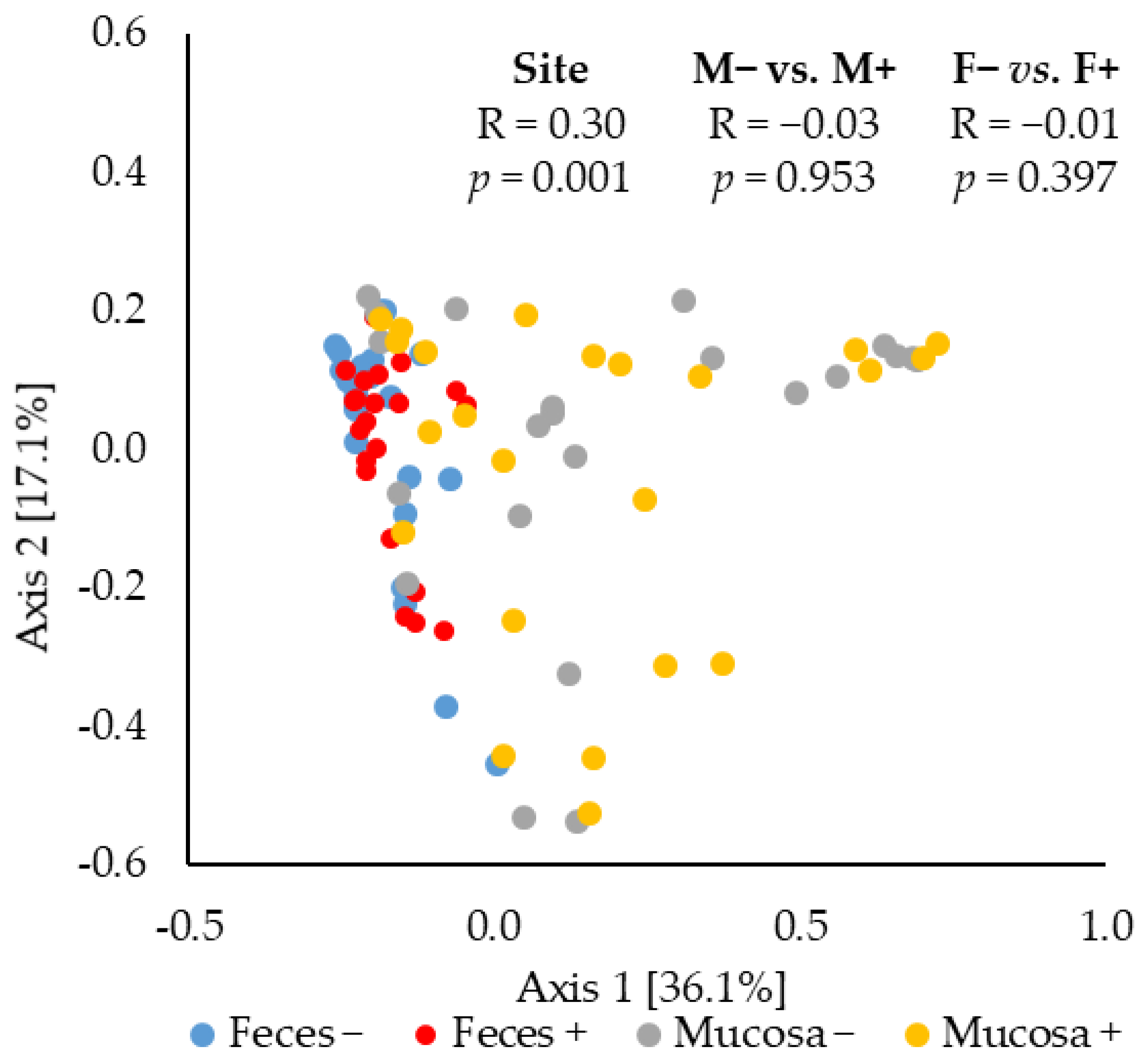
2.3. Beta Diversity of Microbiota

The principal coordinates analysis (PCoA) based on Bray–Curtis distance showed that the jejunal mucosa-associated microbiota was different (ANOSIM, mucosa-associated microbiota vs. fecal microbiota: R = 0.30, p < 0.05) from the fecal microbiota (Figure 3). The F18+ E. coli challenge did not affect the beta diversity of fecal or jejunal mucosa-associated microbiota at d 21 post-challenge.

2.4. Relative Abundance of Microbiota

Firmicutes, Proteobacteria, and Bacteroidetes were the most abundant phylum, accounting for 94 and 95% of all microbiota in mucosa and feces, respectively (Table 1). Proteobacteria was the most abundant phylum in the jejunal mucosa of pigs followed by Firmicutes, and Bacteroidetes. In feces, Firmicutes was the most abundant, followed by Bacteroidetes and Proteobacteria. The relative abundance of Firmicutes and Bacteroidetes was lower (p < 0.05) in jejunal mucosa than in feces. The relative abundance of Proteobacteria was greater (p < 0.05) in jejunal mucosa than in feces. The F18+ E. coli challenge did not affect the relative abundance of fecal and jejunal mucosa-associated microbiota at phylum level.
The relative abundance of Prevotellaceae, Veillonellaceae, Lachnospiraceae, Succinivibrionaceae, Ruminococcaceae, Acidaminococcaceae, Porphyromonadaceae, Erysipelotrichaceae, Eubacteriaceae, Cytophagaceae, Spirochaetaceae, and Rikenellaceae was lower (p < 0.05) in jejunal mucosa than in feces (Table 2). The relative abundance of Helicobacteraceae, Campylobacteraceae, Pseudomonadaceae, Enterobacteriaceae, Moraxellaceae, Pseudanabaenaceae, and Caulobacteraceae was greater (p < 0.05) in jejunal mucosa than in feces. The relative abundance of Lactobacillaceae, Clostridiaceae, Streptococcaceae, Brachyspiraceae, Nostocaceae, Bacillaceae, Bifidobacteriaceae, Eubacteriaceae, Hyphomicrobiaceae, Bacteroidaceae, Flavobacteriaceae, and Mycoplasmataceae in jejunal mucosa was not different from feces.
The relative abundance of Helicobacteraceae, Pseudomonadaceae, Xanthomonadaceae, and Peptostreptococcaceae was greater (p < 0.05) in jejunal mucosa of nursery pigs challenged with F18+ E. coli. The relative abundance of Campylobacteraceae, Enterobacteriaceae, Brachyspiraceae, and Caulobacteraceae was lower (p < 0.05) in jejunal mucosa of nursery pigs challenged with F18+ E. coli. The relative abundance of Bacillaceae tended to be lower (p = 0.089) in jejunal mucosa of nursery pigs challenged with F18+ E. coli. The relative abundance of Spirochaetaceae, Oscillospiraceae, and Others was lower (p < 0.05) in feces of nursery pigs challenged with F18+ E. coli. The relative abundance of Acidaminococcaceae tended to be greater (p = 0.088) in feces of nursery pigs challenged with F18+ E. coli.
The relative abundance of Prevotella, Succinivibrio, Megasphaera, Faecalibacterium Phascolarctobacterium, Selenomonas, Dialister, Roseburia, Acidaminococcus, Eubacterium, Oscillibacter, Herbaspirillum, Tepidimonas, and Ruminococcus was lower (p < 0.05) in jejunal mucosa than in feces (Table 3). The relative abundance of Helicobacter, Pseudomonas, Chlamydia, Acinetobacter, and Tepidimonas was greater (p < 0.05) in jejunal mucosa than in feces. The relative abundance of Campylobacter (p = 0.071), Chlamydia (p = 0.056), and Massilia (p = 0.051) tended to be greater in jejunal mucosa than in feces. The relative abundance of Lactobacillus, Clostridium, Mitsuokella, Streptococcus, Bifidobacterium, Gemmiger, Bacillus, Coprococcus, Catenibacterium, Anaerovibrio, Dorea, Brachyspira, Enterococcus, and Treponemain in jejunal mucosa was not different from feces.
The relative abundance of Helicobacter and Tepidimonas was greater (p < 0.05) in jejunal mucosa of nursery pigs challenged with F18+ E. coli. The relative abundance of Campylobacter was lower (p < 0.05) in jejunal mucosa of nursery pigs challenged with F18+ E. coli. The relative abundance of Pseudomonas (p = 0.073) and Bacillus (p = 0.084) tended to be lower (p < 0.05) in jejunal mucosa of nursery pigs challenged with F18+ E. coli. The relative abundance of Dialister and Acidaminococcus was greater (p < 0.05) in feces of nursery pigs challenged with F18+ E. coli. The relative abundance of Oscillibacter and Tepidimonas was lower (p < 0.05) in feces of nursery pigs challenged with F18+ E. coli.
The relative abundance of Prevotella copri, Prevotella sp., Succinivibrio dextrinosolvens, Faecalibacterium prausnitzii, Phascolarctobacterium succinatutenswas, Dialister succinatiphilus, Roseburia faecis, Selenomonas lipolytica, Acidaminococcus fermentans, Selenomonas bovis, Selenomonas bovis, and Treponema porcinum was lower (p < 0.05) in jejunal mucosa than in feces (Table 4). The relative abundance of Prevotella stercorea (p = 0.068), Lactobacillus salivarius (p = 0.072), and Campylobacter lanienae (p = 0.057) tended to be lower in jejunal mucosa than in feces. The relative abundance of Helicobacter mastomyrinus, Helicobacter rappini, Acinetobacter radioresistens, and Acinetobacter lwoffii was greater (p < 0.05) in jejunal mucosa than in feces. The relative abundance of Lactobacillus mucosae (p = 0.052), Streptococcus alactolyticus (p = 0.080), Chlamydia suis (p = 0.058), Streptococcus infantarius (p = 0.062), and Others (p = 0.055) tended to be greater in jejunal mucosa than in feces.
The relative abundance of Helicobacter rappini was greater (p < 0.05) in jejunal mucosa of nursery pigs challenged with F18+ E. coli. The relative abundance of Campylobacter coli was lower (p < 0.05) in jejunal mucosa of nursery pigs challenged with F18+ E. coli. The relative abundance of Acinetobacter lwoffii was lower (p < 0.05) in jejunal mucosa of nursery pigs challenged with F18+ E. coli. The relative abundance of Prevotella copri tended to be lower (p = 0.073) in jejunal mucosa of nursery pigs challenged with F18+ E. coli. The relative abundance of Helicobacter mastomyrinus (p = 0.079), Helicobacter equorum (p = 0.054), and Streptococcus hyointestinalis (p = 0.058) tended to be greater in jejunal mucosa of nursery pigs challenged with F18+ E. coli. The relative abundance of Dialister succinatiphilus, Acidaminococcus fermentans, and Prevotella sp. were greater (p < 0.05) in feces of nursery pigs challenged with F18+ E. coli. The relative abundance of Selenomonas lipolytica, Campylobacter lanienae, and Treponema porcinum was lower (p < 0.05) in feces of nursery pigs challenged with F18+ E. coli.
The F18+ E. coli challenge did not affect the concentration of TNF-α and protein carbonyl in serum of nursery pigs at d 21 post-challenge (Table 5). However, at d 21 post-challenge, the concentration of protein carbonyl was greater (p < 0.05) in jejunal mucosa of nursery pigs challenged with F18+ E. coli.
The villus height was reduced (p < 0.05) in nursery pigs challenged with F18+ E. coli (Table 6). The VH:CD ratio was increased (p < 0.05) in nursery pigs challenged with F18+ E. coli. The F18+ E. coli challenge did not affect the jejunal crypt depth and the proliferation of enterocytes in crypts.
The concentration of TNF-α in serum was positively correlated with Veillonellaceae (r = 0.32; p < 0.05), Acidaminococcaceae (r = 0.36; p < 0.05), Porphyromonadaceae (r = 0.43; p < 0.05), Eubacteriaceae (r = 0.33; p < 0.05), Cytophagaceae (r = 0.33; p < 0.05), Pseudanabaenaceae (r = 0.35; p < 0.05), Oscillospiraceae (r = 0.37; p < 0.05), Spiroplasmataceae (r = 0.33; p < 0.05), Lactobacillus mucosae (r = 0.51; p < 0.05), Megasphaera hominis (r = 0.49; p < 0.05), and Campylobacter lanienae (r = 0.49; p < 0.05) in jejunal mucosa (Table 7). The concentration of TNF-α in jejunal mucosa was negatively correlated with Bifidobacteriaceae (r = −0.63; p < 0.05) in jejunal mucosa. The concentration of TNF-α in jejunal mucosa of nursery pigs was positively correlated with Mitsuokella jalaludinii (r = 0.45; p < 0.05), Dialister succinatiphilus (r = 0.52; p < 0.05), Mitsuokella multacida (r = 0.42; p < 0.05), and Acidaminococcus fermentans (r = 0.60; p < 0.05) in jejunal mucosa. The concentration of protein carbonyl in serum was positively correlated with Oscillospiraceae (r = 0.42; p < 0.05), Lactobacillus johnsonii (r = 0.49; p < 0.05), Spiroplasmataceae (r = 0.69; p < 0.05), Megasphaera hominis (r = 0.45; p < 0.05), and Campylobacter lanienae (r = 0.66; p < 0.05) in jejunal mucosa. The concentration of protein carbonyl in jejunal mucosa was positively correlated with Erysipelotrichaceae (r = 0.32; p < 0.05), Spiroplasmataceae (r = 0.44; p < 0.05), Acidaminococcus fermentans (r = 0.30; p < 0.05), and Acinetobacter lwoffii (r = 0.33; p < 0.05) in jejunal mucosa. The concentration of protein carbonyl in jejunal mucosa was negatively correlated with Bifidobacteriaceae (r = −0.33; p < 0.05) in jejunal mucosa. The villus height was positively correlated with Veillonellaceae (r = 0.33; p < 0.05), Brachyspiraceae (r = 0.34; p < 0.05), Pseudomonadaceae (r = 0.33; p < 0.05), Oscillospiraceae (r = 0.42; p < 0.05), Campylobacter coli (r = 0.33; p < 0.05), and Megasphaera hominis (r = 0.37; p < 0.05) in jejunal mucosa. The crypt depth was positively correlated Lactobacillaceae (r = 0.41, 0.006), Pseudanabaenaceae (r = 0.36; p < 0.05), Helicobacter rappini (r = 0.32; p < 0.05), Dialister succinatiphilus (r = 0.30; p < 0.05), Megasphaera hominis (r = 0.41; p < 0.05), Selenomonas bovis (r = 0.33; p < 0.05), and Campylobacter lanienae (r = 0.35; p < 0.05) in jejunal mucosa. The crypt depth was negatively correlated with Prevotellaceae (r = −0.36; p < 0.05), Succinivibrio dextrinosolvens (r = −0.50, 0.001), Succinivibrionaceae (r = −0.39; p < 0.05), Faecalibacterium prausnitzii (r = −0.55; p < 0.05), Lachnospiraceae (r = −0.40; p < 0.05), Ruminococcaceae (r = −0.36; p < 0.05), Roseburia faecis (r = −0.38, 0.013), Clostridiaceae (r = −0.49; p < 0.05), Gemmiger formicilis (r = −0.38; p < 0.05), Pseudomonadaceae (r = −0.30; p < 0.05), Xanthomonadaceae (r = −0.32; p < 0.05), Mycoplasmataceae (r = −0.34; p < 0.05), and Dorea longicatena (r = −0.40; p < 0.05) in jejunal mucosa. The villus height to crypt depth ratio was positively correlated with Lachnospiraceae (r = 0.31; p < 0.05), Clostridiaceae (r = 0.48; p < 0.05), Xanthomonadaceae (r = 0.32; p < 0.05), Succinivibrio dextrinosolvens (r = 0.35; p < 0.05), Helicobacter rappini (r = −0.34; p < 0.05), Faecalibacterium prausnitzii (r = 0.43; p < 0.05), Roseburia faecis (r = 0.34; p < 0.05), Acinetobacter lwoffii (r = 0.33; p < 0.05), and Dorea longicatena (r = 0.35; p < 0.05) in jejunal mucosa. The villus height to crypt depth ratio was negatively correlated with Lactobacillaceae (r = −0.32; p < 0.05) and Helicobacter rappini (r = −0.34; p < 0.05) in jejunal mucosa.
The concentration of TNF-α in serum was positively correlated with Veillonellaceae (r = 0.37; p < 0.05), Streptococcaceae (r = 0.47; p < 0.05), Acidaminococcaceae (r = 0.31; p < 0.05), Eubacteriaceae (r = 0.34; p < 0.05), Cytophagaceae (r = 0.41; p < 0.05), Flavobacteriaceae (r = 0.38; p < 0.05), Prevotella copri (r = 0.49; p < 0.05), Phascolarctobacterium succinatutens (r = 0.36; p < 0.05), Streptococcus alactolyticus (r = 0.36; p < 0.05), Megasphaera hominis (r = 0.46; p < 0.05), Streptococcus hyointestinalis (r = 0.34; p < 0.05), Streptococcus infantarius (r = 0.37; p < 0.05), and Dorea longicatena (r = 0.30; p < 0.05) in feces (Table 8). The concentration of TNF-α in serum was negatively correlated with Oxalobacteraceae (r = −0.31; p < 0.05), Succinivibrio dextrinosolvens (r = −0.49; p < 0.05), Selenomonas lipolytica (r = −0.30; p < 0.05), and Treponema porcinum (r = −0.32; p < 0.05) in feces. The concentration of protein carbonyl was positively correlated with Acidaminococcaceae (r = 0.31; p < 0.05) and Prevotella copri (r = 0.35; p < 0.05) in feces. The villus height was positively correlated with Flavobacteriaceae (r = 0.33; p < 0.05), Dialister succinatiphilus (r = 0.41; p < 0.05), Lactobacillus salivarius (r = 0.34; p < 0.05), and Mitsuokella multacida (r = 0.34; p < 0.05), whereas it was negatively correlated with Treponema porcinum (r = −0.30; p < 0.05) in feces. The crypt depth was positively correlated with Acidaminococcaceae (r = 0.48; p < 0.05), Cytophagaceae (r = 0.34; p < 0.05), Flavobacteriaceae (r = 0.35; p < 0.05), Oscillospiraceae (r = 0.32; p < 0.05), Prevotella copri (r = 0.47; p < 0.05), Prevotella stercorea (r = 0.31; p < 0.05), Prevotella sp. (r = 0.45; p < 0.05), Faecalibacterium prausnitzii (r = 0.34; p < 0.05), and Dialister succinatiphilus (r = 0.42; p < 0.05) in feces. The crypt depth was negatively correlated with Oxalobacteraceae (r = −0.33; p < 0.05) Bifidobacteriaceae (r = −0.32; p < 0.05), Succinivibrio dextrinosolvens (r = −0.42; p < 0.05), Lactobacillus kitasatonis (r = −0.33; p < 0.05), Lactobacillus johnsonii (r = −0.37; p < 0.05), Selenomonas lipolytica (r = −0.45; p < 0.05), and Selenomonas bovis (−0.41; p < 0.05) in feces. The villus height and crypt depth ratio was positively correlated with Lactobacillaceae (r = 0.32; p < 0.05), Succinivibrionaceae (r = 0.32; p < 0.05), Selenomonas lipolytica (r = 0.39; p < 0.05), and Selenomonas bovis (r = 0.46; p < 0.05) in feces. The villus height and crypt depth ratio was negatively correlated with Ruminococcaceae (r = −0.33; p < 0.05), Acidaminococcaceae (r = −0.30; p < 0.05), Oscillospiraceae (r = −0.31; p < 0.05), and Prevotella sp. (r = −0.42; p < 0.05) in feces. The enterocyte proliferation in crypt was positively correlated with Ruminococcaceae (r = 0.48; p < 0.05) and Phascolarctobacterium succinatutens (r = 0.41; p < 0.05) in feces. The enterocyte proliferation in crypt was positively correlated with Oxalobacteraceae (r = −0.52; p < 0.05), Lactobacillus mucosae (r = −0.60; p < 0.05), and Lactobacillus delbrueckii (r = −0.51; p < 0.05) in feces.

3. Discussion

The different physicochemical properties, the substrate availability, and the interaction with the immune system along the gastrointestinal tract play important roles in the modulation of intestinal microbiota [2,11,28]. The microbiota in the intestinal lumen is more related to dietary compounds, whereas the mucosa-associated microbiota directly interacts with the mucus layer and the intestinal cells [6,7,8]. In addition, the motility and the ability to attach to the intestinal mucosa are also determinants for intestinal colonization and consequently differentiate the mucosa-associated microbiota from the fecal microbiota [29,30].
In this study, the similarity index of jejunal mucosa-associated microbiota was only 0.30 when compared with the fecal microbiota. Considering that the similarity of microbiota from the ileal lumen was 0.85 when compared with ileal mucosa-associated microbiota [28], the result in the current study is in accordance with Zhao et al. [11] who reported that the similarity of the luminal digesta in the ileum was 0.38 when compared with fecal microbiota. In addition, the jejunal mucosa-associated microbiota showed a lower alpha diversity when compared with the fecal microbiota. The jejunum is the major site for digestion and absorption of energy, amino acids, and fat [31]. The differences in the oxygen gradient, pH, and nutrient availability in the jejunal mucosa and feces are the main factors altering the composition of microbiota [2,4].
The differences between the fecal and the jejunal mucosa-associated microbiota were mainly due to the changes in the relative abundance of Proteobacteria, Firmicutes, and Bacteroidetes observed in this study. The oxygen gradient decreases from mucosa to the lumen and from the small intestine to the large intestine [9,32]. In addition, the luminal content in the jejunum is different from those in the large intestine. Beyond the digestive functions, the small intestine contains more immune cells than the large intestine [33] which may exert more interaction between the immune system and the microbiota in the small intestine [34]. It has been reported that the mucosa-associated microbiota can directly crosstalk with immune cells regulating the immune response [6,7,35]. In this study, the abundance of Helicobacteraceae, Campylobacteraceae, Pseudomonadaceae, Enterobacteriaceae Xanthomonadaceae, and Moraxellaceae was greater in jejunal mucosa, whereas Prevotellaceae, Veillonellaceae, Lachnospiraceae, Succinivibrionaceae, Ruminococcaceae, Acidaminococcaceae, Erysipelotrichaceae, Eubacteriaceae, and Spiroplasmataceae were greater in feces. Kelly et al. [36] also reported similar differences in microbiota composition in jejunal mucosa and the luminal content in the cecum. Campylobacter and Helicobacter are microaerophilic bacteria with great motility and attaching ability [29,30] which benefits their colonization in the mucosa of the small intestine. Prevotellaceae, Veillonellaceae, Lachnospiraceae, Ruminococcaceae, Erysipelotrichaceae, and Eubacteriaceae have been associated with feces or luminal content of the large intestine of pigs [37,38]. Interestingly, the abundance of Lactobacillaceae and Clostridiaceae did not differ between jejunal mucosa and feces. Lactobacillus and Clostridium are fiber-degrading bacteria that produce short-chain fatty acids (SCFAs), which further would affect the intestinal health of the host [39,40].
The mucosa-associated microbiota provides the first line of defense preventing the growth of opportunistic pathogens [12,13]. The commensal Helicobacteraceae, Campylobacteraceae, Pseudomonadaceae, Enterobacteriaceae, and Clostridiaceae are considered opportunistic pathogens that potentially stimulate the immune system conferring a protecting role on the intestinal barrier [1,2,41]. However, in events such as weaning stress, the balance of mucosa-associated microbiota can be disrupted increasing the susceptibility to overgrowth of these pathogens resulting in enteric diseases [2].
This study evaluated the effect of F18+ E. coli on the modulation of microbiota and its correlation with the health of pigs. Previous studies have demonstrated that post-weaning diarrhea caused by F18+ E. coli disrupts the microbiota composition [17,18]. According to Duarte at al. [18], F18+ E. coli challenge markedly affects the jejunal mucosa-associated microbiota in pigs 14 days post-challenge. In the current study, 21 days post-challenge, the F18+ E. coli did not affect the microbial diversity; however, it increased Helicobateraceae, Pseudomonadaceae, Xanthomonadaceae, and Peptostreptococcaceae, whereas it competitively reduced Campylobacteraceae, Clostridiaceae, Enterobacteriaceae, Brachyspiraceae, and Caulobacteraceae, in jejunal mucosa. In feces, the F18+ E. coli challenge reduced Spirochaetaceae and Oscillospiraceae. Bacteria can directly promote the immune response in the intestine due to the cell wall structures and secreted compounds [42,43,44]. The fimbrial or no-fimbrial adhesins facilitate the adhesion of E. coli to the mucosa [14,15]. The adherence of the F18+ E. coli and the secretion of enterotoxins cause an imbalance in the fluid to the intestinal lumen [44,45], which can increase the inflammatory response and the oxidative stress resulting in damaged intestinal morphology and increased enterocyte proliferation [18,22,25]. The disrupted microbiota can further increase the inflammatory response, increasing the nitrate and oxygen concentration which in turn increases the proliferation of aerobic and facultative anaerobic bacteria and reduce anaerobic bacteria [18,20,21]. Additionally, the mucosa-associated microbiota has been reported to be more susceptible to changes in the small intestine [26,46]. These results suggested that the F18+ E. coli has a long-lasting effect on the intestinal health of pigs, although diarrhea caused by E. coli generally lasts 7 to 11 days [14,18,22,25].
The increased abundance of Proteobacteria, including Helicobacter and Pseudomonas, can reduce the mucous layer [47], increasing the immune response [18]. The increased immune response can increase the production of oxygen reactive subspecies, consequently inducing oxidative stress in jejunal mucosa, as reported in this study. The reduced mucus layer and the increased oxidative stress can lead to enterocyte apoptosis reducing the villus height [48,49]. Proteobacteria species have been previously associated with inflammatory and oxidative stress markers and damaged villi [1,18,50]. In this study Campylobacter lanienae and Helicobacter rappini were positively correlated with TNF-α, protein carbonyl, and crypt depth, whereas they were negatively correlated with the VH:CD. Prevotellaceae, Bifidobacteriaceae, and Faecalibacterium prausnitzii were negatively correlated with TNF-α, protein carbonyl, and crypt depth. The crypt depth and the VH:CD are indicators of enterocyte proliferation and villus damage. Greater crypt depth indicated greater enterocyte proliferation in crypts, whereas a greater VH:CD indicates lower enterocyte proliferation in crypts due to the reduced villus damage [51,52].
In conclusion, the jejunal mucosa-associated microbiota had greater abundance of Proteobacteria and lower abundance of Firmicutes and Bacteroidetes compared with fecal microbiota. F18+ E. coli affected the jejunal mucosa-associated microbiota by increasing Helicobacteraceae, Pseudomonadaceae, Xanthomonadaceae, and Peptostreptococcaceae, competitively reducing Campylobacteraceae, Enterobacteriaceae, Brachyspiraceae, and Caulobacteraceathe. The changes in jejunal mucosa-associated microbiota were correlated with the increased oxidative stress and reduced villus height caused by the F18+ E. coli challenge. Compared with fecal microbiota, jejunal mucosa-associated microbiota was effectively correlated with key intestinal health parameters indicating the importance of mucosa-associate microbiota in the jejunum as biomarkers to potentially predict the deleterious effects of F18+ E. coli infection.

4. Materials and Methods

The experimental protocols used in this study were reviewed and approved by the Institutional Animal Care and Use Committee at North Carolina State University following the North Carolina State Animal Care and Use Procedures (REG 10.10.01).

4.1. Animals, Diets, Experimental Design, and Inoculum

Forty-four newly weaned pigs (22 barrows and 22 gilts) at 21 d of age from three experiments were used in this study. The initial BW of pigs were 4.9 ± 0.1, 6.3 ± 0.1, and 6.2 ± 0.5 for Exp. 1, 2, and 3, respectively. The experiments were conducted at the Metabolism Education Unit at North Carolina State University. Within each experiment, pigs were allotted to two treatments (no challenge and Challenge) in a randomized complete block design with sex and initial BW as blocks. All pigs were fed a common basal diet meeting the nutritional requirements suggested by NRC [53] for 28 d divided in 2 phases (Table 9).
At d 7, pigs were inoculated with an oral inoculation of saline solution or F18+ E. coli at 4 × 10⁹ CFU for pigs in Exp. 1 and 5.2 × 10⁹ CFU for pigs in Exp. 2 and 3. All pigs were purchased from a commercial farm in North Carolina, USA and were not vaccinated against E. coli.
The F18+ E. coli inoculum was prepared using the strain F18ac (O147) producing heat-stable toxin A (STa) and heat-stable toxin B (STb), following our standard protocol as previously described by Duarte et al. [18]. The F18ac (O147) was selected based on its stronger capacity to adhere to the small intestinal receptors in weaned pigs [24,54] causing diarrhea [18,22]. The oral inoculation was divided into two doses.

4.2. Sample Collection and Processing

Fresh fecal samples and blood were collected from all pigs at d 21 post-challenge. Blood samples were collected from the jugular vein of all pigs at d 21 of the experiment. The blood samples (7 mL) were collected into vacutainers without anticoagulant (BD, Franklin Lakes, NJ, USA) and centrifuged at 3000× g for 15 min at room temperature. The serum was aliquoted and stored at −80 °C for further analysis to measure the concentration of tumor necrosis factor α (TNF-α), as an indicator of inflammatory response and protein carbonyl, as an indicator of oxidative stress status.
At d 21, all pigs were euthanized to collect jejunal samples. Tissues from mid-jejunum (3 m after the duodenojejunal junction) were collected, rinsed with a 0.9% saline solution, and fixed in 10% formalin. The jejunal tissue was used for histological measurements and to measure the enterocyte proliferation in crypts. The enterocyte proliferation rate was analyzed by immunohistochemistry using Ki67 staining. A section of mid-jejunum (10 cm) was longitudinally opened, rinsed with a 0.9% saline solution, and scraped to obtain mucosa samples. The samples were placed into 2 mL tubes, snap-freezing in liquid nitrogen, and stored at −80 °C for further analyses to evaluate the microbiota composition and measure the concentration of total protein, TNF-α, and protein carbonyl. Prior to the analysis the samples (0.5 g) were suspended in 1 mL of phosphate-buffered saline (PBS) and homogenized on ice using a tissue homogenizer (Tissuemiser; Thermo Fisher Scientific Inc. Waltham, MA USA). The homogenized mucosa samples were centrifuged at 14,000× g for 15 min. The supernatant was aliquoted in 4 aliquots and stored at −80 °C until analysis to measure the concentration of total protein, TNF-α, and protein carbonyl.

4.3. Inflammatory and Oxidative Stress Parameters

The concentrations of total protein, TNF-α, and protein carbonyl were measured by colorimetric methods using commercial kits as previously described by Cheng et al. [1]. The absorbance was measured using a plate reader (Synergy HT, BioTek Instruments, Winooski, VT, USA) and the software (Gen5 Data Analysis Software, BioTek Instruments). The concentrations were obtained by the standard curves generated from the concentration and absorbance of the standard from each parameter. The concentration of TNF-α and protein carbonyl were measured in serum and mucosa as indicators of inflammatory and oxidative stress status, respectively. Total protein concentration was measured to normalize the concentration of TNF-α and protein carbonyl in the mucosa.
Total protein concentration was measured using the bicinchoninic acid (BCA) Protein Assay (23225#, Thermo Fisher Scientific Inc.). Serum and mucosa samples were diluted (1:80 and 1:50, respectively) in PBS to meet the working range of 20 to 2000 µL. The absorbance was measured at 562 nm. The concentration of TNF-α was measured using the porcine TNF-α ELISA Kit (PTA00, R&D Systems, Inc., Minneapolis, MN, USA) with the working range at 23 to 1500 pg/mL, and the absorbance measured at 450 and 540 nm as previously described by Sun et al. [22]. The concentration of protein carbonyl in serum and jejunal mucosa was measured using the OxiSelect Protein carbonyl ELISA Kit (Cell Biolabs, Inc., San Diego, CA, USA). Prior to the analysis, the samples were diluted with PBS to reach the concentration of protein at 10 µg/mL. The working range was 0.0 to 7.5 nmol/mg of protein, and the absorbance was measured at 450 nm as previously described by Cheng et al. [1].

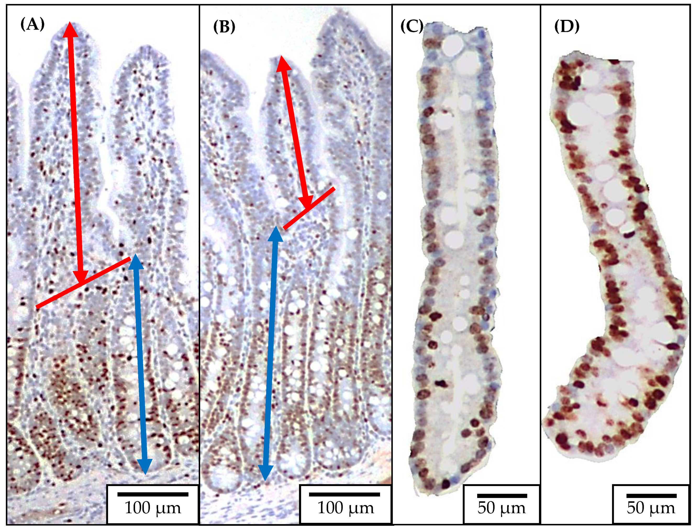
4.4. Jejunal Morphology and Crypt Cell Proliferation

Two sections of jejunal tissue were placed into a cassette in 70% ethanol after 48 h in 10% formalin and sent to the North Carolina State University Histology Laboratory (College of Veterinary Medicine, Raleigh, NC) for Ki67+ staining as previously described by Sun et al. [22] (Figure 4). The jejunal morphology was evaluated by measuring the villus height, and crypt depth using a microscope Olympus CX31 with camera Infinity 2-2 digital CCD. Pictures of well-shaped villi and crypts were taken in 40× magnification to measure the villus height and crypt depth. The villus height to crypt depth ratio (VH:CD) was further calculated. Pictures of jejunal crypts were taken in 100× magnification to measure the enterocyte proliferation rate by measuring the Ki67+ cells. The percentage of Ki67+ cells in the total cells in the crypt was evaluated using the ImageJS software, and an indicator of the enterocyte proliferation in the crypt was used [55].

4.5. Relative Abundance and Diversity of the Fecal and Mucosa-Associated Microbiota in Jejunum

Jejunal mucosa and fecal samples were used for DNA extraction using the QIAamp DNA Stool Mini Kit (51504) following the manufacturer instructions. The extracted DNA samples were sent to the MAKO Medical Laboratories in Raleigh, North Carolina for qPCR analysis of 16S rDNA sequences as previously described by [50]. The Ion Chef instrument was used for the preparation of samples for template and the Ion S5 system was used for sequencing. The Ion 16S Metagenomics Kit (A26216, Thermo Fisher Scientific Inc.) was used to amplify the variable regions V2, V3, V4, V6, V7, V8, and V9 of the 16S rRNA gene. The Ion Xpress Plus Fragment Library Kit (Cat. 4471269, Thermo Fisher Scientific Inc.) was used to prepare the libraries from the amplified regions and the Ion Code Barcode Adapters 1-384 Kit (A29751, Thermo Fisher Scientific Inc.) was used for barcoding and multiplexing of the prepared libraries. The Ion Universal Library Quantitation Kit (A26217, Thermo Fisher Scientific Inc.) was used to quantify the libraries. The Torrent Suite Software (version 5.2.2) was used to process the sequences producing unaligned bam files for further analysis. The Ion Reporter Software Suite (version 5.2) of bioinformatics analysis tools was used for sequence data analysis, alignment to GreenGenes and MicroSeq databases, alpha and beta diversity plot generation, and OTU table generation. All samples had a depth of sequencing coverage greater than 1000×. The OTU data were transformed to relative abundance for further statistical analysis, and the OTU data with less than 0.05% abundance within each level were combined as “others”.

4.6. Statistical Analysis

Data were analyzed using the Mixed procedure of SAS Software. The sites (jejunal mucosa and feces) and the F18+ E. coli challenge (no challenge: (M− and F−) and challenge: (M+ and F+)) were the fixed effects. The experiments were considered random effects. The LSMEANS statement was used to calculate the least squared mean. To evaluate the microbiota data, pre-planned orthogonal contrasts were made to test the effect of the site (jejunal mucosa vs. feces) on microbiota, the effect F18+ E. coli on jejunal mucosa-associated microbiota (M− vs. M+), and the effect F18+ E. coli on fecal microbiota (F− vs. F+). The procedure CORR was used to test the correlation among microbiota (fecal and jejunal mucosa-associated) and inflammatory, oxidative stress, and morphological measurements. Statistical differences were considered significant with p < 0.05 and tendency with 0.05 < p < 0.10.

Author Contributions

Conceptualization, S.W.K.; methodology, S.W.K.; formal analysis, S.W.K. and M.E.D.; investigation, M.E.D.; resources, S.W.K.; data curation, M.E.D. and S.W.K.; writing—original draft preparation, M.E.D. and S.W.K.; writing—review and editing, M.E.D. and S.W.K.; supervision, S.W.K.; project administration, S.W.K.; funding acquisition, S.W.K. All authors have read and agreed to the published version of the manuscript.

Funding

This research was funded by US Dept. of Agriculture (USDA)—National Institute of Food and Agriculture Hatch Project (#02636) and the North Carolina Agricultural Foundation (#660101).

Institutional Review Board Statement

The experimental protocols used in this study were reviewed and approved by the Institutional Animal Care and Use Committee at North Carolina State University. The experiments were performed by trained scientists in full compliance with the North Carolina State Animal Care and Use Procedures (REG 10.10.01).

Informed Consent Statement

Not applicable.

Data Availability Statement

The data presented in this study are available upon request from the corresponding author.

Acknowledgments

K.L. Brooks, H. Chen, J. Guo, K.B. Jang, J.S. Lee, I. Park, W. Parnsen, and L. Zheng of the Kim Lab for their assists in animal management, sampling, and sample analysis.

Conflicts of Interest

The authors declare no conflict of interest.

References

  1. Cheng, Y.-C.; Duarte, M.E.; Kim, S.W. Nutritional and functional values of lysed Corynebacterium glutamicum cell mass for intestinal health and growth of nursery pigs. J. Anim. Sci. 2021, 99, skab331. [Google Scholar] [CrossRef] [PubMed]
  2. Duarte, M.E.; Kim, S.W. Intestinal microbiota and its interaction to intestinal health in nursery pigs. Anim. Nutr. 2022, 8, 169–184. [Google Scholar] [CrossRef]
  3. Albenberg, L.; Esipova, T.V.; Judge, C.P.; Bittinger, K.; Chen, J.; Laughlin, A.; Grunberg, S.; Baldassano, R.N.; Lewis, J.D.; Li, H.; et al. Correlation between Intraluminal Oxygen Gradient and Radial Partitioning of Intestinal Microbiota. Gastroenterology 2014, 147, 1055–1063.e8. [Google Scholar] [CrossRef] [Green Version]
  4. Adhikari, B.; Kim, S.; Kwon, Y. Characterization of microbiota associated with digesta and mucosa in different regions of gastrointestinal tract of nursery pigs. Int. J. Mol. Sci. 2019, 20, 1630. [Google Scholar] [CrossRef] [PubMed] [Green Version]
  5. De Rodas, B.; Youmans, B.P.; Danzeisen, J.L.; Tran, H.; Johnson, T.J. Microbiome profiling of commercial pigs from farrow to finish. J. Anim. Sci. 2018, 96, 1778–1794. [Google Scholar] [CrossRef]
  6. Arpaia, N.; Campbell, C.; Fan, X.; Dikiy, S.; van der Veeken, J.; DeRoos, P.; Liu, H.; Cross, J.R.; Pfeffer, K.; Coffer, P.J.; et al. Metabolites produced by commensal bacteria promote peripheral r egulatory T-cell generation. Nature 2013, 504, 451–455. [Google Scholar] [CrossRef]
  7. Belkaid, Y.; Hand, T.W. Role of the Microbiota in Immunity and Inflammation. Cell 2014, 157, 121–141. [Google Scholar] [CrossRef] [Green Version]
  8. Wang, X.; Tsai, T.; Deng, F.; Wei, X.; Chai, J.; Knapp, J.; Apple, J.; Maxwell, C.V.; Lee, J.A.; Li, Y.; et al. Longitudinal investigation of the swine gut microbiome from birth to market reveals stage and growth performance associated bacteria. Microbiome 2019, 7, 109. [Google Scholar] [CrossRef] [Green Version]
  9. Friedman, E.S.; Bittinger, K.; Esipova, T.V.; Hou, L.; Chau, L.; Jiang, J.; Mesaros, C.; Lund, P.J.; Liang, X.; FitzGerald, G.A.; et al. Microbes vs. chemistry in the origin of the anaerobic gut lumen. Proc. Natl. Acad. Sci. USA 2018, 115, 4170–4175. [Google Scholar] [CrossRef] [Green Version]
  10. Etienne-Mesmin, L.; Chassaing, B.; Desvaux, M.; De Paepe, K.; Gresse, R.; Sauvaitre, T.; Forano, E.; van de Wiele, T.; Schüller, S.; Juge, N.; et al. Experimental models to study intestinal microbes–mucus interactions in health and disease. FEMS Microbiol. Rev. 2019, 43, 457–489. [Google Scholar] [CrossRef] [PubMed] [Green Version]
  11. Zhao, W.; Wang, Y.; Liu, S.; Huang, J.; Zhai, Z.; He, C.; Ding, J.; Wang, J.; Wang, H.; Fan, W.; et al. The dynamic distribution of porcine microbiota across different ages and gastrointestinal tract segments. PLoS ONE 2015, 10, e0117441. [Google Scholar] [CrossRef] [Green Version]
  12. Ma, N.; Guo, P.; Zhang, J.; He, T.; Kim, S.W.; Zhang, G.; Ma, X. Nutrients mediate intestinal bacteria-mucosal immune crosstalk. Front. Immunol. 2018, 9, 1–16. [Google Scholar] [CrossRef]
  13. Shi, N.; Li, N.; Duan, X.; Niu, H. Interaction between the gut microbiome and mucosal immune system. Mil. Med. Res. 2017, 4, 14. [Google Scholar] [CrossRef] [PubMed]
  14. Luise, D.; Lauridsen, C.; Bosi, P.; Trevisi, P. Methodology and application of Escherichia coli F4 and F18 encoding infection models in post-weaning pigs. J. Anim. Sci. Biotechnol. 2019, 10, 53. [Google Scholar] [CrossRef] [PubMed]
  15. Sun, Y.; Kim, S.W. Intestinal challenge with enterotoxigenic Escherichia coli in pigs, and nutritional intervention to prevent postweaning diarrhea. Anim. Nutr. 2017, 3, 322–330. [Google Scholar] [CrossRef] [PubMed]
  16. Nguyen, U.V.; Coddens, A.; Melkebeek, V.; Devriendt, B.; Goetstouwers, T.; van Poucke, M.; Peelman, L.; Cox, E. High susceptibility prevalence for F4+ and F18+ Escherichia coli in Flemish pigs. Vet. Microbiol. 2017, 202, 52–57. [Google Scholar] [CrossRef]
  17. Gresse, R.; Chaucheyras-Durand, F.; Fleury, M.A.; Van de Wiele, T.; Forano, E.; Blanquet-Diot, S. Gut microbiota dysbiosis in postweaning piglets: Understanding thekeys to health. Trends Microbiol. 2017, 25, 851–873. [Google Scholar] [CrossRef]
  18. Duarte, M.E.; Tyus, J.; Kim, S.W. Synbiotic effects of enzyme and probiotics on intestinal health and growth of newly weaned pigs challenged with enterotoxigenic F18+ Escherichia coli. Front. Vet. Sci. 2020, 7, 573. [Google Scholar] [CrossRef]
  19. Boeckman, J.X.; Sprayberry, S.; Korn, A.M.; Suchodolski, J.S.; Paulk, C.; Genovese, K.; Rech, R.R.; Giaretta, P.R.; Blick, A.K.; Callaway, T.; et al. Effect of chronic and acute enterotoxigenic E. coli challenge on growth performance, intestinal inflammation, microbiome, and metabolome of weaned piglets. Sci. Rep. 2022, 12, 5024. [Google Scholar] [CrossRef]
  20. Winter, S.E.; Winter, M.G.; Xavier, M.N.; Thiennimitr, P.; Poon, V.; Keestra, A.M.; Laughlin, R.C.; Gomez, G.; Wu, J.; Lawhon, S.D.; et al. Host-Derived Nitrate Boosts Growth of E. coli in the Inflamed Gut. Science 2013, 339, 708–711. [Google Scholar] [CrossRef] [Green Version]
  21. Zeng, M.Y.; Inohara, N.; Nuñez, G. Mechanisms of inflammation-driven bacterial dysbiosis in the gut. Mucosal Immunol. 2017, 10, 18–26. [Google Scholar] [CrossRef] [PubMed] [Green Version]
  22. Sun, Y.; Duarte, M.E.; Kim, S.W. Dietary inclusion of multispecies probiotics to reduce the severity of post-weaning diarrhea caused by Escherichia coli F18+ in pigs. Anim. Nutr. 2021, 7, 326–333. [Google Scholar] [CrossRef]
  23. McLamb, B.L.; Gibson, A.J.; Overman, E.L.; Stahl, C.; Moeser, A.J. Early Weaning Stress in Pigs Impairs Innate Mucosal Immune Responses to Enterotoxigenic E. coli Challenge and Exacerbates Intestinal Injury and Clinical Disease. PLoS ONE 2013, 8, e59838. [Google Scholar] [CrossRef] [PubMed] [Green Version]
  24. Cutler, S.A.; Lonergan, S.M.; Cornick, N.; Johnson, A.K.; Stahl, C.H. Dietary inclusion of colicin E1 is effective in preventing postweaning diarrhea caused by F18-positive Escherichia coli in pigs. Antimicrob. Agents Chemother. 2007, 51, 3830–3835. [Google Scholar] [CrossRef] [PubMed] [Green Version]
  25. Kim, K.; He, Y.; Xiong, X.; Ehrlich, A.; Li, X.; Raybould, H.; Atwill, E.R.; Maga, E.A.; Jørgensen, J.; Liu, Y. Dietary supplementation of Bacillus subtilis influenced intestinal health of weaned pigs experimentally infected with a pathogenic E. coli. J. Anim. Sci. Biotechnol. 2019, 10, 52. [Google Scholar] [CrossRef] [Green Version]
  26. Levesque, C.L.; Yu, H.; Gong, J.; de Lange, C.F.M. Ileal mucosa-associated—But not ileal digesta—Bacterial profiles in grower pigs are influenced by nutrition and use of antibiotics for weaner pigs1. J. Anim. Sci. 2012, 90, 448–450. [Google Scholar] [CrossRef]
  27. Liu, H.; Zeng, X.; Zhang, G.; Hou, C.; Li, N.; Yu, H.; Shang, L.; Zhang, X.; Trevisi, P.; Yang, F.; et al. Maternal milk and fecal microbes guide the spatiotemporal development of mucosa-associated microbiota and barrier function in the porcine neonatal gut. BMC Biol. 2019, 17, 106. [Google Scholar] [CrossRef] [Green Version]
  28. Zhang, L.; Wu, W.; Lee, Y.K.; Xie, J.; Zhang, H. Spatial heterogeneity and co-occurrence of mucosal and luminal microbiome across swine intestinal tract. Front. Microbiol. 2018, 9, 48. [Google Scholar] [CrossRef] [Green Version]
  29. Leser, T.D.; Mølbak, L. Better living through microbial action: The benefits of the mammalian gastrointestinal microbiota on the host. Environ. Microbiol. 2009, 11, 2194–2206. [Google Scholar] [CrossRef]
  30. Beeby, M. Motility in the epsilon-proteobacteria. Curr. Opin. Microbiol. 2015, 28, 115–121. [Google Scholar] [CrossRef] [Green Version]
  31. Mace, O.J.; Marshall, F. Gut chemosensing and the regulation of nutrient absorption and energy supply. J. Anim. Sci. 2013, 91, 1932–1945. [Google Scholar] [CrossRef] [Green Version]
  32. Espey, M.G. Role of oxygen gradients in shaping redox relationships between the human intestine and its microbiota. Free Radic. Biol. Med. 2013, 55, 130–140. [Google Scholar] [CrossRef] [PubMed]
  33. Wiarda, J.E.; Trachsel, J.M.; Bond, Z.F.; Byrne, K.A.; Gabler, N.K.; Loving, C.L. Intraepithelial T cells diverge by intestinal location as pigs age. Front. Immunol. 2020, 11, 1139. [Google Scholar] [CrossRef] [PubMed]
  34. Jin, J.; Jia, J.; Zhang, L.; Chen, Q.; Zhang, X.; Sun, W.; Ma, C.; Xu, F.; Zhan, S.; Ma, L.; et al. Jejunal inflammatory cytokines, barrier proteins and microbiome-metabolome responses to early supplementary feeding of Bamei suckling piglets. BMC Microbiol. 2020, 20, 169. [Google Scholar] [CrossRef]
  35. Mu, C.; Yang, Y.; Su, Y.; Zoetendal, E.G.; Zhu, W. Differences in microbiota membership along the gastrointestinal tract of piglets and their differential alterations following an early-life antibiotic intervention. Front. Microbiol. 2017, 8, 797. [Google Scholar] [CrossRef] [PubMed]
  36. Kelly, J.; Daly, K.; Moran, A.W.; Ryan, S.; Bravo, D.; Shirazi-Beechey, S.P. Composition and diversity of mucosa-associated microbiota along the entire length of the pig gastrointestinal tract; dietary influences. Environ. Microbiol. 2017, 19, 1425–1438. [Google Scholar] [CrossRef] [Green Version]
  37. Plancade, S.; Clark, A.; Philippe, C.; Helbling, J.-C.; Moisan, M.-P.; Esquerré, D.; Le Moyec, L.; Robert, C.; Barrey, E.; Mach, N. Unraveling the effects of the gut microbiota composition and function on horse endurance physiology. Sci. Rep. 2019, 9, 9620. [Google Scholar] [CrossRef] [Green Version]
  38. Li, Q.; Peng, X.; Burrough, E.R.; Sahin, O.; Gould, S.A.; Gabler, N.K.; Loving, C.L.; Dorman, K.S.; Patience, J.F. Dietary Soluble and Insoluble Fiber with or without Enzymes Altered the Intestinal Microbiota in Weaned Pigs Challenged with Enterotoxigenic E. coli F18. Front. Microbiol. 2020, 11, 1110. [Google Scholar] [CrossRef]
  39. Guo, P.; Zhang, K.; Ma, X.; He, P. Clostridium species as probiotics: Potentials and challenges. J. Anim. Sci. Biotechnol. 2020, 11, 1–10. [Google Scholar] [CrossRef]
  40. Heeney, D.D.; Gareau, M.G.; Marco, M.L. Intestinal Lactobacillus in health and disease, a driver or just along for the ride? Curr. Opin. Biotechnol. 2018, 49, 140–147. [Google Scholar] [CrossRef] [Green Version]
  41. Moita, V.H.C.; Duarte, M.E.; da Silva, S.N.; Kim, S.W. Supplemental Effects of Functional Oils on the Modulation of Mucosa-Associated Microbiota, Intestinal Health, and Growth Performance of Nursery Pigs. Animals 2021, 11, 1591. [Google Scholar] [CrossRef] [PubMed]
  42. Van Den Broeck, W.; Cox, E.; Oudega, B.; Goddeeris, B.M. The F4 fimbrial antigen of Escherichia coli and its receptors. Vet. Microbiol. 2000, 71, 223–244. [Google Scholar] [CrossRef]
  43. Devriendt, B.; Stuyven, E.; Verdonck, F.; Goddeeris, B.M.; Cox, E. Enterotoxigenic Escherichia coli (K88) induce proinflammatory responses in porcine intestinal epithelial cells. Dev. Comp. Immunol. 2010, 34, 1175–1182. [Google Scholar] [CrossRef] [PubMed]
  44. Wang, H.; Zhong, Z.; Luo, Y.; Cox, E.; Devriendt, B. Heat-stable enterotoxins of enterotoxigenic escherichia coli and their impact on host immunity. Toxins 2019, 11, 24. [Google Scholar] [CrossRef] [Green Version]
  45. Loos, M.; Geens, M.; Schauvliege, S.; Gasthuys, F.; van der Meulen, J.; Dubreuil, J.D.; Goddeeris, B.M.; Niewold, T.; Cox, E. Role of heat-stable enterotoxins in the induction of early immune responses in piglets after infection with enterotoxigenic Escherichia coli. PLoS ONE 2012, 7, e41041. [Google Scholar] [CrossRef] [Green Version]
  46. Levesque, C.L.; Hooda, S.; Swanson, K.S.; De Lange, K. Alterations in ileal mucosa bacteria related to diet complexity and growth performance in young pigs. PLoS ONE 2014, 9, e108472. [Google Scholar] [CrossRef] [Green Version]
  47. Zhang, W.; Zhu, Y.-H.; Zhou, D.; Wu, Q.; Song, D.; Dicksved, J.; Wang, J.-F. Oral Administration of a Select Mixture of Bacillus Probiotics Affects the Gut Microbiota and Goblet Cell Function following Escherichia coli Challenge in Newly Weaned Pigs of Genotype MUC4 That Are Supposed to Be Enterotoxigenic E. coli F4ab/ac Receptor. Appl. Environ. Microbiol. 2017, 83, 1–18. [Google Scholar] [CrossRef] [Green Version]
  48. Sido, A.; Radhakrishnan, S.; Kim, S.W.; Eriksson, E.; Shen, F.; Li, Q.; Bhat, V.; Reddivari, L.; Vanamala, J.K.P. A food-based approach that targets interleukin-6, a key regulator of chronic intestinal inflammation and colon carcinogenesis. J. Nutr. Biochem. 2017, 43, 11–17. [Google Scholar] [CrossRef] [Green Version]
  49. Duarte, M.E.; Zhou, F.X.; Dutra, W.M.; Kim, S.W. Dietary supplementation of xylanase and protease on growth performance, digesta viscosity, nutrient digestibility, immune and oxidative stress status, and gut health of newly weaned pigs. Anim. Nutr. 2019, 5, 351–358. [Google Scholar] [CrossRef]
  50. Duarte, M.E.; Sparks, C.; Kim, S.W. Modulation of jejunal mucosa-associated microbiota in relation to intestinal health and nutrient digestibility in pigs by supplementation of β-glucanase to corn–soybean meal-based diets with xylanase. J. Anim. Sci. 2021, 99, skab190. [Google Scholar] [CrossRef]
  51. Kim, S.W.; Duarte, M.E. Understanding intestinal health in nursery pigs and the relevant nutritional strategies. Anim. Biosci. 2021, 34, 338–344. [Google Scholar] [CrossRef]
  52. Zheng, L.; Duarte, M.E.; Sevarolli Loftus, A.; Kim, S.W. Intestinal health of pigs upon weaning: Challenges and nutritional intervention. Front. Vet. Sci. 2021, 8, 628258. [Google Scholar] [CrossRef] [PubMed]
  53. NRC. Nutrient Requirements of Swine, 11th ed.; National Academies Press: Washington, DC, USA, 2012; ISBN 978-0-309-22423-9. [Google Scholar]
  54. Nagy, B.; Whipp, S.C.; Imberechts, H.; Bertschinger, H.U.; Dean-Nystrom, E.A.; Casey, T.A.; Salajka, E. Biological relationship between F18ab and F18ac fimbriae of enterotoxigenic and verotoxigenic Escherichia coli from weaned pigs with oedema disease or diarrhoea. Microb. Pathog. 1997, 22, 1–11. [Google Scholar] [CrossRef] [PubMed]
  55. Jang, K.B.; Kim, S.W. Supplemental effects of dietary nucleotides on intestinal health and growth performance of newly weaned pigs. J. Anim. Sci. 2019, 97, 4875–4882. [Google Scholar] [CrossRef] [PubMed]
Figure 1. Fecal score of pigs challenged with enterotoxigenic F18+ E. coli on d 7 post-weaning. * d 8 to 14: p < 0.05; * d 15 to 21: p < 0.05.
Pathogens 11 00589 g001
Figure 2. Alpha diversity of fecal and jejunal mucosa-associated microbiota estimated with Chao1 richness (A), Shannon diversity (B), and Simpson diversity (C) in pigs at d 21 after challenge with enterotoxigenic E. coli F18+. Site: mucosa and feces. F18+ E. coli challenge: no challenge: (−) and challenge: (+). Mucosa vs. feces: effect of site on microbiota; M− vs. M+: effect of F18+ E. coli on jejunal mucosa-associated microbiota; F− vs. F+: effect of F18+ E. coli on fecal microbiota.
Pathogens 11 00589 g002
Figure 3. Beta diversity of fecal and jejunal mucosa-associated microbiota in nursery pigs challenged with enterotoxigenic F18+ E. coli. Principal coordinates analysis (PCoA) plot based on Bray–Curtis distance showed distinct clusters in the mucosa-associated microbiota (orange) and fecal microbiota (blue). The analysis of similarity (ANOSIM) procedure was used for the significance of the clustering pattern between jejunal mucosa-associated and fecal microbiota. Site: mucosa and feces. F18+ E. coli challenge: no challenge: (−) and challenge: (+). Mucosa vs. feces: effect of site on microbiota; M− vs. M+: effect of F18+ E. coli on jejunal mucosa-associated microbiota; F− vs. F+: effect of F18+ E. coli on fecal microbiota.
Pathogens 11 00589 g003
Figure 4. Representative images of the immunohistochemistry (Ki67) staining for jejunal morphology and crypt cell proliferation. Ten images at 40× of well-oriented villi and their associated crypts ((A): no challenged; (B): F18+ E. coli challenged) were obtained for measuring villus height (from the top to the base of villus as indicated with double arrow line in red) and crypt depth (from the base of villus to the bottom of the crypt as indicated with double arrow line in blue). Ten images at 100× of the crypts ((C): no challenged; (D): F18+ E. coli challenged) were obtained for measuring the percentage of positive Ki67 staining cells.
Pathogens 11 00589 g004
Table 1. Relative abundance of fecal and jejunal mucosa-associated microbiota at the phylum level in pigs at d 21 after challenge with enterotoxigenic E. coli F18+.
Site 1MucosaFeces p Value 3
Challenge 2++SEMSiteM− vs. M+F− vs. F+
Firmicutes36.1135.0748.6146.735.400.0140.8780.781
Proteobacteria35.9343.4712.2715.105.60<0.0010.2870.689
Bacteroidetes21.5516.0435.2332.153.24<0.0010.2300.500
Chlamydiae3.722.91<0.011.712.050.2030.7690.523
Spirochaetes1.530.111.090.340.640.8670.1220.410
Actinobacteria0.410.571.170.550.360.3160.7540.231
Cyanobacteria0.331.510.030.030.740.2230.2540.996
Others0.310.211.213.101.570.3570.9870.167
1 Site: mucosa and feces. 2 F18+ E. coli challenge: no challenge: (−) and challenge: (+). 3 Mucosa vs. feces: effect of site on microbiota; M− vs. M+: effect of F18+ E. coli on jejunal mucosa-associated microbiota; F− vs. F+: effect of F18+ E. coli on fecal microbiota.
Table 2. Relative abundance of fecal and jejunal mucosa-associated microbiota at the family level in pigs at d 21 after challenge with enterotoxigenic E. coli F18+.
Site 1MucosaFeces p Value 3
Challenge 2++SEMSiteM− vs. M+F− vs. F+
Helicobacteraceae14.3129.890.620.775.21<0.0010.0170.982
Prevotellaceae19.8314.7729.3928.812.92<0.0010.2190.888
Lactobacillaceae13.3714.349.3711.623.440.3310.8430.645
Veillonellaceae8.277.1618.9517.142.96<0.0010.7130.550
Campylobacteraceae8.302.041.290.411.980.0390.0270.757
Pseudomonadaceae1.804.74<0.01<0.011.60<0.0010.0340.951
Clostridiaceae4.472.271.601.671.870.2350.2880.975
Enterobacteriaceae5.241.440.122.141.650.0380.0210.140
Streptococcaceae2.964.311.311.362.160.1380.5350.982
Chlamydiaceae3.562.270.02<0.011.900.0650.5870.979
Lachnospiraceae1.311.944.814.730.84<0.0010.5410.942
Succinivibrionaceae0.790.467.4810.003.68<0.0010.9170.428
Ruminococcaceae1.251.345.644.860.64<0.0010.9080.319
Brachyspiraceae2.440.080.550.030.780.2140.0350.634
Moraxellaceae1.130.84<0.010.030.520.0290.6670.980
Oxalobacteraceae0.471.180.570.340.720.5010.3670.780
Acidaminococcaceae1.351.102.653.550.73<0.0010.6430.088
Nostocaceae0.291.25<0.01<0.010.600.1970.2540.999
Xanthomonadaceae0.351.03<0.01<0.010.25<0.0010.0130.969
Porphyromonadaceae0.960.742.542.620.54<0.0010.7170.893
Pseudanabaenaceae0.600.870.070.080.450.0110.4580.970
Erysipelotrichaceae0.450.761.231.420.330.0330.5140.694
Bacillaceae1.190.030.080.030.480.2440.0890.944
Bifidobacteriaceae0.340.210.990.290.360.2890.7860.146
Eubacteriaceae0.640.542.422.580.37<0.0010.8240.707
Hyphomicrobiaceae0.290.290.260.430.120.5920.9730.299
Bacteroidaceae0.250.110.270.240.080.3260.2290.816
Flavobacteriaceae0.270.090.270.360.100.1730.2140.547
Cytophagaceae0.270.191.631.140.670.0120.9000.446
Spirochaetaceae0.070.020.700.250.170.0050.7890.041
Oscillospiraceae0.090.060.520.340.09<0.0010.7380.037
Peptostreptococcaceae0.060.490.060.120.150.0980.0080.698
Mycoplasmataceae0.130.070.350.150.130.1320.6230.139
Spiroplasmataceae0.010.010.240.310.06<0.0010.9280.368
Caulobacteraceae0.430.08<0.01<0.010.140.0060.0160.968
Rikenellaceae0.050.050.280.280.130.0110.9950.980
Others1.791.884.131.940.720.1000.9210.016
1 Site: mucosa and feces. 2 F18+ E. coli challenge: no challenge: (−) and challenge: (+). 3 Mucosa vs. feces: effect of site on microbiota; M− vs. M+: effect of F18+ E. coli on jejunal mucosa-associated microbiota; F− vs. F+: effect of F18+ E. coli on fecal microbiota.
Table 3. Relative abundance of fecal and jejunal mucosa-associated microbiota at the genus level in pigs at d 21 after challenge with enterotoxigenic E. coli F18+.
Site 1MucosaFeces p Value 3
Challenge 2++SEMSiteM− vs. M+F− vs. F+
Prevotella22.1515.0133.7830.103.60<0.0010.1650.472
Lactobacillus16.0418.4412.6118.444.310.6880.6910.335
Helicobacter16.8332.830.510.655.20<0.0010.0150.983
Succinivibrio0.740.409.8112.904.83<0.0010.9300.420
Campylobacter10.072.552.250.702.550.0710.0400.669
Clostridium4.742.621.251.172.510.1520.3810.972
Megasphaera1.901.276.164.441.680.0020.7030.297
Mitsuokella2.751.382.994.911.400.1020.3970.240
Pseudomonas3.536.57<0.01<0.011.74<0.0010.0730.963
Faecalibacterium1.581.344.704.080.97<0.0010.8090.532
Streptococcus3.494.761.451.922.340.1350.5790.839
Phascolarctobacterium1.781.123.492.910.820.0030.4120.466
Selenomonas0.350.334.392.511.15<0.0010.9890.249
Chlamydia4.122.44<0.01<0.012.070.0560.5150.967
Dialister1.170.711.703.961.01<0.0010.5180.002
Roseburia0.540.492.031.940.40<0.0010.9100.867
Bifidobacterium0.240.422.230.460.850.2200.8830.133
Acidaminococcus0.300.300.832.360.530.0010.9970.005
Eubacterium0.330.381.340.940.440.0410.9220.454
Oscillibacter<0.01<0.011.680.540.48<0.0010.8800.004
Acinetobacter1.431.05<0.010.020.590.0240.6190.922
Gemmiger0.400.440.470.780.200.3040.8900.278
Herbaspirillum<0.01<0.011.080.690.39<0.0010.9980.270
Massilia0.521.05<0.01<0.010.500.0510.8500.984
Bacillus1.440.02<0.01<0.010.570.2190.0840.999
Coprococcus0.010.061.000.150.410.1770.9280.134
Catenibacterium0.130.160.160.690.250.2540.9200.126
Anaerovibrio0.090.220.440.270.150.1150.4340.339
Dorea0.250.260.230.370.120.6510.9680.356
Tepidimonas<0.01<0.010.800.100.270.0270.9730.027
Ruminococcus0.060.210.280.350.090.0490.2620.563
Blautia0.160.50<0.01<0.010.150.0040.0390.879
Brachyspira0.190.100.510.050.320.4610.7440.207
Enterococcus0.600.01<0.01<0.010.290.2930.1470.999
Treponema0.130.050.330.80.150.4110.7150.207
Others1.741.771.121.540.390.2250.8120.445
1 Site: mucosa and feces. 2 F18+ E. coli challenge: no challenge: (−) and challenge: (+). 3 Mucosa vs. feces: effect of site on microbiota; M− vs. M+: effect of F18+ E. coli on jejunal mucosa-associated microbiota; F− vs. F+: effect of F18+ E. coli on fecal microbiota.
Table 4. Relative abundance of fecal and jejunal mucosa-associated microbiota at the species level in pigs at d 21 after challenge with enterotoxigenic E. coli F18+.
Site 1MucosaFeces p Value 3
Challenge 2++SEMSiteM− vs. M+F− vs. F+
Prevotella copri23.6314.6433.5228.234.880.0010.0730.288
Succinivibrio dextrinosolvens2.040.9112.5410.265.640.0020.7990.607
Lactobacillus kitasatonis4.135.623.398.753.200.6640.7010.169
Helicobacter mastomyrinus7.7514.40<0.01<0.012.67<0.0010.0790.999
Helicobacter rappini5.2315.590.901.073.960.0010.0110.965
Faecalibacterium prausnitzii3.542.675.245.541.100.0270.5460.834
Prevotella stercorea2.912.243.903.911.150.0680.5110.990
Lactobacillus mucosae4.224.420.702.201.580.0520.9230.469
Campylobacter coli9.030.431.110.272.650.1430.0210.824
Phascolarctobacterium succinatutens2.651.453.923.430.970.0240.2320.622
Lactobacillus delbrueckii1.551.863.142.601.230.2650.8370.715
Mitsuokella jalaludinii2.401.052.143.701.050.2530.2880.358
Dialister succinatiphilus1.931.192.355.121.290.0050.4840.010
Roseburia faecis0.741.083.482.720.56<0.0010.6740.346
Streptococcus alactolyticus3.323.361.221.012.070.0800.9810.908
Lactobacillus johnsonii1.031.371.641.540.900.5160.6930.908
Chlamydia suis3.042.49<0.01<0.011.750.0580.7970.935
Lactobacillus salivarius0.651.011.482.380.890.0720.6690.294
Selenomonas lipolytica0.51<0.012.960.850.870.0150.6210.036
Gemmiger formicilis0.921.090.631.660.510.9780.7630.195
Mitsuokella multacida1.620.690.311.300.540.5220.2260.199
Megasphaera hominis0.500.302.620.891.090.1300.8770.172
Acidaminococcus fermentans0.200.330.832.300.450.0030.8260.015
Helicobacter equorum0.053.42<0.010.041.210.1630.0540.980
Selenomonas bovis0.250.082.170.900.600.0040.8270.235
Prevotella sp.0.600.310.851.790.620.0050.5040.028
Streptococcus hyointestinalis0.011.870.150.780.690.4850.0580.516
Streptococcus infantarius1.111.720.300.270.890.0620.4780.974
Acinetobacter radioresistens1.480.80<0.01<0.010.710.0440.4320.982
Acinetobacter lwoffii1.990.42<0.01<0.010.540.0130.0270.991
Campylobacter lanienae0.290.241.510.210.310.0580.9100.004
Treponema porcinum0.130.081.480.310.370.0160.9150.011
Campylobacter upsaliensis0.911.080.070.090.740.1270.7360.996
Dorea longicatena0.510.600.260.450.170.2470.7230.428
Others9.4411.565.666.412.610.0550.5160.816
1 Site: mucosa and feces. 2 F18+ E. coli challenge: no challenge: (−) and challenge: (+). 3 Mucosa vs. feces: effect of site on microbiota; M− vs. M+: effect of F18+ E. coli on jejunal mucosa-associated microbiota; F− vs. F+: effect of F18+ E. coli on fecal microbiota.
Table 5. Immune and oxidative stress status in pigs after challenge with enterotoxigenic E. coli F18+.
ItemNo challenge (A)Challenge (B)SEMp value
Serum
TNF-α, pg/mL98.6113.725.10.172
Protein carbonyl, nmol/mg of protein1.742.100.450.396
Jejunal mucosa
TNF-α, pg/mg of protein1.151.310.270.551
Protein carbonyl, nmol/mg of protein2.333.170.260.026
Table 6. Intestinal morphology and enterocyte proliferation in jejunum of pigs at d 21 post-challenge with enterotoxigenic E. coli F18+.
ItemNo Challenge (A)Challenge (B)SEMp Value
Jejunal mucosa
Villus Height, µm494434230.026
Crypt Depth, µm209224300.169
VH:CD 12.602.140.300.044
Ki67+, %515580.632
1 Villus height to crypt depth ratio.
Table 7. Pearson correlation coefficients (r) between jejunal mucosa-associated microbiota and other variables measured in pigs after challenge with enterotoxigenic E. coli F18+.
Item 1Family (r, p Value)Species (r, p Value)
TNF-α, serumLactobacillaceae (0.30, 0.048)Succinivibrio dextrinosolvens (−0.33, 0.030)
Veillonellaceae (0.32, 0.034)Faecalibacterium prausnitzii (−0.36, 0.017)
Enterobacteriaceae (−0.32, 0.037)Lactobacillus mucosae (0.51, <0.001)
Acidaminococcaceae (0.36, 0.018)Megasphaera hominis (0.49, 0.001)
Pseudomonadaceae (−0.49, 0.001)Campylobacter lanienae (0.49, 0.001)
Porphyromonadaceae (0.43, 0.004)
Eubacteriaceae (0.33, 0.027)
Cytophagaceae (0.33, 0.027)
Xanthomonadaceae (−0.32, 0.036)
Pseudanabaenaceae (0.35, 0.018)
Oscillospiraceae (0.37, 0.015)
Spiroplasmataceae (0.33, 0.028)
Caulobacteraceae (−0.41, 0.005)
TNF-α, mucosaBifidobacteriaceae (−0.63, <0.001)Mitsuokella jalaludinii (0.45, 0.009)
Dialister succinatiphilus (0.52, 0.002)
Mitsuokella multacida (0.42, 0.016)
Acidaminococcus fermentans (0.60, <0.001)
PC, serumOscillospiraceae (0.42, 0.005)Lactobacillus johnsonii (0.49, 0.001)
Spiroplasmataceae (0.69, <0.001)Megasphaera hominis (0.45, 0.024)
Campylobacter lanienae (0.66, <0.001)
PC, mucosaErysipelotrichaceae (0.32, 0.034)Acidaminococcus fermentans (0.30, 0.044)
Bifidobacteriaceae (−0.33, 0.031)Acinetobacter lwoffii (0.33, 0.031)
Spiroplasmataceae (0.44, 0.003)
Villus heightVeillonellaceae (0.33, 0.031)Campylobacter coli (0.33, 0.027)
Brachyspiraceae (0.34, 0.023)Megasphaera hominis (0.37, 0.013)
Pseudomonadaceae (0.33, 0.026)
Oscillospiraceae (0.42, 0.041)
Crypt depthPrevotellaceae (−0.36, 0.019)Succinivibrio dextrinosolvens (−0.50, 0.001)
Lactobacillaceae (0.41, 0.006)Helicobacter rappini (0.32, 0.039)
Succinivibrionaceae (−0.39, 0.010)Faecalibacterium prausnitzii (−0.55, <0.001)
Lachnospiraceae (−0.40, 0.008)Dialister succinatiphilus (0.30, 0.047)
Ruminococcaceae (−0.36, 0.019)Roseburia faecis (−0.38, 0.013)
Clostridiaceae (−0.49, 0.001)Gemmiger formicilis (−0.38, 0.013)
Pseudomonadaceae (−0.30, 0.049)Megasphaera hominis (0.41, 0.007)
Xanthomonadaceae (−0.32, 0.034)Selenomonas bovis (0.33, 0.035)
Pseudanabaenaceae (0.36, 0.017)Campylobacter lanienae (0.35, 0.020)
Mycoplasmataceae (−0.34, 0.028)Dorea longicatena (−0.40, 0.008)
VH:CDLactobacillaceae (−0.32, 0.034)Succinivibrio dextrinosolvens (0.35, 0.021)
Lachnospiraceae (0.31, 0.046)Helicobacter rappini (−0.34, 0.024)
Clostridiaceae (0.48, 0.001)Faecalibacterium prausnitzii (0.43, 0.004)
Xanthomonadaceae (0.32, 0.036)Roseburia faecis (0.34, 0.024)
Acinetobacter lwoffii (0.33, 0.032)
Dorea longicatena (0.35, 0.020)
1 TNF-α: tumor necrosis alpha; PC: protein carbonyl; VH:CD: villus height to crypt depth ratio.
Table 8. Pearson correlation coefficients (r) between fecal microbiota and other variables measured in pigs after challenge with enterotoxigenic E. coli F18+.
Item 1Family (r, p Value)Species (r, p Value)
TNF-α, serumVeillonellaceae (0.37, 0.013)Prevotella copri (0.49, <0.001)
Succinivibrionaceae (−0.33, 0.031)Succinivibrio dextrinosolvens (−0.49, <0.001)
Streptococcaceae (0.47, 0.001)Phascolarctobacterium succinatutens (0.36, 0.016)
Acidaminococcaceae (0.31, 0.041)Streptococcus alactolyticus (0.36, 0.015)
Eubacteriaceae (0.34, 0.025)Selenomonas lipolytica (−0.30, 0.046)
Oxalobacteraceae (−0.31, 0.044)Megasphaera hominis (0.46, 0.002)
Cytophagaceae (0.41, 0.006)Streptococcus hyointestinalis (0.34, 0.024)
Flavobacteriaceae (0.38, 0.010)Streptococcus infantarius (0.37, 0.013)
Treponema porcinum (−0.32, 0.037)
Dorea longicatena (0.30, 0.049)
PC, serumAcidaminococcaceae (0.31, 0.043)Prevotella copri (0.35, 0.022)
Villus heightFlavobacteriaceae (0.33, 0.030)Dialister succinatiphilus (0.41, 0.005)
Lactobacillus salivarius (0.34, 0.025)
Mitsuokella multacida (0.34, 0.022)
Treponema porcinum (−0.30, 0.049)
Crypt depthPrevotellaceae (0.45, 0.003)Prevotella copri (0.47, 0.002)
Lactobacillaceae (−0.34, 0.026)Succinivibrio dextrinosolvens (−0.42, 0.004)
Succinivibrionaceae (−0.42, 0.005)Lactobacillus kitasatonis (−0.33, 0.030)
Ruminococcaceae (0.35, 0.023)Faecalibacterium prausnitzii (0.34, 0.023)
Acidaminococcaceae (0.48, 0.001)Prevotella stercorea (0.31, 0.040)
Oxalobacteraceae (−0.33, 0.028)Dialister succinatiphilus (0.42, 0.005)
Cytophagaceae (0.34, 0.026)Lactobacillus johnsonii (−0.37, 0.016)
Bifidobacteriaceae (−0.32, 0.034)Selenomonas lipolytica (−0.45, 0.003)
Flavobacteriaceae (0.35, 0.020)Selenomonas bovis (−0.41, 0.006)
Oscillospiraceae (0.32, 0.034)Prevotella sp. (0.45, 0.003)
VH:CDPrevotellaceae (−032, 0.034)Selenomonas lipolytica (0.39, 0.009)
Lactobacillaceae (0.32, 0.036)Selenomonas bovis (0.46, 0.002)
Succinivibrionaceae (0.32, 0.038)Prevotella sp. (−0.42, 0.005)
Ruminococcaceae (−0.33, 0.028)
Acidaminococcaceae (−0.30, 0.049)
Oscillospiraceae (−0.31, 0.045)
Ki67+Lactobacillaceae (−0.42, 0.042)Lactobacillus mucosae (−0.60, 0.002)
Ruminococcaceae (0.48, 0.018)Lactobacillus delbrueckii (−0.51, 0.011)
Oxalobacteraceae (−0.52, 0.009)Phascolarctobacterium succinatutens (0.41, 0.044)
1 TNF-α: tumor necrosis alpha; PC: protein carbonyl; VH:CD: villus height to crypt depth ratio; Ki67+: enterocyte proliferation rate in the crypts.
Table 9. Composition of basal diets (Exp. 1, 2, and 3; as-fed basis).
ItemPhase 1Phase 2
Ingredient, %
Corn, yellow dent40.7154.80
Soybean meal, 48% CP22.0023.50
Whey permeate 20.0010.00
Blood plasma6.003.00
Poultry meal5.004.00
Poultry fat 3.501.80
L-Lys HCl0.480.46
DL-Met0.220.18
L-Thr0.160.14
Dicalcium phosphate0.250.68
Limestone1.281.04
Vitamin premix 10.030.03
Mineral premix 20.150.15
Salt0.220.22
Calculated composition:
Dry matter, %91.590.4
ME, kcal/kg34883398
Crude protein, %23.221.7
SID 3 Lys, %1.501.35
SID Met + Cys, %0.820.74
SID Trp, %0.250.22
SID Thr, %0.880.79
Ca, %0.850.80
STTD 4 P, %0.450.40
Total P, %0.650.64
1 Vitamin premix: the vitamin premix provided the following per kilogram of complete diet: 6613.8 IU of vitamin A as vitamin A acetate, 992.0 IU of vitamin D3, 19.8 IU of vitamin E, 2.64 mg of vitamin K as menadione sodium bisulfate, 0.03 mg of vitamin B12, 4.63 mg of riboflavin, 18.52 mg of D-pantothenic acid as calcium pantothenate, 24.96 mg of niacin, and 0.07 mg of biotin. 2 Mineral premix: the trace mineral premix provided the following per kilogram of complete diet: 4.0 mg of Mn as manganous oxide, 165 mg of Fe as ferrous sulfate, 165 mg of Zn as zinc sulfate, 16.5 mg of Cu as copper sulfate, 0.30 mg of I as ethylenediamine di-hydroiodide, and 0.30 mg of Se as sodium selenite. 3 SID: standardized ileal digestible. 4 STTD: standardized total tract digestible.
Publisher’s Note: MDPI stays neutral with regard to jurisdictional claims in published maps and institutional affiliations.

