
Rectovaginal Colonization with Serotypes of Group B Streptococci with Reduced Penicillin Susceptibility among Pregnant Women in León, Nicaragua

 , , , ,
Abstract
:1. Introduction
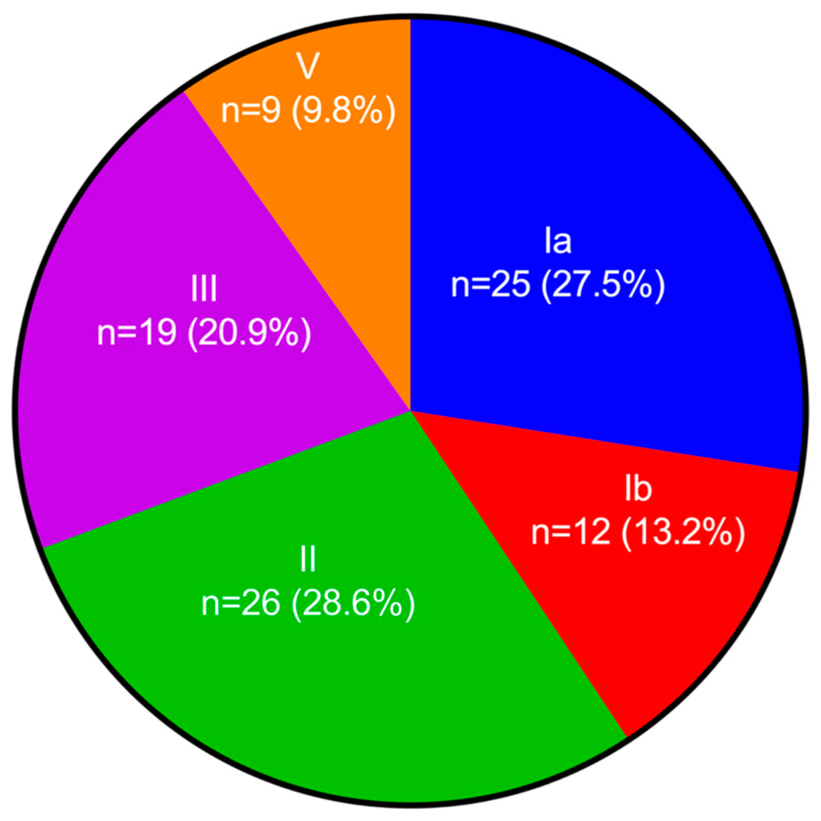
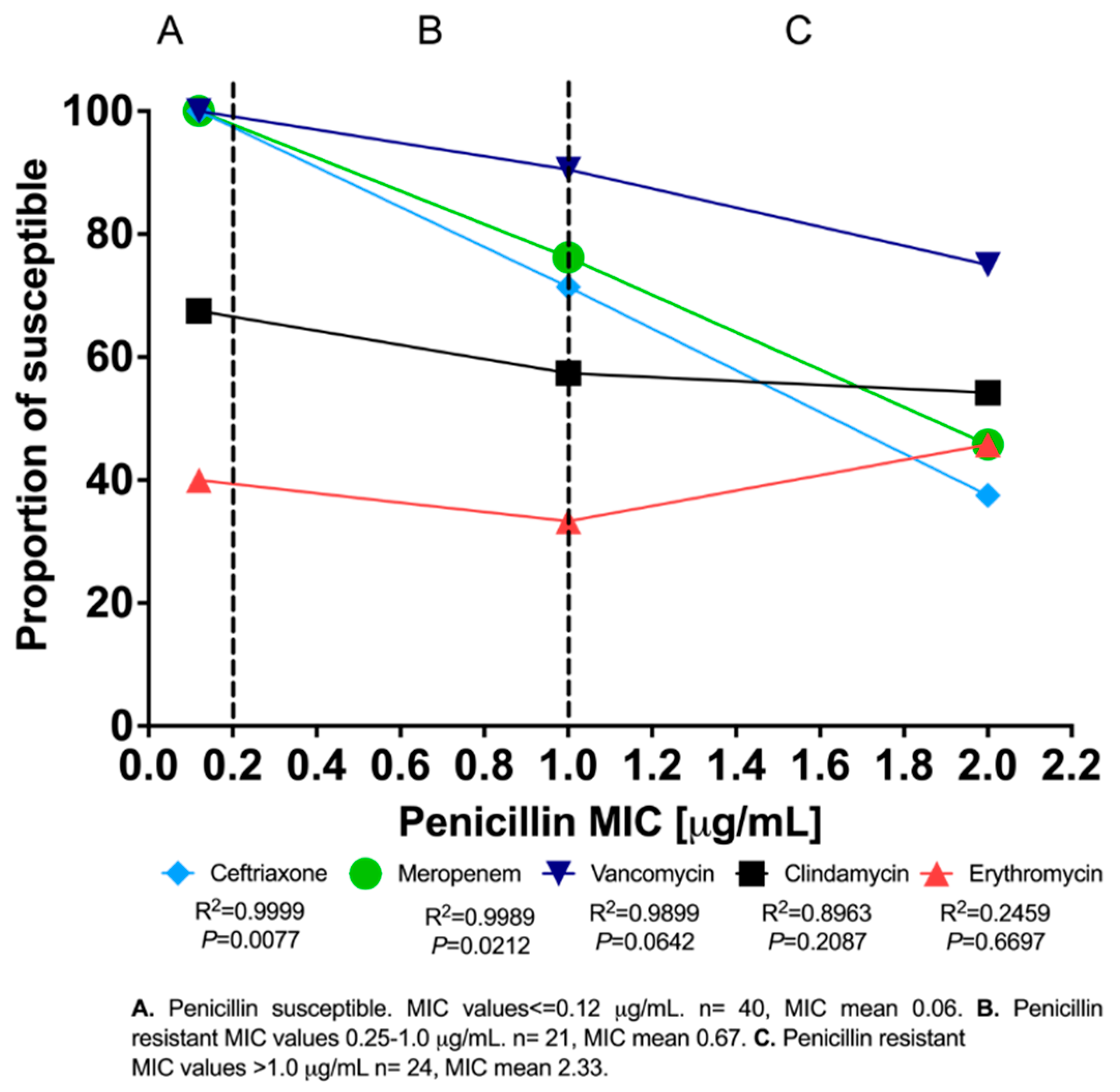
2. Results
3. Discussion
4. Materials and Methods
4.1. Study Design
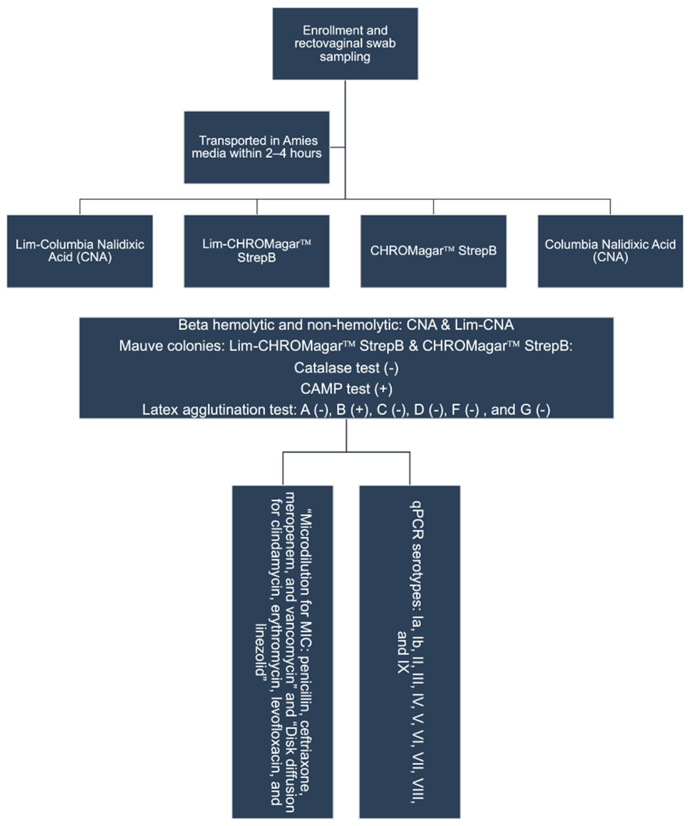
4.2. Data Collection and Handling, Transport, and Processing of Samples
4.3. Identification and Serotyping of Group B Streptococci
4.4. qPCR Serotyping
4.5. Antibiotic Susceptibility Testing
4.6. Statistical Analysis
5. Conclusions
Supplementary Materials
Author Contributions
Funding
Institutional Review Board Statement
Informed Consent Statement
Data Availability Statement
Conflicts of Interest
References
- World Health Organization. Global Report on the Epidemiology and Burden of Sepsis: Current Evidence, Identifying Gaps and Future Directions; World Health Organization: Geneva, Switzerland, 2020; Available online: http://apps.who.int/bookorders.%0Ahttps://apps.who.int/iris/bitstream/handle/10665/334216/9789240010789-eng.pdf (accessed on 14 February 2022).
- Committee on Obstetric Practice. Prevention of Group B Streptococcal Early-Onset Disease in Newborns: ACOG Committee Opinion, Number 797. Obstet. Gynecol. 2020, 135, e51–e72. [Google Scholar] [CrossRef] [PubMed]
- Bank, T.C.; Nuss, E.; Subedi, K.; Hoffman, M.K.; Sciscione, A. Outcomes associated with antibiotic administration for isolated maternal fever in labor. Am. J. Obstet. Gynecol. 2022, 226, 255.e1–255.e7. [Google Scholar] [CrossRef] [PubMed]
- Berardi, A.; Trevisani, V.; Di Caprio, A.; Bua, J.; China, M.; Perrone, B.; Pagano, R.; Lucaccioni, L.; Fanaro, S.; Iughetti, L.; et al. Understanding factors in group b streptococcus late-onset disease. Infect. Drug Resist. 2021, 14, 3207–3218. [Google Scholar] [CrossRef] [PubMed]
- Rao, G.G.; Khanna, P. To screen or not to screen women for Group B Streptococcus (Streptococcus agalactiae) to prevent early onset sepsis in newborns: Recent advances in the unresolved debate. Ther. Adv. Infect. Dis. 2020, 7, 2049936120942424. [Google Scholar] [CrossRef] [PubMed]
- United Nations. Sustainable Development Goals. Available online: https://sdgs.un.org/goals/goal3 (accessed on 25 February 2022).
- Swamy, G.K.; Metz, T.D.; Edwards, K.M.; Soper, D.E.; Beigi, R.H.; Campbell, J.D.; Grassano, L.; Buffi, G.; Dreisbach, A.; Margarit, I.; et al. Safety and immunogenicity of an investigational maternal trivalent group B streptococcus vaccine in pregnant women and their infants: Results from a randomized placebo-controlled phase II trial. Vaccine 2020, 38, 6930–6940. [Google Scholar] [CrossRef]
- Absalon, J.; Segall, N.; Block, S.L.; Center, K.J.; Scully, I.L.; Giardina, P.C.; Peterson, J.; Watson, W.J.; Gruber, W.C.; Jansen, K.U.; et al. Safety and immunogenicity of a novel hexavalent group B streptococcus conjugate vaccine in healthy, non-pregnant adults: A phase 1/2, randomised, placebo-controlled, observer-blinded, dose-escalation trial. Lancet Infect. Dis. 2021, 21, 263–274. [Google Scholar] [CrossRef]
- Fischer, P.; Pawlowski, A.; Cao, D.; Bell, D.; Kitson, G.; Darsley, M.; Johansson-Lindbom, B. Safety and immunogenicity of a prototype recombinant alpha-like protein subunit vaccine (GBS-NN) against Group B Streptococcus in a randomised placebo-controlled double-blind phase 1 trial in healthy adult women. Vaccine 2021, 39, 4489–4499. [Google Scholar] [CrossRef]
- World Health Organization. Group B Streptococcus Vaccine: Full Value of Vaccine Assessment; World Health Organization: Geneva, Switzerland, 2021; Available online: https://apps.who.int/iris/rest/bitstreams/1386120/retrieve (accessed on 14 February 2022).
- Ministerio de Salud. Mapa Nacional de la Salud en Nicaragua. 2021. Available online: http://mapasalud.minsa.gob.ni/mapa-de-padecimientos-de-salud-de-nicaragua/ (accessed on 14 February 2022).
- Vielot, N.A.; Toval-Ruíz, C.E.; Weber, R.P.; Becker-Dreps, S.; Rivera, T.D.J.A. Rectovaginal group B streptococcus colonization among pregnant women in Nicaragua: A systematic review and meta-analysis. J. Matern. Neonatal Med. 2019, 34, 2418–2426. [Google Scholar] [CrossRef]
- Russell, N.J.; Seale, A.; O’Driscoll, M.; O’Sullivan, C.; Bianchi-Jassir, F.; Gonzalez-Guarin, J.; Lawn, J.; Baker, C.J.; Bartlett, L.; Cutland, C.; et al. Maternal Colonization with Group B Streptococcus and Serotype Distribution Worldwide: Systematic Review and Meta-analyses. Clin. Infect. Dis. 2017, 65, S100–S111. [Google Scholar] [CrossRef]
- Carreras-Abad, C.; Ramkhelawon, L.; Heath, P.T.; le Doare, K. A vaccine against group b streptococcus: Recent advances. Infect. Drug Resist. 2020, 13, 1263–1272. [Google Scholar] [CrossRef]
- le Doare, K. GBS: Towards Licensure of a Maternal Vaccine. 2018. Available online: https://www.who.int/immunization/research/meetings_workshops/19_LeDoare_GroupBStrep.pdf?ua=1 (accessed on 14 February 2022).
- Hayes, K.; O’Halloran, F.; Cotter, L. A review of antibiotic resistance in Group B Streptococcus: The story so far. Crit. Rev. Microbiol. 2020, 46, 253–269. [Google Scholar] [CrossRef] [PubMed]
- Bolukaoto, J.Y.; Monyama, C.M.; Chukwu, M.O.; Lekala, S.M.; Nchabeleng, M.; Maloba, M.R.B.; Mavenyengwa, R.T.; Lebelo, S.L.; Monokoane, S.T.; Tshepuwane, C.; et al. Antibiotic resistance of Streptococcus agalactiae isolated from pregnant women in Garankuwa, South Africa. BMC Res. Notes 2015, 8, 364. [Google Scholar] [CrossRef] [PubMed] [Green Version]
- Teatero, S.; Ferrieri, P.; Martin, I.; Demczuk, W.; Geer, A.C.; Fittipaldi, N. Serotype distribution, population structure, and antimicrobial resistance of group b streptococcus strains recovered from colonized pregnant women. J. Clin. Microbiol. 2017, 55, 412–422. [Google Scholar] [CrossRef] [Green Version]
- Tor-Udom, S.; Tor-Udom, P.; Hiriote, W. The prevalence of streptococcus agalactiae (group B) colonization in pregnant women at Thammasat Hospital. J. Med. Assoc. Thail. 2006, 89, 411–414. [Google Scholar]
- Méndez, N.D.; Altamirano, S.; de Jesús, T.; Rivera, A. Streptococo del Grupo B en Mujeres Embarazadas Atendidas en el Centro de Salud Primero de Mayo. Abril–Agosto 2007; Universidad Nacional Autónoma de Nicaragua: León, Nicaragua, 2008; pp. 29–32. [Google Scholar]
- Ortiz Castillo, L.D. Colonización por Estreptococo B en Pacientes con 35–40 Semanas de Gestación, HEODRA—León 2002–2004: Características Sociodemográficas, Patologías Asociadas y Prevalencia. 2005. Available online: http://riul.unanleon.edu.ni:8080/jspui/handle/123456789/1162 (accessed on 24 September 2018).
- Cruz, J.C.G.; Cisneros, L.O.M. Frecuencia de Colonización por Streptococcus β-Hemolítico del Grupo B en Mujeres Embarazadas Entre 35–37 Semanas de Gestación Provenientes del Hospital Escuela Oscar Danilo Rosales (HEODRA)-León y del Hospital Regional Asunción (HRA)- Juigalpa en el Perío. 2011. Available online: http://riul.unanleon.edu.ni:8080/jspui/handle/123456789/3614 (accessed on 24 September 2018).
- Baca, M.P.; Pineda, E.P. Frecuencia de Colonización por Estreptococo del Grupo B, en Mujeres con 35–40 Semanas de Gestación, que Asistieron al Hospital Materno Infantil de. 2008. Available online: http://riul.unanleon.edu.ni:8080/jspui/bitstream/123456789/6476/1/217536.pdf (accessed on 24 September 2018).
- Mengist, H.M.; Zewdie, O.; Belew, A.; Dabsu, R. Prevalence and drug susceptibility pattern of group B Streptococci (GBS) among pregnant women attending antenatal care (ANC) in Nekemte Referral Hospital (NRH), Nekemte, Ethiopia. BMC Res. Notes 2017, 10, 388. [Google Scholar] [CrossRef] [PubMed] [Green Version]
- Mudzana, R.; Mavenyengwa, R.T.; Gudza-Mugabe, M. Analysis of virulence factors and antibiotic resistance genes in group B streptococcus from clinical samples. BMC Infect. Dis. 2021, 21, 125. [Google Scholar] [CrossRef]
- Dahesh, S.; Hensler, M.E.; Van Sorge, N.M.; Gertz, R.E.; Schrag, S.; Nizet, V.; Beall, B.W. Point mutation in the group B streptococcal pbp2x gene conferring decreased susceptibility to β-lactam antibiotics. Antimicrob. Agents Chemother. 2008, 52, 2915–2918. [Google Scholar] [CrossRef] [Green Version]
- Li, Y.; Metcalf, B.J.; Chochua, S.; Li, Z.; Gertz, R.E.; Walker, H.; Hawkins, P.A.; Tran, T.; Whitney, C.G.; McGee, L.; et al. Penicillin-binding protein transpeptidase signatures for tracking and predicting β-lactam resistance levels in Streptococcus pneumoniae. MBio 2016, 7, e00756-16. [Google Scholar] [CrossRef] [Green Version]
- Ge, Y.; Pan, F.; Bai, R.; Mao, Y.; Ji, W.; Wang, F.; Tong, H. Prevalence of group B streptococcus colonization in pregnant women in Jiangsu, East China. BMC Infect. Dis. 2021, 21, 492. [Google Scholar] [CrossRef]
- Rosa-Fraile, M.; Spellerberg, B. Reliable detection of Group B streptococcus in the clinical laboratory. J. Clin. Microbiol. 2017, 55, 2590–2598. [Google Scholar] [CrossRef] [Green Version]
- Amaya, E.; Caceres, M.; Fang, H.; Ramirez, A.T.; Palmgren, A.-C.; E Nord, C.E.; Weintraub, A. Antibiotic resistance patterns in gram-negative and gram-positive bacteria causing septicemia in newborns in León, Nicaragua: Correlation with environmental samples. J. Chemother. 2010, 22, 25–29. [Google Scholar] [PubMed]
- Amaya, E.; Caceres, M.; Fang, H.; Ramirez, A.T.; Palmgren, A.-C.; E Nord, C.E.; Weintraub, A. Extended-spectrum beta-lactamase-producing Klebsiella pneumoniae in a neonatal intensive care unit in León, Nicaragua. Int. J. Antimicrob. Agents 2009, 33, 386–387. [Google Scholar] [CrossRef] [PubMed]
- Breeding, K.M.; Ragipani, B.; Lee, K.U.D.; Malik, M.; Randis, T.M.; Ratner, A.J. Real-time PCR-based serotyping of Streptococcus agalactiae. Sci. Rep. 2016, 6, 38523. [Google Scholar] [CrossRef] [PubMed] [Green Version]
- Clinical and Laboratory Standards Institute. CLSI Supplement M100. In Performance Standards for Antimicrobial Susceptibility Testing, 31st ed.; Clinical and Laboratory Standards Institute: Malvern, PA, USA, 2021; p. 352. [Google Scholar]
- World Health Organization. WHONET. 2022. Available online: https://whonet.org/ (accessed on 25 February 2022).


| Characteristics | Total (%) N = 305 | GBS Colonization Detected | pa | |
|---|---|---|---|---|
| Yes N = 63 (20.7%) | No N = 242 (79.3%) | |||
| Age | 0.036 | |||
| Mean (±STD) | 25.1 (± 6.14) | 26.8 (± 6.69) | 24.6 (± 5.91) | |
| <20 years | 69 (22.6) | 10 (15.9) | 59 (24.4) | |
| 20–24 years | 83 (27.2) | 15 (23.8) | 68 (28.1) | |
| 25–29 years | 72 (23.6) | 15 (23.8) | 57 (23.6) | |
| 30–34 years | 56 (18.4) | 12 (19.0) | 44 (18.2) | |
| >35 years | 25 (8.2) | 11 (17.5) | 14 (5.8) | |
| Gestational age at screening | ||||
| By LMP b (N = 225) | ||||
| <37 weeks | 168 (74.7) | 37 (84.1) | 131 (80.6) | 0.47 |
| 37–41 weeks | 57 (25.3) | 10 (15.9) | 47 (19.4) | |
| By ultrasound b (N = 205) | ||||
| <37 weeks | 176 (85.9) | 41 (91.1) | 135 (84.4) | 0.34 |
| 37–41 weeks | 29 (14.1) | 4 (8.9) | 25 (15.6) | |
| Number of prior pregnancies | 0.12 | |||
| 0 | 117 (38.4) | 22 (34.9) | 95 (39.3) | |
| 1 | 103 (33.8) | 16 (25.4) | 87 (36.0) | |
| 2 | 55 (18.0) | 18 (28.6) | 37 (15.3) | |
| ≥3 | 30 (9.8) | 7 (11.1) | 23 (9.5) | |
| Number of prior births | 0.73 | |||
| 0 | 176 (57.7) | 32 (50.8) | 144 (59.5) | |
| 1 | 82 (26.9) | 21 (33.3) | 61 (25.2) | |
| 2–5 | 47 (15.5) | 10 (15.9) | 37 (15.3) | |
| Number of prior pregnancy losses | 0.92 | |||
| 0 | 265 (86.9) | 50 (79.4) | 215 (88.8) | |
| ≥1 | 40 (13.1) | 13 (20.6) | 27 (11.2) | |
| Number of prior Cesarean deliveries | 0.11 | |||
| 0 | 246 (80.7) | 50 (79.4) | 196 (81.0) | |
| ≥1 | 59 (19.3) | 13 (10.6) | 46 (19.0) | |
| Pregnancy complications associated with maternal GBS colonization or infant GBS disease c | ||||
| PROM > 18 h | 2 (0.7) | 2 (3.2) | - | 0.042 |
| Risk of preterm birth | 31 (10.2) | 8 (12.7) | 23 (9.5) | 0.430 |
| Risk of pregnancy loss | 43 (14.1) | 11 (17.5) | 32 (13.2) | 0.396 |
| Fever > 38 °C | 30 (9.8) | 5 (7.9) | 25 (10.3) | 0.570 |
| Urinary tract infection | 85 (27.9) | 15 (23.8) | 70 (28.9) | 0.410 |
| Present in Current Pregnancy | Present in Prior Pregnancy n (%) | Odds Ratio (95% CI) | pa | |
|---|---|---|---|---|
| Yes | No | |||
| Prolonged rupture of membranes | ||||
| Yes (n = 1) | 0 (0) | 1 (100.0) | N/A | 0.94 |
| No (n = 187) | 1 (0) | 186 (100.0) | ||
| Risk of pregnancy loss | ||||
| Yes (n = 35) | 7 (20.0) | 28 (80.0) | 1.8 (0.7, 4.6) | 0.24 |
| No (n = 153) | 19 (12.4) | 134 (87.6) | ||
| Risk of preterm birth | ||||
| Yes (n = 21) | 4 (19.0) | 17 (81.0) | 3.6 (1.0–13.1) | 0.03 |
| No (n = 167) | 10 (6.0) | 157 (94.0) | ||
| Fever > 38 °C | ||||
| Yes (n = 17) | 1 (5.9) | 16 (94.1) | 1.5 (0.2–12.7) | 0.72 |
| No (n = 171) | 7 (4.1) | 164 (95.9) | ||
| Urinary tract infection | ||||
| Yes (n = 49) | 12 (24.5) | 37 (75.5) | 2.5 (1.1–5.7) | 0.02 |
| No (n = 139) | 16 (11.5) | 123 (88.5) | ||
| Antimicrobial | MIC50 | MIC90 | MIC Range | Resistant c n (%) | Intermediate d n (%) | Susceptible e n (%) |
|---|---|---|---|---|---|---|
| Clindamycin a | N/A | N/A | N/A | 27 (31.7) | 6 (7.1) | 52 (61.2) |
| Erythromycin a | N/A | N/A | N/A | 32 (37.6) | 19 (22.4) | 34 (40.0) |
| Penicillin b | 0.25 | 2 | 0.032–4.00 | 45 (52.9) | 0 | 40 (47.1) |
| Ceftriaxone b | 0.064 | 2 | 0.032–2.00 | 21 (24.7) | 0 | 64 (75.3) |
| Meropenem b | 0.064 | 2 | 0.064–2.00 | 18 (21.2) | 0 | 67 (78.8) |
| Levofloxacin a | N/A | N/A | N/A | 0 | 1 (1.2) | 84 (98.8) |
| Linezolid a | N/A | N/A | N/A | 1 (1.2) | 0 | 84 (98.8) |
| Vancomycin b | 0.5 | 1 | 0.25–2.00 | 8 (9.4) | 0 | 77 (90.6) |
| Serotype | Sequence (5′–3′) | Gene Target | Size (bp) |
|---|---|---|---|
| Ia-F | GTTTAAAAATCCTGATTTTGATAGAATTTTAGCAGCTTTTAAC | cpsH | 207 |
| Ia-R | CTGATATTTTGAATATTATTATGCAAACAATAATAATATGTTCCCCCTA | ||
| Ia-P | 6-FAM-TCGTTGATT/ZEN/ATCGGTATAGTATCATTG GCT-IAbFQ | ||
| Ib-F | GTATTAAATTCGTTATTTAGAAGTCCAGAATTTCATAGAGTCATTGC | cpsH | 195 |
| Ib-R | GGCATAATAATATAGAAATCCTAAACAAGACAAAATAATTGCATTAAAC | ||
| Ib-P | 6-FAM-TGC ATT CAA/ZEN/TTCACTGGCAGTAGGG- IAbFQ | ||
| II-F | CACATATATATTAAAGTTCACCCTAGAGATAACATTGACTACTCTAATC | cpsK | 151 |
| II-R | CTAATGCCGTGGAAAAATATGTAATCCCAACATCAAATT | ||
| II-P | 6-FAM-AATGCAACA/ZEN/GTAATACAAAGGAACATC CCT- IAbFQ | ||
| III-F | GGAATTGTTCTTTATTTTTCTGCCT | cpsI | 170 |
| III-R | ACTATACCAAAAGTTGAGAATAATAATACAATACTCCAATGA | ||
| III-P | 6-FAM-ATGTTACAC/ZEN/GCTCTTTGAGGAAATAGATCC- IAbFQ | ||
| IV-F | GAAGAAAATATATATTTGCCATACAGTATATCATCTCCTTATTACAATTATCA | cpsK | 159 |
| IV-R | CATAGAATACCTTCTTTATTGGTACGTTTACATAAATCATCAATATTAAC | ||
| IV-P | 6-FAM-AGGGAACAG/ZEN/AGGAGATCAATAATTATATTGGC- IAbFQ | ||
| V-F | CAAAATTCAATGAGAGAATGTTGTATTTTTTTGAGGCAATTC | cpsO | 153 |
| V-R | CAATCATCTTCCCACATATATCTATTCCACCAAATACTTC | ||
| V-P | 6-FAM-ATTTTCCAC/ZEN/ATAATACATCTTTAATCTCTGCTG T- IAbFQ | ||
| VI-F | GACAGTCTATTACGAAAGTATAAGAGCGATT | cpsH | 219 |
| VI-R | AGCTTGTAGATTATCCTGTTTTGTTTGATAGCTTCTCTATATAG | ||
| VI-P | 6-FAM-CCCTCCAGT/ZEN/GTGGGAATATTTTTAGGTTCAC- IAbFQ | ||
| VII-F | GAGGGCTTACCTCACGACAGGAGAAGTAAAAAATATAAAG | cpsK | 160 |
| VII-R | GCTGCGTTAATAACAATACTGACTTTGGAGC | ||
| VII-P | 6-FAM-AGTCTTACC/ZEN/CAAGAACAAAAGTCTCTGATT- IAbFQ | ||
| VIII-F | GACTAATGGTTAAGTATGCTAACTTGCTAATTTGTGATAGTAA | cpsR | 152 |
| VIII-R | CTTGTCCTTAAAATTGTGTTTTGACTTTGTCAGATCAGTC | ||
| VIII-P | 6-FAM-ATGCTCCTA/ZEN/AAACAACCTACATCGCCTATG- IAbFQ | ||
| IX-F | CATTGAGCAAAGAGAAAACAGTATATGTCAAAGGGC | cpsO | 128 |
| IX-R | ATGTTCAAGGATAAAATCTCTATTATGTTGCATTGCTTCA | ||
| IX-P | 6-FAM-AGTACTACC/ZEN/AGACAGTCATACAAAGAGAAT- IAbFQ |
Publisher’s Note: MDPI stays neutral with regard to jurisdictional claims in published maps and institutional affiliations. |
© 2022 by the authors. Licensee MDPI, Basel, Switzerland. This article is an open access article distributed under the terms and conditions of the Creative Commons Attribution (CC BY) license (https://creativecommons.org/licenses/by/4.0/).
Share and Cite
Alemán, T.; Vielot, N.A.; Herrera, R.; Velasquez, R.; Berrios, T.; Toval-Ruíz, C.; Téllez, E.; Herrera, A.; Aguilar, S.; Becker-Dreps, S.; et al. Rectovaginal Colonization with Serotypes of Group B Streptococci with Reduced Penicillin Susceptibility among Pregnant Women in León, Nicaragua. Pathogens 2022, 11, 415. https://doi.org/10.3390/pathogens11040415
Alemán T, Vielot NA, Herrera R, Velasquez R, Berrios T, Toval-Ruíz C, Téllez E, Herrera A, Aguilar S, Becker-Dreps S, et al. Rectovaginal Colonization with Serotypes of Group B Streptococci with Reduced Penicillin Susceptibility among Pregnant Women in León, Nicaragua. Pathogens. 2022; 11(4):415. https://doi.org/10.3390/pathogens11040415
Chicago/Turabian StyleAlemán, Teresa, Nadja A. Vielot, Roberto Herrera, Reymundo Velasquez, Tatiana Berrios, Christian Toval-Ruíz, Evert Téllez, Andres Herrera, Samir Aguilar, Sylvia Becker-Dreps, and et al. 2022. "Rectovaginal Colonization with Serotypes of Group B Streptococci with Reduced Penicillin Susceptibility among Pregnant Women in León, Nicaragua" Pathogens 11, no. 4: 415. https://doi.org/10.3390/pathogens11040415