Article

Molecular Survey and Spatial Distribution of Rickettsia spp. in Ticks Infesting Free-Ranging Wild Animals in Pakistan (2017–2021)

1
Department of Zoology, Abdul Wali Khan University Mardan, Mardan 23200, Pakistan
2
Department of Biosciences, COMSATS University Islamabad (CUI), Islamabad 45550, Pakistan
3
Centro de Biotecnologia, Universidade Federal do Rio Grande do Sul, Porto Alegre 91501-970, Brazil
4
Laboratory of Infectious Diseases, Joint Faculty of Veterinary Medicine, Kagoshima University, 1-21-24 Korimoto, Kagoshima 890-0065, Japan
*
Authors to whom correspondence should be addressed.
Pathogens 2022, 11(2), 162; https://doi.org/10.3390/pathogens11020162
Submission received: 28 December 2021 / Revised: 17 January 2022 / Accepted: 24 January 2022 / Published: 26 January 2022
(This article belongs to the Collection Regional Impact of Ticks and Tick-Borne Diseases)

Abstract

:
Rickettsia spp. associated with ticks infesting wild animals have been mostly neglected in several countries, including Pakistan. To address this knowledge gap, ticks were collected during 2017 to 2021 from wild animals including cats (Felis chaus), Indian hedgehogs (Paraechinus micropus), and wild boars (Sus scrofa). The collected ticks were morpho-molecularly identified and screened for the detection of Rickettsia spp. Morphologically identified ticks were categorized into four species of the genus Rhipicephalus: Rhipicephalus haemaphysaloides, Rh. turanicus, Rh. sanguineus sensu lato (s.l), and Rh. microplus. Among 53 wild animals examined, 31 were infested by 531 ticks, an overall prevalence of 58.4%. Adult female ticks were predominant (242 out of 513 ticks collected, corresponding to 46%) in comparison with males (172, 32%), nymphs (80, 15%) and larvae (37, 7%). The most prevalent tick species was Rh. turanicus (266, 50%), followed by Rh. microplus (123, 23%), Rh. sanguineus (106, 20%), and Rh. haemaphysaloides (36, 7%). Among the screened wild animals, wild boars were the most highly infested, with 268 ticks being collected from these animals (50.4%), followed by cats (145, 27.3%) and hedgehogs (118, 22.3%). Tick species Rh. haemaphysaloides, Rh. turanicus, and Rh. sanguineus were found on wild boars, Rh. haemaphysaloides, and Rh. microplus on cats, and Rh. turanicus on hedgehogs. In a phylogenetic analysis, mitochondrial cytochrome C oxidase 1 (cox1) sequences obtained from a subsample (120) of the collected ticks clustered with sequences from Bangladesh, China, India, Iran, Myanmar, and Pakistan, while 16S ribosomal DNA (16S rDNA) sequences clustered with sequences reported from Afghanistan, Egypt, India, Pakistan, Romania, Serbia, and Taiwan. Among Rickettsia infected ticks (10/120, 8.3%), Rh. turanicus (7/10, 70%), and Rh. haemaphysaloides (3/10, 30%) were found infesting wild boars in the districts Mardan and Charsadda. The obtained rickettsial gltA gene sequences showed 99% and ompA gene sequences showed 100% identity with Rickettsia massiliae, and the phylogenetic tree shows ompA clustered with the same species reported from France, Greece, Spain, and USA. This study emphasizes the need for effective surveillance and control programs in the region to prevent health risks due to tick-borne pathogens, and that healthy infested wild animals may play a role in the spread of these parasites.

Graphical Abstract

1. Introduction

Interactions between domestic and wild animals have increased due to urbanization, deforestation, and anthropogenic activities which enhance the risk of emergence of zoonotic diseases [1]. Ticks’ harmful effects are not only restricted to livestock and humans, but are also a major threat to wild animals and thus are important from a conservation point of view [2]. Moreover, wild animals serve as bridges for pathogen transmission between wildlife and humans. Tick-infested wild and domestic animals exchange ticks, and thus pathogenic organisms, upon sharing habitats [2].
Free roaming behavior of wild cats in search of food increases the chances of interaction with diverse habitats and animals, and also enhances the chances of exposure to different ticks and tick-associated pathogens. Cats have been found infested with different tick species including Ixodes ricinus, I. hexagonus, I. trianguliceps in Great Britain [3], I. scapularis, I. pacificus, I. banksi, Amblyomma americanum, A. maculatum, Dermacentor occidentalis, Otobius megnini, I. affinis I. angustus, I. cookie, D. variabilis, Haemaphysalis longicornis in USA [4,5,6], Rhipicephalus sanguineus in Pakistan [7] A. testudinarium in Japan [8], D. albipictus, D. andersoni, and H. hystricis in Belgium [9]. Tick-infested cats have been found infected with Anaplasma spp., Borrelia spp., Babesia spp., Ehrlichia spp., Bartonella spp., and hemoplasma species [3,10,11]. Hedgehogs occupy diverse habitats, mostly living in burrows, and studies of their potential as a host for ectoparasites are limited due to their nocturnal behavior [12]. Hedgehog domestication has become increasingly common in the last few decades, which has enhanced the risk of several tick-borne pathogens in humans and domesticated animals. Hedgehogs have been found infested with species of different tick genera including Amblyomma spp., Dermacentor spp., Hyalomma spp., Haemaphysalis spp., Rhipicephalus spp., and Ornithodoros spp. [13,14]. Hedgehogs infested by ticks may carry various tick-borne pathogens including tick-borne encephalitis virus, Borrelia spp., Anaplasma marginale, and A. phagocytophilum and may serve as a reservoir for various other unknown infectious agents [14,15,16]. The movement of wild boars towards suburban and urban areas has been observed, resulting in their interaction with domestic animals, spreading ticks and tick-associated pathogens [17]. Wild boars have been observed parasitized by different tick species such as D. atrosignatus, D. steini, D. compactus, D. marginatus, D. reticulatus, Rh. turanicus, Rh. sanguineus, I. ricinus, and H. hystricis in the Asian and Southeast Asian countries [18,19,20,21,22]. Reports have shown the occurrence of tick-borne pathogens such as A. marginale, A. phagocytophilum, and B. burgdorferi sensu lato (s.l.) associated with ticks infesting wild boar in Europe, Portugal, and Iran [14,23].
Surveillance of ticks in birds, reptiles, mammals, and vegetation has led to the identification of known and yet-to-be-described pathogens belonging to the genus Rickettsia. Several tick species including Hya. anatolicum, Hya. hussaini [24], Rh. sulcatus, Rh. lunulatus, Rh. muhsamae, Rh. senegalensis, Rh. turanicus, Rh. sanguineus [25,26], D. reticulatus, H. punctata [27], and I. ricinus [28] have been reported associated with Rickettsia massiliae. Infections caused by tick-borne R. massiliae in humans have been reported in various countries [28,29]. In Pakistan, research has been carried out on ticks collected from various mammals, reptiles, and birds [7,20,30,31,32,33]. Various tick-borne pathogens have been investigated in ticks infesting domestic animals. We have recently reported R. massiliae in Rhipicephalus ticks infesting equids [34]. However, studies so far have neglected to screen Rickettsia species associated with ticks infesting free-ranging wild animals in Pakistan. To fill this gap, the present study was designed to screen Rickettsia species in ticks infesting wild animals such as cats (Felis chaus), Indian hedgehogs (Paraechinus micropus), and wild boars (Sus scrofa) in selected districts of Khyber Pakhtunkhwa (KP), Pakistan.

2. Results

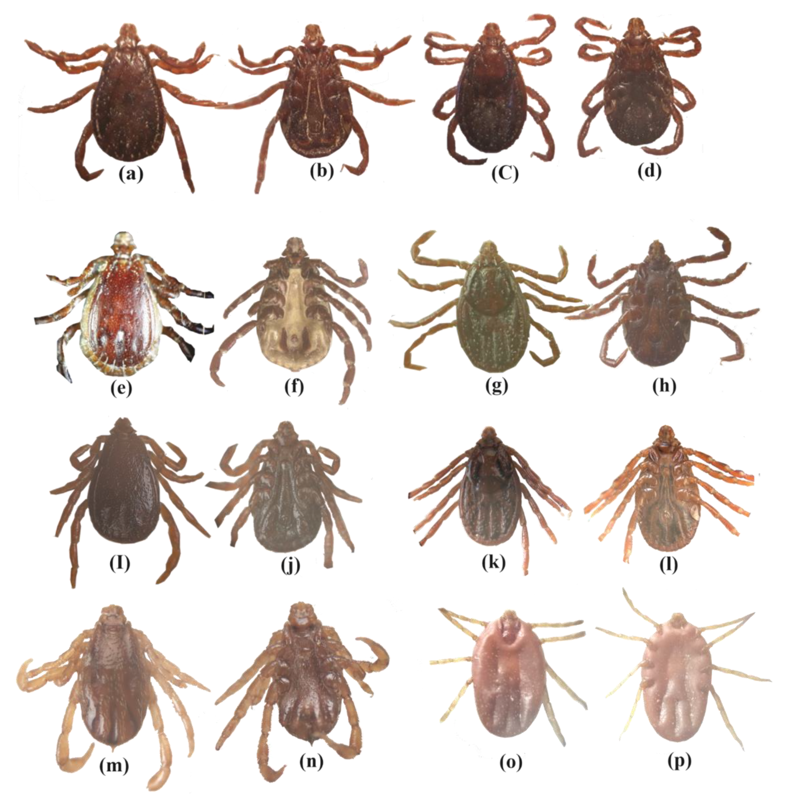
2.1. Ticks’ Morphological Description

The collected ticks were identified according to their distinguishing features, e.g., basis capituli of male Rh. haemaphysaloides bear slightly sharp and pointed cornua and sickle-shaped adanal plates. The female Rh. haemaphysaliodes scutum has nearly the same length as width, with a slightly sinuous posterior margin. The genital opening is narrowly U-shaped on the ventral side. The width of capituli in male Rh. sanguineus sensu lato (s.l) is greater than its length with acutely-curved lateral angles of the basis capituli. Adanal plates are subtriangular or rounded posteriorly. The length of scutum in female Rh. sanguineus is more than its width, having a sinuous posterior margin. The posterior lip genital aperture is broad and U-shaped. Basis capituli of male Rh. turanicus have sharp and pointed cornua with comma-shaped cervical grooves. The adanal plates may be broad and shortened or sharp and longer posteriorly. The posterior margin of the scutum in female Rh. turanicus is distinctly sinuous, having a small genital aperture and U-shaped to broadly V-shaped posterior lip. Male Rh. microplus has a distinct cornua, and ventrally the coxa 1 bears long and distinct spurs. In female Rh. microplus the scutum is pear shaped, with a broader U-shaped genital aperture (Figure 1).

2.2. Tick Infestation and Wild Animals

Among 53 examined wild animals in Charsadda district (10 wild boars), Mardan (10 cats and 10 wild boars), Peshawar (11 cats and 4 hedgehogs), and Swabi (4 cats and 4 hedgehogs), 31 were infested with 531 ticks at different life stages (Table 1). Adult females were the most prevalent (number of ticks 242, corresponding to 46%) followed by males (172, 32%), nymphs (80, 15%) and larvae (37, 7%) (Table 1). The collected ticks were categorized into four species of the genus Rhipicephalus: Rh. haemaphysaloides, Rh. microplus, Rh. sanguineus, and Rh. turanicus. Overall prevalence of tick infestation among wild animals was 58.4%, with the highest tick burden being observed in wild animals of Mardan district (201, 37.7%) followed by Peshawar (153, 28.7%), Charsadda (112, 21.4%), and Swabi (65, 12.2%). A significant difference in tick infestation was observed among wild animals from different districts (p 0.0361). The most prevalent tick species was Rh. turanicus (266, 50%) followed by Rh. microplus (123, 23%), Rh. sanguineus (106, 20%), and Rh. haemaphysaloides (36, 7%). Wild boars were highly infested (268, 50.4%) by ticks including Rh. turanicus (148, 55%), Rh. sanguineus (106, 40%) and Rh. haemaphysaloides (14, 5%). Following wild boars, cats were infested by 145 ticks (27%) including Rh. microplus (123, 85%) and Rh. haemaphysaloides (22, 15%) (Table 1). Hedgehogs were the least infested, with 118 (22.2%) Rh. turanicus ticks. Districtwide tick infestation in wild animals was found to be statistically significant (p 0.0001).

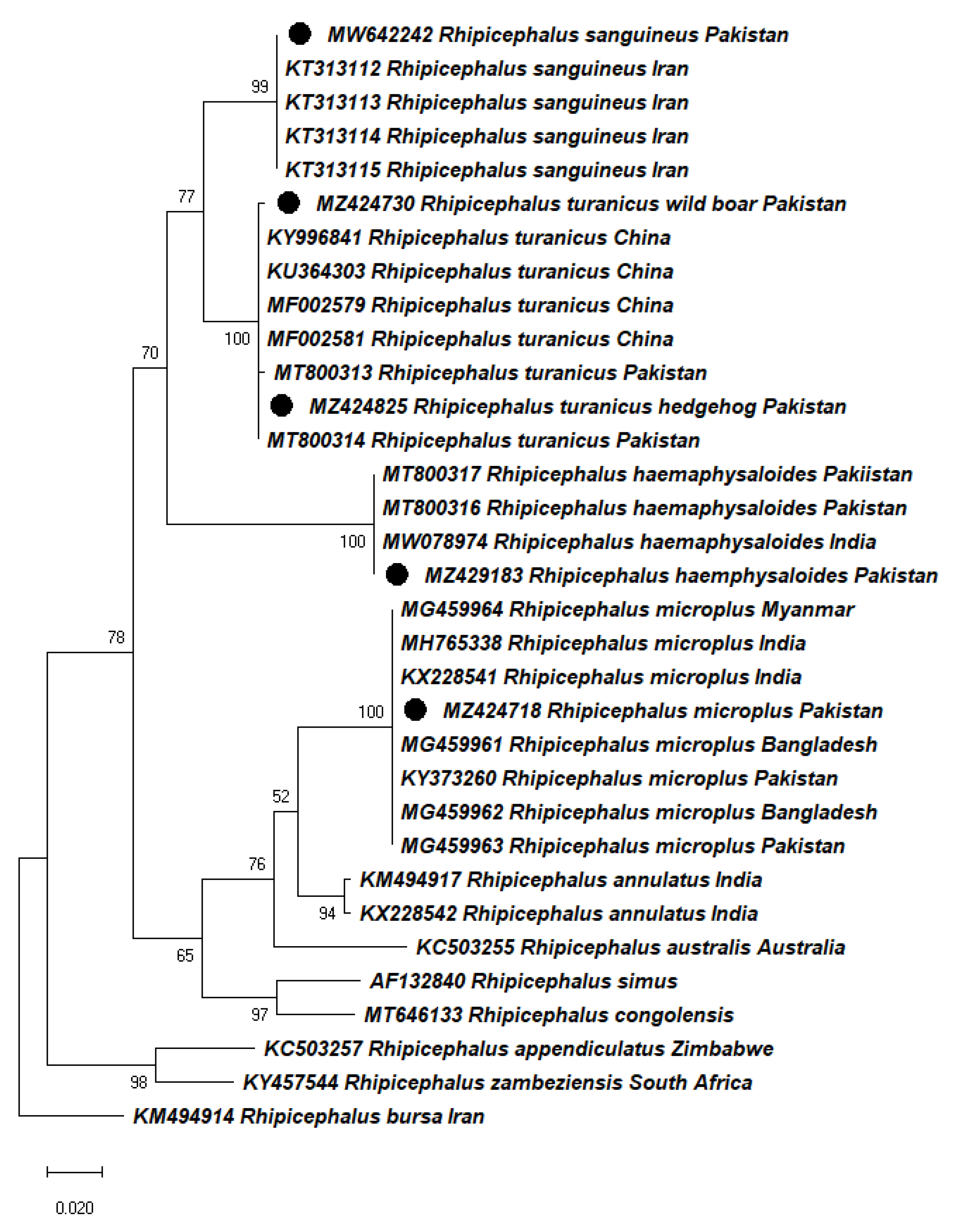
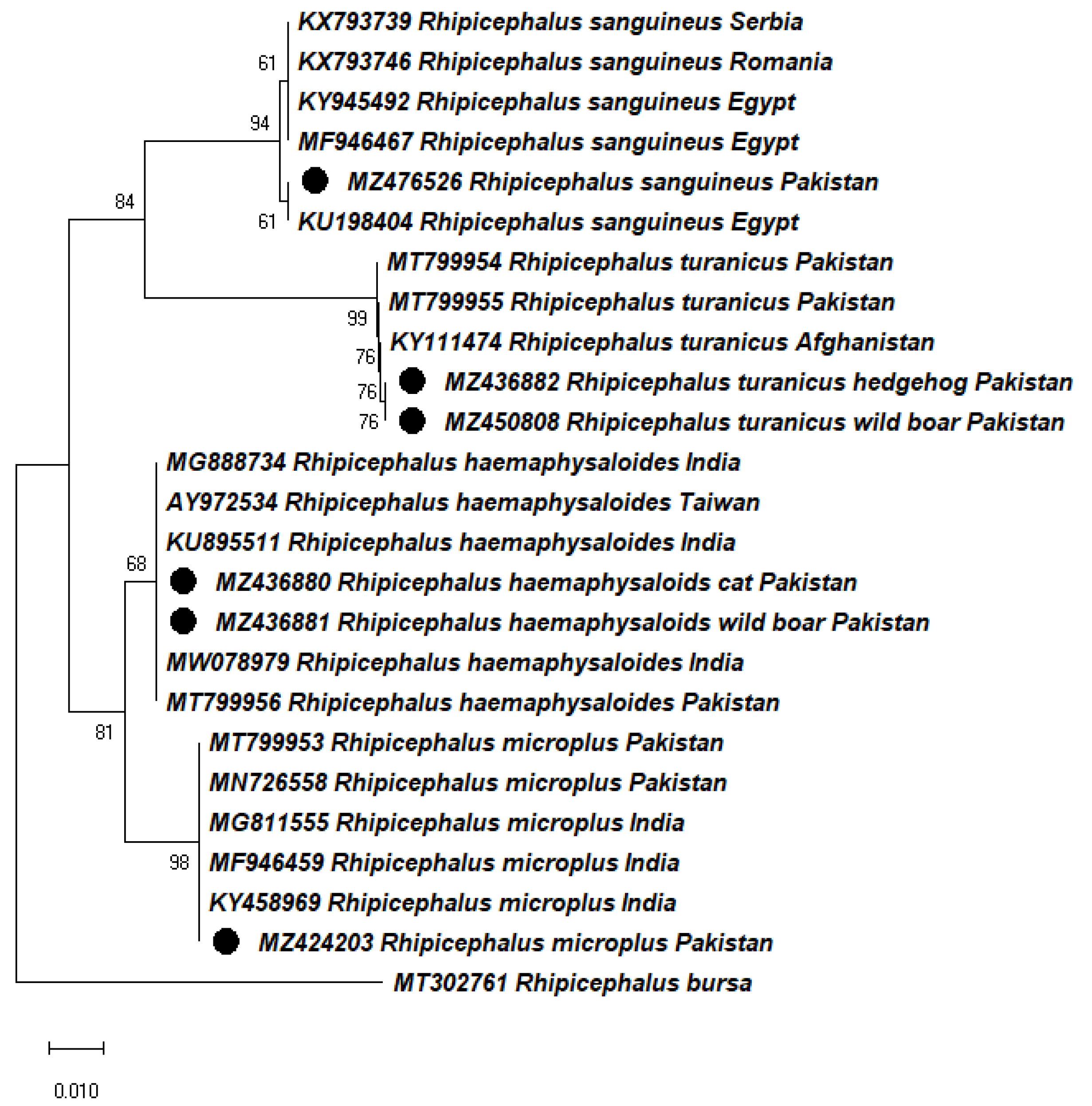
2.3. Molecular Identification and Phylogeny of Ticks

The cox1 sequence fragments obtained from morphologically identified Rh. haemaphysaloides, Rh. sanguineus, Rh. turanicus (wild boar), Rh. turanicus (hedgehog), and Rh. microplus were 521 bp, 603 bp, 778 bp, 659 bp and 780 bp, respectively. The 16S rDNA sequence fragments were 388 bp and 390 bp for Rh. haemaphysaloides from cat and wild boar, respectively; 408 bp and 419 bp for Rh. turanicus from hedgehog and wild boar, respectively; and 340 bp for Rh. sanguineus, and 407 bp for Rh. microplus.
The BLAST analysis of partial cox1 gene sequences from Rh. haemaphysaloides, Rh. turanicus, Rh. sanguineus, and Rh. microplus showed 98-100% identity with sequences of the same species previously reported from Bangladesh, China, Iran, India, Myanmar, and Pakistan. In a phylogenetic tree (Maximum Likelihood), the obtained sequences of Rh. haemaphysaloides clustered with previously reported same-species sequences from India (MW078974) and Pakistan (MT800316, MT800317); Rh. turanicus sequences clustered with reported same-species sequences from China (KY996841, KU364303, MF002579, MF002581), and Pakistan (MT800313, MT800314); Rh. sanguineus sequences clustered with reported same-species sequences from Iran (KT313112, KT313113, KT313114, KT313115); and Rh. microplus sequences clustered with reported same-species sequences from Bangladesh (MG459961, MG459962), India (MH765338, KX228541), Myanmar (MG459964), and Pakistan (KY373260, MG459963) (Figure 2). The sequence data were analyzed by different methods in MEGA-X and similar phylogenetic results were recovered (data not shown).
The BLAST analysis of 16S rDNA sequences showed 97–100% identity with sequences previously reported for the respective tick species from Afghanistan, Egypt, India, Pakistan, Romania, Serbia, and Taiwan. In the phylogenetic tree, the obtained sequences of Rh. haemaphysaloides clustered with the same-species sequences from India (MG888734, KU895511, MW078979), Pakistan (MT799956), and Taiwan (AY972534), Rh. turanicus with Afghanistan (KY111474) and Pakistan (MT799954, MT799955), Rh. sanguineus with Egypt (KY945492, MF946467), Romania (KX793746), and Serbia (KX793739) and Rh. microplus with India (MG811555, MF946459, KY458969) and Pakistan (MT799953, MN726558) (Figure 3). The analysis of cox1 and 16S rRNA gene sequences and subsequent phylogenetic trees supported their monophyly, with identical sequences for each tick species found in previous reports from different countries.
The obtained cox1 and 16S rRNA gene sequences for each tick species were deposited in GenBank under accession numbers: MZ429183 and MZ436880, MZ436881 (Rh. haemaphysaloides), MZ424825, MZ424730, and MZ436882, MZ450808 (Rh. turanicus), MW642242 and MZ476526 (Rh. sanguineus), and MZ424718 and MZ424203 (Rh. microplus).

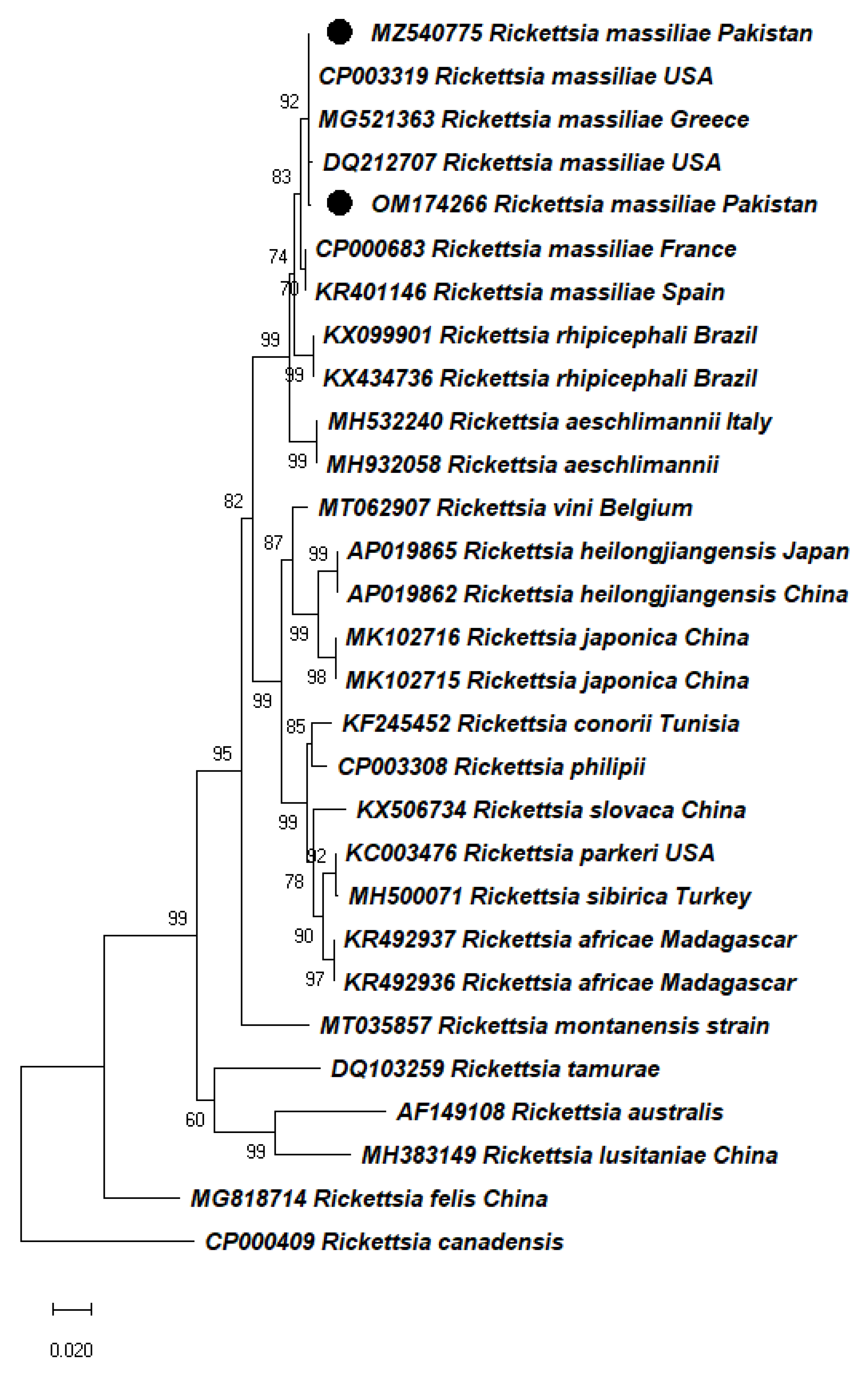
2.4. Detection of Rickettsia spp. in Ticks

Among the 120 ticks screened for Rickettsia spp., Rh. turanicus and Rh. haemaphysaloides collected from wild boars were found positive for rickettsial DNA, as determined by the amplification of both gltA (377 bp) and ompA (576 bp and 503 bp) partial sequences (Table 1). The overall prevalence of Rickettsia spp. was 8.3% (10/120) based on both gltA and ompA genes. The rickettsial DNA sequences were amplified from Rh. turanicus (7/10, 70%) and Rh. haemaphysaloides (3/10, 30%). The presence of Rickettsia spp. in the Charsadda district (6/10, 60%) was detected in Rh. turanicus (3/10, 30%) and Rh. haemaphysaloides (3/10, 30%), while in Mardan district (4/10, 40%) it was found in Rh. turanicus only. Rickettsia spp. was not detected in Rh. microplus and Rh. sanguineus ticks.
BLAST analysis of the obtained gltA gene (Rickettsia) sequences from Rh. turanicus and Rh. haemaphysaloides infesting wild boars showed 99% identity with R. massiliae sequences from China. On the other hand, ompA gene sequences detected in Rh. turanicus and Rh. haemaphysaloides infesting wild boars showed 99-100% identities with previously reported sequences of R. massiliae from France, Greece, Spain, and the USA. In a phylogenetic tree, the obtained sequences clustered with R. massiliae from France (CP000683), Greece (MG521363), Spain (KR401146), and USA (CP003319, DQ212707) (Figure 4). The resulting gltA and ompA sequences for R. massiliae were deposited in GenBank under accession numbers: (OM066912) and (MZ540775 and OM174266), respectively.

3. Discussion

Climate change, urbanization, and other anthropogenic activities have led to the destruction of wildlife habitats, which in turn has increased the chances of interaction between wild and domestic animals [19]. Diverse geographical regions comprising mountainous ranges and agro–wildlife localities serve as habitats for several wildlife species in Pakistan. Studies have been conducted on ticks infesting domestic animals, but research has often neglected ticks infesting wild animals in Pakistan. In this study, we inspected cats, hedgehogs, and wild boars for tick infestation in four districts of KP, Pakistan. The collected ticks were morpho-molecularly identified as Rh. haemaphysaloides, Rh. turanicus, Rh. sanguineus and Rh. microplus and screened for tick-associated Rickettsia species. Rickettsia massiliae was detected in Rh. turanicus and Rh. haemaphysaloides infesting wild boars in the Charsadda and Mardan districts.
Cats infested by ticks can bolster the dispersion of ticks and tick-borne pathogens to predisposed owners and other domestic animals [4]. In the current study, we observed the infestation of Rh. microplus and Rh. haemaphysaloides on cats. Cats and other wild animals generally acquire ticks from natural habitats and their inside access may create a risk of tick infestation to indoor domesticated animals, pets, and humans [4,6]. In this study, Rh. turanicus ticks were found infesting hedgehogs, and other tick species including Rh. haemaphysaloides, Rh. sanguineus, and Rh. turanicus were found infesting wild boars. Rh. turanicus has been previously reported as infesting hedgehogs in Iran [14] and Turkey [13]. Accordingly, Rh. turanicus infestation in hedgehogs, as observed in this study, provides evidence that hedgehogs are not accidental hosts for this tick. In wild boar, Rh. sanguineus and Rh. turanicus infestation has been previously reported in Sri Lanka [19], and Rh. sanguineus in KP, Pakistan [20]. The variety of ticks found infesting wild boar may be due to the free movement of this host and contact with other wild and domestic animals.
Morphological identification of the tick species was confirmed by sequencing fragments of mitochondrial genes (cox1 and 16S rRNA). Using morphology alone is insufficient for the precise identification of tick species due to morphological similarities, the presence of engorged as well as immature stages, and damaged specimens [20,35,36,37]. In several studies, both morphological and molecular identification of ticks have been implemented to achieve accurate taxonomic classification [34,36]. Molecular markers, including mitochondrial cox1 and 16S rRNA, have been reported in the successful determination of the evolution and phylogeny of ticks [37]. Among genetic markers, 16S rRNA and cox1 are useful for understanding interspecific phylogenetic and intraspecific genetic variabilities among ticks [20,37]. In this study, phylogenetic analysis of the identified Rhipicephalus species was performed using cox1 and 16S rDNA partial sequences, which revealed close evolutionary relationship with ticks of the same species reported from Afghanistan, Bangladesh, China, Egypt, India, Iran, Myanmar, Pakistan, Romania, and Taiwan.
Previously, R. massiliae has been detected in Rhipicephalus species including Rh. haemaphysaloides, Rh. microplus, Rh. turanicus [26,34], Rh. sanguineus, Rh. sulcatus, Rh. lunulatus, Rh. muhsamae, and Rh. senegalensis [25,27]. In this study R. massiliae was detected in Rh. haemaphysaloides and Rh. turanicus ticks collected from wild boars. R. massiliae has been described as infecting Rh. turanicus and Rh. sanguineus ticks collected from wild boars [26,38]. To date, there has been a lack of information about the detection of R. massiliae in Rh. haemaphysaloides ticks infesting wild boars. In Pakistan, the presence of R. massiliae was reported in Rh. microplus, Rh. haemaphysaloides, Hya. anatolicum and Hya. hussaini [24,34]. Free roaming of tick-infested wild boars into human residential areas can enhance the exposure of domestic animals and humans to rickettsial infection [17]. Rh. turanicus has been implicated as a vector of several medically important pathogens, such as Babesia spp., Theileria spp., Anaplasma spp., and Rickettsia spp. [14,39,40]. Rh. haemaphysaloides has been found infected with multiple pathogens comprising Anaplasma spp., Babesia spp., Rickettsia spp., Borrelia spp., and Ehrlichia spp. [34,41,42,43]. An increase in the free movement of tick-infested wild animals toward urban and suburban areas have resulted in the transmission of tick-associated Rickettsia spp. from wild animals to humans, pet animals, and livestock [19]. Therefore, the so-far neglected surveillance of tick-borne pathogens in ticks parasitizing wild animals demands immediate attention.

4. Materials and Methods

4.1. Ethical Approval

The experimental design of the present study was approved by the Advance Studies Research Board members of Abdul Wali Khan University, Pakistan (Dir/A&R/AWKUM/2018/1410).

4.2. Study Area

The rural areas of the Charsadda, Mardan, Peshawar, and Swabi districts were selected for the collection of wild animals, including cats, Indian hedgehogs, and wild boars, during 2017–2021. The study area comprising selected districts in the KP northern province have their highest (33.4 °C) and lowest (11.7 °C) mean temperatures in July and December, respectively (climate-data.org) accessed on 27 May 2021. The exact geographical coordinates of sample locations were obtained using Global Positioning System (GPS) and added to the attribute table for tagging on the study area map using ArcGIS v. 10.3 (Figure 5).

4.3. Tick Collection and Morphological Identification

Wild animals including cats, hedgehogs, and wild boars found dead on highways, killed or captured by local farmers to secure their crops, were screened for ticks. Ticks found on the host body were carefully collected to avoid any damage to the specimens. All collected ticks were preserved in 100% ethanol. Morphological identification of the collected ticks was done using morphological features under Stereozoom microscope (BIOBASE, Jinan, China), by comparing with standard available morpho-taxonomic keys [44,45].

4.4. DNA Extraction and PCR

All ticks were morphologically identified, and 120 ticks comprising 10 specimens (different life stages) of each species from all districts were further processed for genomic DNA extraction (Table 1). Ticks were washed with distilled water followed by 70% ethanol and PBS for the removal of any surface contaminants. Washed ticks were individually kept in 1.5 ml tubes and dried in an incubator. Holes were made with needles, and the whole body of each tick was cut into small pieces using sterile scissors and homogenized by micro pestle for DNA extraction using phenol chloroform method [46]. The concentration of extracted DNA was measured using NanoQ (Optizen, Daejeon, South Korea), and samples were maintained at -20 ℃ for further analysis.
Mitochondrial cytochrome C oxidase 1 (cox1) and 16S ribosomal RNA (16S rRNA) genes’ partial sequences were amplified for the molecular identification of ticks. The PCR was performed in a total volume of 25 µL reaction mixture comprised of 1 µL each forward and reverse primers (10 µM), 2 µL template DNA (50 ng), 8.5 µL PCR water, and 12.5 µL DreamTaq PCR Master Mix (2×) (Thermo Scientific, Waltham, MA, USA). Primers used in the present study are given in Table 2, and thermocycling conditions were set as previously described [47,48].

4.5. Detection of Rickettsia

All extracted genomic DNA samples were screened for the presence of any Rickettsia spp. targeting the amplification of rickettsial citrate synthase (gltA) and outer membrane protein (ompA) partial genes. The PCR reaction was performed in a total volume of 25 µL reaction mixture comprised of 1 µL each forward and reverse primers (10 µM), 2 µL template DNA (50 ng), 8.5 µL PCR water, and 12.5 µL DreamTaq PCR Master Mix (2×) (Thermo Scientific, Waltham, MA, USA). Primers used in the present study are given in (Table 2), and thermocycling conditions were set as previously described [49,50]. All genomic DNA samples that yielded visible amplicons for gltA PCR were subjected to second PCR assay for the amplification of ompA gene. The amplified PCR products were electrophoresed on 1.5% agarose gel and results were visualized under UV light using a GelDoc (UVP BioDoc-It imaging system, Upland, CA, USA).

4.6. DNA Purification and Sequencing

Prior to sequencing, the positive PCR products were purified with GeneClean II DNA purification Kit (Qbiogene, Illkirch, France) following the protocol provided by the manufacturer. All 120 purified PCR products for each cox1 and 16S rRNA gene of ticks, and 10 positive samples for each gltA and ompA of Rickettsia spp. were sent for bidirectional sequencing (Macrogen Inc., Seoul, South Korea).

4.7. Phylogenetic Analysis

The obtained sequences were trimmed in SeqMan V. 5.00 (DNASTAR) for the removal of unnecessary nucleotides and primer contamination. Redundant sequences (100% identity) were excluded from further analysis. Sequences with maximum identities were retrieved from NCBI (National Center for Biotechnology Information) using BLAST (Basic Local Alignment Search Tool) [51]. The obtained sequences were aligned in BioEdit V. 7.0.5 [52]. Phylogenetic trees were constructed in MEGA X [53], and different phylogenetic methods (Maximum likelihood, Neighbor-Joining, Minimum-Evolution, Parsimony, and UPGMA) were tested for consistency, efficiency, and robustness. The Maximum likelihood method was used for the phylogenetic tree, with bootstrap 1000 replicates, and an outgroup was used for estimating tree stability and validity, respectively. Finally, the sequences of cox1, 16S rDNA, gltA and ompA were submitted to NCBI.

4.8. Statistical Analysis

The recorded data was organized in spreadsheets using Microsoft Excel V. 2016 (Microsoft). A chi-square test was performed using GraphPad prism software V. 5.00 (GraphPad Software Inc) considering a significant p value < 0.05.

5. Conclusions

The present study reported tick infestation in wild animals in KP, Pakistan, and for the first-time detected R. massiliae in Rh. turanicus and Rh. haemaphysaloides ticks infesting wild boars in Charsadda and Mardan. These results improve our knowledge of the circulation of R. massiliae in Rhipicephalus ticks infesting both domestic and wild animals. These findings reinforce the need to further understand the diversity of ticks infesting wild animals, tick-associated Rickettsia spp. and other pathogens across the country.

Author Contributions

A.A., I.d.S.V.J. and T.T. experimental design of the study. A.A., S.S., H.Z., F.U., I.Z. and H.A. collected the tick samples. A.A., S.S., H.Z., F.U. and I.Z. performed the experiments. I.Z. and H.A. perform statistical analysis. All authors have read and agreed to the published version of the manuscript.

Funding

The authors are thankful for the financial support provided by Pakistan Science Foundation and Higher Education Commission of Pakistan and CAPES and CNPq (Brazil). This work was supported by the JST Adaptable and Seamless Technology Transfer Program through Target-driven R&D (A-STEP) Grant Number JPMJTM20SV.

Institutional Review Board Statement

The experimental design of the present study was approved by the Advance Studies Research Board members of Abdul Wali Khan University, Pakistan (Dir/A&R/AWKUM/2018/1410).

Data Availability Statement

Details regarding data supporting reported results can be found https://www.ncbi.nlm.nih.gov/nuccore/?term= (accessed on 26 December 2021).

Conflicts of Interest

The authors declare no conflict of interest.

References

  1. Clark, N.J.; Sedden, J.M.; Slapeta, J.; Wells, K. Parasite spread at the domestic animal- wildlife interface: Anthropogenic habitat use, phylogeny and body mass drive risk of cat and dog flea (Ctenocephlides spp.) infestation in wild mammals. Parasit. Vectors 2018, 11, 8. [Google Scholar] [CrossRef] [PubMed]
  2. Daszak, P.; Cunningham, A.A.; Hyatt, A.D. Emerging infectious diseases of wildlife--threats to biodiversity and human health. Science 2000, 287, 443–449. [Google Scholar] [CrossRef] [PubMed]
  3. Davies, S.; Abdullah, S.; Helps, C.; Tasker, S.; Newbury, H.; Wall, R. Prevalence of ticks and tick-borne pathogens: Babesia and Borrelia species in ticks infesting cats of Great Britain. Vet. Parasitol. 2017, 244, 129–135. [Google Scholar] [CrossRef] [PubMed] [Green Version]
  4. Little, S.E.; Barrett, A.W.; Nagamori, Y.; Herrin, B.H.; Normile, D.; Heaney, K.; Armstrong, R. Ticks from cats in the United States: Patterns of infestation and infection with pathogens. Vet. Parasitol. 2018, 257, 15–20. [Google Scholar] [CrossRef]
  5. Ghosh, P.; Saleh, M.N.; Sundstrom, K.D.; Ientile, M.; Little, S.E. Ixodes spp. from dogs and cats in the United States: Diversity, seasonality, and prevalence of Borrelia burgdorferi and Anaplasma phagocytophilum. Vector-Borne Zoonotic Dis. 2021, 21, 11–19. [Google Scholar] [CrossRef] [PubMed]
  6. Saleh, M.N.; Sundstrom, K.D.; Duncan, K.T.; Ientile, M.M.; Jordy, J.; Ghosh, P.; Little, S.E. Show us your ticks: A survey of ticks infesting dogs and cats across the USA. Parasit. Vectors 2019, 12, 595. [Google Scholar] [CrossRef] [PubMed]
  7. Karim, S.; Budachetri, K.; Mukherjee, N.; Williams, J.; Kausar, A.; Hassan, M.J.; Adamson, S.; Dowd, S.E.; Apanskevich, D.; Arijo, A.; et al. A study of ticks and tick-borne livestock pathogens in Pakistan. PLoS Negl. Trop. Dis. 2017, 11, 0005681. [Google Scholar] [CrossRef] [PubMed]
  8. Shimada, Y.; Inokuma, H.; Beppu, T.; Okuda, M.; Onishi, T. Survey of ixodid tick species on domestic cats in Japan. Vet. Parasitol. 2003, 111, 231–239. [Google Scholar] [CrossRef]
  9. Iwakami, S.; Ichikawa, Y.; Inokuma, H. A nationwide survey of ixodid tick species recovered from domestic dogs and cats in Japan in 2011. Ticks Tick-Borne Dis. 2014, 5, 771–779. [Google Scholar] [CrossRef]
  10. Duplan, F.; Davies, S.; Filler, S.; Abdullah, S.; Keyte, S.; Newbury, H.; Helps, C.R.; Wall, R.; Tasker, S. Anaplasma phagocytophilum, Bartonella spp., haemoplasma species and Hepatozoon spp. in ticks infesting cats: A large-scale survey. Parasit. Vectors 2018, 11, 201. [Google Scholar] [CrossRef]
  11. Nguyen, V.L.; Colella, V.; Greco, G.; Fang, F.; Nurcahyo, W.; Hadi, U.K.; Venturina, V.; Tong, K.B.Y.; Tsai, Y.L.; Taweethavonsawat, P.; et al. Molecular detection of pathogens in ticks and fleas collected from companion dogs and cats in East and Southeast Asia. Parasit. Vectors 2020, 13, 420. [Google Scholar] [CrossRef] [PubMed]
  12. Földvári, G.; Rigó, K.; Jablonszky, M.; Biró, N.; Majoros, G.; Molnár, V.; Tóth, M. Ticks and the city: Ectoparasites of the Northern white-breasted hedgehog (Erinaceus roumanicus) in an urban park. Ticks Tick-Borne Dis. 2011, 2, 231–234. [Google Scholar] [CrossRef] [PubMed]
  13. Goz, Y.; Yilmaz, A.B.; Aydin, A.; Dicle, Y. Ticks and fleas infestation on east hedgehogs (Erinaceus concolor) in Van Province, Eastern Region of Turkey. J. Arthropod. Borne Dis. 2016, 10, 50. [Google Scholar] [PubMed]
  14. Khodadadi, N.; Nabavi, R.; Sarani, A.; Saadati, D.; Ganjali, M.; Mihalca, A.D.; Otranto, D.; Sazmand, A. Identification of Anaplasma marginale in long-eared hedgehogs (Hemiechinus auritus) and their Rhipicephalus turanicus ticks in Iran. Ticks Tick-Borne Dis. 2021, 12, 101641. [Google Scholar] [CrossRef] [PubMed]
  15. Skuballa, J.; Oehme, R.; Hartelt, K.; Petney, T.; Bucher, T.; Kimmig, P.; Taraschewski, H. European hedgehogs as hosts for Borrelia spp., Germany. Emerg. Infect. Dis. 2007, 13, 952. [Google Scholar] [CrossRef]
  16. Silaghi, C.; Skuballa, J.; Thiel, C.; Pfister, K.; Petney, T.; Pfaffle, M.; Taraschewski, H.; Passos, L.M. The European hedgehog (Erinaceus europaeus)–A suitable reservoir for variants of Anaplasma phagocytophilum? Ticks Tick-Borne Dis. 2012, 3, 49–54. [Google Scholar] [CrossRef]
  17. Sgroi, G.; Iatta, R.; Lia, R.P.; D’Alessio, N.; Manoj, R.R.S.; Veneziano, V.; Otranto, D. Spotted fever group Rickettsiae in Dermacentor marginatus from wild boars in Italy. Transbound. Emerg. Dis. 2020, 68, 2111–2120. [Google Scholar] [CrossRef]
  18. Ortuno, A.; Quesada, M.; Lopez, S.; Miret, J.; Cardenosa, N.; Castella, J.; Anton, E.; Segura, F. Prevalence of Rickettsia slovaca in Dermacentor marginatus ticks removed from wild boar (Sus scrofa) in northeastern Spain. Ann. N. Y. Acad. Sci. 2006, 1078, 324–327. [Google Scholar] [CrossRef]
  19. Liyanaarachchi, D.R.; Rajakaruna, R.S.; Dikkumbura, A.W.; Rajapakse, R.P.V.J. Ticks infesting wild and domestic animals and humans of Sri Lanka with new host records. Acta Trop. 2015, 142, 64–70. [Google Scholar] [CrossRef]
  20. Ali, A.; Khan, M.A.; Zahid, H.; Yaseen, P.M.; Khan, M.Q.; Nawab, J.; Rehman, Z.U.; Ateeq, M.; Khan, S.; Ibrahim, M. Seasonal dynamics, record of ticks infesting humans, wild and domestic animals and molecular phylogeny of Rhipicephalus microplus in Khyber Pakhtunkhwa Pakistan. Front. Physiol. 2019, 10, 793. [Google Scholar] [CrossRef] [Green Version]
  21. Ciebiera, O.; Lopinska, A.; Gabrys, G. Ticks on game animals in the fragmented agricultural landscape of western Poland. Parasitol. Res. 2021, 120, 1781–1788. [Google Scholar] [CrossRef] [PubMed]
  22. Hrazdilova, K.; Lesiczka, P.M.; Bardon, J.; Vyroubalova, S.; Simek, B.; Zurek, L.; Modry, D. Wild boar as a potential reservoir of zoonotic tick-borne pathogens. Ticks Tick-Borne Dis. 2021, 12, 101558. [Google Scholar] [CrossRef] [PubMed]
  23. Lim, F.S.; Khoo, J.J.; Tan, K.K.; Zainal, N.; Loong, S.K.; Khor, C.S.; AbuBakar, S. Bacterial communities in Haemaphysalis, Dermacentor and Amblyomma ticks collected from wild boar of an Orang Asli Community in Malaysia. Ticks Tick-Borne Dis. 2020, 11, 101352. [Google Scholar] [CrossRef] [PubMed]
  24. Ghafar, A.; Cabezas-Cruz, A.; Galon, C.; Obregon, D.; Gasser, R.B.; Moutailler, S.; Jabbar, A. Bovine ticks harbour a diverse array of microorganisms in Pakistan. Parasit. Vectors 2020, 13, 1. [Google Scholar] [CrossRef] [PubMed] [Green Version]
  25. Eremeeva, M.E.; Bosserman, E.A.; Demma, L.J.; Zambrano, M.L.; Blau, D.M.; Dasch, G.A. Isolation and identification of Rickettsia massiliae from Rhipicephalus sanguineus ticks collected in Arizona. Appl. Environ. Microbiol. 2006, 72, 5569–5577. [Google Scholar] [CrossRef] [Green Version]
  26. Keysary, A.; Eremeeva, M.E.; Leitner, M.; Din, A.B.; Wikswo, M.E.; Mumcuoglu, K.Y.; Inbar, M.; Wallach, A.D.; Shanas, U.; King, R.; et al. Spotted fever group rickettsiae in ticks collected from wild animals in Israel. Am. J. Trop. Med. Hyg. 2011, 85, 919. [Google Scholar] [CrossRef] [Green Version]
  27. Olivieri, E.; Wijnveld, M.; Bonga, M.; Berger, L.; Manfredi, M.T.; Veronesi, F.; Jongejan, F. Transmission of Rickettsia raoultii and Rickettsia massiliae DNA by Dermacentor reticulatus and Rhipicephalus sanguineus (s.l.) ticks during artificial feeding. Parasit. Vectors 2018, 11, 494. [Google Scholar] [CrossRef]
  28. Fernandez-Soto, P.; Perez-Sanchez, R.; Martin, V.D.; Encinas-Grandes, A.; Sanz, R.A. Rickettsia massiliae in ticks removed from humans in Castilla y León, Spain. Eur. J. Clin. Microbiol. Infect. Dis. 2006, 25, 811–813. [Google Scholar] [CrossRef]
  29. Garcia-Garcia, J.C.; Portillo, A.; Nunez, M.J.; Santibanez, S.; Castro, B.; Oteo, J.A. A patient from Argentina infected with Rickettsia massiliae. Am. J. Trop. Med. Hyg. 2010, 82, 691–692. [Google Scholar] [CrossRef] [Green Version]
  30. Ali, A.; Mulenga, A.; Vaz Jr, I. Tick and Tick-Borne Pathogens: Molecular and Immune Targets for Control Strategies. Front. Physiol. 2020, 11, 00744. [Google Scholar] [CrossRef]
  31. Kamran, K.; Ali, A.; Villagra, C.; Siddiqui, S.; Alouffi, A.S.; Iqbal, A. A cross-sectional study of hard ticks (acari: Ixodidae) on horse farms to assess the risk factors associated with tick-borne diseases. Zoonoses Public Health 2021, 68, 247–262. [Google Scholar] [CrossRef] [PubMed]
  32. Kamran, K.; Ali, A.; Villagra, C.A.; Bazai, Z.A.; Iqbal, A.; Sajid, M.S. Hyalomma anatolicum resistance against ivermectin and fipronil is associated with indiscriminate use of acaricides in southwestern Balochistan, Pakistan. Parasitol. Res. 2021, 120, 15–25. [Google Scholar] [CrossRef] [PubMed]
  33. Zahid, H.; Munoz-Leal, S.; Khan, M.Q.; Alouffi, A.S.; Labruna, M.B.; Ali, A. Life Cycle and genetic identification of Argas persicus Infesting Domestic Fowl in Khyber Pakhtunkhwa, Pakistan. Front. Vet. Sci. 2021, 8, 302. [Google Scholar] [CrossRef] [PubMed]
  34. Ali, A.; Zahid, H.; Zeb, I.; Tufail, M.; Khan, S.; Haroon, M.; Tufail, M.; Bilal, M.; Hussain, M.; Alouffi, S.A.; et al. Risk Factors Associated with Tick Infestations on Equids in Khyber Pakhtunkhwa, Pakistan, with Notes on Rickettsia massiliae Detection. Parasit. Vectors 2021, 14, 363. [Google Scholar] [CrossRef]
  35. Ali, A.; Parizi, L.F.; Ferreira, B.R.; Vaz, I., Jr. A revision of two distinct species of Rhipicephalus: R. microplus and R. australis. Cienc. Rural 2016, 46, 1240–1248. [Google Scholar] [CrossRef] [Green Version]
  36. Estrada-Pena, A.; D’Amico, G.; Palomar, A.M.; Dupraz, M.; Fonville, M.; Heylen, D.; Habela, M.A.; Hornok, S.; Lempereur, L.; Madder, M.; et al. A comparative test of ixodid tick identification by a network of European researchers. Ticks Tick-Borne Dis. 2017, 8, 540–546. [Google Scholar] [CrossRef] [Green Version]
  37. Zhao, L.; Lv, J.; Li, F.; Li, K.; He, B.; Zhang, L.; Han, X.; Wang, H.; Johnson, N.; Lin, X.; et al. Identification and molecular analysis of ixodid ticks (Acari: Ixodidae) infesting domestic animals and tick-borne pathogens at the Tarim Basin of Southern Xinjiang, China. Korean J. Parasitol. 2020, 58, 37. [Google Scholar] [CrossRef] [Green Version]
  38. Castillo-Contreras, R.; Magen, L.; Birtles, R.; Varela-Castro, L.; Hall, J.L.; Conejero, C.; Aguilar, X.F.; Colom-Cadena, A.; Lavín, S.; Mentaberre, G.; et al. Ticks on wild boar in the metropolitan area of Barcelona (Spain) are infected with spotted fever group rickettsiae. Transbound. Emerg. Dis. 2021, 1–14. [Google Scholar] [CrossRef]
  39. Eremeeva, M.E.; Stromdahl, E.Y. New spotted fever group Rickettsia in a Rhipicephalus turanicus tick removed from a child in eastern Sicily, Italy. Am. J. Trop. Med. Hyg. 2011, 84, 99–101. [Google Scholar] [CrossRef]
  40. Jahfari, S.; Ruyts, S.C.; Frazer-Mendelewska, E.; Jaarsma, R.; Verheyen, K.; Sprong, H. Melting pot of tick-borne zoonoses: The European hedgehog contributes to the maintenance of various tick-borne diseases in natural cycles urban and suburban areas. Parasit. Vectors 2017, 10, 134. [Google Scholar] [CrossRef] [Green Version]
  41. Zhang, J.; Liu, Q.; Wang, D.; Li, W.; Beugnet, F.; Zhou, J. Epidemiological survey of ticks and tick-borne pathogens in pet dogs in south-eastern China. Parasite 2017, 24, 35. [Google Scholar] [CrossRef] [PubMed]
  42. Kuo, C.C.; Huang, J.L.; Chien, C.H.; Shih, H.C.; Wang, H.C. First molecular detection of Anaplasma phagocytophilum in the hard tick Rhipicephalus haemaphysaloides in Taiwan. Exp. Appl. Acarol. 2018, 75, 437–443. [Google Scholar] [CrossRef] [PubMed]
  43. Fang, L.Q.; Liu, K.; Li, X.L.; Liang, S.; Yang, Y.; Yao, H.W.; Sun, R.X.; Sun, Y.; Chen, W.J.; Zuo, S.Q.; et al. Emerging tick-borne infections in mainland China: An increasing public health threat. Lancet Infect. Dis. 2015, 15, 1467–1479. [Google Scholar] [CrossRef] [Green Version]
  44. Walker, A. The Genus Rhipicephalus (Acari, Ixodidae): A Guide to the Brown Ticks of the World; Walker, J.B., Keirans, J.E., Horak, I.G., Eds.; Kluwer Academic Publishers: Drodrecht, The Netherlands, 2000; pp. 417–418. [Google Scholar]
  45. Roy, B.C.; Estrada-Pena, A.; Krucken, J.; Rehman, A.; Nijhof, A.M. Morphological and phylogenetic analyses of Rhipicephalus microplus ticks from Bangladesh, Pakistan and Myanmar. Ticks Tick-Borne Dis. 2018, 9, 1069–1079. [Google Scholar] [CrossRef] [PubMed]
  46. Sambrook, J.; Russell, D.W. Molecular Cloning: A Laboratory Manual, 3rd ed.; Cold Spring Harbor Laboratory Press: Long Island, NY, USA, 2001; pp. 23–44. [Google Scholar]
  47. Chitimia, L.; Lin, R.Q.; Cosoroaba, I.; Wu, X.Y.; Song, H.Q.; Yuan, Z.G.; Zhu, X.Q. Genetic characterization of ticks from southwestern Romania by sequences of mitochondrial cox 1 and nad 5 genes. Exp. Appl. Acarol. 2010, 52, 305–311. [Google Scholar] [CrossRef]
  48. Mangold, A.J.; Bargues, M.D.; Mas-Coma, S. Mitochondrial 16S rDNA sequences and phylogenetic relationships of species of Rhipicephalus and other tick genera among Metastriata (Acari: Ixodidae). Parasitol. Res. 1998, 84, 478–484. [Google Scholar] [CrossRef]
  49. Labruna, M.B.; Whitworth, T.; Horta, M.C.; Bouyer, D.H.; McBride, J.W.; Pinter, A.; Popov, V.; Gennari, S.M.; Walker, D.H. Rickettsia species infecting Amblyomma cooperi ticks from an area in the state of Sao Paulo, Brazil, where Brazilian spotted fever is endemic. J. Clin. Microbiol. 2004, 42, 90–98. [Google Scholar] [CrossRef] [Green Version]
  50. Roux, V.; Fournier, P.E.; Raoult, D. Differentiation of spotted fever group rickettsiae by sequencing and analysis of restriction fragment length polymorphism of PCR-amplified DNA of the gene encoding the protein rOmpA. J. Clin. Microbiol. 1996, 34, 2058–2065. [Google Scholar] [CrossRef] [Green Version]
  51. Altschul, S.F.; Gish, W.; Miller, W.; Myers, E.W.; Lipman, D.J. Basic local alignment search tool. J. Mol. Biol. 1990, 215, 403–410. [Google Scholar] [CrossRef]
  52. Hall, T.; Biosciences, I.; Carlsbad, C. BioEdit: An important software for molecular biology. GERF Bull. Biosci. 2011, 2, 60–61. [Google Scholar]
  53. Kumar, S.; Stecher, G.; Li, M.; Knyaz, C.; Tamura, K. MEGA X: Molecular evolutionary genetics analysis across computing platforms. Mol. Biol. Evol. 2018, 35, 1547–1549. [Google Scholar] [CrossRef] [PubMed]
Figure 1. Rhipicephalus haemaphysaliodes: (a) male dorsal, (b) male ventral, (c) female dorsal, (d) female ventral; Rhipicephalus sanguineus: (e) male dorsal, (f) male ventral, (g) female dorsal, (h) female ventral; Rhipicephalus turanicus: (i) male dorsal, (j) male ventral, (k) female dorsal, (l) female ventral; Rhipicephalus microplus: (m) male dorsal, (n) male ventral, (o) female dorsal, (p) female ventral.
Pathogens 11 00162 g001
Figure 2. Phylogenetic tree (Maximum Likelihood) based on cox1 gene partial sequences of the collected ticks Rh. haemaphysaloides, Rh. turanicus, Rh. sanguineus, and Rh. microplus in Khyber Pakhtunkhwa (KP) Pakistan. Rhipicephalus bursa was used as an outgroup. Bootstrap values are presented at each node (1000). GenBank accession numbers are followed by species name and country of collection at each terminal taxon. Sequences obtained in this study are labeled with black circles.
Pathogens 11 00162 g002
Figure 3. Phylogenetic (Maximum Likelihood) tree based on 16S rRNA gene partial sequences of Rh. haemaphysaloides, Rh. turanicus, Rh. sanguineus, and Rh. microplus of Khyber Pakhtunkhwa (KP) Pakistan. Rhipicephalus bursa was used as an outgroup. Bootstrap values are presented at each node (1000). GenBank accession numbers are followed by species name and country of collection. Sequences obtained in the present study are labeled with black circles.
Pathogens 11 00162 g003
Figure 4. Maximum likelihood tree inferred from partial sequences of the ompA gene for Rickettsia spp. Rickettsia canadensis was used as an outgroup. Bootstrap values are presented at each node (1000). Accession numbers are followed by species and country name. Sequences obtained in the present study were labeled with black circles.
Pathogens 11 00162 g004
Figure 5. Vegetation map of the study area, indicating locations where different ticks were collected in each district of Khyber Pakhtunkhwa (KP), Pakistan.
Pathogens 11 00162 g005
Table 1. Abundance of ticks, and screening for rickettsial DNA associated with ticks infesting wild animals.
DistrictsHostTick SpeciesExamined Hosts (%)Infested Hosts (%)Collected Ticks
(%)
Tick Life StagesTicks Molecularly Analyzed *Rickettsia gltA and ompA
MardanCatsRh. microplus
Rh. haemaphysaloides
10 (18.8)4 (40)33 (73.3)
12 (36.6)
14F, 11M, 5N, 3L8F, 2N0
5F, 4M, 3N5F, 2M, 3N0
Wild boarRh. turanicus
Rh. sanguineus
10 (18.8)9 (90)90 (57.6)
66 (42.4)
32F, 28M, 21N, 9L6F, 4N4
27F, 19M, 13N, 7L7F, 3N
PeshawarCatsRh. microplus11 (20.7)6 (54.5)55 (36)18F, 15M, 12N, 10L8F, 2N0
HedgehogsRh. turanicus4 (7.54)2 (50)98 (64)64F, 34M10F0
CharsaddaWild boarRh. turanicus
Rh. sanguineus
Rh. haemaphysaloides
10 (18.8)5 (50)58 (52)
40 (36)
14 (12)
21F, 17M, 14N, 6L
19F, 17M, 4N
9F, 1N
8F, 2N
3
0
6F, 4M, 2N, 2L6F, 2M, 2N3
SwabiCatsRh. microplus4 (7.54)3 (75)35 (54)19F, 12M, 4N8F, 2N0
Rh. haemaphysaloides10 (15.3)5F, 3M, 2N5F,3M,2N0
HedgehogsRh. turanicus4 (7.54)2 (50)20 (30.7)12F, 8M6F, 4M0
Total 53 (100)31 (58.4)531 (Mean 44.25)242F, 172M, 80N, 37L86F, 11M, 23N
Total: 120
10(8.3%)
Note: F = Adult females, M = males, N = nymphs, L = larvae, * ticks molecularly tested and screened for Rickettsia spp.
Table 2. Primers used for the amplification of ticks and rickettsial DNA.
OrganismGenePrimerSequenceAmplicon bpReferences
Tickcox 1cox1FGGAACAATATATTTAATTTTTGG850[47]
cox1RATCTATCCCTACTGTAAATATATG
16S16S+1CCGGTCTGAACTCAGATCAAGT460[48]
16S-1GCTCAATGATTTTTTAAATTGCTGT
Rickettsia spp.gltACS-78GCAAGTATCGGTGAGGATGTAAT401[49]
CS-323GCTTCCTTAAAATTCAATAAATCAGGAT
ompARrl9O.70ATGGCGAATATTTCTCCAAAA631[50]
Rr190.701nGTTCCGTTAATGGCAGCATCT
Publisher’s Note: MDPI stays neutral with regard to jurisdictional claims in published maps and institutional affiliations.

