Article

Pb103 Regulates Zygote/Ookinete Development in Plasmodium berghei via Double Zinc Finger Domains

Department of Tropical Medicine and Parasitology, Faculty of Medicine, Juntendo University, 2-1-1 Hongo Bunkyo-ku, Tokyo 113-8421, Japan
*
Author to whom correspondence should be addressed.
Equal contribution.
Pathogens 2021, 10(12), 1536; https://doi.org/10.3390/pathogens10121536
Submission received: 4 November 2021 / Revised: 22 November 2021 / Accepted: 22 November 2021 / Published: 24 November 2021
(This article belongs to the Collection Feature Papers on Parasitic Pathogens)

Abstract

:
Sexual reproduction of Plasmodium parasites takes place in anopheline mosquitoes, where male and female gametes fuse to form zygotes and then ookinetes. These processes are orchestrated by stage-specific protein expression, which is mediated in part by translational repression. Accumulating evidence shows that RNA binding proteins (RBPs) play crucial roles in these processes. Here, we report the characterization of P. berghei 103 (Pb103), which encodes a protein possessing double zinc finger domains (ZFs), an RBP. Reporter parasites expressing azami green fluorescent protein (AGFP) under the endogenous Pb103 gene promoter (Pb103-AGFP reporter) showed that the AGFP fluorescent signal was detected from gametes to ookinetes, while AGFP mRNA was translationally repressed in female gametocytes. The Pb103-disrupted parasites (Pb103(−)) grew and produced gametocytes with similar efficiencies to those of wild-type parasites. However, no oocysts were formed in mosquitoes fed Pb103(−). An in vitro fertilization assay showed abortion at the zygote stage in Pb103(−), suggesting that Pb103 plays a critical role in zygote/ookinete development. Cross-fertilization assays with Pb103(−) and male- or female-sterile parasites revealed that Pb103 was essential exclusively for female gametes. To identify the domains critical for zygote/ookinete development, transgenic parasites expressing partially deleted Pb103 were generated and assayed for ookinete maturation. As a result, deleting either of two ZFs but not the C-terminal region abolished zygote/ookinete development, highlighting the indispensable roles of ZFs in parasite sexual development, most likely via translational repression.

1. Introduction

Malaria is one of the most life-threatening parasitic diseases. There are 229 million clinical cases and 409,000 deaths, and the main victims are children under 5 years old living in sub-Saharan Africa [1]. The causative agents of malaria are Plasmodium spp., which are transmitted by anopheline mosquitoes. In vertebrate hosts, a subpopulation of asexually proliferating malaria parasites differentiates into male and female gametocytes. Once gametocytes enter the mosquito midgut by blood feeding, they develop into mature gametes in 8 to 15 minutes. The male gametes undergo three rounds of DNA replication and release eight flagella through a process called exflagellation [2,3]. Each male flagellum fertilizes a female gamete via membrane fusion factors, such as HAP2/GCS1 [4,5,6], in one hour. After fertilization, the zygote is converted into a motile ookinete in the next 16–18 h [7]. Uncovering the molecular mechanisms behind these developmental processes would provide a clue for the identification of transmission-blocking vaccine molecules that target parasite development in mosquitoes [8,9].
Recent accumulating evidence shows that the parasite’s life cycle is tightly coordinated by stage-specific transcription and translation. For transcriptional regulation, apicomplexan Apetala-2 (Api-AP2) transcription factors [10] regulate the expression of the genes essential for each developmental stage. AP2-G determines sexual commitment (gametocytogenesis) [11,12]. In female gametocytes, AP2-G2 represses asexual gene expression [11,13], while AP2-FG activates female-specific genes [14], whereby gametogenesis and zygote development proceed. Subsequent ookinete development is regulated by the AP2-O family [15,16]. Apart from transcriptional regulation, translational regulation also plays a crucial role in the parasite life cycle, especially at the moment of host switching. It has been reported that female gametocytes store mRNAs, such as P25, P28, CCP2, and AP2-O. These mRNAs are translationally silent to preadapt to drastic environmental changes incurred by introducing gametocytes into the mosquito body [17]. It has been reported that the DDX6-class DEAD-box RNA-helicase DOZI and its interacting partner, CITH (CAR-I/Trailer Hitch Homolog) of P. berghei, play a central role in the translational repression of the above-mentioned mRNAs at gametocyte stages [18,19]. DOZI colocalizes with CITH as messenger ribonucleotide proteins (mRNPs) in stress granules, where transcripts are stored in the quiescent state for translation for a later time [18]. Deletion mutants of DOZI and CITH can fertilize and differentiate to zygote but fail in ookinete development [19]. DOZI and CITH deletion mutants showed that the expression level of 370 or 183 transcripts, respectively, dropped to less than half because of the instability of these transcripts in the absence of DOZI and CITH [18,19]. These demonstrate that the fine tuning of DOZI/CITH-mediated translation is critical for ookinete development. In eukaryotes, RNA-binding proteins (RBPs) play crucial roles in post-transcriptional regulation, such as translational repression, RNA transport, and degradation. In particular, it has been reported that RBPs repress the translation of maternal mRNAs, which are necessary for early gamete development immediately after fertilization [20]. The importance of RBPs for cell development and differentiation is corroborated by the high degree of conservation of RBPs in a wide variety of eukaryotes from yeast to humans [21] as well as Plasmodium [22,23]. Among a dozen RNA binding proteins, RNA helicases, zinc-finger domains (ZFs), K homology (KH), Pumilio and Fem-3 binding factor (Puf), RNA recognition motifs, and acetylation lowers binding affinity (Alba) families have been reported in Plasmodium [18,24,25,26,27]. To understand RBP-mediated translational repression in gametocytes, we searched for genes coding for RBPs whose expression is predominant in gametocytes. Among several candidate genes, we report the characterization of Pb103-containing double ZF domains. Employing a gene targeting strategy, the function of Pb103 on the parasite life cycle was analyzed with an emphasis on detailed structural characteristics by using partially deleted Pb103 mutants.

2. Results and Discussion

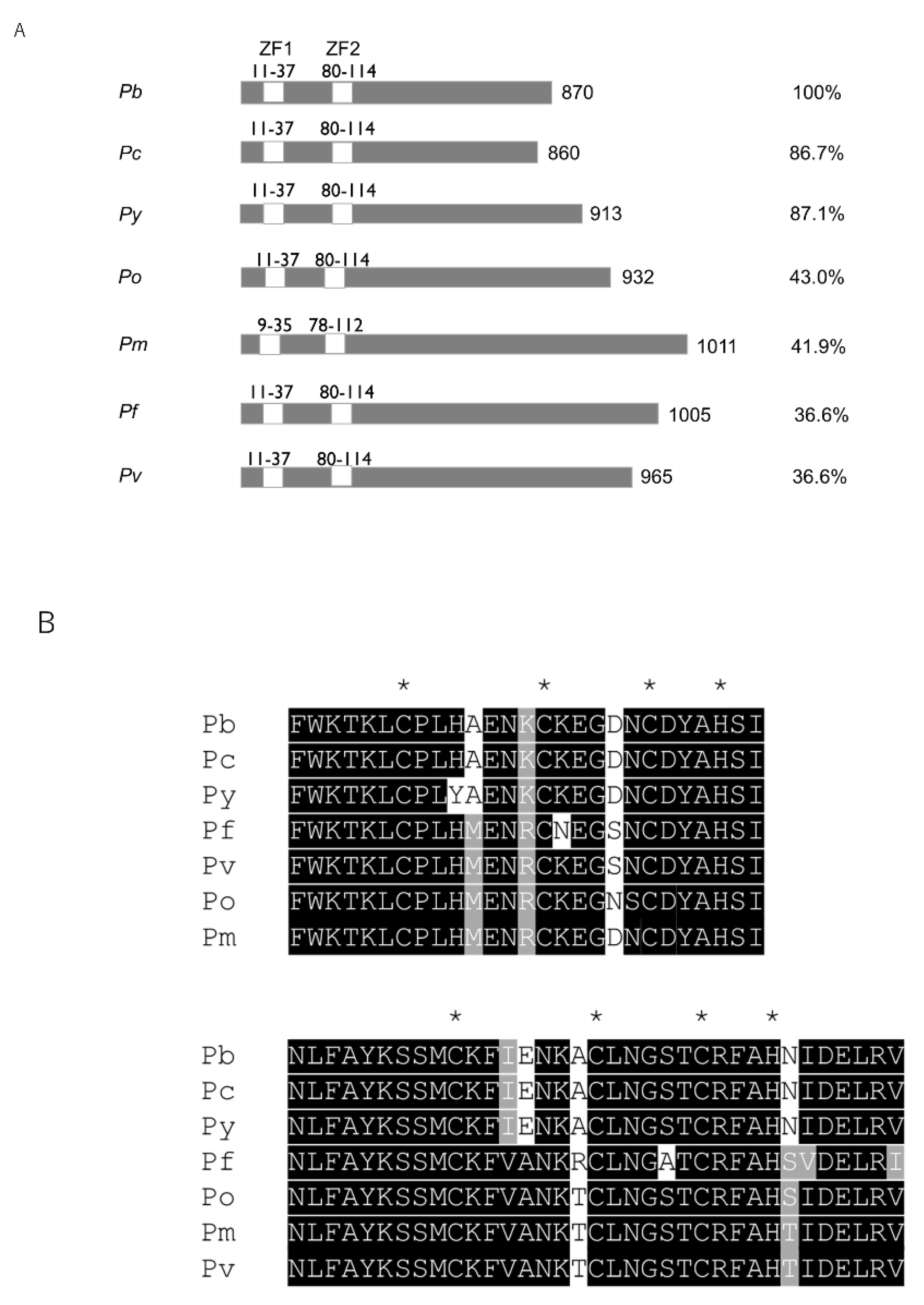
2.1. Pb103 Is Conserved in Plasmodium

We searched PlasmoDB for RBP-coding genes whose expression is high in gametocytes to identify novel translation regulators functioning in parasite reproduction stages. Among several candidate genes, we focused on PBANKA_1134900, which encodes 870 amino acids with a molecular weight of approximately 103 kDa, and designated it Pb103. InterProScan analysis revealed that Pb103 possesses two CCCH-type ZF domains. A database search showed that Pb103 is highly conserved among rodent malaria parasites (Figure 1A). While orthologous genes are also detected in human malaria parasites with low identity at the entire sequence level, two ZF domains are highly conserved among rodent and human malaria parasites (Figure 1B). All these genes contain a single exon, which is another conserved feature in these orthologous genes. The presence of a Pb103 ortholog in various Plasmodium species suggests that the Pb103 ortholog may have conserved roles in parasite development.

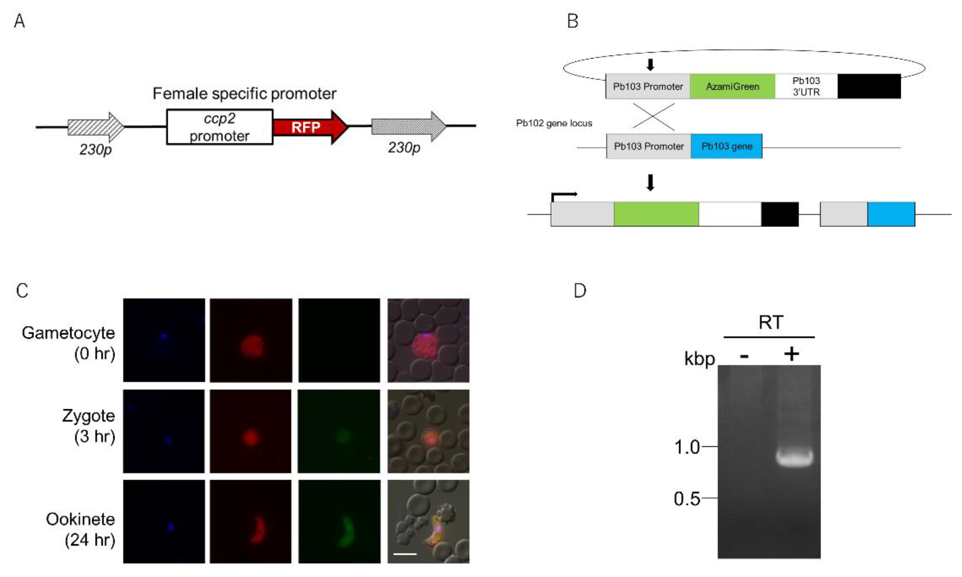
2.2. Expression Analysis of Pb103 by Using the Pb103-AGFP Reporter

According to RNA-seq data [28], the Pb103 gene is exclusively expressed in female gametocytes. To confirm this, we attempted to generate a P. berghei reporter line expressing AGFP fused to the C-terminal of the Pb103 protein under the endogenous Pb103 promoter. The resulting transgenic parasite did not show any AGFP signal during the whole life cycle (data not shown). This could be due to the steric hindrance of AGFP caused by Pb103 fusion or proteolytic processing. Then, we changed our strategy to a generation of another transgenic line expressing AGFP driven by the endogenous Pb103 promoter. To detect female gametocytes easily, we first generated a selection marker-free female RFP reporter that expresses RFP throughout female gametocytes to ookinete under the control of the ccp2 promoter, which is known to be active in those developmental stages [29] (Figure 2A). Next, we transformed the female RFP reporter with the Pb103-AGFP plasmid (Figure 2B), whereby AGFP expression in RFP-positive cells could be monitored easily. Tail blood was taken from mice infected with the Pb103-AGFP reporter, and the fluorescent signal was observed. Unexpectedly, no AGFP signal was detected in RFP-positive or RFP-negative cells (Figure 2C, upper panels). This suggests that AGFP is not transcribed or AGFP mRNA is not translated. To investigate whether the AGFP gene is transcribed, we performed RT-PCR using RNA isolated from gametocyte-rich cell fractions. As a result, AGFP mRNA was detected (Figure 2D), indicating that AGFP mRNA is in a translationally quiescent state. Next, the same infected blood was incubated in a gametogenesis-inducing medium. The AGFP signal was exclusively detected in RFP-positive cells at 3 and 24 h postinduction (Figure 2C, middle and bottom panels), demonstrating that AGFP is translated in zygotes and ookinetes. Considering the translational repression of AGFP mRNA, the results suggest that Pb103 mRNA translation could also be repressed and then initiated after gametogenesis. Such stage-specific translational regulation suggests that Pb103 may have crucial roles in parasite development after gametogenesis.

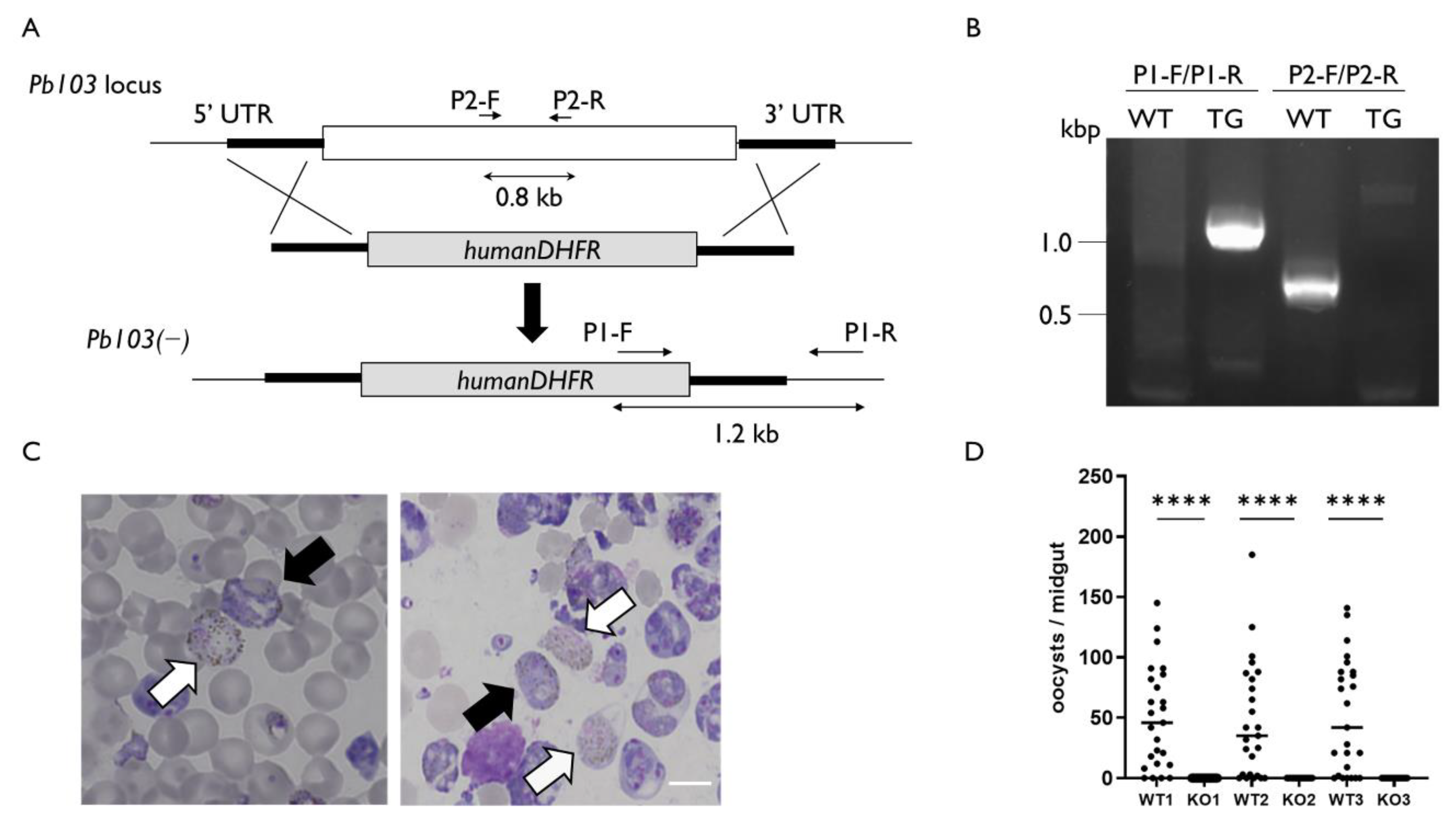
2.3. Pb103 Is Essential for Parasite Reproduction

To address the functions of Pb103, we generated Pb103(−) parasites. The disrupting construct, which contains the selectable marker human DHFR, targeted 500 bp upstream and downstream of the Pb103-coding region (Figure 3A, upper and middle panels). Pb103 gene deletion was conducted by double-crossover homologous recombination, whereby the Pb103 gene was replaced by the selectable marker (Figure 3A, lower panel). The correct gene replacement in the Pb103(−) clone was confirmed by diagnostic PCR (Figure 3B). The morphology of Pb103(−) gametocytes was indistinguishable from that of wild-type parasites (Figure 3C). Pb03(−) differentiated into gametocytes with similar efficiency as wild type (Table 1). We investigated the infectivity of Pb103(−) in mosquitoes. Mosquitoes were fed on mice infected with either Pb103(−) or wild type and were then dissected for the evaluation of parasite development on day 16 post feeding. Three independent experiments showed that oocysts in the midgut were detected in mosquitoes fed on mice carrying wild-type parasites. In contrast, no oocysts were detected in the mosquitoes fed on mice carrying Pb103(−) (Figure 3D). This result indicates that Pb103 is required for parasite development in mosquitoes. To ensure that no infectious Pb103(−) sporozoites existed in the salivary glands, we prepared salivary gland extracts from mosquitoes fed Pb103(−) or wild type and injected them into mice intravenously. As a result, the mice injected with the extracts from four to six mosquitoes carrying wild-type oocysts were infected, while no infection was detected in mice injected with the extract prepared from 15 and 20 mosquitoes fed with Pb103(−) (Table 2). Since Pb103(−) could not develop into oocysts and no infectious sporozoites were produced, it is likely that Pb103(−) parasites may halt development before oocyst stages. To investigate which steps of the developmental process are defective in Pb103(−), we performed an in vitro fertilization assay that mimics the gametogenesis and fertilization taking place in the mosquito midgut [30]. In the wild type, 54.3% of female gametes were fertilized with male gametes and then transformed into ookinetes (Figure 4A,B, left panel). In Pb103(−), on the other hand, no mature ookinetes but zygotes were detected (Figure 4A,B, right panel). These results suggest that Pb103 has critical roles after zygote development.
As shown in the protein expression analysis of the Pb103-AGFP reporter, Pb103 is translated after gametogenesis in females. Therefore, the developmental failure observed in Pb103(−) parasites could be exclusively attributed to females. To investigate this possibility, we performed in vitro cross-fertilization experiments between Pb103(−) and CDPK4() (male infertility) or Nek4() (female infertility) gametes. The self-fertilization of CDPK4() and Nek4(−) did not produce ookinetes, while CDPK4() × Nek4() cross produced ookinetes, which was equivalent to 35.1% of CDPK4() female gametes fertilized to Nek4() males (Figure 4C). Cross-fertilization of Pb03(−) × CDPK4() produced ookinetes with slightly lower efficiency compared to CDPK4() × Nek4() crosses (p < 0.01; Figure 4C). On the other hand, the Pb03(−) × Nek4() cross failed to generate mature ookinetes. These results demonstrate that Pb103 is exclusively required in females.

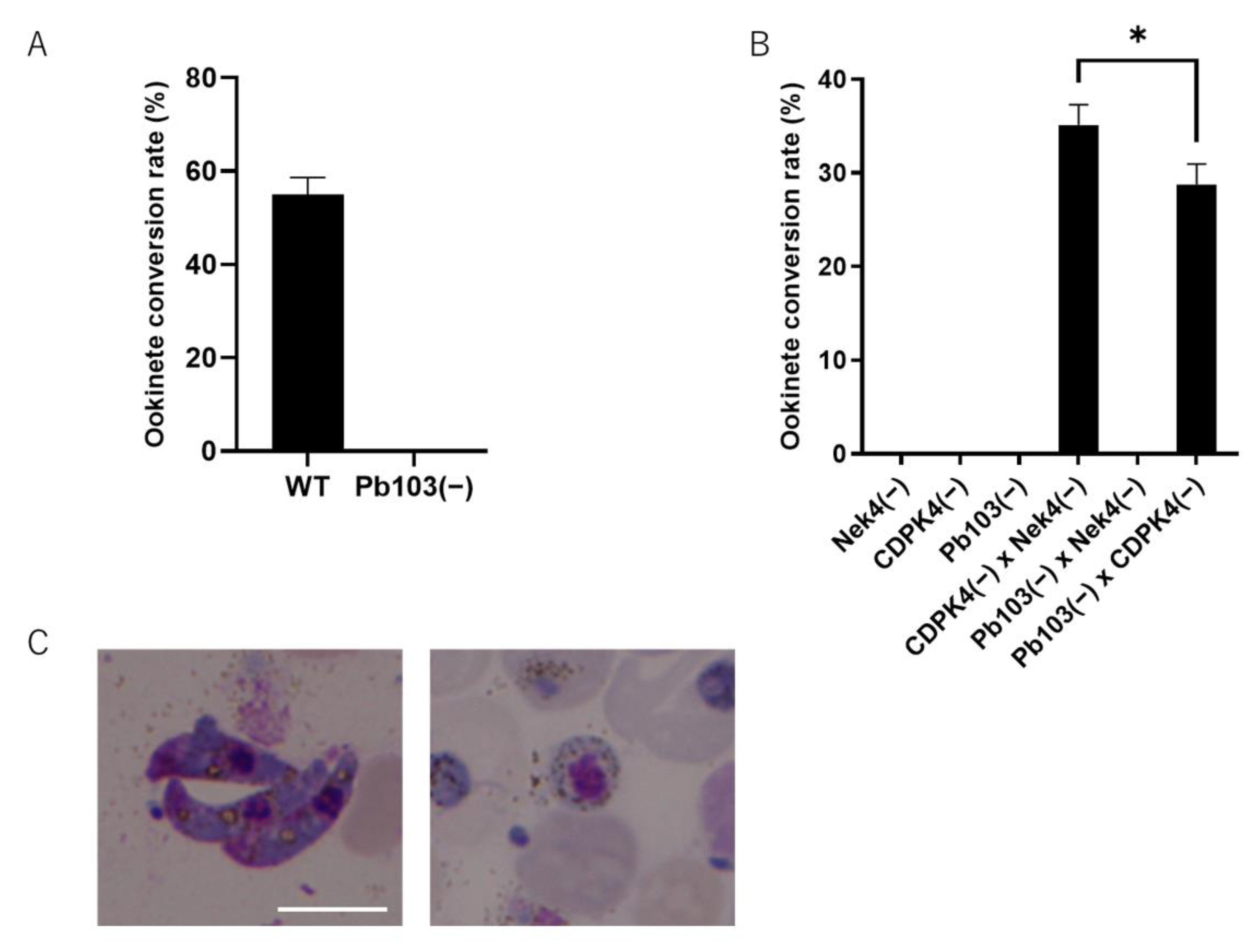
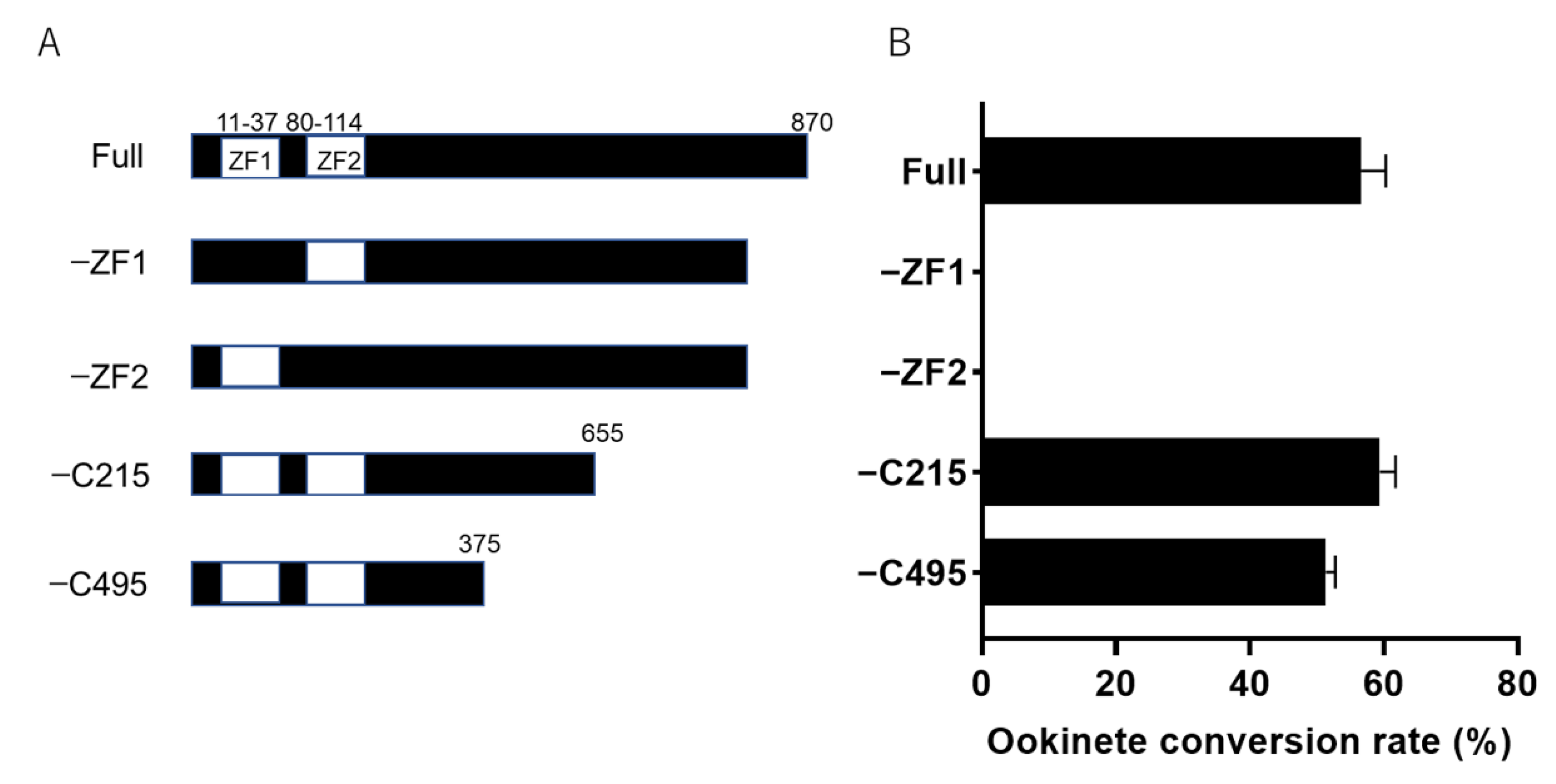
2.4. Zinc Finger Domains Are Indispensable for Ookinete Maturation

To identify crucial domains for ookinete maturation, we made four Pb103 deletion mutants and designated them −ZF1, −ZF2, −C215, and −C495 (Figure 5A). The −ZF-1 or −ZF-2 lacks the first or second ZF domain, while −C215 or −C495 lacks the respective numbers of amino acids from the C-terminus. These mutants were generated by genome edition, and the desired deletions in all clones were confirmed by Sanger sequencing. These mutants were subjected to an in vitro fertilization assay to investigate the functional domain based on the morphological maturation of ookinetes. As a result, obvious mature ookinetes were frequently detected in −C495 and −C215 with similar efficiency to that of wild-type (full) parasites (Figure 5B). Notably, mature ookinetes were generated in −C495, which lost more than half of the full length from the C-terminus. As we mentioned in the construction of the Pb103-AGFP reporter, the reporter parasites expressing the Pb103 protein fused to AGFP did not show a fluorescent signal. Considering this and the successful ookinete maturation of the −C495 mutant together, it is conceivable that Pb103 may be cleaved in the C-terminus. This could be the reason for the lack of signal detection in the Pb103-AGFP-expressing parasites. In addition, the −ZF1 and −ZF2 parasites did not produce mature ookinetes. These results highlight the importance of the ZF domains in Pb103 for ookinete maturation.

2.5. Mechanisms of Post-Translation Repression of Pb103

By using a Pb103-AGFP reporter, we showed that AGFP mRNA was translationally repressed in gametocytes and that this repression was released by gametogenesis. In the Pb103-AGFP reporter, the AGFP gene is under the control of the 5′ and 3′ UTRs of the Pb103 gene, suggesting that translational regulatory elements, if any, would exist in these UTRs. It has been reported that a U-rich element in the 5′ or 3′ UTR is essential for the translational repression of P28 and P25 in female gametocytes [31,32]. Several U-rich regions are detected in the 5′ and 3′ UTRs of Pb103. However, the genome of Plasmodium is extremely AT-rich (www.plasmodb.org, accessed on 12 December 2016). Thus, it is difficult to identify such elements based on U-richness. Further study is needed for functional identification of the elements to which regulatory factors get access to halt the translation of Pb103 mRNA. It has been reported that the DOZI/CITH RNA helicase is a central factor in translational repression in gametocytes [18]. The DOZI/CITH complex is a component of mRNPs, which prevents target mRNAs from translation and degradation. RNA immunoprecipitation assays and microarrays revealed that 731 mRNAs were associated with DOZI/CITH, and half of them were translated into ookinetes. The functions of these proteins are speculated to be involved in ookinete maturation, such as motility, adhesion, and cell traversal [33]. In addition, both DOZI and CITH deletion mutants could fertilize and differentiate into zygotes but failed in ookinete formation [19]. Importantly, the RNA immunoprecipitation assay data showed that Pb103 mRNA was bound by DOZI/CITH, and the level of Pb103 mRNA was a 6.57-fold reduction in the DOZI mutant [32]. These results suggest that the stability and translation of Pb103 mRNA could be under the control of DOZI/CITH. Among 731 mRNAs of potential targets for DOZI/CITH, only a single gene deletion, Pb103 deletion, showed a severe defect in ookinete maturation, suggesting the functional importance of Pb103 for DOZI/CITH-mediated ookinete maturation. In the current study, our main effort was made to elucidate the structural importance of the Pb103 protein by making four Pb103 deletion mutants and their phenotypic study. Our data revealed that over half of the C-terminal region was not necessary for ookinete maturation. In contrast, we found that either of the ZF domains was essential for ookinete maturation. It is plausible that double ZF domains of Pb103 function as RNA binding to regulate the translation of target mRNAs. A previous study revealed that 15 RBP-coding mRNAs in addition to Pb103 are bound to the DOZI/CITH complex [32]. It is interesting to understand the interrelationship among these RBPs as well as other putative targets of DOZI/CITH to uncover the DOZI/CITH-mediated translational regulation cascade. In this context, for further work, the identification of target mRNAs for Pb103 is of special interest because this would provide clues for an in-depth understanding of the molecular machinery of DOZI/CITH and Pb103-mediated posttranscriptional regulation. In addition, this study may also help to identify new candidate molecules of transmission-blocking vaccine strategies that target parasite development in mosquitoes.

3. Methods

3.1. Ethics Statement

The protocols for all animal and recombinant DNA experiments were approved by the Ethics of Experimental Animals Committee and Recombinant DNA Committee of School of Medicine, Juntendo University, and the assigned numbers were no. 2021036 and no. 25-115, respectively.

3.2. Animals and Parasites

BALB/c mice (female, 5 weeks old) were purchased from Japan SLC, Inc. and were used throughout the experiments. The rodent malaria parasite Plasmodium berghei ANKA (clone 2.34) was used for transgenic parasite generation. Anopheles stephensi (SDA 500 strain) was reared under a photoperiod of 14:10 h (light:dark) at 26 °C.

3.3. Generation and Expression Analysis of the Pb103-AGFP Reporter

The pL1186 plasmid was previously designed to make a transgenic line that expresses azami green (AGFP) and red fluorescent proteins (RFPs) for male and female gametocytes to ookinete, respectively [29]. The pL1186 plasmid was digested with EcoRV/NotI and self-ligated to remove the male-specific promoter and AGFP. The resulting plasmid (female-RFP) was digested with two SacII sites and introduced into the 230p gene locus by double-crossover homologous recombination by a previously reported protocol [34]. Subsequently, the transgenic parasite clones were subjected to negative selection using 5-fluorocytosine to remove the selectable marker gene. The resulting parasite clone (female-RFP reporter) expresses RFP throughout female gametocytes to ookinetes [35] and was used as a recipient parasite of the Pb103-AGFP plasmid described below. For the construction of the Pb103-AGFP plasmid, fragments covering the 5′ untranslated region (UTR) (−1.5 kb) and 3′ UTR (−1.5 kb) of Pb103 and AGFP were amplified using Pb103-5′ UTR-F1/Pb103-5′ UTR-R1, Pb103-3′ UTR-F1/Pb103-3′ UTR-R1, and AGFP-F/AGFP-R. These three fragments were serially cloned in the order of Pb103-5′ UTR, AGFP, and Pb103-3′ UTR into HindIII/PstI-digested pL0006 by an In-Fusion HD cloning kit (TaKaRa). The final plasmid, Pb103-AGFP, was linearized by the PacI site in the 5′ UTR of Pb103 and integrated into the recipient parasites by a single crossover. The final transformant, the Pb103-AGFP reporter, was selected by pyrimethamine. The fluorescent signal was monitored in asexual and gametocyte stages. To monitor the signal after gametocyte stages, gametogenesis and fertilization of the reporter were induced by a previously described protocol [4,30]. The fluorescent signal was observed at 0, 3, and 24 h postinduction under a fluorescence microscope (AxioImager M2, Zeiss, Tokyo, Japan).

3.4. Reverse Transcription PCR (RT-PCR)

The mice were intraperitoneally injected with 100 μL of phenylhydrazine hydrochloride (2.5 mg) on day 0 and were then injected with the freezing stock of wild-type parasites on day 2. The mice were fed water containing sulfadiazine (15 mg/L) on days 5 and 6 to kill asexual stage parasites. On day 7, whole blood (1 mL) was collected by cardiac puncture and mixed with 5 mL of lysis buffer (150 mM ammonium chloride, 10 mM potassium carbonate, and 1 mM EDTA) on ice to induce hemolysis. The blood was then centrifuged at 600× g for 6 min and washed with phosphate-buffered saline. Total RNA was extracted from this gametocyte-rich cell fraction and then treated with DNase-I using the FastGene™ RNA Premium Kit (NIPPON Genetics Co., Ltd., Tokyo, Japan). Total RNA (1 μg) was reverse-transcribed using the PrimeScript™ RT–PCR Kit (TAKARA BIO Inc., Shiga, Japan) and was used as a template for PCR to amplify the full-length AGFP gene using a pair of primers (AGFP-F/AGFP-R). RNA without reverse transcription was used as a negative control.

3.5. Targeting Disruption of Pb103, PbCDPK4, and PbNek-4 Genes

To generate a construct for Pb103 gene disruption, two PCR fragments covering 500 bp of the 5′ and 3′ UTRs of the Pb103 gene were amplified by two pairs of primers, Pb03(−)-F1/Pb03(−)-R1 and Pb03(−)-F2/Pb03(−)-R2, respectively, and P. berghei genomic DNA as a template. Then, each PCR product was conjugated to either end of PstI/HindIII digested-pL0006 (MRA-775; BEI Resources.) by fusion PCR using primer Pb03(−)-F1/Pb03(−)-R2 [36]. Similarly, knockout constructs for the CDPK4 [37] and Nek-4 [38] genes, which are essential for male and female fertility, were generated as follows. The 5′ and 3′ UTRs of CDPK4 and Nek-4 were amplified using CDPK4(−)-F1, -R1, -F2, and -R2 or Nek4(−)-F1, -R1, -F2, and -R2. Each of these fragments was fused to either the 5′ or 3′ end of PstI/HindIII-digested pL0006 using the primers CDPK4(−)-F1/CDPK4(−)-R2 and Nek4(−)-F1/Nek4(−)-R2. The final constructs were introduced to P. berghei, and the transformant was cloned.

3.6. Development of Pb03(−) in Mosquitoes and Transmission to Mice

A phenotypic study of Pb03(−) in mosquitoes was performed by following a previously described procedure [39]. In brief, female mosquitoes (4–7 days old) were fed on mice carrying either Pb03(−) or wild type, and fully blood-fed mosquitoes were collected. Sixteen days after feeding, the mosquitoes were dissected, and the numbers of oocysts in the midgut were counted. Mosquito feeding and subsequent cultivation were performed at 21 °C. To test parasite transmission from mosquitoes to mice, salivary glands from mosquitoes carrying Pb03(−) and wild-type parasites were collected in 100 μL of phosphate-buffered saline on ice. Salivary gland extracts (100 μL) were injected into each mouse. On day 5 after the injection, the mice were checked for infection by observing Giemsa-stained thin blood film smear slides. For the mice that were negative for infection, a blood smear test was conducted on day 16. The slides were observed under a microscope (×1000).

3.7. In Vitro Cross-Fertilization Assay

Fertilization among Pb03(−), CDPK4(−), Nek4(−), or wild type was performed by an in vitro cross-fertilization assay, as described previously [4]. A pair of 5 μL of tail blood infected with either Pb03(−), CDPK4(−), Nek4(−), or wild type was added immediately to 1 mL of gametogenesis-inducing medium (10% fetal bovine serum in RPMI1640, pH 8.2) at 21 °C for 12 min to induce gametogenesis and check for exflagellation. This mixture was further incubated for 24 h to induce fertilization and ookinete formation. The ookinete conversion rate was determined by the percentage of female gametocytes fertilized with males and transformed into a mature ookinete. The female gametocytes and ookinetes were stained with Giemsa and counted under a microscope (×1000).

3.8. Generation of Partially Truncated Pb103 Mutants

Four Pb103 partial deletion mutants were generated by using CRISPR/Cas9. For the construction of −C215, two fragments covering 455 to 655 a.a. (Frag. A) and 0.5 kb of 3′ UTR (Frag. B) were amplified by two pairs of primers: −C215-F/−C215-R and −C215-3UTR-F/−C215-3UTR-R. These two fragments were serially inserted as a donor into AflII/HindIII of pBS-1, which was a CRISPR/Cas9 plasmid customized to P. berghei [39]. For the −C495 mutant, two fragments covering 189–375 a.a. (Frag. C) and 0.5 kb of 3′ UTR (Frag. D) were amplified by two pairs of primers: −C495-F/−C495-R and −C495-3UTR-F/−C495-3UTR-R. These two fragments were inserted into pBS-1, as described above. For the −ZF1 mutant, fragments covering the 5′ UTR (0.5 kb) to the first 10 a.a. (Frag. E) and 38 to 220 a.a. motif (Frag. F) were amplified using −ZF1-F1/−ZF1-R1 and −ZF1-F2/−ZF1-R2. For the −ZF2 mutant construct, fragments covering the 5′ UTR (0.2 kb) to the first 79 a.a. (Frag. G), and 115 a.a. to 270 a.a. (Frag. H) were amplified using −ZF2-F1/−ZF2-R1 and −ZF2-F2/−ZF2-R2, respectively. The fragment, E/F, and G/H were inserted as donors into pBS-1. The PAM sites were searched using CHOPCHOP (http://chopchop.cbu.uib.no, accessed on 12 December 2018). The double-stranded DNA coding for gRNA was inserted into the BsmBI site in pBS-1. The resultant plasmids were introduced into P. berghei, and transformants were cloned. All clones were subjected to Sanger sequencing to confirm that the expected regions in the Pb103 gene were deleted. The sequences of primers and gRNAs are listed in Supplementary Table S1.

3.9. Statistical Analyses

An unpaired t-test or one-way ANOVA with Tukey’s multiple comparison test of means was used to compare the ookinete conversion rate. The Mann–Whitney U-test was used to analyze oocyst density (oocyst number/midgut). Statistical analyses were carried out using GraphPad Prism 9 (GraphPad Software Inc., San Diego, CA, USA, version 9.3.0).

Supplementary Materials

The following are available online at https://www.mdpi.com/article/10.3390/pathogens10121536/s1, Table S1: List of oligos used in this study.

Author Contributions

Conceptualization, M.H., A.M. and T.M. (Toshiyuki Mori); methodology, M.H., A.M. and T.M. (Toshiyuki Mori); formal analysis, M.H., A.M., T.M. (Toshiyuki Mori) and T.M. (Toshihiro Mita); writing—original draft preparation, M.H.; writing—review and editing, M.H.; supervision, M.H. All authors have read and agreed to the published version of the manuscript.

Funding

This work was supported by JSPS KAKENHI for Scientific Research (B) and (C) to TMo (17H03707) and MH (18K07095), respectively. This work was also supported by the Research Program on Emerging and Re-emerging Infectious Diseases (21fk0108138s0402) from the Japan Agency for Medical Research and Development, AMED, to MH.

Data Availability Statement

Not applicable.

Acknowledgments

We would like to thank Kaisaki I. for helping with animal and molecular experiments. We also thank the Laboratory of Molecular and Biochemical Research, Research Support Centre, Juntendo University Graduate School of Medicine, for technical support with Sanger sequencing.

Conflicts of Interest

The authors declare no conflict of interest.

References

  1. WHO. 2020 World Malaria Report; WHO Press: Geneva, Switzerland, 2020. [Google Scholar]
  2. Kubo-Irie, M.; Hirai, M.; Irie, M.; Mohri, H. Postulated Process of Axoneme Organization in the Male Gametogenesis of Malaria Parasite Plasmodium berghei. Zool. Sci. 2020, 38, 187–192. [Google Scholar] [CrossRef]
  3. Billker, O.; Shaw, M.K.; Margos, G.; Sinden, R.E. The Roles of Temperature, PH and Mosquito Factors as Triggers of Male and Female Gametogenesis of Plasmodium berghei in Vitro. Parasitology 1997, 115, 1–7. [Google Scholar] [CrossRef]
  4. Hirai, M.; Arai, M.; Mori, T.; Miyagishima, S.; Kawai, S.; Kita, K.; Kuroiwa, T.; Terenius, O.; Matsuoka, H. Male Fertility of Malaria Parasites Is Determined by GCS1, a Plant-Type Reproduction Factor. Curr. Biol. 2008, 18, 607–613. [Google Scholar] [CrossRef] [PubMed] [Green Version]
  5. Liu, Y.; Tewari, R.; Ning, J.; Blagborough, A.M.; Garbom, S.; Pei, J.; Grishin, N.V.; Steele, R.E.; Sinden, R.E.; Snell, W.J.; et al. The Conserved Plant Sterility Gene HAP2 Functions after Attachment of Fusogenic Membranes in Chlamydomonas and Plasmodium Gametes. Gene Dev. 2008, 22, 1051–1068. [Google Scholar] [CrossRef] [PubMed] [Green Version]
  6. Van Dijk, M.R.; Janse, C.J.; Thompson, J.; Waters, A.P.; Braks, J.A.M.; Dodemont, H.J.; Stunnenberg, H.G.; van Gemert, G.-J.; Sauerwein, R.W.; Eling, W. A Central Role for P48/45 in Malaria Parasite Male Gamete Fertility. Cell 2001, 104, 153–164. [Google Scholar] [CrossRef] [Green Version]
  7. Ruiz, J.L.; Gómez-Díaz, E. The Second Life of Plasmodium in the Mosquito Host: Gene Regulation on the Move. Brief. Funct. Genom. 2019, 18, 313–357. [Google Scholar] [CrossRef] [PubMed]
  8. Coelho, C.H.; Rappuoli, R.; Hotez, P.J.; Duffy, P.E. Transmission-Blocking Vaccines for Malaria: Time to Talk about Vaccine Introduction. Trends Parasitol. 2019, 35, 483–486. [Google Scholar] [CrossRef]
  9. Hirai, M.; Mori, T. Fertilization Is a Novel Attacking Site for the Transmission Blocking of Malaria Parasites. Acta Trop. 2010, 114, 157–161. [Google Scholar] [CrossRef]
  10. Painter, H.J.; Campbell, T.L.; Llinás, M. The Apicomplexan AP2 Family: Integral Factors Regulating Plasmodium Development. Mol. Biochem. Parasit. 2011, 176, 1–7. [Google Scholar] [CrossRef] [Green Version]
  11. Sinha, A.; Hughes, K.R.; Modrzynska, K.K.; Otto, T.D.; Pfander, C.; Dickens, N.J.; Religa, A.A.; Bushell, E.; Graham, A.L.; Cameron, R.; et al. A Cascade of DNA-Binding Proteins for Sexual Commitment and Development in Plasmodium. Nature 2014, 507, 253–257. [Google Scholar] [CrossRef]
  12. Kafsack, B.F.; Rovira-Graells, N.; Clark, T.G.; Bancells, C.; Crowley, V.M.; Campino, S.G.; Williams, A.E.; Drought, L.G.; Kwiatkowski, D.P.; Baker, D.A.; et al. A Transcriptional Switch Underlies Commitment to Sexual Development in Malaria Parasites. Nature 2014, 507, 248–252. [Google Scholar] [CrossRef] [Green Version]
  13. Yuda, M.; Iwanaga, S.; Kaneko, I.; Kato, T. Global Transcriptional Repression: An Initial and Essential Step for Plasmodium Sexual Development. Proc. Natl. Acad. Sci. USA 2015, 112, 12824–12829. [Google Scholar] [CrossRef] [PubMed] [Green Version]
  14. Yuda, M.; Kaneko, I.; Iwanaga, S.; Murata, Y.; Kato, T. Female-specific Gene Regulation in Malaria Parasites by an AP2-family Transcription Factor. Mol. Microbiol. 2020, 113, 40–51. [Google Scholar] [CrossRef] [PubMed]
  15. Kaneko, I.; Iwanaga, S.; Kato, T.; Kobayashi, I.; Yuda, M. Genome-Wide Identification of the Target Genes of AP2-O, a Plasmodium AP2-Family Transcription Factor. PLoS Pathog. 2015, 11, e1004905. [Google Scholar] [CrossRef] [PubMed] [Green Version]
  16. Modrzynska, K.; Pfander, C.; Chappell, L.; Yu, L.; Suarez, C.; Dundas, K.; Gomes, A.; Goulding, D.; Rayner, J.C.; Choudhary, J.; et al. A Knockout Screen of ApiAP2 Genes Reveals Networks of Interacting Transcriptional Regulators Controlling the Plasmodium Life Cycle. Cell Host Microbe 2017, 21, 11–22. [Google Scholar] [CrossRef] [PubMed] [Green Version]
  17. Lasonder, E.; Rijpma, S.R.; van Schaijk, B.C.L.; Hoeijmakers, W.A.M.; Kensche, P.R.; Gresnigt, M.S.; Italiaander, A.; Vos, M.W.; Woestenenk, R.; Bousema, T.; et al. Integrated Transcriptomic and Proteomic Analyses of P. Falciparum Gametocytes: Molecular Insight into Sex-Specific Processes and Translational Repression. Nucleic Acids Res. 2016, 44, 6087–6101. [Google Scholar] [CrossRef] [PubMed] [Green Version]
  18. Mair, G.R.; Braks, J.A.M.; Garver, L.S.; Wiegant, J.C.A.G.; Hall, N.; Dirks, R.W.; Khan, S.M.; Dimopoulos, G.; Janse, C.J.; Waters, A.P. Regulation of Sexual Development of Plasmodium by Translational Repression. Science 2006, 313, 667–669. [Google Scholar] [CrossRef] [Green Version]
  19. Mair, G.R.; Lasonder, E.; Garver, L.S.; Franke-Fayard, B.; Carret, C.K.; Wiegant, J.; Dirks, R.W.; Dimopoulos, G.; Janse, C.J.; Waters, A.P. Universal Features of Post-Transcriptional Gene Regulation Are Critical for Plasmodium Zygote Development. PLoS Pathog. 2010, 6, e1000767. [Google Scholar] [CrossRef] [Green Version]
  20. Nguyen-Chi, M.; Morello, D. RNA-Binding Proteins, RNA Granules, and Gametes: Is Unity Strength? Reproduction 2011, 142, 803–817. [Google Scholar] [CrossRef] [Green Version]
  21. Beckmann, B.M.; Horos, R.; Fischer, B.; Castello, A.; Eichelbaum, K.; Alleaume, A.-M.; Schwarzl, T.; Curk, T.; Foehr, S.; Huber, W.; et al. The RNA-Binding Proteomes from Yeast to Man Harbour Conserved EnigmRBPs. Nat. Commun. 2015, 6, 10127. [Google Scholar] [CrossRef]
  22. Reddy, B.N.; Shrestha, S.; Hart, K.J.; Liang, X.; Kemirembe, K.; Cui, L.; Lindner, S.E. A Bioinformatic Survey of RNA-Binding Proteins in Plasmodium. BMC Genom. 2015, 16, 890. [Google Scholar] [CrossRef] [PubMed] [Green Version]
  23. Ngwa, C.J.; Farrukh, A.; Pradel, G. Zinc Finger Proteins of Plasmodium Falciparum. Cell Microbiol. 2021, e13387. [Google Scholar] [CrossRef] [PubMed]
  24. Miao, J.; Fan, Q.; Parker, D.; Li, X.; Li, J.; Cui, L. Puf Mediates Translation Repression of Transmission-Blocking Vaccine Candidates in Malaria Parasites. PLoS Pathog. 2013, 9, e1003268. [Google Scholar] [CrossRef] [Green Version]
  25. Chêne, A.; Vembar, S.S.; Rivière, L.; Lopez-Rubio, J.J.; Claes, A.; Siegel, T.N.; Sakamoto, H.; Scheidig-Benatar, C.; Hernandez-Rivas, R.; Scherf, A. PfAlbas Constitute a New Eukaryotic DNA/RNA-Binding Protein Family in Malaria Parasites. Nucleic Acids Res. 2012, 40, 3066–3077. [Google Scholar] [CrossRef] [PubMed]
  26. Komaki-Yasuda, K.; Okuwaki, M.; Nagata, K.; Kawazu, S.; Kano, S. Identification of a Novel and Unique Transcription Factor in the Intraerythrocytic Stage of Plasmodium Falciparum. PLoS ONE 2013, 8, e74701. [Google Scholar] [CrossRef]
  27. Müller, K.; Silvie, O.; Mollenkopf, H.-J.; Matuschewski, K. Pleiotropic Roles for the Plasmodium berghei RNA Binding Protein UIS12 in Transmission and Oocyst Maturation. Front. Cell Infect. Microbiol. 2021, 11, 624945. [Google Scholar] [CrossRef] [PubMed]
  28. Yeoh, L.M.; Goodman, C.D.; Mollard, V.; McFadden, G.I.; Ralph, S.A. Comparative Transcriptomics of Female and Male Gametocytes in Plasmodium berghei and the Evolution of Sex in Alveolates. BMC Genom. 2017, 18, 734. [Google Scholar] [CrossRef] [Green Version]
  29. Ponzi, M.; Sidén-Kiamos, I.; Bertuccini, L.; Currà, C.; Kroeze, H.; Camarda, G.; Pace, T.; Franke-Fayard, B.; Laurentino, E.C.; Louis, C.; et al. Egress of Plasmodium berghei Gametes from Their Host Erythrocyte Is Mediated by the MDV-1/PEG3 Protein. Cell Microbiol. 2009, 11, 1272–1288. [Google Scholar] [CrossRef]
  30. Rosales-Ronquillo, M.C.; Silverman, P.H. In Vitro Ookinete Development of the Rodent Malarial Parasite, Plasmodium berghei. J. Parasitol. 1974, 60, 819–824. [Google Scholar] [CrossRef]
  31. Braks, J.A.M.; Mair, G.R.; Franke-Fayard, B.; Janse, C.J.; Waters, A.P. A Conserved U-Rich RNA Region Implicated in Regulation of Translation in Plasmodium Female Gametocytes. Nucleic Acids Res. 2008, 36, 1176–1186. [Google Scholar] [CrossRef] [Green Version]
  32. Guerreiro, A.; Deligianni, E.; Santos, J.M.; Silva, P.; Louis, C.; Pain, A.; Janse, C.J.; Franke-Fayard, B.; Carret, C.K.; Siden-Kiamos, I.; et al. Genome-Wide RIP-Chip Analysis of Translational Repressor-Bound MRNAs in the Plasmodium Gametocyte. Genome Biol. 2014, 15, 493. [Google Scholar] [CrossRef] [PubMed] [Green Version]
  33. Sebastian, S.; Brochet, M.; Collins, M.O.; Schwach, F.; Jones, M.L.; Goulding, D.; Rayner, J.C.; Choudhary, J.S.; Billker, O. A Plasmodium Calcium-Dependent Protein Kinase Controls Zygote Development and Transmission by Translationally Activating Repressed MRNAs. Cell Host Microbe. 2012, 12, 9–19. [Google Scholar] [CrossRef] [PubMed] [Green Version]
  34. Janse, C.J.; Ramesar, J.; Waters, A.P. High-Efficiency Transfection and Drug Selection of Genetically Transformed Blood Stages of the Rodent Malaria Parasite Plasmodium berghei. Nat. Protoc. 2006, 1, 346–356. [Google Scholar] [CrossRef]
  35. Braks, J.A.M.; Franke-Fayard, B.; Kroeze, H.; Janse, C.J.; Waters, A.P. Development and Application of a Positive–Negative Selectable Marker System for Use in Reverse Genetics in Plasmodium. Nucleic Acids Res. 2006, 34, e39. [Google Scholar] [CrossRef] [PubMed] [Green Version]
  36. Kuwayama, H.; Obara, S.; Morio, T.; Katoh, M.; Urushihara, H.; Tanaka, Y. PCR-Mediated Generation of a Gene Disruption Construct without the Use of DNA Ligase and Plasmid Vectors. Nucleic Acids Res. 2002, 30, e2. [Google Scholar] [CrossRef] [PubMed]
  37. Billker, O.; Dechamps, S.; Tewari, R.; Wenig, G.; Franke-Fayard, B.; Brinkmann, V. Calcium and a Calcium-Dependent Protein Kinase Regulate Gamete Formation and Mosquito Transmission in a Malaria Parasite. Cell 2004, 117, 503–514. [Google Scholar] [CrossRef] [Green Version]
  38. Reininger, L.; Billker, O.; Tewari, R.; Mukhopadhyay, A.; Fennell, C.; Dorin-Semblat, D.; Doerig, C.; Goldring, D.; Harmse, L.; Ranford-Cartwright, L.; et al. A NIMA-Related Protein Kinase Is Essential for Completion of the Sexual Cycle of Malaria Parasites*. J. Biol. Chem. 2005, 280, 31957–31964. [Google Scholar] [CrossRef] [Green Version]
  39. Yamauchi, M.; Hirai, M.; Tachibana, S.-I.; Mori, T.; Mita, T. Fitness of Sulfadoxine-Resistant Plasmodium berghei Harboring a Single Mutation in Dihydropteroate Synthase (Dhps). Acta Trop. 2021, 222, 106049. [Google Scholar] [CrossRef]
Figure 1. Comparison of the structure of Pb103 and orthologs. (A) Primary structure of Pb103 orthologs in malaria parasites. White boxes and numbers indicate the position of the first and the second zinc finger domains. The right number indicates the amino acid length. The right-most number is the identity of each ortholog to Pb103. Gene accession numbers are as follows: Pb (P. berghei), PBANKA_1134900; Pc (P. chabaudi), PCHAS_1134400; Py (P. yoelii), PY17X_1136400; Po (P. ovale), PocGH01_11021900; Pm (P. malariae), PmUG01_11027400; Pf (P. falciparum), PF3D7_1358600; Pv (P. vivax), PVP01_11133009. (B) Comparison of the amino acid sequences of 1st (upper) and 2nd (lower) zinc finger domains among Pb103 orthologs. Identical and conserved residues are shaded black and gray, respectively. CCCH motif is indicated by asterisks.
Pathogens 10 01536 g001
Figure 2. Generation of reporters. (A) Generation of female RFP reporter, which is a recipient parasite. Rough- and fine-dashed arrows indicate 5′ and 3′ UTR of the 230p gene, respectively. The red arrow indicates the RFP gene. The white box indicates the ccp2 gene promoter, which is exclusively active throughout female gametocyte to ookinete stages. This DNA fragment was integrated into the 230p gene locus by double-crossover homologous recombination. (B) Schematic representation of Pb103-AGFP reporter generation using a plasmid integration through single crossover homologous recombination. The boxes indicate the Pb103 promoter (gray), AGFP (green), 3′ UTR of the Pb103 gene (white), and a selectable marker (black). For the transfection, the plasmid was linearized by PacI site located in Pb103 promoter and introduced in a female RFP reporter, as shown in (A). (C) Expression analysis of the Pb103-AGFP reporter. The female RFP reporter was transfected with the Pb103-AGFP plasmid, as described in (B). The resulting Pb103-AGFP reporter was subjected to in vitro fertilization assay. The fluorescent signal was observed at 0 (top), 3 (middle), and 24 h (bottom) post-gametogenesis. The nucleus was stained with DAPI (1st column). RFP (2nd column), AGFP (3rd column), and merged images (last column) were shown. The white bar represents 5 μm. (D) Detection of AGFP mRNA by RT-PCR. RNA was prepared from gametocyte-rich fraction and treated with DNase-I. A reverse transcribed (+) sample was used as a template for PCR. RNA without reverse transcription was used as negative control (−).
Pathogens 10 01536 g002
Figure 3. Targeted disruption of the Pb103 gene and phenotypes of Pb103(−). (A) Schematic representation of replacement strategy to generate Pb103(−). The coding region of the Pb103 gene is replaced with human DHFR, which is a selectable marker by double-crossover homologous recombination through the 5′ and 3′ UTR of the Pb103 gene. The directional and double arrows indicate the primer position and the expected product size, as shown in (B). (B) Diagnostic PCR. Genomic DNA of Pb103(−) (TG) and wild-type (WT) were used as templates. P1-F/P1-R and P2-F/P2-R are specific to TG and WT, respectively. The expected size is 1.2 kb for TG and 0.8 kb for WT. (C) Thin blood smear prepared from mice infected with wild-type (left) or Pb103(−) parasites (right). The slides were stained with Giemsa. Black and white arrows indicate female and male gametocytes, respectively. The bar represents 5 μm. (D) Infectivity of Pb103(−) (KO) and wild-type (WT) to mosquitoes. The oocyst numbers per mosquito carrying KO or WT are plotted. The results of three independent experiments are shown. Horizontal bars indicate the median. Stars represent a statistically significant difference between WT and KO in each experiment (Mann–Whitney test: **** p < 0.0001).
Pathogens 10 01536 g003
Figure 4. In vitro fertilization assay of Pb103(−) and wild type. (A) The ratio of females that successfully fertilized males and differentiated into mature ookinetes is expressed as ookinete formation rate. The bar represents mean ± SD (n = 3). (B) Giemsa staining of the samples after in vitro fertilization assay of Pb103(−) (right) and wild type (left). The mature ookinete (left) and zygote (right) are shown. The bar represents 5 μm. (C) In vitro fertilization assay. Self-fertilization of CDPK4(−) (female fertile), Nek4(−) (male fertile), and Pb103(−), and cross-fertilization between two lines among those three parasites was performed. The ookinete formation rate is shown. Data are from three independent experiments. (t-test: ∗ p < 0.02).
Pathogens 10 01536 g004
Figure 5. Characterization of Pb103 domains essential for ookinete maturation. (A) Grammatic representation of the partially deleted Pb103 products. The top bar is the normal Pb103 protein (Full). The −ZF1 and −ZF2 lack respective domains. −C215 and −C495 indicate that the respective numbers of amino acids are deleted from the C-terminal. The position of ZF1 and ZF2 are indicated by white boxes. (B) In vitro fertilization assay of four transgenic and wild-type parasites. Error bars represent mean ± SD (n = 3).
Pathogens 10 01536 g005
Table 1. Gametocytemia and sex ratio of Pb103() and wild-type.
Gametocytemia (%)Sex Ratio (♂:♀)
Pb103()0.241:2.38
WT0.251:1.96
The numbers of male and female gametocytes per 10,000 RBC were counted (n = 3).
Table 2. Infectivity of salivary gland extract from the mosquitoes infected with Pb103(−) or wild-type.
ParasitesNo. of Salivary GlandsInfected/Injected
WT51/1
61/1
41/1
Pb103(−)150/1
200/1
150/1
Mice were intravenously injected with the pooled salivary gland extract from mosquitoes being infected with Pb103(−) or wild-type. The blood smear slides were made on day 5 and 16 after injection.
Publisher’s Note: MDPI stays neutral with regard to jurisdictional claims in published maps and institutional affiliations.

