Article

Co-Infection with Anaplasma Species and Novel Genetic Variants Detected in Cattle and Goats in the Republic of Korea

1
Laboratory of Veterinary Internal Medicine, BK21 PLUS Program for Creative Veterinary Science Research, Research Institute for Veterinary Science and College of Veterinary Medicine, Seoul National University, 1 Gwanak-ro, Gwanak-gu, Seoul 08826, Korea
2
College of Ecology and Environmental Science, Kyungpook National University, Sangju 37224, Korea
*
Author to whom correspondence should be addressed.
Pathogens 2021, 10(1), 28; https://doi.org/10.3390/pathogens10010028
Submission received: 23 November 2020 / Revised: 17 December 2020 / Accepted: 29 December 2020 / Published: 1 January 2021
(This article belongs to the Collection Regional Impact of Ticks and Tick-Borne Diseases)

Abstract

:
Anaplasmosis, a tick-borne disease with multiple reservoirs, has been evolving in its pathogenesis, increasing domestic ruminants susceptibility to simultaneous infections with multiple pathogens. However, there is limited information regarding anaplasmosis in domestic ruminants in the Republic of Korea (ROK). We aimed to evaluate the role of Korean cattle and goats in Anaplasma infection maintenance. Polymerase chain reaction was performed to investigate the prevalence and genetic diversity of Anaplasma spp. from 686 whole blood samples collected from different ROK provinces. Anaplasma infection was mostly caused by A. phagocytophilum (21.1%) in cattle, and A. bovis (7.3%) in goats. Co-infection cases were found in cattle: A. bovis and A. phagocytophilum (16.7%), and in goats: A. bovis and A. capra (1.0%). Notably, a triple co-infection with A. bovis, A. phagocytophilum, and A. capra was found in one cow. Phylogenetic analysis revealed novel variants of the A. phagocytophilum 16S rRNA and A. capra gltA genes. This research contributes to the ratification of cattle as a potential reservoir of A. capra and demonstrates Anaplasma co-infection types in Korean domestic ruminants. As anaplasmosis is a zoonotic disease, our study could be crucial in making important decisions for public health.

Graphical Abstract

1. Introduction

Anaplasmosis, an infectious but non-contagious tick-borne disease, classically related as a disease of ruminants, is caused by obligate intra-erythrocytic bacteria of the genus Anaplasma [1] discovered in 1910 by Theiler [2]. This genus belongs to the Anaplasmataceae family and is composed of six confirmed species based on host cell tropism: A. phagocytophilum, A. bovis, and A. platys, which infect neutrophils [3], monocytes [4], and platelets [5], respectively, whereas A. marginale, A. centrale, and A. ovis infect erythrocytes [6]. Furthermore, the newly recognized Anaplasma species, A. capra might infect endothelial cells [7].
Anaplasmosis in domestic ruminants is caused by two main etiological agents: A. marginale in cattle [8] and A. ovis in sheep and goats [9]. However, additional studies on the genus Anaplasma have detected other Anaplasma spp. [10] that may act as a causative agent of anaplasmosis in ruminants. In the Republic of Korea (ROK), A. phagocytophilum [11] and A. bovis [12] infections have been recently detected in Holstein cattle. Moreover, a recent study that was conducted in this country suggests that cattle may serve as potential reservoirs of A. capra [13]. Currently, A. centrale has not been detected in the ROK [13], but this does not rule it out as a causative agent of bovine anaplasmosis, since a clinical case associated with this species has been reported in the European continent [14]. Regarding caprine anaplasmosis, in addition to A. ovis infection, A. phagocytophilum and A. bovis infections have been detected in goats from Central and Southern China [15] and A. capra infection in Korean native goats (Capra aegagrus hircus) [16].
Additionally, co-infection cases with Anaplasma species have been identified in Chinese domestic ruminants using polymerase chain reaction (PCR), such as A. ovis plus A. bovis, A. ovis plus A. phagocytophilum, and A. bovis plus A. phagocytophilum in goats [15], A. bovis plus A. phagocytophilum in cattle, and A. ovis plus A. phagocytophilum in sheep [17]. In the ROK, co-infection with A. phagocytophilum and A. phagocytophilum-like Anaplasma spp. has been detected in cattle [18].
Tick-borne zoonoses have been observed since the second half of the 19th century [19]. To date, there are three Anaplasma species that have been recognized as zoonotic pathogens: A. phagocytophilum [20], A. ovis [21], and A. capra, recently isolated from 28 human patients during acute phase illness in Heilongjiang, China [7]. In the ROK, serological evidence of A. phagocytophilum infection in human subjects with acute fever was first described in 2002, with a seropositivity rate of 1.8%. These results were also confirmed by western blotting and TaqMan real-time PCR [22]. Moreover, a recent study in the ROK reported a clinical case of human granulocytic anaplasmosis in a patient with a history of tick bite, which was confirmed by seroconversion, PCR, and sequence analysis of A. phagocytophilum [20].
Anaplasma spp. are mainly transmitted to humans and animals by specific species of ticks belonging to the Ixodidae family [23]. In the ROK, A. phagocytophilum and A. bovis infections have been identified not only in ticks of the Haemaphysalis genus, such as: H. longicornis, and H. flava, but also in ticks belonging to the Ixodes genus, such as: I. persulcatus, and I. nipponensis [24]. H. longicornis has been recognized as the most common tick species that infests Korean native goats [25] and Korean grazing cattle [26]. This arthropod vector is considered the most prevalent tick species associated with tick-borne pathogens, followed by Ixodes spp., throughout Korea [27].
Considering that ticks transmit more pathogenic species than any other group of blood-feeding arthropods worldwide [28], and given the increase in the human population along with the emergence of new animal species acting as reservoir hosts, it is expected that over the years, humans will become one of the most common blood sources for ticks [29], thus representing an impact on public health and a disadvantage to livestock farming due to the economic losses. The recognition of Anaplasma as a genus of public health significance has promoted an interest in these bacteria, translating into remarkable information about their molecular biology, genetics, and pathobiology [23]. However, in the ROK there is limited information regarding Anaplasma infection in domestic ruminants. Therefore, we aimed to investigate the prevalence and genetic variability of Anaplasma species circulating in cattle and goats according to geographic distribution to provide epidemiological information that could be crucial in making important decisions for animal and public health.

2. Results

2.1. Distribution of Anaplasma Infection

Anaplasma infection in domestic ruminants was detected in five out of the seven tested provinces and one metropolitan city. The highest infection rate by causative agents of bovine anaplasmosis was found in the Gyeongsangbuk-do province (92.0%), where 83 out of 90 cattle were carriers of one or more Anaplasma species. In particular, the cattle tested in this province presented severe tick infestation, which were visible on different body parts of the animal (Figure 1). Chungbuk-do was reported to have an infection rate of 3.7%, followed by Chungnam-do and Jeollanam-do, which were provinces with a lower prevalence of Anaplasma species at 1.7% and 1.5%, respectively. In the Gyeongsangnam-do and Gyeonggi-do provinces, neither single infection nor co-infection cases were detected. In goats, the highest prevalence was observed in Jeollabuk-do (27.0%), followed by Gwangju Metropolitan City (7.3%) and Jeollanam-do province (4.6%), which had the lowest infection rates (Figure 2).

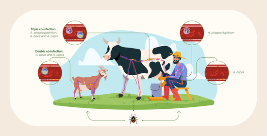
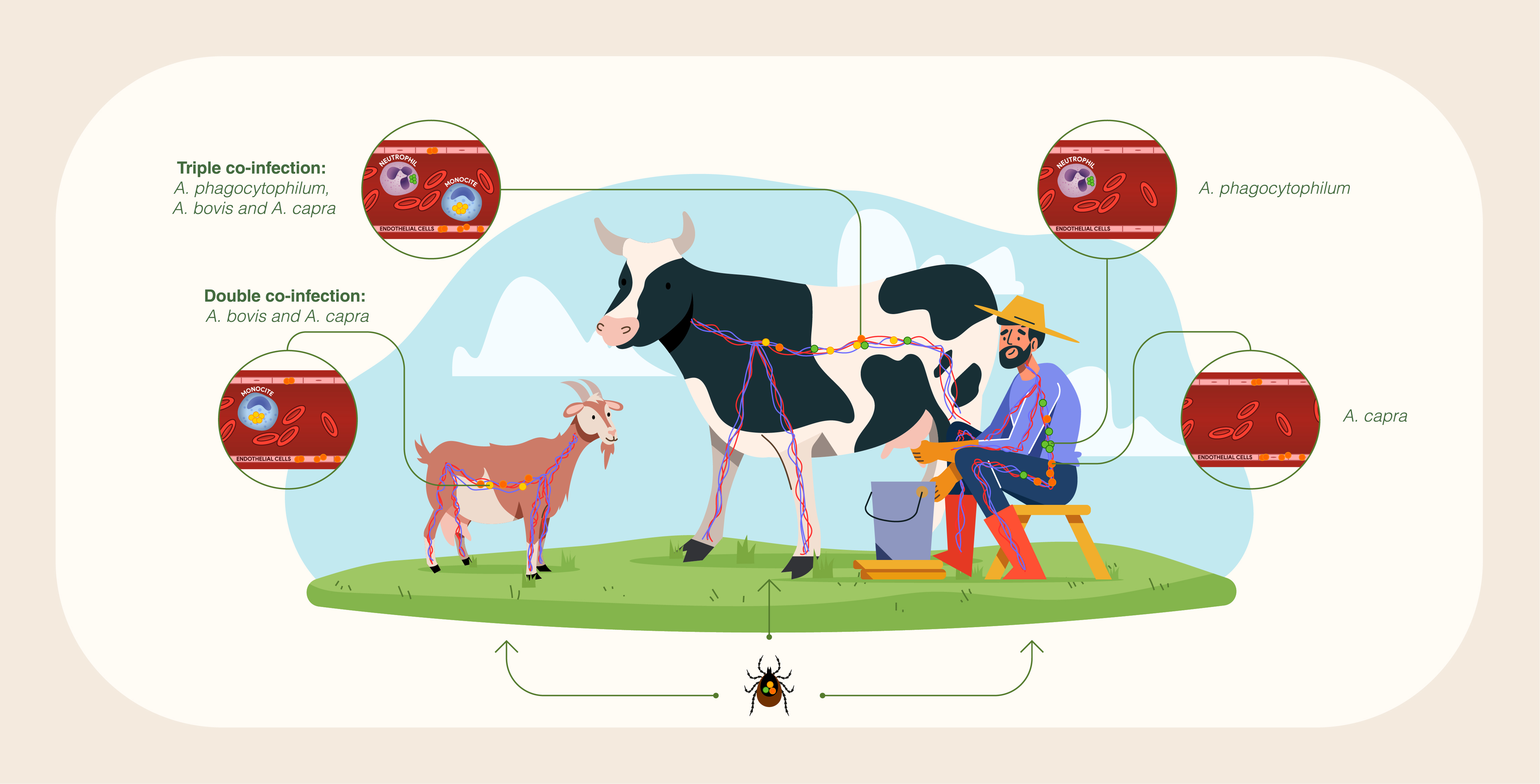
2.2. Anaplasma Species Prevalence: Single Infection and Co-Infection Cases

As described in Table 1, 16 out of 384 cattle blood samples tested positive for A. phagocytophilum (4.2%), which was the dominant single infection among the analyzed Anaplasma species, followed by A. bovis with 1.3% (5/384). Single infection with A. capra occurred in only one cow (0.3%) from the Chungbuk-do province. A. ovis infection was not found in any of the tested cattle blood samples. Interestingly, co-infection with two pathogens (A. bovis and A. phagocytophilum) was found in 64 cattle (16.7%). Additionally, a special case of triple infection caused by the pathogens A. bovis, A. phagocytophilum, and A. capra, was identified in only one cow (0.3%) from Gyeongsangbuk-do province.
On the other hand, goats were infected with A. bovis and A. capra. The dominant species in all the single infection cases was A. bovis with 6.3% (19/302), followed by A. capra, which was present in 0.3% of the total number of goats analyzed. Neither A. phagocytophilum nor A. ovis was detected in the analyzed goat blood samples. In contrast with the cattle results, co-infection cases in goats were caused by A. bovis and A. capra, reaching an infection rate of 1.0% (3/302). These findings indicate that the overall infection rate per host was 22.7% (87/384) in cattle and 8.0% (23/302) in goats, as shown in Table 1.

2.3. Total Number of Animals Infected per Anaplasma Species Analyzed

Infection rates of Anaplasma spp. were also calculated for each etiological agent. In the present study, 81 (21.1%) cattle were positive for A. phagocytophilum, 70 (18.2%) for A. bovis, and 2 (0.5%) for A. capra. On the other hand, 22 (7.3%) goats were positive for A. bovis and four (1.3%) for A. capra (Table 2).

2.4. PCR and Molecular Identification

PCR amplification of the 16S rRNA gene was successfully achieved for the identification of A. phagocytophilum, A. bovis, and A. capra, generating fragments of 925 bp, 547 bp, and 1499 bp, respectively. For multilocus genotyping of A. phagocytophilum, no samples were positive for ankyrin-related protein (ankA) and major surface protein 2 (msp2) gene fragments. Amplification of the A. bovis heat shock protein (groEL) gene was successfully achieved with a fragment length of 845 bp. Regarding A. capra, two cattle and four goats were positive for its 16S rRNA gene (1499 bp) as well as for the citrate synthase (gltA) gene (594 bp). However, only one goat was positive for groEL gene (874 bp) and two goats were positive for the major surface protein 4 (msp4) gene fragment with a length of 656 bp.

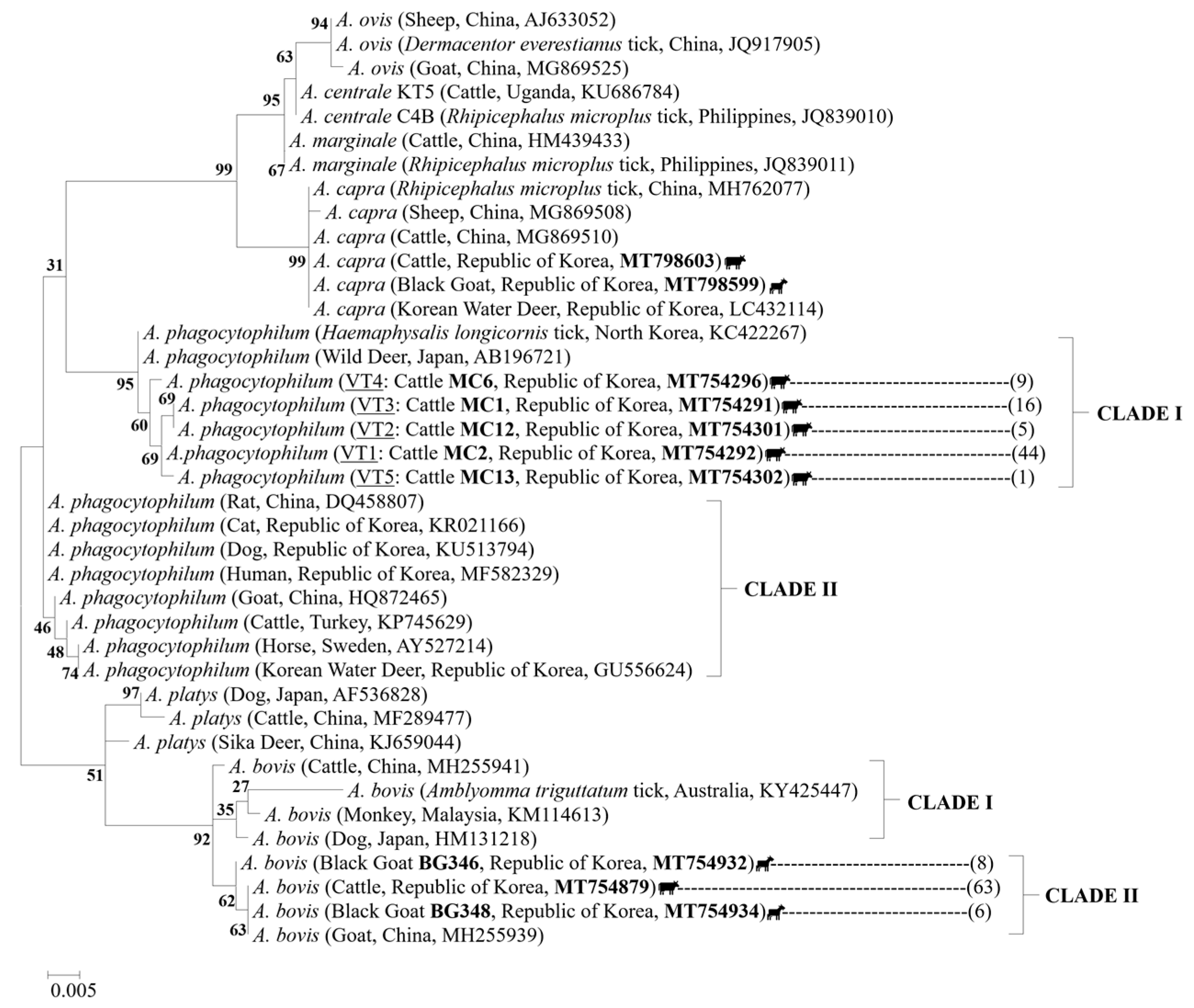
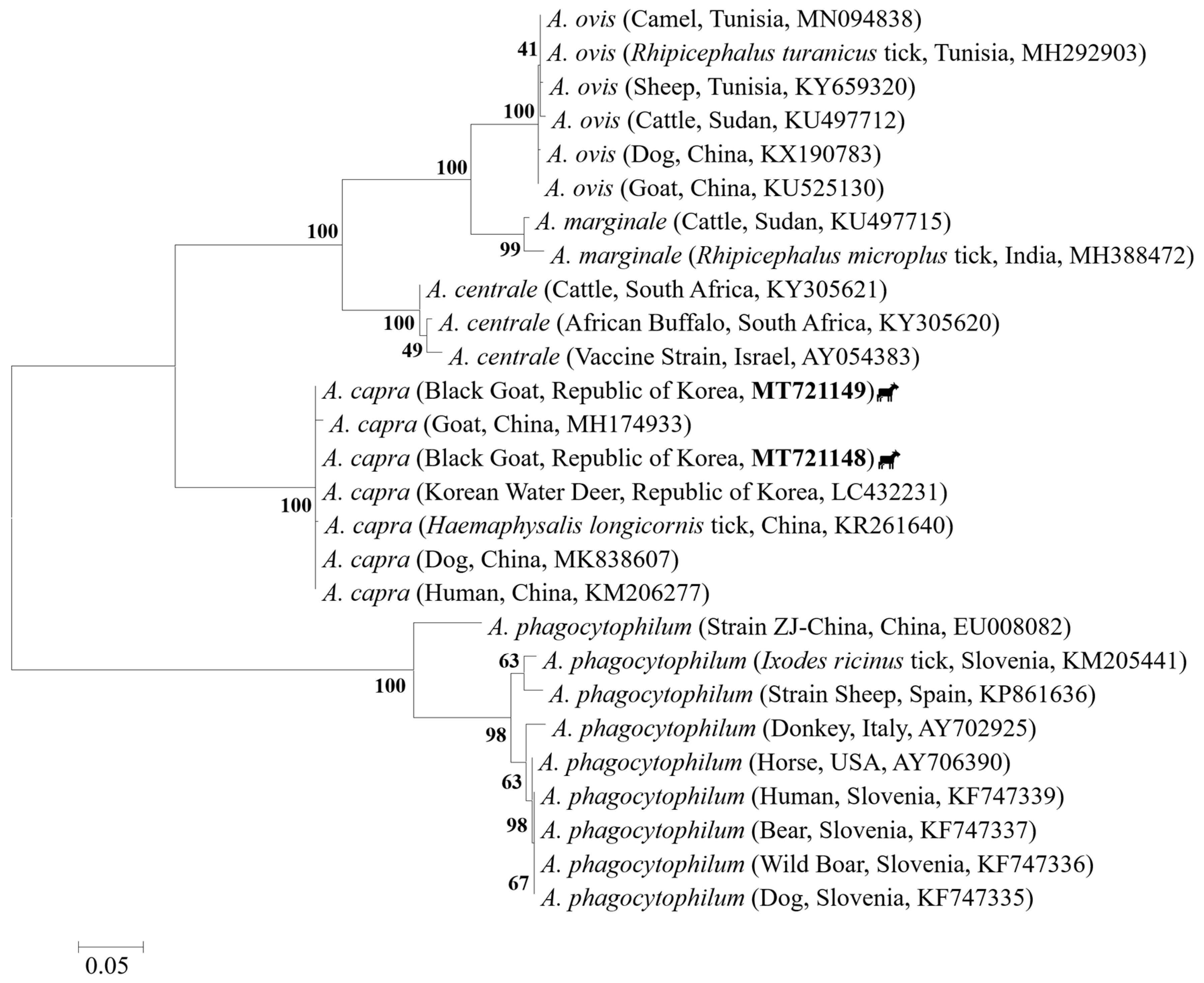
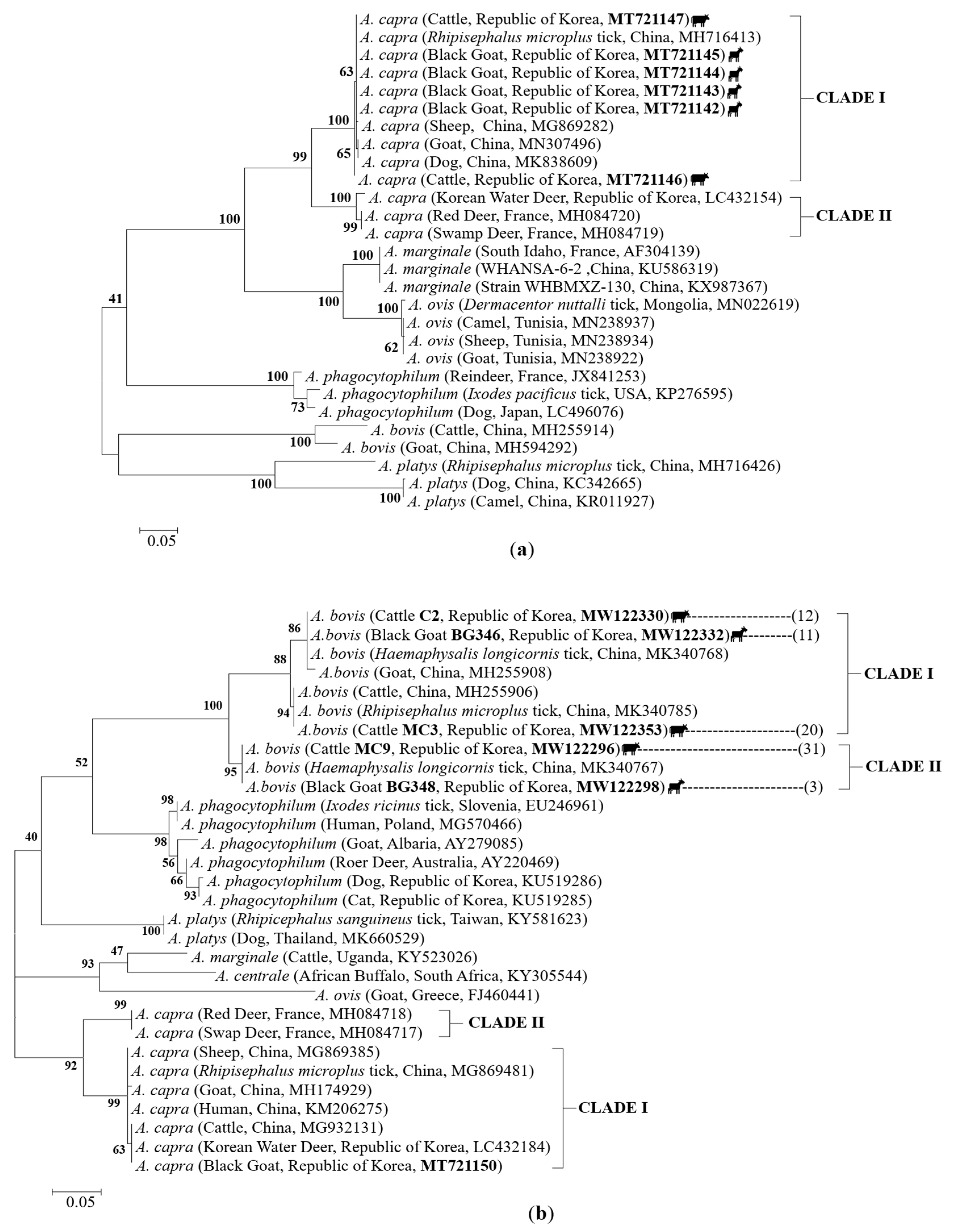
2.5. Sequences and Phylogenetic Analysis

To investigate the genetic variability of A. phagocytophilum, the 75 gene sequences obtained from cattle were analyzed and compared with sequences downloaded from the National Center for Biotechnology Information (NCBI). Analysis based on the A. phagocytophilum 16S rRNA gene fragments (925 bp) revealed the presence of five distinct strains, named variant type (VT): VT1 (n = 44), VT2 (n = 5), VT3 (n = 16), VT4 (n = 9) and VT5 (n = 1). The phylogenetic analysis demonstrated that these strains were classified into the first clade together with sequences previously isolated from North Korean H. longicornis ticks (KC422267) and Japanese wild deer (AB196721), sharing an identity percentage range of 98.0–99.6% and 96.3–99.6%, respectively (Figure 3).
Further analyses of A. phagocytophilum 16S rRNA gene sequences revealed that the strains obtained in this study shared a high degree of identity (99.5–99.6%) with North Korean ticks isolates (Table S1); however, they presented single and double nucleotide polymorphisms, in which one or two consecutive nucleotides were altered compared with the reference sequence (KC422267; Figure 4), suggesting that all of them are novel variants of the A. phagocytophilum 16S rRNA gene.
The phylogenetic analysis of A. bovis 16S rRNA gene enabled classification of this Anaplasma species into two different clades. Clade I included isolates from China, Australia, Malaysia, and Japan, while clade II contained the 63 cattle sequences and the two different strains found in goat blood samples tested in this study [BG346 (n = 8) and BG348 (n = 6)], along with those from Chinese goats (MH255939; Figure 3), which were highly related, sharing an identity percentage of 100% with cattle isolates, 99.8% with variant BG346, and 100% with the variant BG348 (Table S2). The variant BG346 exhibited the highest sequence identity (100%) with H. longicornis tick isolates (GU064901) from the ROK.
A. capra 16S rRNA sequences obtained from cattle and goats showed 100% identity with the sequences detected in Rhipicephalus microplus ticks (MH762077) and cattle (MG869510) from China and Korean water deer (LC432114; Table S3). Although only two goat sequences were generated through amplification of the A. capra msp4 gene (Figure 5), the results of the phylogenetic classification were identical to those shown by amplification of the 16S rRNA gene (Figure 3). In other words, all A. capra sequences of these two genes were classified into an independent clade clearly distinct from other members of Anaplasma species. The msp4 sequences showed 100% identity with the sequences isolated from Korean water deer (LC432231), dogs (MK838607), and humans (KM206277) from China (Table S4).
In contrast, phylogenetic analysis of A. capra based on gltA and groEL genes classified the sequences into two major clades (Figure 6). Sequence analysis of the A. capra gltA gene revealed two different variants in this study. Five isolates (MT721147, MT721145, MT721144, MT721143, and MT721142) were found to be 100% identical to those obtained from Chinese R. microplus ticks (MH716413), while the isolate MT721146 obtained from a cattle showed an identity of 99.8% (with one substitution, G/A at position 460) with Chinese tick isolates (MH716413), and 99.6% with the rest of the clustered sequences, which were isolated from Chinese goats, dogs, and sheep (Figure 6a, Table S5), suggesting that this is a novel variant of the A. capra gltA gene, which has never been reported before (Figure S1).
For the A. capra groEL gene, one sequence was acquired from a goat (MT721150). This sequence shared 100% identity with the isolates from Chinese cattle (MG932131) and Korean water deer (LC432184) belonging to clade I, together with samples isolated from humans (99.6%, KM206275), sheep (99.6%, MG869385), R. microplus ticks (99.6%, MG869481), and goats (99.2%, MH174929) from China (Figure 6b, Table S6).
For A. bovis groEL gene sequences, three different variants were obtained: variant I (represented by the isolates C2 [cattle] and BG346 [goat]), variant II (represented by the isolate MC3 [cattle]), and variant III (represented by the isolates MC9 [cattle] and BG348 [goat]). Variant I shared 100% identity with H. longicornis ticks (MK340768), while variant II was 100% identical to R. microplus ticks (MK340785). Both variants belonged to clade I together with Chinese isolates. Finally, variant III showed 100% identity with the sequences from Chinese H. longicornis ticks (MK340767), thus representing the second clade (Figure 6b, Table S7).

3. Discussion

In recent years, environmental factors such as global warming and deforestation have favored the increase in tick populations due to changes in their seasonal activity. However, the introduction of new animal species acting as a potential reservoir of one or even multiple pathogen species has led to a rapid distribution and expansion in the number of ticks. This phenomenon can be exemplified by anaplasmosis, a tick-borne disease distributed worldwide that has evolved in its pathogenesis over time. Despite recent reports of new species belonging to the genus Anaplasma and the considerable increase in their zoonotic potential, few studies on Anaplasma infection have been carried out in the ROK. The present study was mainly focused on investigating the genetic variability and prevalence of Anaplasma species as single and multiple infection in cattle and goats from different provinces of the ROK.
Our investigation revealed that the Korean cattle were mainly infected with A. phagocytophilum (21.1%, 81/384) instead of A. bovis, whose main hosts are supposed to be cows and buffalo, since its first identification in cattle in 1936 [23]. Of the 81 cattle infected with A. phagocytophilum, 80 were from Gyeongsangbuk-do province. Conversely, a study carried out in 2016 in the same province identified A. phagocytophilum with a relatively low infection rate (4.7%) [18] compared with the results obtained in our study (88.9%). A. bovis was the second dominant pathogen (18.2%, 70/384) identified in this study. Based on the geographic distribution, Gyeongsangbuk-do was also the province with a higher number of carriers of A. bovis (74.4%). Our results differed from those of previous molecular studies conducted in the ROK, which reported a comparatively low prevalence of A. bovis in cattle: 4.2% (3/71) in Jeju Island [12], 1.9% (11/581) in Gyeongsangnam-do, and 0.2% (1/638) in Gyeongsangbuk-do province [13]. This sudden increase in Anaplasma spp. prevalence rates may be largely associated with climate change. It is known that the Korean peninsula is gradually transitioning to a subtropical climate [13], where a hot summer season and changes in rainfall patterns can favor the abundance and spread of ticks to different localities.
In addition, the present study also detected A. capra infection in cattle. In 2018, A. capra was described for the first time in Korean cattle from Gyeongsangnam-do with a low infection rate (0.4%) [13]. These results are similar to those obtained in this study (0.5%). An important point to consider is that one cow positive for A. capra was infected with A. phagocytophilum and A. bovis, which is the first detection of triple infection with Anaplasma species in the ROK. It should also be noted that this is the first molecular study of co-infection cases caused by several Anaplasma species in cattle, in which triple and doble infection cases were detected, with the latter type involving A. phagocytophilum and A. bovis. Our findings, along with previous studies performed in the ROK [13,18], demonstrates that the cattle residing in provinces located at a lower latitude, such as Gyeongsangbuk-do and Gyeongsangnam-do, are naturally infected with Anaplasma spp., thus suggesting that cattle may play an important role in the enzootic maintenance of Anaplasma infection.
On the other hand, goats were found to be carriers of two Anaplasma species: A. bovis (7.3%) and A. capra (1.3%). The results obtained in the current study coincide with those previously described in Ulsan Metropolitan City, ROK, which reported infection rates of 8.6% and 2.2% for A. bovis and A. capra, respectively [16]. Interestingly, we also found three co-infection cases due to A. bovis and A. capra, two of which were in Jeollabuk-do and the other in Jeollanam-do province. Thus, our findings shed light on co-infection types that may be found among Korean goats. Although A. phagocytophilum and A. ovis were not identified in our study, we cannot rule them out as causative agents of caprine anaplasmosis, since a study conducted in China demonstrated the prevalence of these species, even reporting cases of triple infection with A. ovis, A. bovis, and A. phagocytophilum [15]. It is also worth mentioning that a serological study conducted in the ROK detected antibodies against the major surface protein 5 (msp5) of A. marginale, A. centrale, and A. ovis, using a commercial competitive enzyme-linked immunosorbent assay (ELISA). The seroprevalence rate reported in this study was 6.6% (36/544) in native Korean goats [30]. However, identification of a species among A. marginale, A. centrale, and A. ovis was not performed by PCR; these findings compared with our data suggest that Anaplasma infection in goats could be caused by other Anaplasma species, in addition to A. bovis and A. capra pathogens. Additional studies are necessary to corroborate this finding, taking into consideration a large sample size that involves the different Korean provinces.
Despite the fact that the genes that are most often targeted to investigate the genetic diversity of A. phagocytophilum involve the 16S rRNA locus, groESL operon, major surface protein coding genes (msp2 and msp4), and ankA gene [23], PCR amplification of the msp2 and ankA genes was unsuccessful in the present study. This could be due to the complex epidemiological cycles of A. phagocytophilum, which involves different genetic variants, vectors, and host species [23]. However, according to the results obtained by the amplification of the A. phagocytophilum 16S rRNA and its phylogenetic analysis, we demonstrated that five novel strains are circulating among Korean cattle. In addition, the multilocus genotyping of A. capra not only favored the ratification of cattle as a potential reservoir of this Anaplasma species but also contributed to the identification of a new variant of the A. capra gltA gene, thus aiding in elucidation of the genetic variability of A. phagocytophilum and A. capra, two species with zoonotic importance.

4. Material and Methods

4.1. Sample Collection

A total of 686 whole blood samples from domestic ruminants (384 cattle and 302 black goats) were collected from different provinces of the ROK, which were randomly selected between August 2015 and June 2020. These samples were collected in sterile 10 mL tubes containing EDTA anticoagulant and transported to the laboratory in an icepack container. Goat blood samples were collected in Jeollabuk-do (n = 37), Gwangju Metropolitan City (n = 41), and Jeollanam-do (n = 224) province, while cattle blood samples were collected in Gyeongsangbuk-do (n = 90), Gyeongsangnam-do (n = 65), Jeollanam-do (n = 65), Gyeonggi-do (n = 50), Chungbuk-do (n = 54), and Chungnam-do (n = 60) provinces (Figure 1).

4.2. DNA Extraction

Genomic DNA was extracted from 200 µL whole blood samples using a commercial LaboPass DNA Purification Kit (Cosmo Genetech, Seoul, Korea) according to the manufacturer’s instructions. The extracted DNA was stored at −20 °C until further analysis. The quantity and purity of the extracted DNA were calculated using a NanoDrop spectrophotometer (Implen NanoPhotometer Classic, Germany).

4.3. PCR Amplification

DNA samples were subjected to nested PCR (nPCR) to amplify the 16S rRNA gene fragments of A. phagocytophilum and A. bovis. The first round was performed using the primer pair AE1-F/AE1-R, which amplifies the 16S rRNA gene shared by all Anaplasma spp. (Table 3). In the second round, PCR-positive samples were reamplified by employing species-specific primer sets EE3/EE4 and ABKf/AB1r for A. phagocytophilum [31] and A. bovis [32], respectively. For multilocus genotyping, A. phagocytophilum msp2 [33] and ankA [34] gene fragments were amplified using nPCR, and A. bovis groEL gene using semi-nested PCR [35]. For A. capra, the 16S rRNA [7], gltA [36], groEL, and msp4 [37] genes were amplified using the primer sets described in Table 3. The A. ovis msp4 gene was amplified using single-step PCR with the primers pairs MSP45/MSP43 [38]. These reactions were performed in a total volume of 30 µL, containing 10 pmol of each primer, 15 µL of 2x Taq PCR Pre-mix (BioFACT, Daejeon, Korea), 50 to 100 ng of genomic DNA samples for the first round of PCR, and 1 µL of the first PCR product was used as template DNA for the nPCR. Each reaction was conducted in a SimpliAmp Thermal Cycler (Thermo Fisher Scientific, Korea) under optimal cycling conditions (Table 3). The PCR products were visualized under UV light after 1.5% agarose gel electrophoresis, using a 100 bp ladder (SiZer-100 DNA Marker Solution, iNtRON Biotechnology, Korea) as a DNA size marker.

4.4. DNA Sequencing and Phylogenetic Analysis

The PCR products were purified using the DNA Gel Extraction S & V Kit (BIONICS, Daejeon, ROK) and were directly sequenced using an Applied Biosystems 3730 DNA Analyzer (Thermo Fisher Scientific, Foster City, USA). The obtained sequences were evaluated with Chromas software, aligned using the multiple sequence alignment program ClustalX 2.1, compared with reference sequences searched in the NCBI, analyzed using the Basic Local Alignment Search Tool to determine the identity percentage between them, and finally examined with a similarity matrix. Relationships between individuals were assessed by the neighbor-joining method with nucleotide distance (p distance) for 1000 replications with a bootstrap analysis. A phylogenetic tree was constructed based on nucleotide alignment using MEGA 6.06 software.

4.5. Nucleotide Sequence Accession Numbers

The sequences obtained in this study have been deposited in the GenBank database with the following accession numbers: A. phagocytophilum 16S rRNA (MT754291 to MT754365), A. bovis 16S rRNA (MT754858 to MT754934), A. bovis groEL (MW122296 to MW122372), A. capra 16S rRNA (MT798599 to MT798604), A. capra msp4 (MT721148 to MT721149), A. capra gltA (MT721142 to MT721147), and A. capra groEL (MT721150).

5. Conclusions

Based on the results obtained in this study, in the ROK, cattle are mainly infected with A. phagocytophilum, while goats act mainly as carriers of A. bovis. This study has contributed to the ratification of cattle as a potential reservoir of the emerging zoonotic pathogen, A. capra. Although A. ovis was not detected, we cannot rule it out as a causative agent of bovine and caprine anaplasmosis; further studies are needed to corroborate this finding. Our study sheds light on the geographical distribution of Anaplasma infection types; however, supplementary studies are needed to clarify the clinical presentation and epidemiological significance of genetic variants of Anaplasma species to establish effective prevention and control strategies.

Supplementary Materials

The following are available online at https://www.mdpi.com/2076-0817/10/1/28/s1, Figure S1: DNA sequencing electropherogram, Table S1: Genetic identity matrix based on the 16S rRNA gene fragments of A. phagocytophilum (547 bp), Table S2: Genetic identity matrix based on the 16S rRNA gene fragments of A. bovis (547 bp), Table S3: Genetic identity matrix based on the 16S rRNA gene fragments of A. capra (547 bp), Table S4: Genetic identity matrix based on the msp4 gene fragments of A. capra (527 bp), Table S5: Genetic identity matrix based on the gltA gene fragments of A. capra (480 bp), Table S6: Genetic identity matrix based on the groEL gene fragments of A. capra (238 bp), Table S7: Genetic identity matrix based on the groEL gene fragments of A. bovis (238 bp).

Author Contributions

Conceptualization, E.A.M. and J.-S.C.; methodology, E.A.M. and J.-S.C.; software, E.A.M.; validation, E.A.M., K.-S.C. and J.-S.C.; formal analysis, E.A.M.; investigation, E.A.M.; resources, S.-W.H., Y.-K.C., K.-S.C. and J.-S.C.; data curation, E.A.M.; writing—original draft preparation, E.A.M.; writing—review and editing, E.A.M.; visualization, E.A.M. and J.-S.C.; supervision, S.-W.H., Y.-K.C. and J.-S.C.; project administration, J.-S.C.; funding acquisition, J.-S.C. All authors have read and agreed to the published version of the manuscript.

Funding

This research was funded by the Government-wide R&D Fund for Infectious Diseases Research (HG18C0021) in the Republic of Korea.

Institutional Review Board Statement

The study was conducted according to the guidelines of the Declaration of Helsinki, and approved by the Ethics Committee of Seoul National University (Animal Care and Use Committee [IACUC] Decision No. SNU-190524-2-1).

Informed Consent Statement

Informed consent was obtained from all animals involved in the study.

Data Availability Statement

The authors confirm that the data supporting the findings of this study are available within the article and its supplementary materials.

Acknowledgments

This research was supported by the Laboratory of Veterinary Internal Medicine and the BK21 PLUS Program for Creative Veterinary Science Research, Research Institute for Veterinary Science and College of Veterinary Medicine, Seoul National University.

Conflicts of Interest

The authors declare no conflict of interest.

References

  1. Aubry, P.; Geale, D.W. A review of Bovine anaplasmosis. Transbound. Emerg. Dis. 2011, 58, 1–30. [Google Scholar] [CrossRef] [PubMed]
  2. Theiler, A. Anaplasma marginale (Gen. and Spec. Nov.): The Marginal Points in the Blood of Cattle Suffering from a Specific Disease; Report of the Government Veterinary Bacteriologist of the Transvaal; Transvaal Department of Agriculture: Pretoria, South Africa, 1910; pp. 7–64.
  3. Woldehiwet, Z. The natural history of Anaplasma phagocytophilum. Vet. Parasitol. 2010, 167, 104–122. [Google Scholar] [CrossRef] [PubMed]
  4. Sreekumar, C.; Anandan, R.; Balasundaram, S.; Rajavelu, G. Morphology and staining characteristics of Ehrlichia bovis. Comp. Immunol. Microbiol. Infect. Dis. 1996, 1, 79–83. [Google Scholar] [CrossRef]
  5. Uilenberg, G.; Van Vorstenbosch, C.J.A.H.V.; Perié, N.M. Blood parasites of sheep in the netherlands. I. Anaplasma mesaeterum sp.n. (Rickettsiales, Anaplasmataceae). Vet. Q. 1979, 1, 14–22. [Google Scholar] [CrossRef] [PubMed]
  6. Munderloh, U.G.; Lynch, M.J.; Herron, M.J.; Palmer, A.T.; Kurtti, T.J.; Nelson, R.D.; Goodman, J.L. Infection of endothelial cells with Anaplasma marginale and A. phagocytophilum. Vet. Microbiol. 2004, 101, 53–64. [Google Scholar] [CrossRef] [PubMed]
  7. Li, H.; Zheng, Y.C.; Ma, L.; Jia, N.; Jiang, B.G.; Jiang, R.R.; Huo, Q.B.; Wang, Y.W.; Liu, H.B.; Chu, Y.L.; et al. Human infection with a novel tick-borne Anaplasma species in China: A surveillance study. Lancet Infect. Dis. 2015, 15, 663–670. [Google Scholar] [CrossRef]
  8. de la Fuente, J.; Lew, A.; Lutz, H.; Meli, M.L.; Hofmann-Lehmann, R.; Shkap, V.; Molad, T.; Mangold, A.J.; Almazán, C.; Naranjo, V.; et al. Genetic diversity of Anaplasma species major surface proteins and implications for anaplasmosis serodiagnosis and vaccine development. Anim. Health Res. Rev. 2005, 6, 75–89. [Google Scholar] [CrossRef] [Green Version]
  9. Underwood, W.J.; Blauwiekel, R.; Delano, M.L.; Gillesby, R.; Mischler, S.A.; Schoell, A. Chapter 15—Biology and Diseases of Ruminants (Sheep, Goats, and Cattle). In Laboratory Animal Medicine, 3rd ed.; Academic Press: Cambridge, MA, USA, 2015; pp. 623–694. [Google Scholar] [CrossRef]
  10. Ybañez, A.P.; Inokuma, H. Anaplasma species of veterinary importance in Japan. Vet. World 2016, 11, 1190–1196. [Google Scholar] [CrossRef] [Green Version]
  11. Han, D.G.; Ryu, J.H.; Chae, J.B.; Kim, D.W.; Kwon, C.H.; Choi, K.S. First report of Anaplasma phagocytophilum infection in Holstein cattle in the Republic of Korea. Acta Trop. 2018, 183, 110–113. [Google Scholar] [CrossRef]
  12. Park, J.; Han, D.G.; Ryu, J.H.; Chae, J.B.; Chae, J.S.; Yu, D.H.; Park, B.K.; Kim, H.C.; Choi, K.S. Molecular detection of Anaplasma bovis in Holstein cattle in the Republic of Korea. Acta Vet. Scand. 2018, 60, 1–5. [Google Scholar] [CrossRef]
  13. Seo, M.G.; Ouh, I.O.; Lee, H.; Geraldino, P.J.L.; Rhee, M.H.; Kwon, O.D.; Kwak, D. Differential identification of Anaplasma in cattle and potential of cattle to serve as reservoirs of Anaplasma capra, an emerging tick-borne zoonotic pathogen. Vet. Microbiol. 2018, 226, 15–22. [Google Scholar] [CrossRef] [PubMed]
  14. Carelli, G.; Decaro, N.; Lorusso, E.; Paradies, P.; Elia, G.; Martella, V.; Buonavoglia, C.; Ceci, L. First report of bovine anaplasmosis caused by Anaplasma centrale in Europe. Proc. Ann. N. Y. Acad. Sci. 2008, 1149, 107–110. [Google Scholar] [CrossRef] [PubMed]
  15. Liu, Z.; Ma, M.; Wang, Z.; Wang, J.; Peng, Y.; Li, Y.; Guan, G.; Luo, J.; Yin, H. Molecular survey and genetic identification of Anaplasma species in goats from central and southern China. Appl. Environ. Microbiol. 2012, 78, 464–470. [Google Scholar] [CrossRef] [PubMed] [Green Version]
  16. Seo, H.J.; Jin, B.C.; Kim, K.H.; Yoo, M.S.; Seong, K.W.; Jeong, S.J.; Hyun, B.H.; Cho, Y.S. Molecular detection and phylogenetic analysis of Anaplasma spp. in Korean native goats from Ulsan Metropolitan City, Korea. Vector-Borne Zoonotic Dis. 2019, 19, 773–776. [Google Scholar] [CrossRef]
  17. Yang, J.; Li, Y.; Liu, Z.; Liu, J.; Niu, Q.; Ren, Q.; Chen, Z.; Guan, G.; Luo, J.; Yin, H. Molecular detection and characterization of Anaplasma spp. in sheep and cattle from Xinjiang, northwest China. Parasites Vectors 2015, 8, 108. [Google Scholar] [CrossRef] [PubMed] [Green Version]
  18. Seo, M.G.; Ouh, I.O.; Kwon, O.D.; Kwak, D. Molecular detection of Anaplasma phagocytophilum-like Anaplasma spp. and pathogenic A. Phagocytophilum in cattle from South Korea. Mol. Phylogenet. Evol. 2018, 126, 23–30. [Google Scholar] [CrossRef]
  19. Hoogstraal, H. Ticks in relation to human diseases caused by Rickettsia species. Annu. Rev. Entomol. 1967, 12, 377–420. [Google Scholar] [CrossRef]
  20. Kim, K.H.; Yi, J.; Oh, W.S.; Kim, N.H.; Choi, S.J.; Choe, P.G.; Kim, N.J.; Lee, J.K.; Oh, M.D. Human granulocytic anaplasmosis, South Korea, 2013. Emerg. Infect. Dis. 2014, 20, 1708. [Google Scholar] [CrossRef]
  21. Chochlakis, D.; Ioannou, I.; Tselentis, Y.; Psaroulaki, A. Human anaplasmosis and Anaplasma ovis variant. Emerg. Infect. Dis. 2010, 16, 1031–1032. [Google Scholar] [CrossRef]
  22. Heo, E.J.; Park, J.H.; Koo, J.R.; Park, M.S.; Park, M.Y.; Dumler, J.S.; Chae, J.S. Serologic and molecular detection of Ehrlichia chaffeensis and Anaplasma phagocytophila (human granulocytic ehrlichiosis agent) in Korean patients. J. Clin. Microbiol. 2002, 40, 3082–3085. [Google Scholar] [CrossRef] [Green Version]
  23. Battilani, M.; De Arcangeli, S.; Balboni, A.; Dondi, F. Genetic diversity and molecular epidemiology of Anaplasma. Infect. Genet. Evol. 2017, 49, 195–211. [Google Scholar] [CrossRef] [PubMed]
  24. Kang, J.G.; Ko, S.; Kim, H.C.; Chong, S.T.; Klein, T.A.; Chae, J.B.; Jo, Y.S.; Choi, K.S.; Yu, D.H.; Park, B.K.; et al. Prevalence of Anaplasma and Bartonella spp. In ticks collected from Korean water deer (Hydropotes inermis argyropus). Korean J. Parasitol. 2016, 54, 87. [Google Scholar] [CrossRef] [PubMed] [Green Version]
  25. Seo, M.G.; Kwon, O.D.; Kwak, D. Molecular and phylogenetic analysis of tick-borne pathogens in ticks parasitizing native korean goats (Capra hircus coreanae) in South Korea. Pathogens 2020, 9, 71. [Google Scholar] [CrossRef] [Green Version]
  26. Kang, S.W.; Doan, H.T.T.; Choe, S.E.; Noh, J.H.; Yoo, M.S.; Reddy, K.E.; Kim, Y.H.; Kweon, C.H.; Jung, S.C.; Chang, K.Y. Molecular investigation of tick-borne pathogens in ticks from grazing cattle in Korea. Parasitol. Int. 2013, 62, 276–282. [Google Scholar] [CrossRef]
  27. Oh, J.Y.; Moon, B.C.; Bae, B.K.; Shin, E.H.; Ko, Y.H.; Kim, Y.J.; Park, Y.H.; Chae, J.S. Genetic identification and phylogenetic analysis of Anaplasma and Ehrlichia species in Haemaphysalis longicornis collected from Jeju island, Korea. J. Bacteriol. Virol. 2009, 39, 257–267. [Google Scholar] [CrossRef] [Green Version]
  28. Durden, L.A. Taxonomy, host associations, life cycles and vectorial importance of ticks parasitizing small mammals. In Micromammals and Macroparasites: From Evolutionary Ecology to Management; Springer: Tokyo, Japan, 2006; pp. 91–102. [Google Scholar] [CrossRef]
  29. Baneth, G. Tick-borne infections of animals and humans: A common ground. Int. J. Parasitol. 2014, 44, 591–596. [Google Scholar] [CrossRef]
  30. Lee, S.H.; Jung, B.Y.; Kwak, D. Evidence of Anaplasma spp. exposure in native Korean goats (Capra hircus coreanae). Vet. Med. 2015, 60, 248–252. [Google Scholar] [CrossRef] [Green Version]
  31. Barlough, J.E.; Madigan, J.E.; DeRock, E.; Bigornia, L. Nested polymerase chain reaction for detection of Ehrlichia equi genomic DNA in horses and ticks (Ixodes pacificus). Vet. Parasitol. 1996, 63, 319–329. [Google Scholar] [CrossRef]
  32. Kang, J.G.; Ko, S.; Kim, Y.J.; Yang, H.J.; Lee, H.; Shin, N.S.; Choi, K.S.; Chae, J.S. New genetic variants of Anaplasma phagocytophilum and Anaplasma bovis from Korean water deer (Hydropotes inermis argyropus). Vector-Borne Zoonotic Dis. 2011, 11, 929–938. [Google Scholar] [CrossRef]
  33. Lin, Q.; Rikihisa, Y.; Felek, S.; Wang, X.; Massung, R.F.; Woldehiwet, Z. Anaplasma phagocytophilum has a functional msp2 gene that is distinct from p44. Infect. Immun. 2004, 3883–3889. [Google Scholar] [CrossRef] [Green Version]
  34. Massung, R.F.; Owens, J.H.; Ross, D.; Reed, K.D.; Petrovec, M.; Bjoersdorff, A.; Coughlin, R.T.; Beltz, G.A.; Murphy, C.I. Sequence analysis of the ank gene of granulocytic ehrlichiae. J. Clin. Microbiol. 2000, 36, 1090–1095. [Google Scholar] [CrossRef] [PubMed] [Green Version]
  35. Guo, W.P.; Wang, X.; Li, Y.N.; Xu, G.; Wang, Y.H.; Zhou, E.M. GroEL gene typing and genetic diversity of Anaplasma bovis in ticks in Shaanxi, China. Infect. Genet. Evol. 2019, 74, 103927. [Google Scholar] [CrossRef] [PubMed]
  36. Yang, J.; Liu, Z.; Niu, Q.; Liu, J.; Han, R.; Liu, G.; Shi, Y.; Luo, J.; Yin, H. Molecular survey and characterization of a novel Anaplasma species closely related to Anaplasma capra in ticks, northwestern China. Parasites Vectors 2016, 9, 603. [Google Scholar] [CrossRef] [PubMed] [Green Version]
  37. Yang, J.; Liu, Z.; Niu, Q.; Liu, J.; Han, R.; Guan, G.; Hassan, M.A.; Liu, G.; Luo, J.; Yin, H. A novel zoonotic Anaplasma species is prevalent in small ruminants: Potential public health implications. Parasites Vectors 2017, 10, 264. [Google Scholar] [CrossRef]
  38. de la Fuente, J.; Atkinson, M.W.; Naranjo, V.; Fernández de Mera, I.G.; Mangold, A.J.; Keating, K.A.; Kocan, K.M. Sequence analysis of the msp4 gene of Anaplasma ovis strains. Vet. Microbiol. 2007, 119, 375–381. [Google Scholar] [CrossRef]
Figure 1. Cattle from the Gyeongsangbuk-do province with the highest Anaplasma spp. infection rates. A severe tick infestation was evident. (a) Ticks were attached behind the tail, (b) around the anus, where there was an accumulated number of ticks feeding on the host. (c) Ticks were also attached to the legs as well to (d) the dewlap; lesions caused by ticks were perceptible in this region. (e) Teats and (f) rear udder were the areas that presented the heaviest infestation, where different tick stages were evident. Additionally, local redness (red spots) and rashes were noticeable in those sites.
Pathogens 10 00028 g001
Figure 2. Geographical distribution of Anaplasma species in the provinces sampled in the Republic of Korea. Pie charts represent Anaplasma spp. infection rates for single infection and co-infection cases identified in each tested province. The cattle and goat icons indicate the sampled sites for respective animal species. A. phago: Anaplasma phagocytophilum.
Pathogens 10 00028 g002
Figure 3. Phylogenetic tree based on the 16S rRNA gene fragments of the Anaplasma species (547 bp). The sequence alignments were performed among the A. phagocytophilum, A. bovis and A. capra sequences obtained in this study and other members of the family Anaplasmataceae. The phylogenetic tree was constructed using the neighbor-joining method with 1000 replicates of the alignment (MEGA 6.06 software). Cattle and goat icons indicate the sequences found in this study for the respective animal species. Isolate, country, and GenBank accession numbers are shown in parentheses. Novel variants of A. phagocytophilum are indicated by the abbreviation VT (variant type). The numbers in brackets represent the total number of sequences that are identical to the representative sequence. The scale bar represents the number of nucleotide substitutions between sequences. Clades are denoted by roman numbers. Numbers on the branches indicate percent support for each clade.
Pathogens 10 00028 g003
Figure 4. Novel variants of Anaplasma phagocytophilum 16S rRNA gene found in cattle. KC422267: reference sequence. Through the comparison between the reference sequence and the five variants identified in this study, single nucleotide polymorphisms (SNPs) were identified at positions 34, 36, 38, 740, and 743 with variation A/G; and positions 527 and 713 with variation C/T. Double nucleotide polymorphisms (DNPs) were found at positions 38 and 39 variation A/G, which are indicated by bold letters. The haplotypes generated due to the DNA variations found along the sequences are shown.
Pathogens 10 00028 g004
Figure 5. Phylogenetic tree based on the msp4 gene fragments of the Anaplasma species (527 bp). The sequence alignments were performed among the A. capra sequences obtained in this study and other members of the family Anaplasmataceae. The phylogenetic tree was constructed using the neighbor-joining method with 1000 replicates of the alignment (MEGA 6.06 software). Goat icons indicate the sequences found in this study. Isolate, country, and GenBank accession numbers are shown in parentheses. The scale bar represents the number of nucleotide substitutions between sequences. Numbers on the branches indicate percent support for each clade.
Pathogens 10 00028 g005
Figure 6. Phylogenetic tree based on the (a) gltA and (b) groEL gene fragments of the Anaplasma species. The phylogenetic tree was constructed using the neighbor-joining method with 1000 replicates of the alignment (MEGA 6.06 software). The numbers of nucleotides were 480 bp and 238 bp in the final alignment for gltA and groEL genes, respectively. Cattle and goat icons indicate the sequences found in this study. The numbers in brackets represent the total number of sequences that are identical to the representative sequence. Isolate, country, and GenBank accession numbers are shown in parentheses. The scale bar represents the number of nucleotide substitutions between sequences. Clades are denoted by roman numbers. Numbers on the branches indicate percent support for each clade.
Pathogens 10 00028 g006
Table 1. Prevalence of single infection and co-infection cases with Anaplasma species detected in cattle and goat blood samples.
Animal SpeciesNo. TestedNo. of Positive (IR 1, %)Type of Infection Identified in Tested Blood SamplesNot Infected (%)
Single InfectionDouble InfectionTriple Infection
A.
phago2 (%)
A.
bovis (%)
A.
capra (%)
A.
ovis (%)
A. bovis + A. phago 2 (%)A. bovis + A. capra (%)A. phago2 + A. bovis + A. capra (%)
Cattle38487 (22.7)16 (4.2)5 (1.3)1 (0.3)0 (0)64 (16.7)0 (0)1 (0.3)297 (77.3)
Goat30223 (8.0)0 (0)19 (6.3)1 (0.3)0 (0)0 (0)3 (1.0)0 (0)279 (94.4)
Total686110 (16.0)16 (2.3)24 (3.5)2 (0.3)0 (0)64 (9.3)3 (0.4)1 (0.1)576 (83.9)
1 IR, infection rate; 2 A. phago: Anaplasma phagocytophilum.
Table 2. Prevalence rate per Anaplasma species analyzed in domestic ruminants in the Republic of Korea.
HostCollected ProvinceNo. TestedA. phago1 (%)A. bovis (%)A. capra (%)A. ovis (%)
CattleGyeongsangbuk-do9080 (88.9)67 (74.4)1 (1.1)0 (0)
Gyeongsangnam-do650 (0)0 (0)0 (0)0 (0)
Jeollanam-do650 (0)1 (1.5)0 (0)0 (0)
Gyonggi-do500 (0)0 (0)0 (0)0 (0)
Chungbuk-do541 (1.9)1 (1.9)1 (1.9)0 (0)
Chungnam-do600 (0)1 (1.7)0 (0)0 (0)
Subtotal38481 (21.1)70 (18.2)2 (0.5)0 (0)
GoatJeollabuk-do370 (0)10 (27.0)2 (5.4)0 (0)
Gwangju Metropolitan City410 (0)3 (7.3)0 (0)0 (0)
Jeollanam-do2240 (0)9 (4.0)2 (1.0)0 (0)
Subtotal3020 (0)22 (7.3)4 (1.3)0 (0)
Grand total68681 (11.8)92 (13.4)6 (0.9)0 (0)
1A. phago: Anaplasma phagocytophilum.
Table 3. Oligonucleotide primers and polymerase chain reaction (PCR) conditions used in this study.
SpeciesTarget GenePrimer Name and PCR ConditionsPrimer Sequences (5′-3′)CyclesAmplicon Size (bp)References
Denaturation (°C/min)Annealing
(°C/min)
Extension
(°C/min)
Anaplasma spp.16S rRNA1AE1-FAAGCTTAACACATGCAAGTCGAA351406Oh et al. (2009) [27]
AE1-RAGTCACTGACCCAACCTTAAATG
Conditions94/156/172/1.5
A. phagocytophilum16S rRNA2EE3GTCGAACGGATTATTCTTTATAGCTTGC25926Barlough et al. (1996) [31]
EE4CCCTTCCGTTAAGAAGGATCTAATCTCC
Conditions94/0.5056/0.5072/0.75
msp2msp2fullFTCAGAAAGATACACGTGCGCCC351079Lin et al. (2004) [33]
msp2fullRTTATGATTAGGCCTTTGGGCATG
Conditions94/154/172/1
msp2FGGTTACATAAGGGCCGCAAAGGTG25467
msp2RCCGGCGCATGTGTAAGGTGAAA
Conditions94/0.557/0.572/0.5
ankAU7GCGTCTGTAAGGCAGATTGTG351696Massung et al. (2000) [34]
1R1TATACACCTGGAGTAGGAAC
Conditions94/157/172/1.5
U8TAAGATAGGTTTAGTAAGACG25460
1R7TGCATCGTCATTACGCACAAGGTC
Conditions94/0.7557/0.7572/0.75
A. bovis16S rRNA3ABKfTAGCTTGCTATGGGGACAA25547Kang et al. (2011) [32]
AB1rTCTCCCGGACTCCAGTCTG
Conditions94/0.559/0.572/0.5
groELbovis-groEL-F1GTTCGCAGTATTTTGCCAGT30845Guo et al. (2019) [35]
bovisgroEL-RCTGCRTTCAGAGTCATAAATAC
bovis-groEL-F2ATCTGGAAGRCCACTATTGAT
Conditions94/0.756/0.772/1
A. capra16S rRNAForwardTTGAGAGTTTGATCCTGGCTCAGAACG571499Li et al. (2015) [7]
ReverseWAAGGWGGTAATCCAGC
Conditions94/0.7557/0.7572/0.75
gltAOuter-fGCGATTTTAGAGTGYGGAGATTG301031
Outer-rTACAATACCGGAGTAAAAGTCAA
Conditions94/0.7555/0.7572/0.75
Inner-fTCATCTCCTGTTGCACGGTGCCC30594Yang et al. (2016) [36]
Inner-rCTCTGAATGAACATGCCCACCCT
Conditions94/0.7560/0.7572/0.75
groELForwardTGAAGAGCATCAAACCCGAAG30874Yang et al. (2017) [37]
ReverseCTGCTCGTGATGCTATCGG
Conditions94/0.7555/0.7572.0.75
msp4ForwardGGGTTCTGATATGGCATCTTC30656
ReverseGGGAAATGTCCTTATAGGATTCG
Conditions94/0.7553/0.7572/0.75
A. ovismsp4MSP45GGGAGCTCCTATGAATTACAGAGAATTGTTTAC35852De la Fuente et al. (2007) [38]
MSP43CCGGATCCTTAGCTGAACAGGAATCTTGC
Conditions94/0.560/0.568/1
1 Primer pair used in the first round for the amplification of the 16S rRNA gene shared by all Anaplasma spp., 2 species-specific primer sets used in the second round for the amplification of the 16S rRNA gene of A. phagocytophilum, and 3 species-specific primer sets used in the second round for the amplification of the 16S rRNA gene of A. bovis.
Publisher’s Note: MDPI stays neutral with regard to jurisdictional claims in published maps and institutional affiliations.

