
Biocompatibility Assessment of Coatings Obtained in Argon and Nitrogen Atmospheres for TiNi Materials
 , , , and
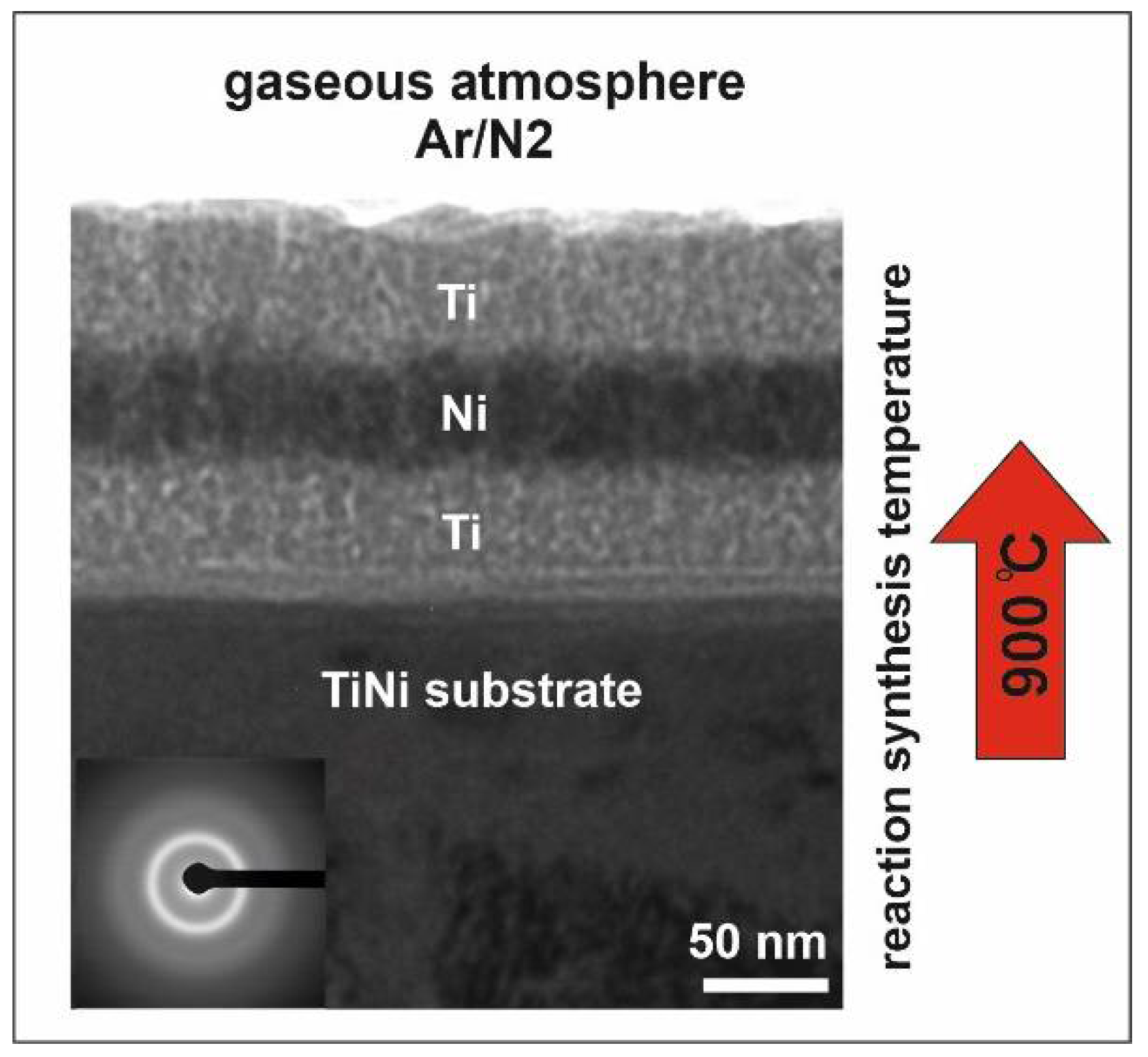
Abstract
:1. Introduction
2. Materials and Methods
2.1. Deposition of the Amorphous Ti/Ni/Ti Nanolaminate and Synthesis of Coating
2.2. Cytocompatibility and Hemolysis Test
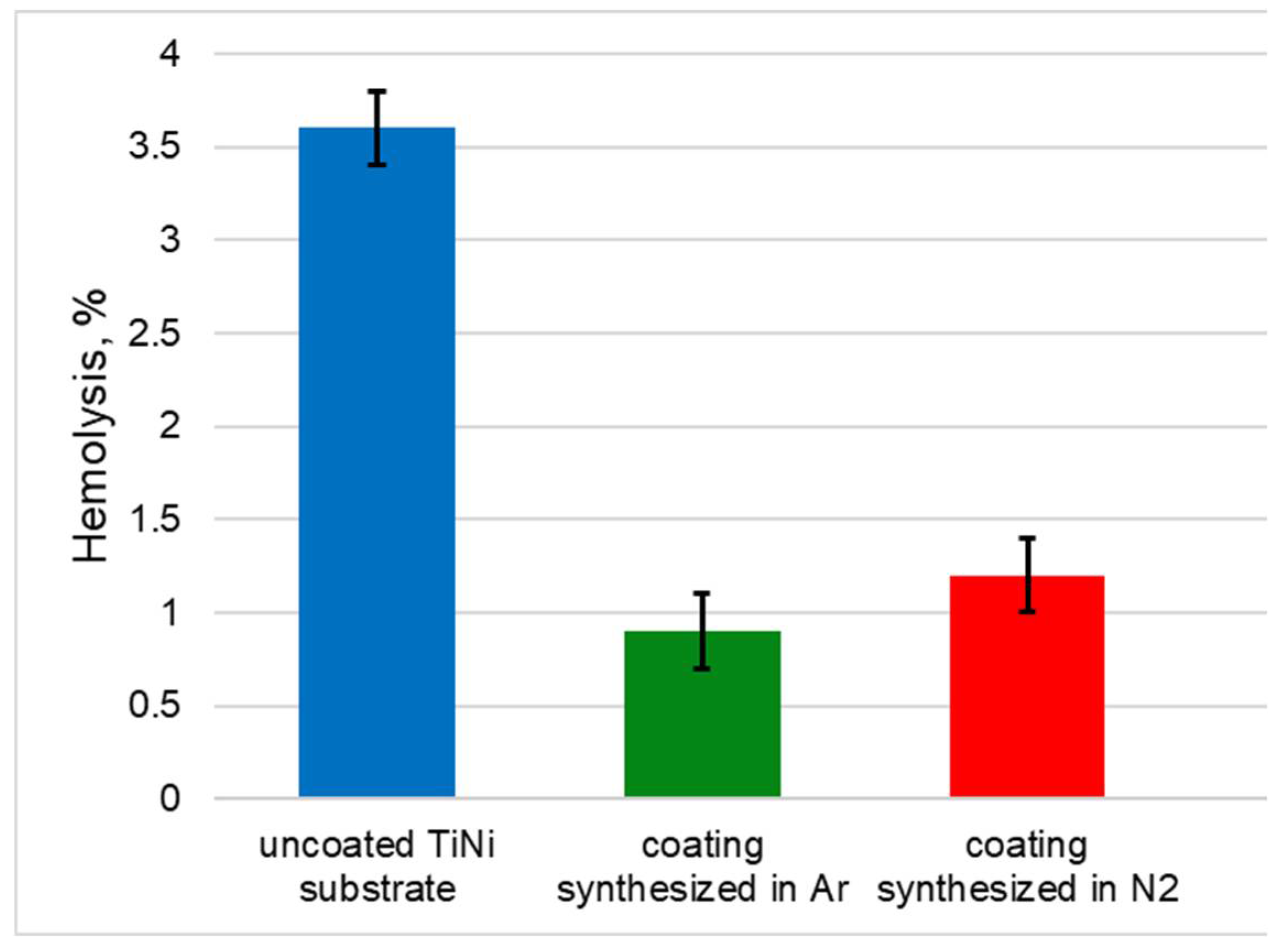
2.2.1. Hemolysis Test
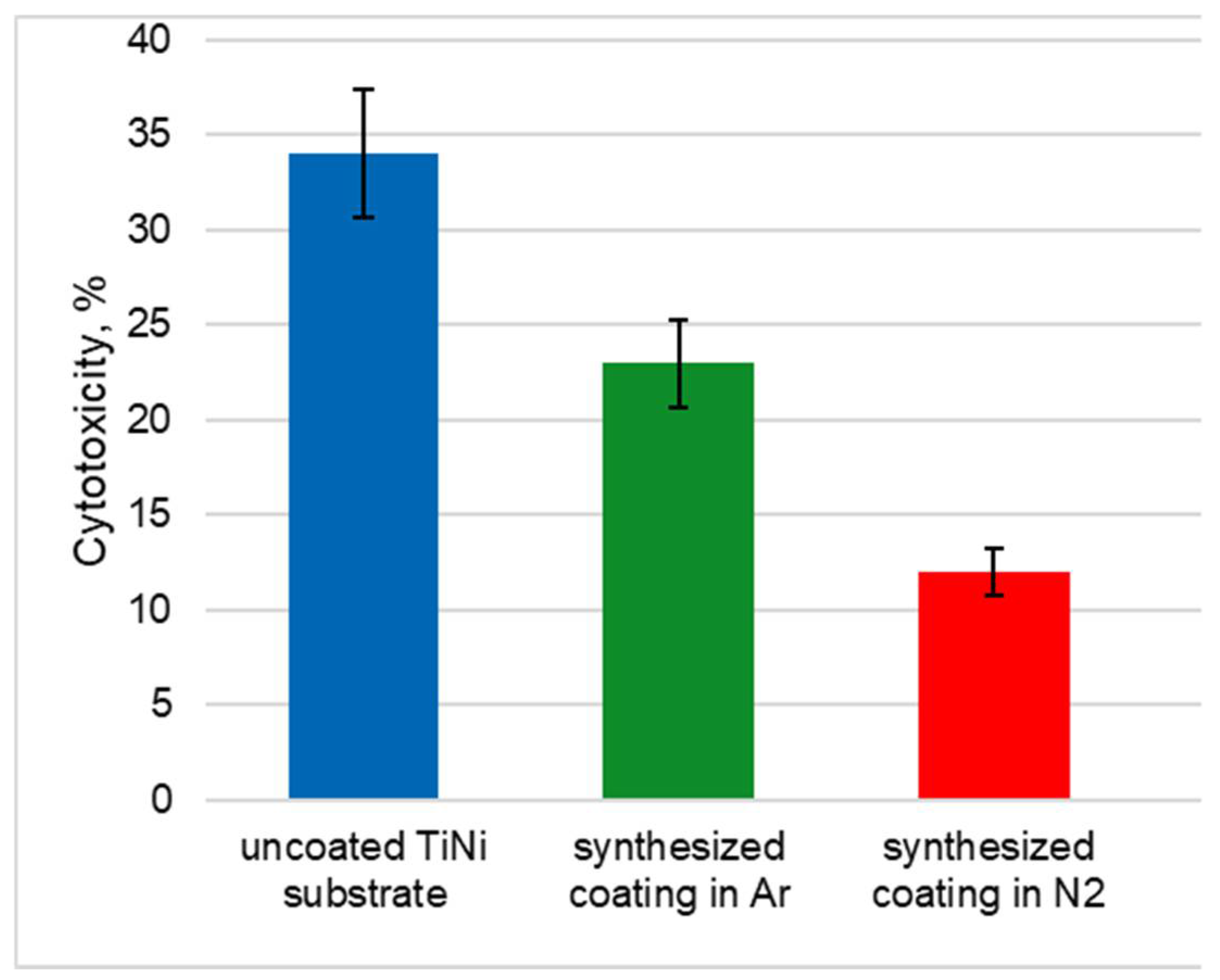
2.2.2. MTT Test
2.3. Surface Characterization Methods
3. Results and Discussion
3.1. Hemolysis and MTT Test
3.2. Structural Studies of Coatings Synthesized in Argon and Nitrogen Atmospheres
3.2.1. Structural Studies
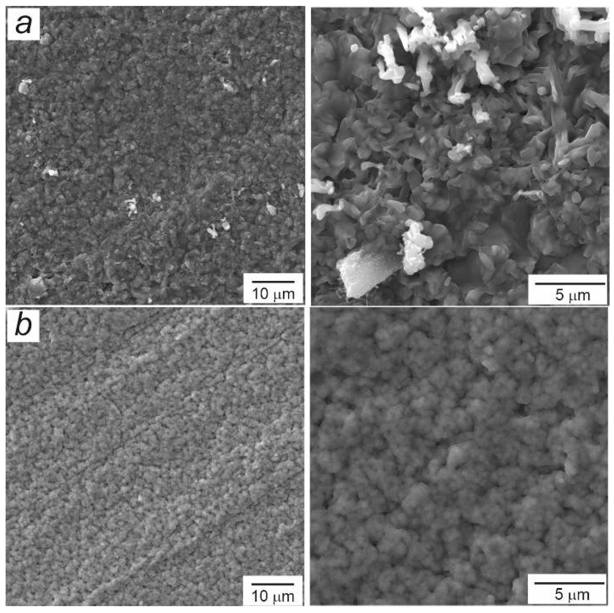
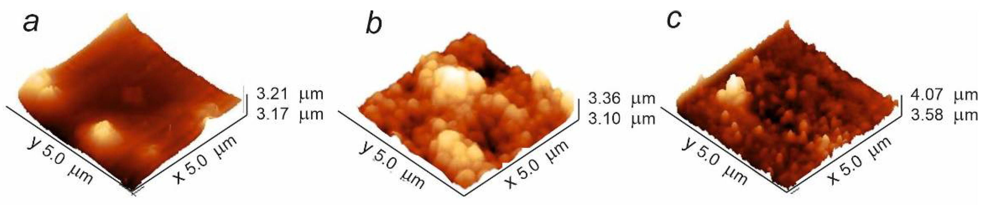
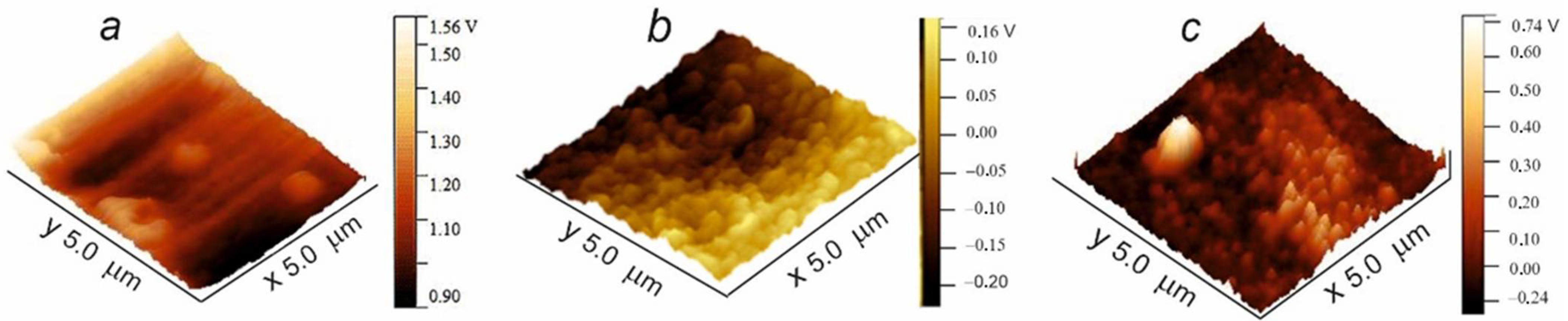
3.2.2. Study on Topographical Evolution and Wettability
4. Conclusions
Author Contributions
Funding
Institutional Review Board Statement
Data Availability Statement
Acknowledgments
Conflicts of Interest
References
- Casaletto, M.P.; Ingo, G.M.; Kaciulis, S.; Mattogno, G.; Pandolfi, L.; Scavia, G. Surface studies of in vitro biocompatibility of titanium oxide coatings. Appl. Surf. Sci. 2001, 172, 167–177. [Google Scholar] [CrossRef]
- Liu, M.; Li, J.; Zhang, Y. Recent advances in corrosion research of biomedical NiTi shape memory alloy. Rare Met. Mater. Eng. 2021, 50, 4165–4173. [Google Scholar]
- Wang, J.; Wang, T.; Dong, S.; Kang, X.; Zhao, S.; Shi, H.; Gao, B.; Ma, S.; Liu, M.; Niu, L.; et al. The effect of Cu-doping on the corrosion behavior of NiTi alloy arch wires under simulated clinical conditions. Mater. Res. Express 2021, 8, 016537. [Google Scholar] [CrossRef]
- Jin, S.; Zhang, Y.; Wang, Q.; Zhang, D.; Zhang, S. Influence of TiN coating on the biocompatibility of medical NiTi alloy. Colloids Surf B Biointerfaces 2013, 101, 343–349. [Google Scholar] [CrossRef]
- Piscanec, S.; Ciacchi, L.C.; Vesselli, E.; Comelli, G.; Sbaizero, O.; Meriani, S.; De Vita, A. Bioactivity of TiN-coated titanium implants. Acta Mater. 2004, 52, 1237–1245. [Google Scholar] [CrossRef]
- Zhao, L.F.; Hong, Y.; Yang, D.Y.; Lü, X.Y.; Xi, T.F.; Zhang, D.Y.; Hong, Y.; Yuan, J.F. The underlying biological mechanisms of biocompatibility differences between bare and TiN-coated NiTi alloys. Biomed. Mater. 2011, 6, 025012. [Google Scholar] [CrossRef]
- Zuo, J.; Xie, Y.; Zhang, J.; Wei, Q.; Zhou, B.; Luo, J.; Wang, Y.; Yu, Z.M.; Tang, Z.G. TiN coated stainless steel bracket: Tribological, corrosion resistance, biocompatibility and mechanical performance. Surf. Coat. Technol. 2015, 277, 227–233. [Google Scholar] [CrossRef]
- Nikolova, M.; Nikolova, V.; Ivanova, V.L.; Valkov, S.; Petrov, P.; Apostolova, M. Mechanical properties and in vitro biocompatibility evaluation of TiN/TiO2 coated Ti6Al4V alloy. Mater. Today Proc. 2020, 33, 1781–1786. [Google Scholar] [CrossRef]
- Luo, Q.; Yang, S.; Cooke, K.E. Hybrid HIPIMS and DC magnetron sputtering deposition of TiN coatings: Deposition rate, structure and tribological properties. Surf. Coat. Technol. 2013, 236, 13–21. [Google Scholar] [CrossRef]
- Aissani, L.; Alhussein, A.; Ayad, A.; Nouveau, C.; Zgheib, E.; Belgroune, A.; Zaabat, M.; Barille, R. Relationship between structure, surface topography and tribo-mechanical behavior of Ti-N thin films elaborated at different N2 flow rates. Thin Solid Film. 2021, 724, 138598. [Google Scholar] [CrossRef]
- Kehal, A.; Saoula, N.; Nouveau, C. Effect of Ar/N2 flow ratio on the microstructure and mechanical properties of Ti-Cr-N coatings deposited by DC magnetron sputtering on AISI D2 tool steels. Surf. Coat. Technol. 2021, 421, 127444. [Google Scholar] [CrossRef]
- Oliveira, J.C.; Fernandes, F.; Serra, R.; Cavaleiro, A. On the role of the energetic species in TiN thin film growth by reactive deep oscillation magnetron sputtering in Ar/N. Thin Solid Film. 2017, 645, 253–264. [Google Scholar] [CrossRef]
- Oghenevweta, J.E.; Wexler, D.; Calka, A. Understanding reaction sequences and mechanisms during synthesis of nanocrystalline Ti2N and TiN via magnetically controlled ball milling of Ti in nitrogen. J. Mater. Sci. 2017, 53, 3064–3077. [Google Scholar] [CrossRef]
- Yue-Lin, L.; Shuo, J.; Ying, Z. Interaction between impurity nitrogen and tungsten: A first-principles investigation. Chin. Phys. B 2012, 21, 016105. [Google Scholar] [CrossRef]
- Çahaa, I.; Alves, A.C.; Affonço, L.J.; Lisboa-Filho, P.N.; da Silva, J.H.D.; Rocha, L.A.; Pinto, A.M.P.; Toptan, F. Corrosion and tribocorrosion behaviour of titanium nitride thin films grown on titanium under different deposition times. Surf. Coat. Technol. 2019, 374, 878–888. [Google Scholar] [CrossRef]
- Chaplanov, A.M.; Shcherbakova, E.N. Structural and phase transformations in thin titanium films under irradiation with nitrogen-hydrogen plasma. Tech. Phys. 1999, 69, 102–108. [Google Scholar] [CrossRef]
- Datta, S.; Das, M.; Balla, V.K.; Bodhak, S.; Murugesan, V.K. Mechanical, wear, corrosion and biological properties of arc deposited titanium nitride coatings. Surf. Coat. Technol. 2018, 344, 214–222. [Google Scholar] [CrossRef]
- Premnath, P.; Tavangar, A.; Tan, B.; Venkatakrishnan, K. Tuning cell adhesion by direct nanostructuring silicon into cell repulsive/adhesive patterns. Exp. Cell Res. 2015, 337, 44–52. [Google Scholar] [CrossRef]
- Liao, S.-C.; Chen, C.-Y.; Hsu, Y.-H.; Li, C.-T.; Hsieh, C.-C.; Tsai, M.-S.; Chan, M.-Y.; Lee, C.-H.; Wang, S.-H.; Ng, S.-K.; et al. In vitro and in vivo biocompatibility study of surface modified TiN deposited on Ti6Al4V using high-power impulse magnetron sputtering technique. Surf. Coat. Technol. 2020, 394, 125814. [Google Scholar] [CrossRef]
- Anusha Thampi, V.V.; Chukwuikea, V.I.; Shtansky, D.V.; Subramanian, B. Biocompatibility study of nanocomposite titanium boron nitride (TiBN) thin films for orthopedic implant applications. Surf. Coat. Technol. 2021, 410, 126968. [Google Scholar] [CrossRef]
- Bai, J.; Gao, J.; Zhang, M.; Zheng, K.; Cao, Y.; Mao, Y.; Yu, S.; He, Z. Microwave plasma oxidation of near-equiatomic NiTi alloy for obtaining low-Ni TiO2 coating. Surf. Coat. Technol. 2021, 428, 127883. [Google Scholar] [CrossRef]
- Mahmud, A.; Wu, Z.; Zhang, J.; Liu, Y.; Yang, H. Surface oxidation of NiTi and its effects on thermal and mechanical properties. Intermetallics 2018, 103, 52–62. [Google Scholar] [CrossRef]
- Wu, Z.; Mahmud, A.; Zhang, J.; Liu, Y.; Yang, H. Surface oxidation of NiTi during thermal exposure in flowing argon environment. Mater. Des. 2018, 140, 123–133. [Google Scholar] [CrossRef]
- Yuan, B.; Li, H.; Gao, Y.; Chung, C.; Zhu, M. In vitro and in vivo evaluation of porous NiTi alloy modified by sputtering a surface TiO2 film. Sci. China Technol. Sci. 2011, 55, 437–444. [Google Scholar] [CrossRef]
- Wong, M.H.; Cheng, F.T.; Man, H.C. Laser oxidation of NiTi for improving corrosion resistance in Hanks’ solution. Mater. Lett. 2007, 61, 3391–3394. [Google Scholar] [CrossRef]
- Huang, C.-F.; Cheng, H.-C.; Liu, C.-M.; Chen, C.-C.; Ou, K.-L. Microstructure and phase transition of biocompatible titanium oxide film on titanium by plasma discharging. J. Alloy. Compd. 2009, 476, 683–688. [Google Scholar] [CrossRef]
- Pana, I.; Braic, V.; Dinu, M.; Mouele, E.S.M.; Parau, A.C.; Petrik, L.F.; Braic, M. In Vitro Corrosion of Titanium Nitride and Oxynitride-Based Biocompatible Coatings Deposited on Stainless Steel. Coatings 2020, 10, 710. [Google Scholar] [CrossRef]
- Hussein, M.A.; Madhan Kumar, A.; Ankah, N.; Azeem, M.A. Thermal treatment effect on the surface and in vitro corrosion characteristics of arc deposited TiN coating on Ti alloy for orthopedic applications. Ceram. Int. 2021, 47, 23203–23213. [Google Scholar] [CrossRef]
- Tsyganov, I.A.; Maitz, M.F.; Richter, E.; Reuther, H.; Mashina, A.I.; Rustichelli, F. Hemocompatibility of titanium-based coatings prepared by metal plasma immersion ion implantation and deposition. Nucl. Instrum. Methods Phys. Res. B 2007, 257, 122–127. [Google Scholar] [CrossRef]
- Surovtseva, M.A. Titanium oxide– and oxynitride–coated nitinol: Effects of surface structure and composition on interactions with endothelial cells. Appl. Surf. Sci. 2022, 578, 152059. [Google Scholar] [CrossRef]
- Karjalainen, P. Neointimal coverage and vasodilator response to titanium-nitrideoxide-coated bioactive stents and everolimus-eluting stents in patients with acute coronary syndrome: Insights from the BASE-ACS trial. Int. J. Cardiovasc. Imaging 2013, 29, 1693–1703. [Google Scholar] [CrossRef] [PubMed]
- Marchenko, E.S.; Baigonakova, G.A.; Dubovikov, K.M.; Yasenchuk, Y.F.; Gunther, S.V. Reaction synthesis of gradient coatings by annealing of three-layer Ti–Ni–Ti nanolaminate magnetron sputtered on the TiNi substrate. Surf. Interfaces 2021, 24, 101111. [Google Scholar] [CrossRef]
- Baigonakova, G.; Marchenko, E.; Yasenchuk, Y.; Kokorev, O.; Vorozhtsov, A.; Kulbakin, D. Microstructural characterization, wettability and cytocompatibility of gradient coatings synthesized by gas nitriding of three-layer Ti/Ni/Ti nanolaminates magnetron sputtered on the TiNi substrate. Surf. Coat. Technol. 2022, 436, 125543. [Google Scholar] [CrossRef]
- Marchenko, E.; Yasenchuk, Y.; Baigonakova, G.; Gunther, S.; Yuzhakov, M.; Zenkin, S.; Potekaev, A.; Dubovikov, K. Phase formation during air annealing of Ti-Ni-Ti laminate. Surf. Coat. Technol. 2020, 388, 125543. [Google Scholar] [CrossRef]
- Liu, X.; Lim, J.Y.; Donahue, H.J.; Dhurjati, R.; Mastro, A.M.; Vogler, E.A. Influence of substratum surface chemistry/energy and topography on the human fetal osteoblastic cell line hFOB 1.19: Phenotypic and genotypic responses observed in vitro. Biomaterials 2007, 28, 4535–4550. [Google Scholar] [CrossRef]
- Ding, X.; Xu, S.; Li, S.; Guo, Z.; Lu, H.; Lai, C.; Wu, J.; Wang, J.; Zeng, S.; Lin, X.; et al. Biological effects of titanium surface charge with a focus on protein adsorption. ACS Omega 2020, 5, 25617–25624. [Google Scholar] [CrossRef]
- Movchan, B.A.; Demchishin, A.V. Structure and properties of thick condensates of nickel, titanium, tungsten, aluminum oxides, and zirconium dioxide in vacuum. Phys. Met. Metallogr. 1969, 28, 653. [Google Scholar]
- Ponsonnet, L.; Reybier, K.; Jaffrezic, N.; Comte, V.; Lagneau, C.; Lissac, M.; Martelet, C. Relationship between surface properties (roughness, wettability) of titanium and titanium alloys and cell behavior. Mater. Sci. Eng. 2003, 23, 551–560. [Google Scholar] [CrossRef]
- Maroudas, N.G. Adhesion and spreading of cells on charged surfaces. J. Theor. Biot. 1975, 49, 417–424. [Google Scholar] [CrossRef]
- Metwally, S.; Stachewicz, U. Surface potential and charges impact on cell responses on biomaterials interfaces for medical applications. Mater. Sci. Eng. 2019, 104, 109883. [Google Scholar] [CrossRef]
- Guo, C.Y.; Matinlinna, J.P.; Hong Tang, T.A. Effects of surface charges on dental implants: Past, present, and future. Int. J. Biomater. 2012, 5, 381535. [Google Scholar] [CrossRef] [PubMed]
- Kieswetter, K.; Schwartz, Z.; Dean, D.D.; Boyan, B.D. The role of implant surface characteristics in the healing of bone. Crit. Rev. Oral. Biol. Med. 1996, 7, 329–345. [Google Scholar] [CrossRef] [PubMed] [Green Version]
- Surmeneva, M.A.; Kleinhans, C.; Vacun, G.; Kluger, P.J.; Schddotonhaar, V.; Muller, M.; Hein, S.B.; Wittmar, A.; Ulbricht, M.; Prymak, O.; et al. Nano-hydroxyapatite-coated metal-ceramic composite of irontricalcium phosphate: Improving the surface wettability, adhesion and proliferation of mesenchymal stem cells in vitro. Colloids Surf. B Biointerfaces 2015, 135, 386–393. [Google Scholar] [CrossRef]
- Redey, S.A.; Nardin, M.; Bernache-Assolant, D.; Rey, C.; Delannoy, P.; Sedel, L.; Marie, P.J. Behavior of human osteoblastic cells on stoichiometric hydroxyapatite and type A carbonate apatite: Role of surface energy. J. Biomed. Mater. Res. 2000, 50, 353–364. [Google Scholar] [CrossRef]
- Ammarullah, M.I.; Santoso, G.; Sugiharto, S.; Supriyono, T.; Kurdi, O.; Tauviqirrahman, M.; Winarni, T.I.; Jamari, J. Tresca Stress Study of CoCrMo-on-CoCrMo Bearings Based on Body Mass Index Using 2D Computational Model. J. Tribol. 2022, 33, 31–38. [Google Scholar]
- Chen, Y.; Guo, H.; Sun, M.; Lv, X. Tensile Mechanical Properties and Dynamic Constitutive Model of Polyurea Elastomer under Different Strain Rates. Polymers 2022, 14, 3579. [Google Scholar] [CrossRef]


Publisher’s Note: MDPI stays neutral with regard to jurisdictional claims in published maps and institutional affiliations. |
© 2022 by the authors. Licensee MDPI, Basel, Switzerland. This article is an open access article distributed under the terms and conditions of the Creative Commons Attribution (CC BY) license (https://creativecommons.org/licenses/by/4.0/).
Share and Cite
Marchenko, E.; Baigonakova, G.; Kokorev, O.; Yasenchuk, Y.; Vorozhtsov, A. Biocompatibility Assessment of Coatings Obtained in Argon and Nitrogen Atmospheres for TiNi Materials. Metals 2022, 12, 1603. https://doi.org/10.3390/met12101603
Marchenko E, Baigonakova G, Kokorev O, Yasenchuk Y, Vorozhtsov A. Biocompatibility Assessment of Coatings Obtained in Argon and Nitrogen Atmospheres for TiNi Materials. Metals. 2022; 12(10):1603. https://doi.org/10.3390/met12101603
Chicago/Turabian StyleMarchenko, Ekaterina, Gulsharat Baigonakova, Oleg Kokorev, Yuri Yasenchuk, and Alexander Vorozhtsov. 2022. "Biocompatibility Assessment of Coatings Obtained in Argon and Nitrogen Atmospheres for TiNi Materials" Metals 12, no. 10: 1603. https://doi.org/10.3390/met12101603