Article

Infection Prevalence of Microsporidia Vairimorpha (Nosema) spp. in Japanese Bumblebees

Department of Agriculture, Tokyo University of Agriculture and Technology, 3-5-8 Saiwai-cho, Fuchu, Tokyo 183-8509, Japan
*
Author to whom correspondence should be addressed.
Insects 2023, 14(4), 340; https://doi.org/10.3390/insects14040340
Submission received: 25 February 2023 / Revised: 27 March 2023 / Accepted: 28 March 2023 / Published: 30 March 2023

Abstract

:

Simple Summary

Microsporidia are intracellular parasites that have detrimental effects on their various invertebrate and vertebrate hosts. The microsporidia pathogenic to honeybees and bumblebees belong to the genus Vairimorpha (previously Nosema). Among these, V. bombi is the most widespread species worldwide and it infects a variety of bumblebee species. In this study, we investigated the infection of V. bombi among Japanese native bumblebees and the alien species, Bombus terrestris, established in Japan, using PCR and microscopy. We discovered that V. bombi infected the subgenus Bombus s. str. species/subspecies with a low prevalence in areas both with and without the presence of B. terrestris, suggesting that V. bombi may be native to Japan. In contrast, a newly discovered microsporidian parasite that is phylogenetically distinct from V. bombi was found to infect the subgenus Diversobombus species/subspecies with a high prevalence. Hence, if the new microsporidian species does not negatively affect the host and is vertically transmitted from queen to offspring, it may coexist with its host. In addition, this may suggest that V. bombi and the new microsporidium have different life history strategies.

Abstract

Microsporidia are spore-forming intracellular parasites of various invertebrates and vertebrates. Vairimorpha bombi negatively affects the fitness of bumblebees and its prevalence correlates with declining bumblebee populations. The invasive alien species Bombus terrestris colonized Japan and possibly introduced new parasites. To assess the infection prevalence of V. bombi in Japanese bumblebees and B. terrestris, we investigated V. bombi infections using PCR and microscopy. The prevalence of sporulating V. bombi infections in three Bombus s. str. species/subspecies was low, whereas that of non/low-sporulating Vairimorpha sp. infections in three Diversobombus species/subspecies was high. Invasive B. terrestris showed low prevalence of non/low-sporulating V. bombi infections and shared the same V. bombi haplotype with B. hypocrita found in Hokkaido, where B. terrestris is present, and in Honshu, where B. terrestris is absent. Although V. bombi may have been introduced with B. terrestris colonies imported from Europe, it seems to be originally distributed in Japan. Furthermore, a new Vairimorpha sp. was found in Japanese bumblebee species. V. bombi and Vairimorpha sp. showed different organ and host specificities in bumblebees. There are no reports on the specific effects of other Vairimorpha spp. on bumblebees; further studies are needed to clarify the individual characteristics of Vairimorpha spp.

1. Introduction

Bumblebees (Apidae: Hymenoptera) are eusocial insects with annual colony cycles. Queens emerge from hibernation in spring and establish new colonies, each producing hundreds of workers during the summer. Declines in wild bumblebee populations have been reported mainly in Europe and the United States; four bumblebee species in Europe have disappeared from eleven countries in the last sixty years [1], and the prevalence of microsporidia was high in bumblebee species with declining populations in the United States [2,3].
Microsporidia are intracellular spore-forming parasites of a variety of invertebrates and vertebrates and sometimes cause extensive damage and death of the hosts [4]. For example, Nosema bombycis, a parasite of silkworms, kills most orally inoculated larvae [5]. Nosema thompsoni, which infects the harlequin ladybird Harmonia axyridis, was found to successfully reduce the survival rate of the European native ladybird Coccinella septempunctata after experimental inoculation [6]. Vairimorpha (Nosema) bombi, redefined by Tokarev et al. [7], is a parasite of bumblebees, first discovered by Fantham and Porter in 1914 [8], and is considered to have spread worldwide with commercialized bumblebee colonies [3]. Infection with V. bombi negatively affects the fitness of bumblebees, shortening adult longevity and reducing colony size [9,10]. During the past decade, surveys of V. bombi infections among bumblebees have been conducted worldwide; their prevalence varied, ranging from 1.2% to 66.5% [2,11,12,13,14,15,16,17,18,19,20,21,22,23,24]. In these studies, light microscopy and/or PCR were used to diagnose infections, but microsporidia infections were often undetectable using microscopy. Rutrecht et al. [25] reported 25% more V. bombi infections using PCR diagnosis than with microscopy, and Blaker et al. [11] found high levels of PCR-positive infections with no detectable spores in bumblebees. Thus, a combination of PCR and microscopy is important for surveying the infection prevalence and status of V. bombi in wild bumblebees.
The European bumblebee Bombus terrestris, introduced to different countries for greenhouse crop pollination, is considered to be one of the factors leading to reduced populations of native pollinators and the species has now been established in New Zealand [26,27,28], Israel [29], Chile [30,31], Argentina [32,33], Australia [34,35], and Japan [36]. In northern Japan, B. terrestris has populated wide areas of Hokkaido [37,38,39] since its introduction in 1991 [40]. In open agricultural areas in southern Hokkaido, a drastic decrease in the native consubgeneric species B. hypocrita sapporoensis was associated with a rapid increase in B. terrestris measured over 3 years from 2003 to 2005, suggesting a competitive replacement of the native by the invading species [36]. The early emergence of its queens in the spring [36], high reproductive ability [41], and superior foraging ability [31,42] could be reasons for B. terrestris’ dominance and could lead to further expansion of this species.
However, few studies exist overall on the parasites introduced to bumblebees, and it remains unclear whether the introduction of new parasites affects native bumblebee species. One previous study showed that V. bombi-like spores were found in commercial colonies of B. terrestris imported from the Netherlands and experimentally transmitted them to the native bumblebees Bombus hypocrita hypocrita and B. diversus diversus [43]. Furthermore, V. bombi detected in B. terrestris collected in Hokkaido had the same nucleotide sequence as V. bombi from bumblebees in Britain [44]. In its native ranges, B. terrestris was infected by V. bombi with a relatively high prevalence at approximately 25%, sometimes more than 50% [13,14,24]. Since introduced for pollination in Chile, B. terrestris also has been established in Argentina’s Patagonia region [45] and was highly infected by V. bombi (37%), suggesting a potential threat to Argentina’s native bumblebees [46].
In this study, we aimed to determine the infection prevalence and status of V. bombi in Japanese bumblebees and B. terrestris collected from Hokkaido, where B. terrestris is established, and Central Japan, where B. terrestris has not established itself. We examined the infected organs, assessed the density of microsporidia, and demonstrated the phylogenetic relationship of the reported V. bombi isolates.

2. Materials and Methods

2.1. Sampling of Bumblebees

To investigate the prevalence of Vairimorpha spp. among bumblebee species, bumblebee sampling was conducted in Hokkaido, where B. terrestris is established, and in Central Japan, where B. terrestris is not established (Table S1). Specimens were collected in Biei and Kamifurano, Central Hokkaido in 2014, 2016, and 2018; Hokuto, Yamanashi and Minamiaiki, Nagano (west side of the Okuchichibu mountain area, west OMA) in 2014 and 2016; Koshu, Yamanashi (east side of the Okuchichibu mountain area, east OMA) in 2016, 2019, and 2020; and in Hinohara, Tokyo in 2016. Additional collections were conducted in Oshino, Yamanashi, and Hara, Nagano in 2016. The midgut of each specimen was removed and crushed in 200 μL 1× PBS (1370 mM NaCl, 27 mM KCl, 81 mM Na2HPO4 12H2O, and 14.7 mM KH2PO4; pH 7.4), and homogenates were stored at −35 °C for the following experiments.

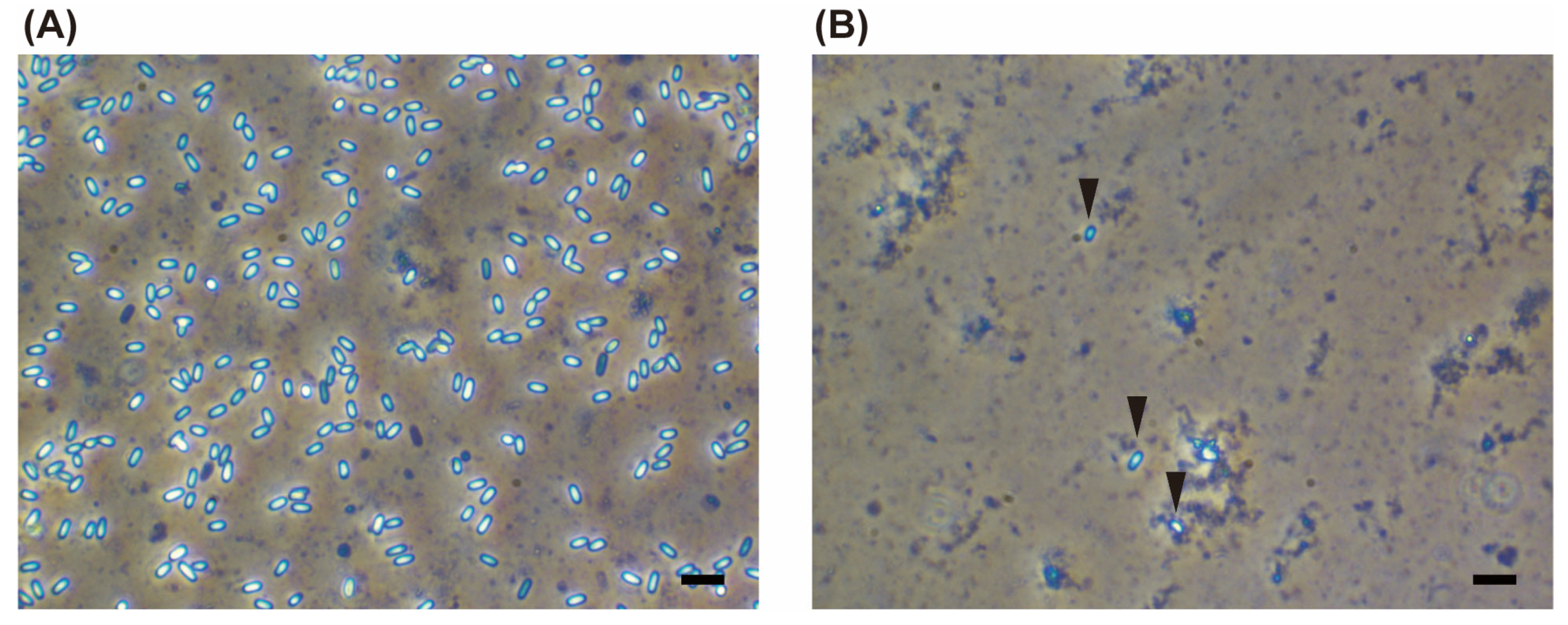
2.2. Detection of Vairimorpha spp. Spores

Using 1 µL of the homogenates spread on a glass slide, spore-forming (herein, “sporulating”, according to Blaker et al. [11]) Vairimorpha spp. were examined across the center of the cover glass from the left edge to the right edge under a phase-contrast microscope (Nikon, Tokyo, Japan) (400× magnification).

2.3. PCR and Genotyping of Vairimorpha spp. Infections

A 50 μL sample of the midgut homogenates was mixed with 250 μL DNAzol (Molecular Research Center, Cincinnati, OH, USA) and 250 mg glass beads (SIGMA, Kanagawa, Japan) and vortexed at 3000 rpm for 5 min (CM-70M-07, ELMI, Riga, Latvia). Then, 250 μL DNAzol and 1 μL proteinase K (20 mg/mL) (Merck Bioscience, Darmstadt, Germany) were added prior to incubation for 1 h at 50 °C and centrifugation at 4 °C, 15,400× g for 10 min. The aqueous layer was transferred to a new tube, 250 μL of 99% ethanol (FUJIFILM Wako Pure Chemical Corporation, Osaka, Japan) was added, and the mixture was centrifuged at 4 °C, 20,400× g for 10 min. The DNA was washed with 70% ethanol, dried for 10 min, resuspended in 20 μL ultrapure water (UPW), and stored at −35 °C.
The PCR assay for Vairimorpha spp. detection was performed using 1.0 μL of 10 × Ex Taq Buffer (TaKaRa, Shiga, Japan), 0.8 μL of 2.5 mM each dNTP mixture (TaKaRa), 0.2 μL of each forward and reverse primer (10 μM) for small subunit (SSU) rRNA, Nbombi-SSU-Jfl, and Nbombi-SSU-Jrl [47] (Table S2), 0.05 μL of 5 U/μL Ex Taq HS (TaKaRa), 1 μL of template DNA, and 6.75 μL of sterilized distilled water (SDW). The PCR profile for the detection of Vairimorpha spp. comprised an initial denaturation step at 94 °C for 3 min, followed by 40 cycles at 94 °C for 30 s, 50 °C for 30 s, and 72 °C for 30 s, and a final extension at 72 °C for 5 min. DNA presence was confirmed by PCR using β-actin primers [48] (Table S2) at an annealing temperature of 55 °C. The PCR products were purified using a QIAquick PCR Purification Kit (Qiagen, Hilden, Germany), and sequencing reactions were performed using the BigDye Terminator Kit ver. 3.1 (Applied Biosystems, Foster City, CA, USA). PCR products were directly sequenced using a 3700 DNA analyzer (Applied Biosystems, Foster City, CA, USA). The cloned plasmid DNA was used as a positive control in all PCR assays: Vairimorpha spp.-like spores were harvested from a male B. hypocrita sapporoensis, which was collected in Hokkaido in 2014, and spore DNA was subjected to SSU rRNA gene amplification using the primers Nbombi-SSU-Jfl and Nbombi-SSU-Jrl; the PCR products were purified. The purified PCR products were ligated with the T-vector pMD20 (TaKaRa) using a ligation mix (TaKaRa) as per the manufacturer’s instructions. Competent cells (Escherichia coli JM109; TaKaRa) were transformed with plasmids. Plasmid DNA was extracted using the QIAprep® Spin Miniprep Kit (Qiagen) (Hilden, Germany) according to the manufacturer’s protocol and stored at −25 °C.
A phylogenetic tree was constructed based on the SSUr RNA gene sequences amplified from Vairimorpha spp. using the primers SSUrRNA-f1 and SSUrRNA-r1c [17] (Table S2) and gene sequences obtained from GenBank using MEGA 7.0 software [49]. After manual sequence editing, they were aligned and the maximum parsimony method was used to construct a phylogenetic tree for clustering haplotypes. To test the reliability of each clade in the tree, 1000 bootstrap resamplings were performed.

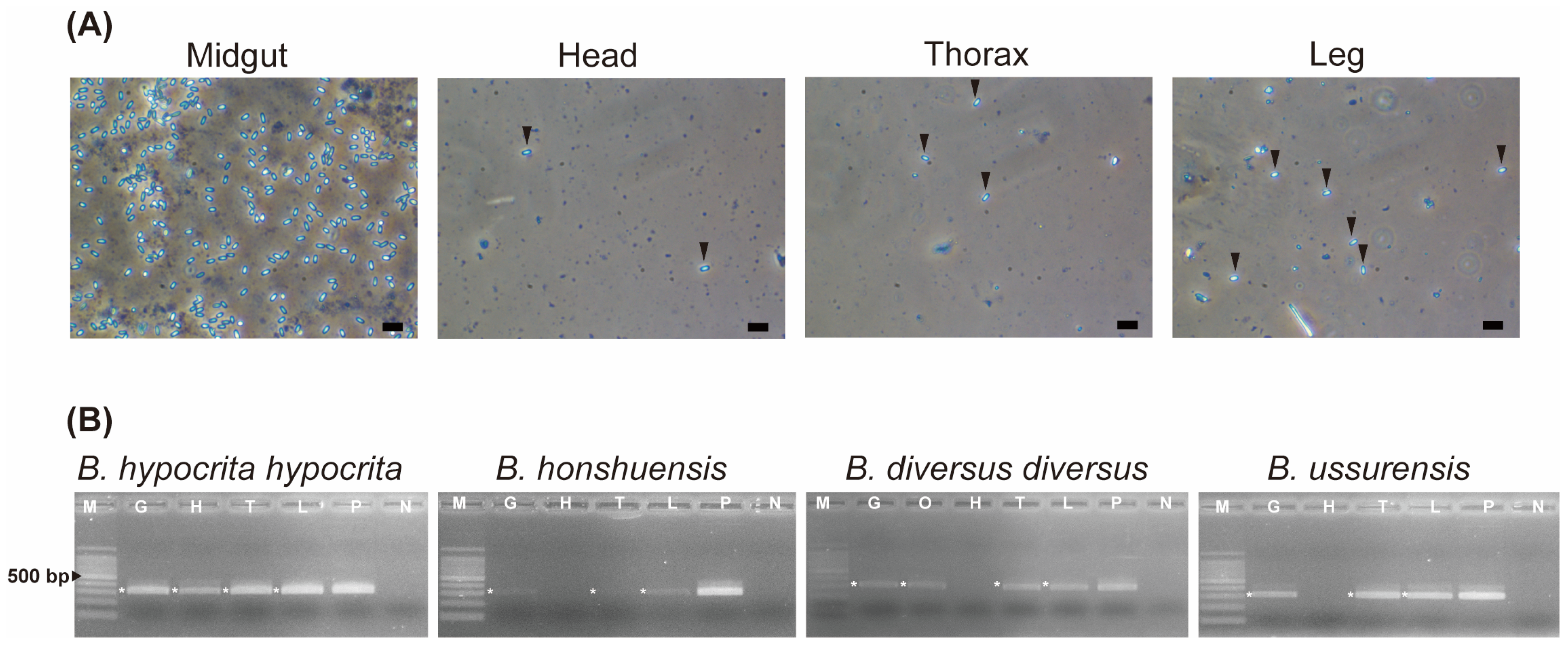
2.4. Organs Infected with Vairimorpha spp.

Four organs (midgut, head, thorax, and legs) of bumblebees infected with Vairimorpha spp. were examined for signs of infection using microscopy and PCR to demonstrate its systemic infectivity. Two individuals from each species were used in subsequent experiments. To eliminate spores from the body surface of bumblebees, they were washed with 1× PBS. Then, their head, thorax, and legs were separated using scissors sterilized by autoclaving; only the femur was used for the “legs” sample after separation from other parts. For B. diversus diversus, an ovary was also isolated and checked for Vairimorpha spp. infection to examine the possibility of vertical transmission. Each organ from two individuals was homogenized using a sterile mortar with 200–400 µL of DNAzol and pooled. The homogenates were then transferred to 1.5 mL microtubes.
DNAzol was added to the homogenates up to the 1 mL mark, and 500 mg of glass beads was added. DNA was extracted as described in Section 2.3, with 4 µL proteinase K used here. PCR was performed with 30 ± 5 ng of DNA.

2.5. Quantitative PCR

To quantify the severity of Vairimorpha spp. infection among bumblebees, qPCR was performed using a StepOnePlus Real-Time PCR System (Applied Biosystems, Foster City, CA, USA). The reaction mixture contained 10 ng DNA, 30 μM of each primer, and 5 μL FastStart Universal SYBR Green Master Mix (Roche, Basel, Switzerland). The cycling conditions were as follows: 2 min at 50 °C and 10 min at 95 °C, followed by 40 cycles of 15 s at 95 °C and 1 min at 60 °C. Dissociation curve analysis of the amplified product was performed after amplification using the specific primers BOMBICAR-F and BOMBICAR-R [50] (Table S2) with the following conditions: 15 s at 95 °C, 20 s at 60 °C, and 15 s at 95 °C. Negative (UPW) controls were used for each run. For each sample, the numbers of Vairimorpha spp. gene copies were calculated using the Ct values (mean of two replicates) measured against a standard curve, which was generated using 10-fold serial dilutions from 3.0 × 10² to 3.0 × 10⁻³ DNA extracted from a solution of 9.8 × 10⁵ spores/mL. The spore density, expressed as the number of spores per 100 ng of DNA, was calculated from duplicate measurements per sample.

3. Results

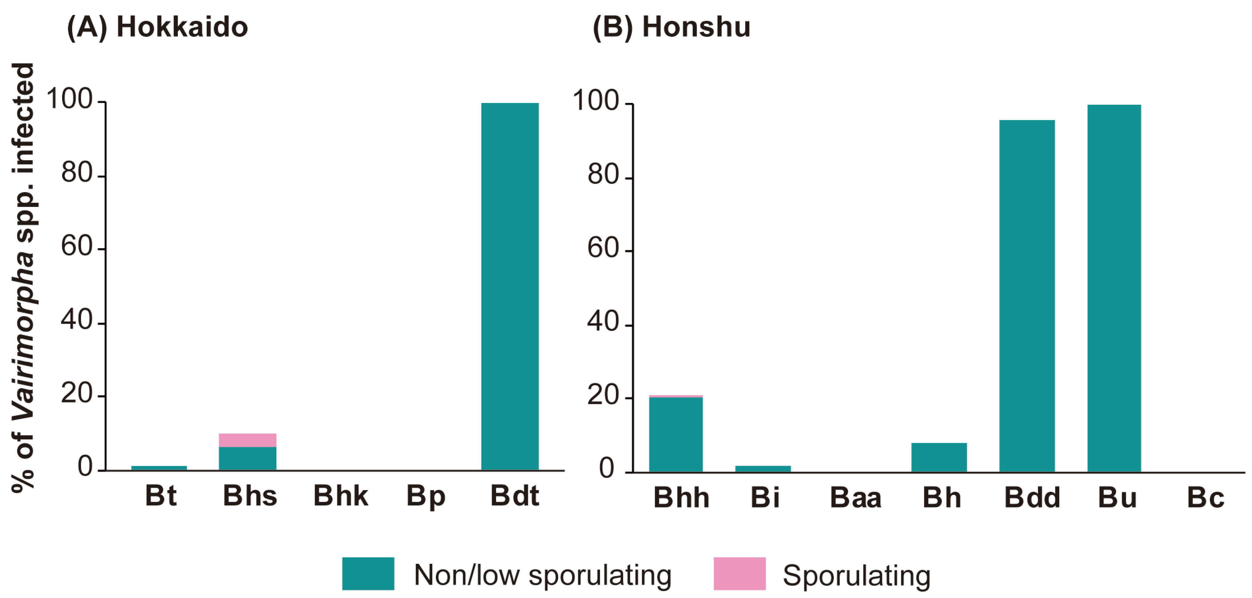
3.1. Prevalence of Vairimorpha spp.

We examined 903 bumblebee specimens belonging to 12 species and subspecies collected in four regions of Japan (Hokkaido, Tokyo, Nagano, and Yamanashi). The overall prevalence of Vairimorpha spp. across all specimens was 19.4%, varying among bumblebee species (Table 1 and Table 2, Figure 1).
Microscopic examination confirmed the presence of Vairimorpha spp. spores in two consubgeneric (Bombus s. str.) subspecies of B. hypocrita: B. hypocrita sapporoensis (nine queens, one male, and two workers collected in 2014 and 2018 in Central Hokkaido) and B. hypocrita hypocrita (five queens, one worker, and three males collected in 2016 and a total of 15 individuals, the caste were not distinguished, collected in 2019 in east OMA) (Figure 2). Microsporidia spores were not observed in any of the remaining samples.
PCR analysis revealed that Vairimorpha spp. exhibited a low prevalence in Central Hokkaido: 0.8%, 1.4%, and 2.4% of B. terrestris specimens and 10.3%, 11.9%, and 6.7% of B. hypocrita sapporoensis specimens from Hokkaido were positive for V. bombi in 2014, 2016, and 2018, respectively. Additionally, one B. diversus tersatus worker collected from Central Hokkaido in 2014 was positive for Vairimorpha spp. In Central Japan, a low prevalence of Vairimorpha spp. was found in two consubgeneric (Bombus s. str.) species B. ignites and B. hypocrita hypocrita: 1.6% of B. ignitus and 7.6% of B. hypocrita hypocrita collected from west OMA in 2014 and 2016, respectively, were positive for Vairimorpha spp. The prevalence of Vairimorpha spp. in B. hypocrita hypocrita collected from east OMA was relatively high at 60% in 2016 but decreased to 4.3% in 2020. The prevalence of Vairimorpha spp. in B. honshuensis was low at 10% in specimens from east OMA and 0% in specimens from west OMA. The highest prevalence of Vairimorpha spp. was found in two consubgeneric (Diversobombus) species, B. diversus diversus and B. ussurensis; in 2014, 100% of B. diversus diversus collected from west OMA were positive, and in 2016, 100%, 100%, 95.6%, 100%, and 77.8% of B. diversus diversus collected from Hinohara, Oshino, west and east OMA, and Hara, respectively, were positive. Similarly, 100% of B. ussurensis collected from west OMA in 2016 showed positive PCR results for Vairimorpha spp.i.

3.2. Organ Specificity of Vairimorpha spp.

The infection of Vairimorpha spp. in four organs of bumblebees (midgut, head, thorax, and legs) was investigated using PCR analysis. Two queens of B. hypocrita hypocrita from Yamanashi were sporulating Vairimorpha spp. PCR-positive specimens, and two queens of B. diversus diversus from Nagano and two workers each of B. ussurensis and B. honshuensis from Yamanashi were non/low-sporulating Vairimorpha spp. PCR-positive specimens.
In the midgut and leg homogenates of two workers of B. hypocrita hypocrita, more than five Vairimorpha spp. spores (++) were observed under a phase-contrast microscope at 400× magnification (Figure 3A), whereas 1–4 spores (+) were observed in the head and thorax homogenates (Figure 3A). Vairimorpha spp. spores were not observed in any of the B. diversus, B. ussurensis, or B. honshuensis specimens.
Using DNA extracted from B. hypocrita hypocrita, PCR analysis revealed amplified Vairimorpha spp.fragments of approximately 300 bp from all four organs (Figure 3B). For B. honshuensis, B. diversus diversus, and B. ussurensis, similar fragments were detected in the midguts, thoraxes, and legs, but not in the heads (Figure 3B). DNA was also extracted from the ovaries of B. diversus diversus, and an amplified Vairimorpha spp. fragment of 300 bp was detected.

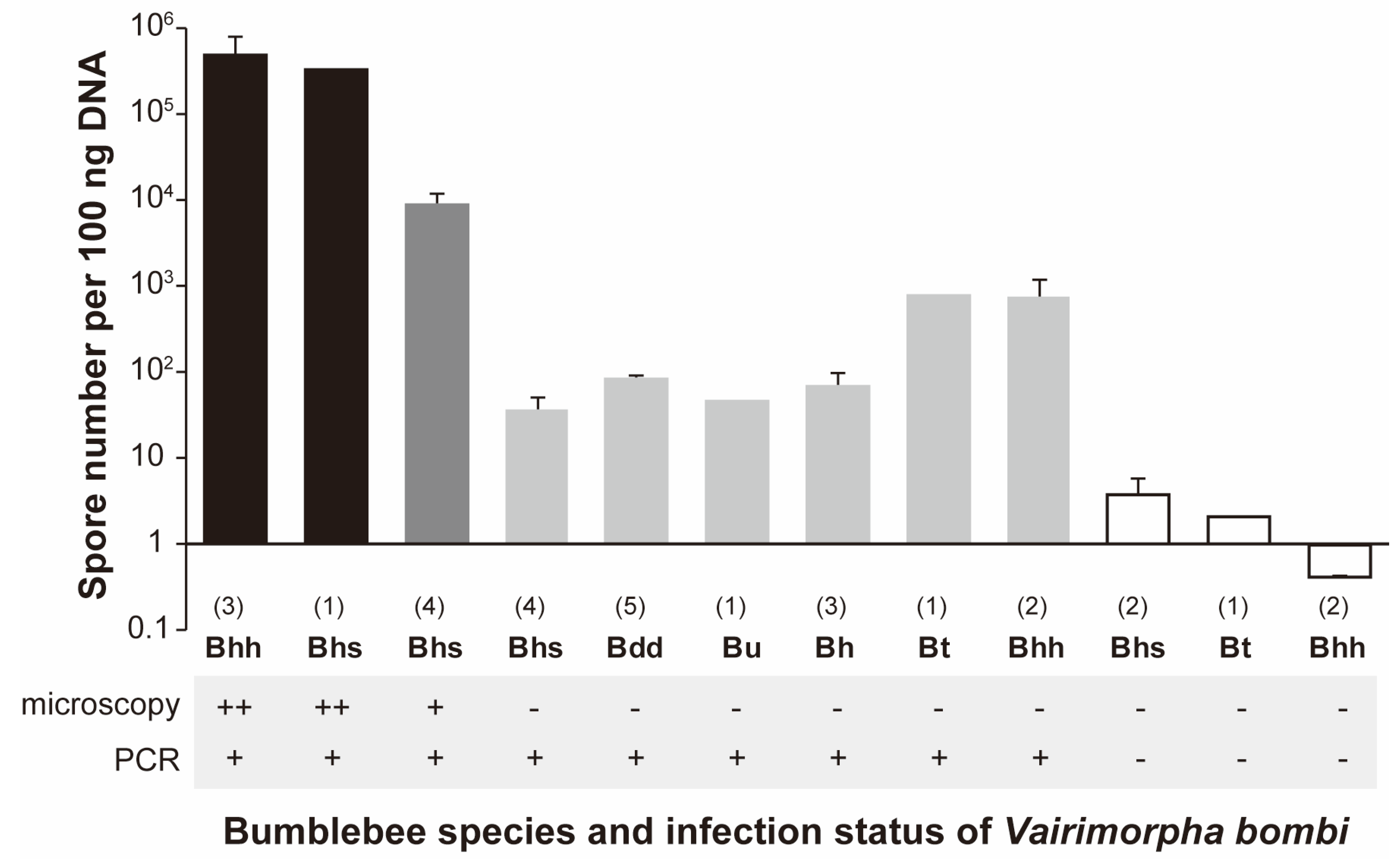
3.3. Spore Numbers of Vairimorpha spp. among Bumblebees

The number of Vairimorpha spp. spores in the midguts per 100 ng DNA containing the host gene were estimated using quantitative PCR in the following specimens: (1) sporulating Vairimorpha spp. PCR-positive bees, (2) non/low-sporulating Vairimorpha spp. PCR-positive bees, and (3) non/low-sporulating Vairimorpha spp. PCR-negative bees (Table S3, Figure 4). The highest spore numbers were found in the sporulating Vairimorpha spp. PCR-positive bees: approximately 1.6 × 10⁵ to 1.1 × 10⁶ spores/100 ng DNA in bees with more than five spores/per visual field of the microscope, and approximately 2.5 × 10⁴ to 1.4 × 10⁵ spores/100 ng DNA in bees with less than five spores/per visual field of the microscope. Conversely, low spore numbers, approximately 8.9 to 1.2 × 10³ spores/100 ng DNA, were found in non-sporulating Vairimorpha spp. PCR-positive bees. The detection limit for spore concentration as assessed by microscopy was <103 spores/100 ng DNA. According to Blaker et al. [11], this number is the threshold for Vairimorpha spp. to be considered non/low-sporulating. As expected, no spores were detected in V. bombi PCR-negative bees, because these bees were not infected with Vairimorpha spp.

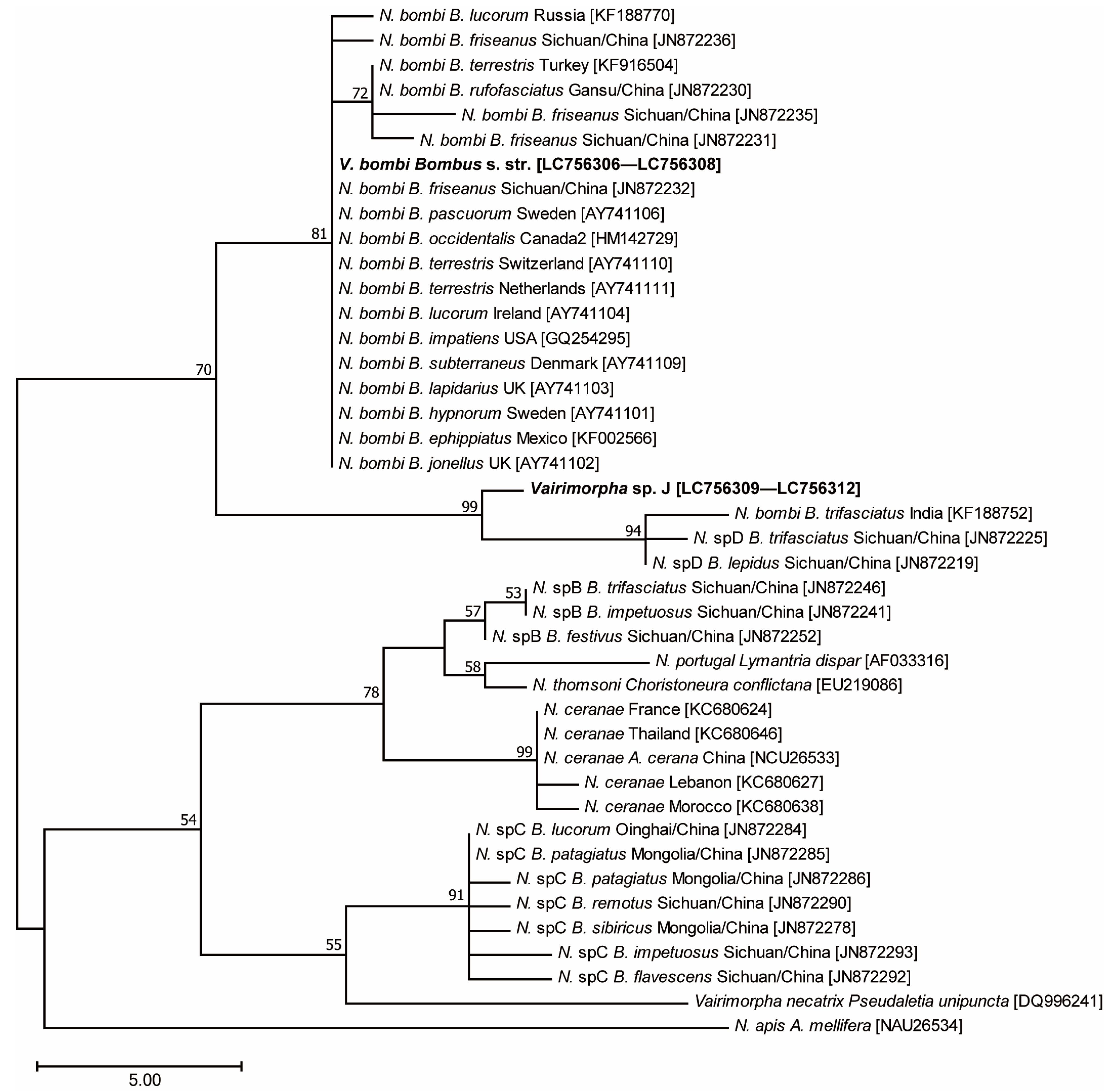
3.4. Phylogenetic Analysis Using Microsporidian SSU rRNA

Sequences of 502 bp were directly amplified from the SSU rRNA genes of 10 individuals of B. hypocrita hypocrita, 11 B. hypocrita sapporoensis, 1 B. terrestris, 16 B. diversus diversus, 6 B. ussurensis, and 2 B. honshuensis. BLAST analysis showed the sequences (GenBank accession number: LC756306—LC756308) from the three Bombus s. str. species were the same and more than 99% identical to the SSUr RNA of V. bombi from Europe, the United States, China, and Russia. The sequences (GenBank accession number: LC756309—LC756312) from the three Diversobombus species and B. honshuensis were the same and shared more than 98% similarity with Nosema sp. from India and China. Phylogenetic analysis using the maximum parsimony method showed that Vairimorpha sp. found in Japan belonged to the same clade as N. bombi, whereas Vairimorpha sp. was close to the clade of Nosema sp. D (Figure 5), suggesting that Vairimorpha sp. is endemic to Japanese bumblebees, and is hereafter referred to as Vairimorpha sp. J.

4. Discussion

This study showed that PCR is a more sensitive and specific method for V. bombi detection than microscopy is. The prevalence of sporulating V. bombi was low (6.8%) among the three Bombus s. str. species/subspecies, B. hypocrita hypocrita, B. hypocrita sapporoensis, and B. ignitus, and the prevalence of non/low-sporulating Vairimorpha sp. J was high (96.1%) among the three Diversobombus species/subspecies, B. diversus diversus, B. diversus tersatus, and B. ussurensis. The invasive B. terrestris showed low prevalence of non/low-sporulating V. bombi infections and shared the same haplotype of V. bombi with B. hypocrita collected in Central Hokkaido and Honshu, Japan.
Previous studies have reported the introduction of alien parasites with B. terrestris commercial colonies [51,52,53,54]. For example, V. bombi and other parasitic species have been detected in worker bees and pollen of commercial colonies from Europe [55]. Therefore, it is possible that V. bombi could have been introduced to and subsequently spread with bees escaping from greenhouses to the field. Consequently, it could have horizontally infected the native bumblebee species. Takahashi et al. [44] reported that the prevalence of V. bombi was 7.5% (n = 120) and 0% (n = 100) in B. terrestris and native Bombus spp., respectively, in other areas of Hokkaido and Nagano from 2010 to 2013, suggesting that V. bombi may have been brought with B. terrestris colonies imported from Europe later. However, considering that V. bombi was detected not only in B. hypocrita sapporoensis in Hokkaido, where B. terrestris is present, but also B. hypocrita hypocrita in Honshu, where B. terrestris is absent, and that the higher prevalence of V. bombi in B. hypocrita than B. terrestris suggests that V. bombi originates from Japan. In the future, it will be necessary to distinguish whether V. bombi is introduced or native to clarify the status of the accompanying V. bombi infestation using molecular genetic techniques.
The prevalence of V. bombi in wild bumblebees is often low. Studies using microscopy reported that the overall prevalence of nine different Bombus spp. in the United States was 2.9% (n = 9909) with the highest prevalence being 37.2% in declining B. occidentalis (n = 172) [15]. In Europe, 56.1% of the natural population of B. terrestris in Sweden (n = 380) [14] and 24.7% of B. terrestris/lucorum with 9.0% overall prevalence in 10 different Bombus spp. and Psithrus spp. (n = 446) in Switzerland [13] were infected with V. bombi. Conversely, PCR analysis suggested the mean prevalence was 1.2% (n = 83) for B. terrestris in Spain [21], 6.1% for 27 different Bombus spp. (n = 1009) in China [16], 12.7% for 22 different Bombus spp. (n = 595) in Russia [18], 10.3% for five different Bombus spp. (n = 39) in India [22], and 13.6% for four different Bombus spp. (n = 280) in Thailand [23]. The 6.8% overall prevalence of V. bombi in our study was similar to that reported in other areas of the world. However, a high prevalence of V. bombi was found in B. hypocrita hypocrita collected from Koshu, Yamanashi from 2016 to 2019, suggesting that the spreading of V. bombi occurred at the site and ended in 2020 for unknown reasons.
While V. bombi has been reported across a wide range of the Northern Hemisphere [3,11,15,56], other Vairimorpha (Nosema) spp. found in bumblebees have been reported in Asian regions—four species from China and two from Russia [16,17]—indicating a high diversity of microsporidia in bumblebees in Asia. Bumblebees also show high species diversity in Asia, and their geographic origin is considered to be the mountainous region of Asia [57,58]. Based on these facts, the microsporidia of bumblebees may have originated in Asia and then dispersed to Europe and North America as bumblebees expanded their habitats. Specifically, this is a possible explanation for the worldwide occurrence of V. bombi.
In this study, a new Vairimorpha sp. J was detected in species endemic to Japan, such as B. diversus, B. honshuensis, and in B. ussurensis, which is also distributed in far east Russia and Korea [59]. Organ specificity in bumblebees differed between V. bombi and Vairimorpha sp. J: V. bombi showed systemic infectivity, whereas Vairimorpha sp. J did not seem to infect the head tissue of bees (Figure 3). It is possible that spores contaminated on the surface of the body were detected, but considering differences in spore density between these two species, this might be a species-specific difference in infections for target organs. For example, nine Nosema species of grasshoppers have organ specificity: N. locustae and N. chorthippi infect only the fat body, showing host and tissue specificity, whereas N. pyrgomorphae infects the midgut, fat body, muscle tissue, and gonad [60]. Alternatively, the difference in infected organs might be related to spore density; the spore density of some specimens with sporulating V. bombi was high, whereas that of others with non/low-sporulating Vairimorpha sp. J was low. Vairimorpha sp. J was highly prevalent in B. diversus and B. ussurensis. Since V. bombi infects multiple subgenera of bumblebees [2,3,11,16,17] and can be artificially inoculated into B. diversus [43], it has a wide host range. In contrast, Vairimorpha spp. reported in China and Russia also infect various bumblebee subgenera with a low prevalence [16,17]. Of these, Nosema sp. D contained microsporidia detected from B. trifasciatus (Diversobombus), B. lepides (Pyrobombus), B. friseanus (Melanobombus), and B. festivus (Festivobombus), and 83% of B. trifasciatus specimens were infected with Nosema sp. D, albeit with a small sample size [16]. Vairimorpha sp. J was mostly found in long-tongued Diversobombus species. These infection patterns, combined with molecular phylogenetic analysis, suggest that Vairimorpha sp. J is a distinct species from V. bombi and is more closely related to Nosema sp. D.
The negative effects of V. bombi infection on bumblebees [9,61] and its high prevalence in declining bumblebee species [2] have been reported; however, there are no reports on the effects of other Vairimorpha spp. Vairimorpha sp. J., which is highly prevalent in Diversobombus species/subspecies, but shows low spore density, may be a covert infection, possibly vertically transmitted from queen to offspring. In this scenario, it may provide a fitness benefit to its host by establishing a mutualistic relationship to spread throughout a host population. To clarify the characteristics of Vairimorpha spp., it is necessary to conduct a comparative study of Vairimorpha spp.-infected and non-infected bumblebees in the future.
In conclusion, we revealed that V. bombi and a new microsporidium, Vairimorpha sp. J., are infecting the Japanese native bumblebees and discovered that they may have different life history strategies.

Supplementary Materials

The following supporting information can be downloaded at: https://www.mdpi.com/article/10.3390/insects14040340/s1, Table S1: List of collection sites; Table S2: Primer list of the study; Table S3: The list of the bumblebees used for quantitative PCR. References [17,43,45,47] are cited in Supplementary Materials.

Author Contributions

Experiments, T.Y. and Y.K.; methodology, T.Y.; investigation, T.Y., Y.K. and M.N.I.; data curation, writing, supervision, and funding acquisition, M.N.I. All authors have read and agreed to the published version of the manuscript.

Funding

This study was supported by a research grant provided by The Oshita Foundation.

Data Availability Statement

The datasets analyzed during the current study are available from the corresponding author upon reasonable request.

Acknowledgments

We thank M. Nakai, Y. Kunimi, S.M. Barribeau, S. Biganski, K. Saar, M. Mitsuhata, M. Ono, H. Iwano, and Y. Hatakeyama for their invaluable comments on the experiments and for their assistance in the field.

Conflicts of Interest

The authors declare no conflict of interest.

References

  1. Kosior, A.; Celary, W.; Olejniczak, P.; Fijal, J.; Krol, W.; Solarz, W.; Plonka, P. The decline of the bumble bees and cuckoo bees (Hymenoptera: Apidae: Bombini) of western and central Europe. Oryx 2007, 41, 79–88. [Google Scholar] [CrossRef] [Green Version]
  2. Cameron, S.A.; Lozier, J.D.; Strange, J.P.; Koch, J.B.; Cordes, N.; Solter, L.F.; Griswold, T.L. Patterns of widespread decline in North American bumble bees. Proc. Natl. Acad. Sci. USA 2011, 108, 662–667. [Google Scholar] [CrossRef] [PubMed] [Green Version]
  3. Cameron, S.A.; Lim, H.C.; Lozier, J.D.; Duennes, M.A.; Thorp, R. Test of the invasive pathogen hypothesis of bumble bee decline in North America. Proc. Natl. Acad. Sci. USA 2016, 113, 4386–4391. [Google Scholar] [CrossRef] [PubMed] [Green Version]
  4. Weiss, L.M.; Becnel, J.J. Microsporidia: Pathogens of Opportunity; Wiley Blackwell: Ames, IA, USA, 2014. [Google Scholar]
  5. Fujiwara, T. Infectivity and pathogenicity of Nosema bombysis to larvae of the silkworm. J. Sericult. Sci. Jpn. 1979, 48, 376–380. [Google Scholar]
  6. Vilcinskas, A.; Stoecker, K.; Schmidtberg, H.; Roehrich, C.R.; Vogel, H. Invasive harlequin ladybird carries biological weapons against native competitors. Science 2013, 340, 862–863. [Google Scholar] [CrossRef]
  7. Tokarev, Y.S.; Huang, W.; Solter, L.F.; Malysh, J.M.; Becnel, J.J.; Vossbrinck, C.R. A formal redefinition of the genera Nosema and Vairimorpha (Microsporidia: Nosematidae) and reassignment of species based on molecular phylogenetics. J. Invertebr. Pathol. 2020, 169, 107279. [Google Scholar] [CrossRef]
  8. Fantham, H.B.; Porter, A. The morphology, biology and economic importance of Nosema bombi, n. sp., parasitic in various humble bees (Bombus Spp.). Ann. Trop. Med. Parasitol. 1914, 8, 623–638. [Google Scholar] [CrossRef]
  9. Otti, O.; Schmid-Hempel, P. Nosema bombi: A pollinator parasite with detrimental fitness effects. J. Invertebr. Pathol. 2007, 96, 118–124. [Google Scholar] [CrossRef]
  10. Rutrecht, S.T.; Brown, M.J.F. Differential virulence in a multiple-host parasite of bumble bees: Resolving the paradox of parasite survival? Oikos 2009, 118, 941–949. [Google Scholar] [CrossRef]
  11. Blaker, E.A.; Strange, J.P.; James, R.R.; Monroy, F.P.; Cobb, N.S. PCR reveals high prevalence of non/low sporulating Nosema bombi (Microsporidia) infections in bumble bees (Bombus) in Northern Arizona. J. Invertebr. Pathol. 2014, 123, 25–33. [Google Scholar] [CrossRef]
  12. Kissinger, C.N.; Cameron, S.A.; Thorp, R.W.; White, B.; Solter, L.F. Survey of bumble bee (Bombus) pathogens and parasites in Illinois and selected areas of northern California and southern Oregon. J. Invertebr. Pathol. 2011, 107, 220–224. [Google Scholar] [CrossRef] [PubMed]
  13. Shykoff, J.A.; Schmidhempel, P. Incidence and effects of four parasites in populations of bumble bees in Switzerland. Apidologie 1991, 22, 117–125. [Google Scholar] [CrossRef]
  14. Huth-Schwarz, A.; Settele, J.; Moritz, R.F.A.; Kraus, F.B. Factors Influencing Nosema bombi infections in natural populations of Bombus terrestris (Hymenoptera: Apidae). J. Invertebr. Pathol. 2012, 110, 48–53. [Google Scholar] [CrossRef] [PubMed]
  15. Cordes, N.; Huang, W.F.; Strange, J.P.; Cameron, S.A.; Griswold, T.L.; Lozier, J.D.; Solter, L.F. Interspecific geographic distribution and variation of the pathogens Nosema bombi and Crithidia species in United States bumble bee populations. J. Invertebr. Pathol. 2012, 109, 209–216. [Google Scholar] [CrossRef]
  16. Li, J.; Chen, W.; Wu, J.; Peng, W.; An, J.; Schmid-Hempel, P.; Schmid-Hempel, R. Diversity of Nosema associated with bumblebees (Bombus spp.) from China. Int. J. Parasitol. 2012, 42, 49–61. [Google Scholar] [CrossRef] [PubMed]
  17. Vavilova, V.; Sormacheva, I.; Woyciechowski, M.; Eremeeva, N.; Fet, V.; Strachecka, A.; Bayborodin, S.I.; Blinov, A. Distribution and diversity of Nosema bombi (Microsporidia: Nosematidae) in the natural populations of bumblebees (Bombus spp.) from west Siberia. Parasitol. Res. 2015, 114, 3373–3383. [Google Scholar] [CrossRef]
  18. Szentgyorgyi, H.; Blinow, A.; Eremeeva, N.; Luzyanin, S.; Grzes, I.M.; Woyciechowski, M. Bumblebees (Bombidae) along pollution gradient—Heavy metal accumulation, species diversity, and Nosema bombi infection level. Pol. J. Ecol. 2011, 59, 599–610. [Google Scholar]
  19. Aytekin, A.M.; Cagatay, N.; Hazir, S. Floral choices, parasites and micro-organisms in natural populations of bumblebees (Apidae: Hymenoptera) in Ankara Province. Turk Zool. Derg. 2002, 26, 149–155. [Google Scholar]
  20. Evison, S.E.F.; Roberts, K.E.; Laurenson, L.; Pietravalle, S.; Hui, J.; Biesmeijer, J.C.; Smith, J.E.; Budge, G.; Hughes, W.O.H. Pervasiveness of parasites in pollinators. PLoS ONE 2012, 7, e30641. [Google Scholar] [CrossRef] [Green Version]
  21. Jabal-Uriel, C.; Martin-Hernandez, R.; Ornosa, C.; Higes, M.; Berriatua, E.; De la Rua, P. First data on the prevalence and distribution of pathogens in bumblebees (Bombus terrestris and Bombus pascuorum) from Spain. Span. J. Agric. Res. 2017, 15. [Google Scholar] [CrossRef] [Green Version]
  22. Vavilova, V.Y.; Konopatskaia, I.; Luzyanin, S.L.; Woyciechowski, M.; Blinov, A.G. Parasites of the genus Nosema, Crithidia and Lotmaria in the honeybee and bumblebee populations: A case study in India. Vavilovskij Žurnal Genet. I Sel. 2017, 21, 943–951. [Google Scholar] [CrossRef] [Green Version]
  23. Sinpoo, C.; Disayathanoowat, T.; Williams, P.H.; Chantawannakul, P. Prevalence of Infection by the microsporidian Nosema spp. in native bumblebees (Bombus spp.) in northern Thailand. PLoS ONE 2019, 14, e0213171. [Google Scholar] [CrossRef] [Green Version]
  24. Votavová, A.; Trněný, O.; Staveníková, J.; Dybová, M.; Brus, J.; Komzáková, O. Prevalence and distribution of three bumblebee pathogens from the Czech Republic. Insects 2022, 13, 1121. [Google Scholar] [CrossRef]
  25. Rutrecht, S.T.; Klee, J.; Brown, M.J.F. Horizontal transmission success of Nosema bombi to its adult bumble bee hosts: Effects of dosage, spore source and host age. Parasitology 2007, 134, 1719–1726. [Google Scholar] [CrossRef] [PubMed]
  26. Donovan, B.J.; Wier, S.S. Development of hives for field population increase, and studies on the life cycles of the four species of introduced bumble bees in New Zealand. N. Z. J. Agric. Res. 1978, 21, 733–756. [Google Scholar] [CrossRef] [Green Version]
  27. Goulson, D.; Hanley, M.E. Distribution and forage use of exotic bumblebees in South Island, New Zealand. N. Z. J. Ecol. 2004, 28, 225–232. [Google Scholar]
  28. MacFarlane, R.P.; Gurr, L. Distribution of bumble bees in New Zealand. N. Z. Entomol. 1995, 18, 29–36. [Google Scholar] [CrossRef]
  29. Dafni, A. The Threat of Bombus terrestris spread. Bee World 1998, 79, 113–114. [Google Scholar] [CrossRef]
  30. Montalva, J.; Sepulveda, V.; Vivallo, F.; Silva, D.P. New records of an invasive bumble bee in northern Chile: Expansion of its range or new introduction events? J. Insect Conserv. 2017, 21, 657–666. [Google Scholar] [CrossRef]
  31. Ruz, L.; Herrera, R. Preliminary observations on foraging activities of Bombus dahlbomii and Bombus terrestris (Hym: Apidae) on native and non-native vegetation in Chile. Acta Hortic. 2001, 561, 165–169. [Google Scholar] [CrossRef]
  32. Morales, C.L.; Arbetman, M.P.; Cameron, S.A.; Aizen, M.A. Rapid ecological replacement of a native bumble bee by invasive species. Front. Ecol. Environ. 2013, 11, 529–534. [Google Scholar] [CrossRef]
  33. Torretta, J.P.; Medan, D.; Abrahamovich, A.H. First record of the invasive bumblebee Bombus terrestris (L.) (Hymenoptera, Apidae) in Argentina. Trans. Am. Entomol. Soc. 2006, 132, 285–289. [Google Scholar]
  34. Hingston, A.B.; McQuillan, P.B. Does the recently introduced bumblebee Bombus terrestris (Apidae) threaten Australian ecosystems? Aust. J. Ecol. 1998, 23, 539–549. [Google Scholar] [CrossRef]
  35. Semmens, T.D.; Turner, E.; Buttermore, R. Bombus terrestris (L.) (Hymenoptera: Apidae) now established in Tasmania. Aust. J. Entomol. 1993, 32, 346. [Google Scholar] [CrossRef]
  36. Inoue, M.N.; Yokoyama, J.; Washitani, I. Displacement of Japanese native bumblebees by the recently introduced Bombus terrestris (L.) (Hymenoptera: Apidae). J. Insect Conserv. 2008, 12, 135–146. [Google Scholar] [CrossRef]
  37. Matsumura, C.; Yokoyama, J.; Washitani, I. Invasion status and potential ecological impacts of an invasive alien bumblebee, Bombus terrestris L. (Hymenoptera: Apidae) naturalized in southern Hokkaido, Japan. Glob. Environ. Res. 2004, 8, 51–66. [Google Scholar]
  38. Inari, N.; Nagamitsu, T.; Kenta, T.; Goka, K.; Hiura, T. Spatial and temporal pattern of introduced Bombus terrestris abundance in Hokkaido, Japan, and its potential impact on native bumblebees. Popul. Ecol. 2005, 47, 77–82. [Google Scholar] [CrossRef]
  39. Nukatsuka, Y.; Yokoyama, J. Environmental factors and land uses related to the naturalization of Bombus terrestris in Hokkaido, northern Japan. Biol. Invasions 2010, 12, 795–804. [Google Scholar] [CrossRef]
  40. Ono, M. Why Are Now the Bumblebees? Nat. Insects 1998, 33, 2–3. (In Japanese) [Google Scholar]
  41. Inoue, M.N.; Yokoyama, J.; Tsuchida, K. Colony growth and reproductive ability of feral nests of the introduced bumblebee Bombus terrestris in northern Japan. Insectes Soc. 2010, 57, 29–38. [Google Scholar] [CrossRef]
  42. Inoue, M.N.; Makino, T.T.; Yokoyama, J.; Sakai, S. Is Bombus terrestris (Hymenoptera: Apidae) a stronger competitor against native Japanese species? A comparison of foraging efficiency. Appl. Entomol. Zool. 2010, 45, 71–75. [Google Scholar] [CrossRef] [Green Version]
  43. Niwa, S.; Iwano, H.; Asada, S.; Matsuura, M.; Goka, K. Microsporidian pathogen isolated from a Colony of the European Bumblebees, Bombus Terrestris, and Infectivity on Japanese Bumblebee. Jap. J. Appl. Entomol. Zool. 2004, 48, 60–64. (In Japanese) [Google Scholar] [CrossRef] [Green Version]
  44. Takahashi, J.; Takeuchi, M.; Matsumoto, K.; Nomura, T. Prevalence of microsporidia isolated from honeybee and bumblebees in Japan. Bull. Res. Inst. Adv. Technol. Kyoto Sangyo Univ. 2013, 12, 59–68. (In Japanese) [Google Scholar]
  45. Schmid-Hempel, E.; Eckhardt, M.; Goulson, D.; Heinzmann, D.; Lange, C.; Plischuk, S.; Escudero, L.R.; Salathe, R.; Scriven, J.J.; Schmid-Hempel, P. The invasion of southern South America by imported bumblebees and associated parasites. J. Anim. Ecol. 2014, 83, 823–837. [Google Scholar] [CrossRef] [Green Version]
  46. Arismendi, N.; Riveros, G.; Zapata, N.; Smagghe, G.; González, C.; Vargas, M. Occurrence of bee viruses and pathogens associated with emerging infectious diseases in native and non-native bumble bees in southern Chile. Biol. Invasions 2021, 23, 1175–1189. [Google Scholar] [CrossRef]
  47. Klee, J.; Tay, W.T.; Paxton, R.J. Specific and sensitive detection of Nosema bombi (Microsporidia: Nosematidae) in bumble bees (Bombus spp.; Hymenoptera: Apidae) by PCR of partial rRNA gene sequences. J. Invertebr. Pathol. 2006, 91, 98–104. [Google Scholar] [CrossRef]
  48. Tsugeno, Y.; Koyama, H.; Takamatsu, T.; Nakai, M.; Kunimi, Y.; Inoue, M.N. Identification of an early male-killing agent in the oriental tea tortrix, Homona magnanima. J. Hered. 2017, 108, 553–560. [Google Scholar] [CrossRef]
  49. Kumar, S.; Stecher, G.; Tamura, K. MEGA7: Molecular evolutionary genetics analysis version 7.0 for bigger datasets. Mol. Biol. Evol. 2016, 33, 1870–1874. [Google Scholar] [CrossRef] [Green Version]
  50. Plischuk, S.; Martin-Hernandez, R.; Prieto, L.; Lucia, M.; Botias, C.; Meana, A.; Abrahamovich, A.H.; Lange, C.; Higes, M. South American native bumblebees (Hymenoptera: Apidae) infected by Nosema ceranae (Microsporidia), an emerging pathogen of honeybees (Apis Mellifera). Environ. Microbiol. Rep. 2009, 1, 131–135. [Google Scholar] [CrossRef] [PubMed]
  51. Goka, K.; Okabe, K.; Yoneda, M.; Niwa, S. Bumblebee commercialization will cause worldwide migration of parasitic mites. Mol. Ecol. 2001, 10, 2095–2099. [Google Scholar] [CrossRef] [PubMed]
  52. Colla, S.R.; Otterstatter, M.C.; Gegear, R.J.; Thomson, J.D. Plight of the bumble bee: Pathogen spillover from commercial to wild populations. Biol. Conserv. 2006, 129, 461–467. [Google Scholar] [CrossRef]
  53. Meeus, I.; Brown, M.J.F.; De Graaf, D.C.; Smagghe, G. Effects of invasive parasites on bumble bee declines. Conserv. Biol. 2011, 25, 662–671. [Google Scholar] [CrossRef] [PubMed]
  54. Szabo, N.D.; Colla, S.R.; Wagner, D.L.; Gall, L.F.; Kerr, J.T. Do Pathogen spillover, pesticide use, or habitat loss explain recent North American bumblebee declines? Conserv. Lett. 2012, 5, 232–239. [Google Scholar] [CrossRef]
  55. Graystock, P.; Yates, K.; Evison, S.E.F.; Darvill, B.; Goulson, D.; Hughes, W.O.H. The trojan hives: Pollinator pathogens, imported and distributed in bumblebee colonies. J. Appl. Ecol. 2013, 50, 1207–1215. [Google Scholar] [CrossRef]
  56. Tay, W.T.; O’Mahony, E.M.; Paxton, R.J. Complete rRNA gene sequences reveal that the microsporidium Nosema bombi infects diverse bumblebee (Bombus spp.) hosts and contains multiple polymorphic sites. J. Eukaryot. Microbiol. 2005, 52, 505–513. [Google Scholar] [CrossRef] [PubMed]
  57. Williams, P.H. A preliminary cladistic investigation of relationships among the bumble bees (Hymenoptera, Apidae). Syst. Entomol. 1985, 10, 239–255. [Google Scholar] [CrossRef]
  58. Woodard, S.H.; Lozier, J.D.; Goulson, D.; Williams, P.H.; Strange, J.P.; Jha, S. Molecular tools and bumble bees: Revealing hidden details of ecology and evolution in a model system. Mol. Ecol. 2015, 24, 2916–2936. [Google Scholar] [CrossRef] [PubMed]
  59. Katayama, E.; Ochiai, H.; Takamizawa, K. Supplementary notes on the nests of some japanese bumblebees: II. Bombus ussurensis. Jap. J. Entomol. 1990, 58, 335–346. [Google Scholar]
  60. Sokolova, Y.Y.; Lange, C.E. An ultrastructural study of Nosema locustae canning (Microsporidia) from three species of Acrididae (Orthoptera). Acta Protozool. 2002, 41, 229–236. [Google Scholar]
  61. Van der Steen, J.J.M. Infection and transmission of Nosema bombi in Bombus terrestris colonies and its effect on hibernation, mating and colony founding. Apidologie 2008, 39, 273–282. [Google Scholar] [CrossRef]
Figure 1. Prevalence of Vairimorpha spp. in different bumblebee species. Species names as abbreviations: Bt, B. terrestris; Bhs, B. hypocrita sapporoensis; Bhk, B. hypnorum koropokkrus; Bp, B. pseudobaicalensis; Bdt, B. diversus tersatus; Bhh, B. hypocrita hypocrita; Bi, B. ignitus; Baa, B. ardens ardens; Bh, B. honshuensis; Bdd, B. diversus diversus; Bu, B. ussurensis; and Bc, B. consobrinus.
Insects 14 00340 g001
Figure 2. Images of Vairimorpha spp.-infected midgut homogenates of B. hypocrita hypocrita under a phase contrast microscopy (400×): (A) >five spores and (B) 1–4 spores. Arrowheads indicate microsporidian spores. Scale bar = 10 μm.
Insects 14 00340 g002
Figure 3. Organ specificity of Vairimorpha spp. (A) Images of the Vairimorpha spp.-infected organs of B. hypocrita hypocrita under a phase-contrast microscope (×400): midgut, head, thorax, and leg. Arrowheads indicate microsporidian spores. Scale bar = 10 μm. (B) PCR detection of Vairimorpha spp. in wild species: B. hypocrita hypocrita, B. honshuensis, B. diversus diversus, and B. ussurensis. M: DNA Ladder, G: midgut, O: ovary, H: head, T: thorax, L: leg, P: positive control, N: negative control. Asterisks indicate PCR-positive results.
Insects 14 00340 g003
Figure 4. Spore numbers per 100 ng DNA as bar graph quantified using quantitative PCR in each bumblebee species and their Vairimorpha spp. infection statuses. Black bar indicates sporulating Vairimorpha spp. PCR-positive bumblebees (with more than five spores in a visual field); dark gray bar, sporulating Vairimorpha spp. PCR-positive bumblebees (with less than five spores in visual field); light gray bar, non/low-sporulating Vairimorpha spp. PCR-positive bumblebees; white bar, Vairimorpha spp. PCR-negative bumblebees. Species names as abbreviations: Bhh, B. hypocrita hypocrita; Bhs, B. hypocrita sapporoensis; Bt, B. terrestris; Bdd, B. diversus diversus; Bu, B. ussurensis; and Bh, B. honshuensis. The number of samples is indicated in brankets. Error bars represent standard errors.
Insects 14 00340 g004
Figure 5. Maximum parsimony of the relationships among Vairimorpha spp. Samples collected from Hokkaido and Honshu based on alignments of the SSUr RNA gene. Bootstrap values exceeding 50% are shown (1000 replicates). The names indicate Vairimorpha spp., host names, collection site, and GenBank accession numbers were in square brackets.
Insects 14 00340 g005
Table 1. List of bumblebee species collected in Biei and Kamifurano, Hokkaido and results of the PCR amplification of small subunit (SSU) rRNA and microscopy.
YearSpecies *
BtBhaBhkBpBdtTotal Specimen Number
2014Q79 (1/0)
W30 (0/0)
M19 (0/0)
Q106 (11/9)
W56 (7/0)
M33 (2/1)
Q1 (0/0)W11 (0/0)
M1 (0/0)
W1 (1/0)337 (22/10)
2016Q61 (1/0)
W9 (0/0)
Q65 (8/0)
W2 (0/0)
Q1 (0/0)
W10 (0/0)
148 (9/0)
2018W42 (1/0)W45 (3/2) 87 (4/2)
Total number240 (3/0)307 (31/12)12 (0/0)12 (0/0)1 (1/0)572 (35/12)
* Species names and castes as abbreviations: Bt, B. terrestris; Bhs, B. hypocrita sapporoensis; Bhk, B. hypnorum koropokkrus; Bp, B. pseudobaicalensis; Bdt, B. diversus tersatus; Q, queen; W, worker; and M: male.
Table 2. List of bumblebee species collected in Honshu and results of the PCR amplification of small subunit (SSU) rRNA and microscopy.
YearSpecies *Total Number
BhhBiBaaBhBddBuBc
Hinohara, Tokyo
2016Q1 (0/0)
W1 (0/0)
Q1 (0/0)
W23 (0/0)
Q7 (7/0) 33 (7/0)
Oshino, Yamanashi
2016Q10 (0/0) Q10 (0/0) Q1 (1/0) 21 (1/0)
east OMA
2016Q4 (4/4)
W11 (4/1)
M5 (4/2)
W27 (4/0)
M13 (0/0)
W14 (14/0) W2 (0/0)62 (30/7)
2019Q18
W27
M4
(-/15) **
49 (-/15)
2020Q2 (0/0)
W18 (1/0)
M3 (0/0)
23 (0/1)
west OMA
2014 W8 (1/0)
M7 (0/0)
M1(0/0)W17(17/0)
M5(5/0)
38 (23/0)
2016W5 (0/0)
M6 (1/0)
W46 (0/0) W5(0/0)W68(65/0)W6(6/0) 136 (72/0)
Hara, Nagano
2016M2 (0/0)W3 (0/0) W3 (0/0)
M1 (0/0)
W9 (7/0) 18 (7/0)
Total number117
(14+/24)
64 (1/0)34 (0/0)50 (4/0)121 (116/0)6 (6/0)2 (0/0)380
(140+/23)
* Species names and castes as abbreviations: Bhh, B. hypocrita hypocrita; Bi, B. ignitus; Baa, B. ardens ardens; Bh, B. honshuensis; Bdd, B. diversus diversus; Bu, B. ussurensis; and Bc, B. consobrinus; Q, queen; W, worker; and M: male. ** PCR was not conducted, and sex/caste was not distinguished.
Disclaimer/Publisher’s Note: The statements, opinions and data contained in all publications are solely those of the individual author(s) and contributor(s) and not of MDPI and/or the editor(s). MDPI and/or the editor(s) disclaim responsibility for any injury to people or property resulting from any ideas, methods, instructions or products referred to in the content.

