Article

Phylogeny and Evolutionary Timescale of Muscidae (Diptera: Calyptratae) Inferred from Mitochondrial Genomes

1
College of Plant Protection, China Agricultural University, Beijing 100193, China
2
The Institute of Scientific and Technical Research on Archives, National Archives Administration of China, Beijing 100050, China
3
College of Horticulture and Plant Protection, Henan University of Science and Technology, Luoyang 471023, China
4
College of Plant Science and Technology, Huazhong Agricultural University, Wuhan 430070, China
5
College of Life Science, Shenyang Normal University, Shenyang 110034, China
*
Authors to whom correspondence should be addressed.
Insects 2023, 14(3), 286; https://doi.org/10.3390/insects14030286
Submission received: 9 January 2023 / Revised: 10 March 2023 / Accepted: 12 March 2023 / Published: 15 March 2023
(This article belongs to the Section Insect Systematics, Phylogeny and Evolution)

Abstract

:

Simple Summary

Reconstruction of the phylogenetic relationship within Muscidae inferred by 43 mitochondrial genomes has revealed the split of the family into three major groups. The best phylogenetic tree recovers the monophyly of all subfamilies except Mydaeinae. Genus Helina Robineau-Desvoidy, 1830 was synonymized with Phaonia Robineau-Desvoidy, 1830. We prefer the subfamily status of Azeliinae and Reinwardtiinae and separate Stomoxyinae from Muscinae. Muscidae diverged in the early Eocene (51.59 Ma) and most subfamilies diverged at around 41 Ma.

Abstract

House flies (Muscidae) comprise the most species-rich family of the muscoid grade with over 5000 described species worldwide, and they are abundant in various terrestrial and aquatic ecosystems. The high number of species, varied appearances, complex feeding habits, and wide distributions have hindered researchers from understanding their phylogeny and evolutionary history. Here, we newly sequenced fifteen mitochondrial genomes and reconstructed the phylogenetic relationships and divergence time among eight subfamilies of Muscidae (Diptera). The best phylogenetic tree, which was inferred by IQ-Tree, recovered the monophyly for seven out of eight subfamilies (except for Mydaeinae). Based on phylogenetic analyses and morphological characteristics, we prefer the subfamily status of Azeliinae and Reinwardtiinae, and separate Stomoxyinae from Muscinae. Genus Helina Robineau-Desvoidy, 1830 was synonymized with Phaonia Robineau-Desvoidy, 1830. The divergence time estimation indicated Muscidae originated at 51.59 Ma (early Eocene). Most subfamilies had originated around 41 Ma. We provided a mtgenomic viewpoint on the phylogenetic relationships and divergence time estimation of Muscidae.

1. Introduction

The Muscidae family, commonly known as house flies, are one of the most prominent dipteran families. They are recognized as essential for pollination, recycling, and as a food resource for birds, spiders, and many other animals. As one of the most species-rich families among the muscoid grade, they contain 5579 described species belonging to 207 genera worldwide [1]. They can be found in a variety of terrestrial and aquatic habitats, spanning most continents of the world, i.e., from arctic to tropical regions, with only limited exceptions for the most arid environments (e.g., Synthesiomyia Brauer et Bergenstamm, 1893; Graphomya Robineau-Desvoidy, 1830; Limnophora Robineau-Desvoidy, 1830; Lispe Latreille, 1796; etc.) [2,3,4]. Adult muscid flies include predators, bloodsuckers, and detritivores. Many species are also anthophilous, while larvae are saprophagous, coprophagous, predaceous, or more rarely phytophagous [5]. They also feed on numerous exudates from plants or animals [6]. The association between humans and Muscidae comprises agricultural, medical, and veterinary aspects [3,7]. There are many reports about Muscidae in forensic entomology, disease transmission, and livestock parasitism [2,4,6,8]. About 200 Muscidae species are known to visit human bodies or animal carrion [4]. Different species represent various hazards to livestock and humans, such as sucking blood, parasitism, and spreading of disease [6]. Additionally, muscid flies can be used as laboratory animals to test the toxic effects of pharmaceuticals and nanomaterials [9,10].
The small body size (2–8 mm) and significant variation in appearance make identification of many species within Muscidae difficult. The subfamily classification in Muscidae, as proposed by various authors, has been incongruent for a long time. Skidmore (1985) presented a fundamental ten-subfamily classification system mainly based on characteristics of the immature and mature stages (Achanthipterinae, Atherigoninae, Azeliinae, Coenosiinae, Eginiinae, Reinwardtiinae, Stomoxyinae, Muscinae, Mydaeinae, and Phaoniinae) [3]. Carvalho (1989) proposed a seven-subfamily classification based on adult characteristics (Achanthipterinae, Atherigoninae, Azeliinae, Coenosiinae, Muscinae, Mydaeinae, and Phaoniinae) [11]. Comparable to Skidmore (1985), who treated Stomoxyini, Reinwardtiini, and Eginiini as subfamilies [12], Xue and Chao (1998) divided Muscidae species in China into ten subfamilies. Carvalho et al. (2005) suggested a seven-subfamily classification for the Neotropical region, promoted a new subfamily Cyrtoneurininae, and downgraded Stomoxyinae and Reinwardtiinae to tribes [13]. Fan (2008) also proposed a ten-subfamily system that was nearly the same as that of Xue and Chao (1998), except for its treatment of Ophyra Robineau-Desvoidy, 1830 as a valid genus which was later shown to be a junior synonym of Hydrotaea Robineau-Desvoidy, 1830 [14,15]. Besides those based on traditional morphological characteristics, some researchers have presented hypotheses based on molecular data. Haseyama et al. (2015) proposed a new classification of three subfamilies based on the COI, AATS, CAD, and EF1-α genes (Cyrtoneurininae, Muscinae, and Mydaeinae) [16]. Grzywacz et al. (2021) implied a classification with seven subfamilies, which were divided into two clades based on a re-interpretation of the morphology of immature stages; they raised Reinwardtiinae to a subfamily [17].
With developing sequencing technology, usage of gene markers has increased for muscid phylogenetic relationship reconstruction, from multi-locus [16,17,18,19] to whole genome level [17]. However, the muscid phylogenetic relationships established by mitochondrial genome data are still incomplete. Mitochondrial genomes (mtgenomes) have been shown to be effective and powerful markers for phylogenetic relationship reconstruction [20,21,22,23]. The utilities of complete mtgenomes in higher taxon-level phylogenetics have produced some remarkable achievements, e.g., recovering most deep branches of Holometabola [20,24], accurately resolving intraordinal relationships within Diptera [25], and the phylogeny and divergence time estimation of Megaloptera [21]. The Arthropoda are one of the best-studied phyla with an analysis based on mitochondrial genes [26]. Cameron et al. (2014) also demonstrated that there has been a large increase in the amount of new mtgenome data produced each year since 2005, and that the mtgenome is the most extensively studied genomic system in insects [27]. However, 88 muscid mtgenomes were accessible on the NCBI website (www.ncbi.nlm.nih.gov/nuccore, search date: 4 July 2022, keywords: Muscidae mitochondrion genome), which is not enough for understanding muscid phylogeny and evolution considering the existence of over 5000 described species in the Muscidae family.
In our study, we inferred the phylogeny of Muscidae by sequencing fifteen new mtgenomes and combined these with twenty-three available mtgenomes; all samples covered six out of seven subfamilies in Muscidae. Furthermore, we provided a mtgenomic viewpoint on the divergence time estimation based on our new phylogenetic relationships.

2. Materials and Methods

2.1. Sampling and DNA Extraction

The newly sequenced specimens were identified by Prof. Chuntian Zhang and Xin Li. The samples and taxonomic information are listed in Table 1. We also retrieved more muscid mtgenomes from the NCBI database (https://www.ncbi.nlm.nih.gov, accessed on 28 July 2021). To reduce the unbalanced sampling bias, a part of the sequences was chosen to represent species-rich and well-studied taxa. In total, there were 43 sequences sampled in our analyses, including 15 new sequenced samples, as well as 23 Muscidae sequences and five outgroup sequences retrieved from the NCBI database. The sampling covered 6 of 7 Muscidae subfamilies following the taxonomic system promoted by Carvalho et al. (2005) [13].
DNA was extracted from the muscle tissue of the specimen using the DNeasy Blood and Tissue Kit (Qiagen China (Shanghai) Co., Ltd.), and the DNA samples were stored at −20 °C. The DNA library procedures used a total of 0.2 g of DNA per sample as raw material. The sequencing library was created using the NEBNext® UltraTM DNA Library Prep Kit for Illumina (NEB, Ipswich, MA, USA, Catalog #: E7370L) and index numbers were applied to each sample. Sonication was used to split the genomic DNA material to a size of 350 bp. Next, the DNA pieces were end polished, A-tailed, and ligated with the full-length adaptor for Illumina sequencing, which was followed by additional PCR amplification. Subsequently, the PCR results were purified using the AMPure XP technology (Beverly, MA, USA). Following this, the quality of the collection was evaluated using the Agilent 5400 system (Agilent, Santa Clara, CA, USA) and measured using QPCR (1.5 nM). The libraries were then pooled to the flow cell based on the effective concentration and target offline data volume (4–6 Gb). The cBOT was clustered and sequenced using the Illumina high-throughput sequencing platform NovaSeq 6000 with PE150 strategy in Novogene Bioinformatics Technology Co., Ltd. Beijing, China (except for OP528689, which was sequenced by the Sanger method). The raw sequence data of new mtgenomes were submitted into the SRA database with BioProject ID (PRJNA937892) and the SRA accession numbers (SRR23603891–SRR23603904) (except for OP528689).

2.2. Sequences Assembly and Annotation

Data cleaning, quality control, and assembling used the “all” submodule of MitoZ software [29]. The genetic code was invertebrate mitochondrial code (genetic_code = 5), and the assembly was conducted with quick mode (run_mode = 2). The annotation of genes was executed in the MITOS2 web server [42]. All protein-coding genes were checked for reading frames following the invertebrate genetic codes in Geneious v9 [43]. The annotations of tRNA genes were checked using the tRNAscan-SE Search Server (http://lowelab.ucsc.edu/tRNAscan-SE/, accessed on 31 August 2021) [44] and ARWEN version 1.2 (http://130.235.244.92/ARWEN/, accessed on 31 August 2021) [45]. Finally, all genes’ annotations were checked by hand following Cameron (2014) [46], who provided basic rules for annotating the mitochondrial genome.

2.3. Phylogeny Analyses

All dataset operations were carried out using the Phylosuite v1.2.2 software [47]. The alignments were executed in MAFFT with L-INS-I strategy [48]. In contrast with the RNA genes that were aligned through the nucleotide base, the protein-coding genes were aligned with the codon model (the MAFFT-based reading frame considering method [47]). The aligned protein-coding genes were also trimmed using gblocks with the codon strategy to extract conserved loci [49]. By comparing the pre-reconstructed phylogenetic results, the trimmed dataset was chosen for the following analyses.
PartitionFinder2 recommended the best-fit partitioning schemes (Table S1) in maximum likelihood (ML) [50]. We chose the criteria of “the corrected Akaike Information Criterion” (AICc) and the “greedy” algorithm with branch lengths estimated as “linked” to search for the optimal schemes.
The maximum likelihood phylogenies were inferred by IQ-TREE v1.6.8 [51]. We conducted ML analyses based on IQ-TREE with 5000 ultrafast bootstraps tests [52] and 1000 replications of the Shimodaira–Hasegawa-like approximate likelihood ratio test [53], and the minimum correlation coefficient was set to 0.90. The “-spp.” parameter was used to indicate that each partition could have a separate evolution rate.
In total, 12 datasets were produced through combinations of gene markers and partition schemes (Table 2). The best result tree was used for systematic relationship discussion and divergence time estimation. Phylogenetic trees were annotated on the iTOL website [54].

2.4. Divergence Time Estimation

To explore the evolutionary history, the MCMCTree, a software package of PAML v4.9j, was used to estimate the divergence time of Muscidae [55]. The relaxed-clock model (clock = 2) was used, and the divergence estimation was conducted using the quick model. First, codeml was used to run multiple sequence alignment (usedata = 3). Then, the approximation likelihood analyses were performed based on the previous results (usedata = 2). Four fossil calibration records were chosen from the mindat website (http://www.mindat.org, accessed on 31 August 2021) to estimate the divergence time within the Muscidae family: Phoridae (125–129 Ma), Muscoidea (47.8–56.0 Ma), Muscidae (46.2–50.3 Ma), and Hydrotaea (23.0–28.1 Ma). The phylogenetic tree of Figure 1 and the dataset “C123TR-P2PF” were used as input files to estimate the divergence time of Muscidae. By utilizing the quick model, we first set the parameter “usedata = 3”, then changed to “usedata = 2; ndata = 1; seqtype = 0; RootAge ≤ 129 Ma; nsample = 20,000; burnin = 20,000; sampfreq = 10”, while other parameters were set to default according to the user guidebook for MCMCTree. Two MCMCTree analyses were run independently to confirm that the results converged. Finally, the divergence result was parsed and shown using three R packages. The treeio package read the MCMCTree result tree [56], the ggtree package drew the tree [57], and the deeptime package illustrated the geographic time scale of the time tree [58].

3. Results

3.1. Mtgenome Assembly

There were fifteen newly sequenced mtgenomes of muscid species belonging to five subfamilies and twelve genera. We also retrieved related mtgenomes from the NCBI database, including twenty-five Muscidae species and five outgroup species from Anthomyiidae, Fanniidae, Scathophagidae, Hippoboscidae, and Phoridae. The lengths of all these mtgenomes ranged from 14,187 to 17,676 bp, and their A-T contents ranged from 77.8% to 80%. All canonical 37 genes were annotated for each mtgenome except for Atherigona ateripraepeda (“OP528680”, trnC missed), Azelia zetterstedtii (“OP528689”, trnI and trnQ missed), and Dasyphora quadrisetosa (“OP528694”, trnQ missed). The gene arrangements of Muscidae are similar to those found in previous dipteran mtgenome studies [59,60]. All tRNA genes were predicted and folded as cloverleaf structures.

3.2. Phylogenetic Effect of RNA Sequences

Studies using RNA data for phylogenetic analysis are relatively common, and some articles proved that RNA data might provide effective phylogeny signals in various groups (Diptera, Hymenoptera, Hemiptera, and Nematoda) by focusing on different systematic levels of phylogenetic relationships (from tribe to phylum) [22,61,62,63]. We found that the genetic differences in tRNA were not significant, and the trimmed RNAs were reduced by 3–28% (C123: 1–16%). We also examined the effects of using RNA data and trimming by gblocks in phylogenetic analyses (Figure 2). There are no significant phylogenetic differences in RNA sequences trimmed by gblocks. Adding both tRNA and rRNA genes rather than a single type of RNA noticeably improved the resolution of the Muscidae phylogenetic tree.

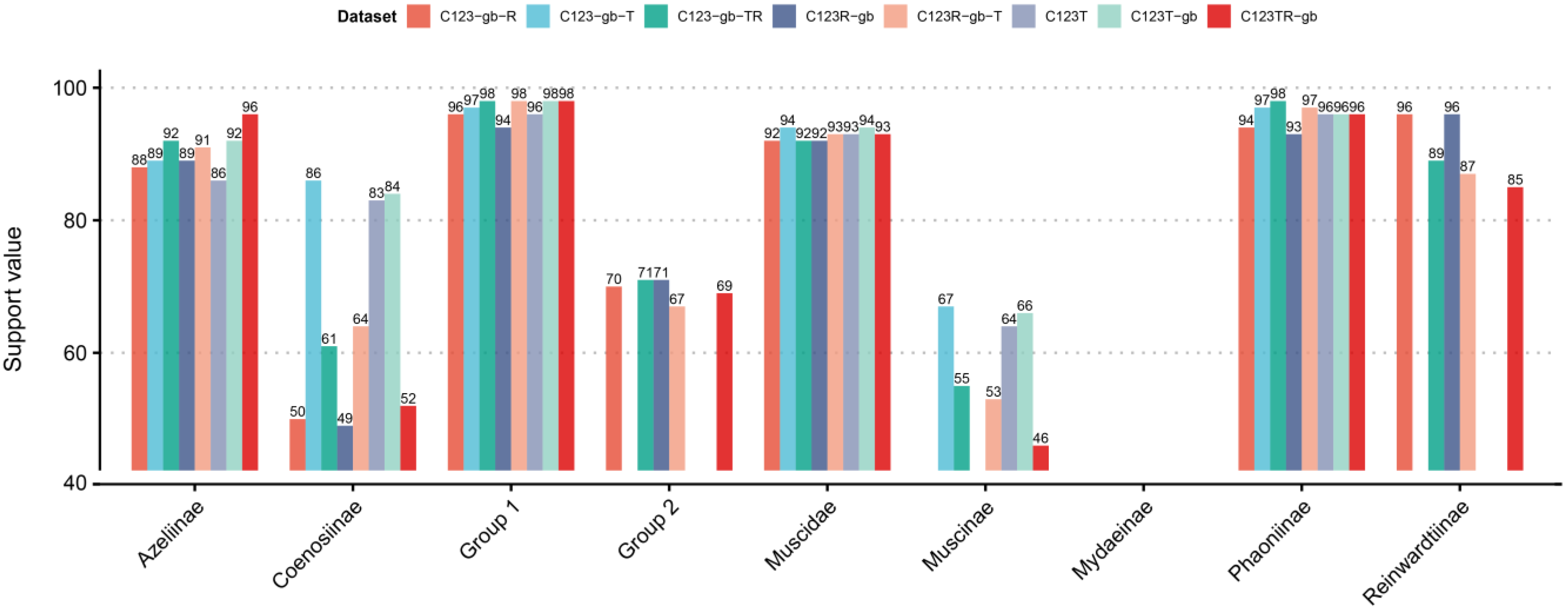
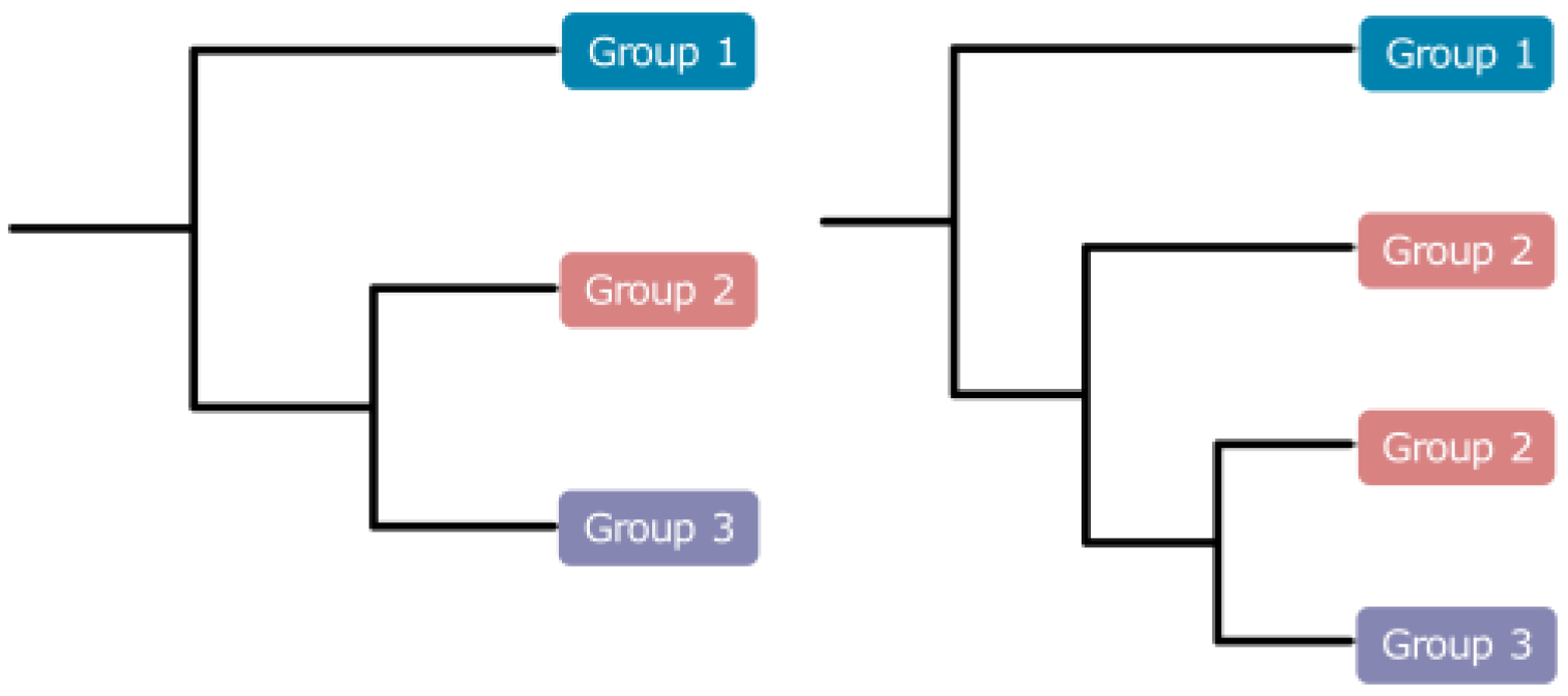
3.3. Phylogenetic Analyses of Muscidae

3.3.1. Phylogenetic Analyses Based on IQ-Tree

To obtain the most consentaneous phylogeny, we utilized 12 datasets based on different genes or codon loci combinations (Table 2). Comparing all the results, we hypothesized that Muscidae were divided into three major groups: group one containing subfamilies Azeliinae, Muscinae and Stomoxyinae; group two consisting of subfamilies Atherigoninae and Reinwardtiinae; group three comprising subfamilies Mydaeinae, Phaoniinae, and Coenosiinae (Figure 1). Two topologies appeared across the result trees (Figure 3). The major difference was whether group two was monophyletic or not. Ten of the twelve trees built in IQ-Tree supported tree topology one, and the other two trees supported topology two (Figure 1 and Figure 3). Topology two did not cluster Atherigoninae and Reinwardtiinae as a monophyletic clade. In group one, Azeliinae were a sister group to Muscinae sensu lato (Muscinae and Stomoxyinae) in all IQ-Tree analyses, and these three subfamilies were recovered as monophyletic groups in most of the IQ-Tree results except for Muscinae in datasets “C123R-P2”, “C12R-P2”, and “C12TR-P2” (Table 2). In group two, most of the datasets supported the sister relationship between Atherigoninae and Reinwardtiinae, while “C123T-P1” and “C123T-P2” recovered them as paraphyletic groups. The Reinwardtiinae were separated: Atherigoninae along with the species Synthesiomyia nudiseta (Reinwardtiinae) and the remaining species of Reinwardtiinae. In group three, the phylogenetic relationships among three subfamilies were poorly reconstructed (Coenosiinae, Mydaeinae, and Phaoniinae). Nearly all datasets indicated Phaoniinae were monophyletic. The paraphyletic Phaoniinae and two species of Mydaeinae were mixed in two datasets (“C12T-P2” and “C12R-P2”), and one Mydaeinae species (Graphomya rufitibia) always nested in Coenosiinae in all datasets. When Graphomya rufitibia was taken out of consideration, eight datasets supported the monophyly of Coenosiinae. Mydaeinae were a non-monophyletic group in all datasets, and the two Mydaeinae species (except for Graphomya rufitibia) were close to each other but not as a clade in all IQ-Tree results.

3.3.2. The Best Phylogenetic Tree

The best tree was ultimately determined to be the one with the highest support values. The “C123TR-P2PF” dataset with thirteen protein-coding genes, twenty-two tRNAs, and two rRNAs, and each locus of the protein-coding genes, were partitioned separately; this was used to estimate the best tree. As shown in Figure 1, Muscidae consisted of three major groups (Figure 2 and Figure 3): Azeliinae, Muscinae, and Stomoxyinae were in group one; Atherigoninae and Reinwardtiinae were in group two; and Mydaeinae, Phaoniinae, and Coenosiinae were in group three. In a few phylogenetic trees, Reinwardtiinae were assigned to group three (topology two in Figure 3). Most of the analyses supported the phylogenetic relationship of topology one: group one + (group two + group three). Therefore, we assumed that this tree topology tends to be stable.

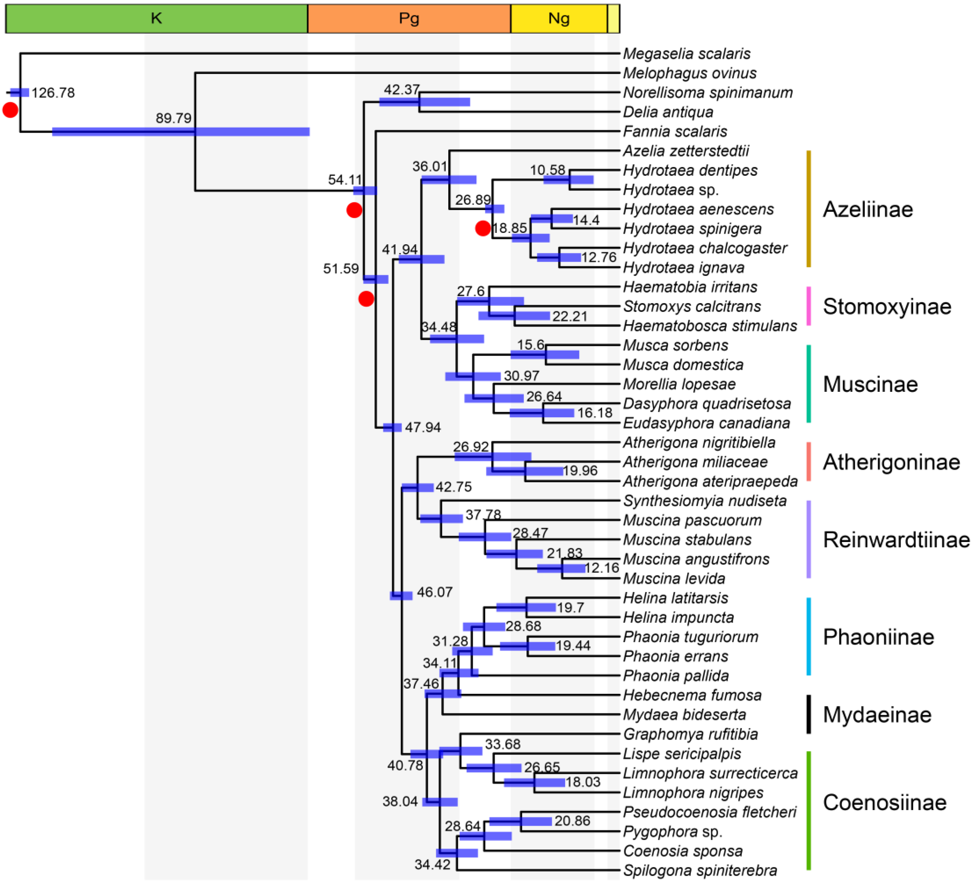
3.4. Time Frame Based on Mtgenomes

The result revealed that Muscidae diverged from the rest of the muscoid grade at 51.59 Ma (95% HPD: 48.93–54.22 Ma) (Figure 4), which is similar to the proposed time frame of all major Muscidae lineages [16]. First, group one (defined in Figure 1) split with groups two and three at 47.94 Ma (95%HPD: 46.10–50.03 Ma); then the split between group two and group three was dated to 46.07 Ma (95%HPD: 43.81–48.60 Ma). The Azeliinae were split from Muscinae and Stomoxyinae at 41.94 Ma (95%HPD: 37.06–46.73 Ma). The Stomoxyinae split with Muscinae at 34.48 Ma (95%HPD: 28.68–40.10 Ma). The subfamily Atherigoninae and Reinwardtiinae diverged at 42.75 Ma (95%HPD: 39.32–46.07 Ma). Coenosiinae diverged with Mydaeinae and Phaoniinae at 40.78 Ma (95%HPD: 37.33–44.26 Ma), and the split between Phaoniinae (Mydaea Robineau-Desvoidy, 1830 and Hebecnema Schnabl, 1889) and Mydaeinae occurred at 34.11 Ma (95%HPD: 29.85–38.12 Ma).

4. Discussion

We confirmed the early divergence of Muscidae based on mtgenomic data, and also reported the split of Muscidae into three major groups (Figure 1 and Figure 3) which were discovered in previous studies through multi-markers (mitochondrial and nuclear genes) [16,17,18], AHE, and RAD-seq datasets [17]. The morphological characteristics and molecular data provide conclusive evidence for the monophyly of Muscidae [11,16,17,18,19,64,65,66]. However, the monophylies of some subfamilies (i.e., Azeliinae sensu lato (including Reinwardtiinae), Muscinae sensu lato (including Stomoxyinae), Mydaeinae, and Phaoniinae) have not been fully supported [16,17,18,65,66,67].
The divisions of subfamilies Azeliinae sensu lato (including Azeliinae and Reinwardtiinae) and Muscinae sensu lato (including Muscinae and Stomoxyinae) in Muscidae were inconsistent in the past. Couri and Carvalho (2003) revealed the cladistic relationship of seven subfamilies and rebuilt Muscinae sensu lato as a sister group to the rest of the subfamilies (Muscinae + (Azeliinae + (Phaoniinae + (Reinwardtiinae + (Dichaetomyiinae + (Mydaeinae + Coenosiinae)))))) [64]. Schuehli et al. (2007) pointed out that subfamily Azeliinae sensu lato were paraphyletic and mixed with the polyphyletic Muscinae sensu lato in their strict consensus tree of combined data for four gene markers [19]. However, Grzywacz et al. (2017) considered that their gene trees were anomalous and that the position of Hydrotaea and Ophyra within Azeliinae could not be supported [15]. Kutty et al. (2008, 2010) offered phylogenetic analyses with eight gene markers; they obtained a split between genus Muscina Robineau-Desvoidy, 1830 (Reinwardtiinae) and the rest of Azeliinae sensu lato, and their results failed to support a sister-group relationship between Muscinae and Stomoxyinae [65,66]. With complete tribal sampling, Kutty et al. (2014) also confirmed the clear split between Azeliinae and Reinwardtiinae as well as the non-sister-group relationship between Muscinae and Stomoxyinae [18]. Haseyama et al. (2015) proposed a new classification where Muscidae consisted of three subfamilies (Muscinae + (Cyrtoneurininae + Mydaeinae)), which represented all tribes of Muscidae and all biogeographic regions [16]. The phylogeny showed a significant cladistic distance between Azeliini and Reinwardtiini and rejected the monophyly of Azeliinae. They also provided a time frame based on four gene markers: the housefly subfamilies originated during Paleocene to Eocene. Ding et al. (2015) indicated that Muscidae split from Fanniidae at 37.65 Ma (late Eocene) based on three muscid mtgenomes [22]. Ren et al. (2019) inferred phylogenetic relationships within Muscidae based on fifteen mtgenomes of four subfamilies and recovered Mydaeinae as a basal clade: Mydaeinae + (Reinwardtiinae + (Muscinae sensu lato + Azeliinae)) [23]. Kutty et al. (2019) corroborated the monophyly of Muscinae sensu lato and Coenosiinae and revealed a close relationship between Mydaeinae and Phaoniinae in their Calyptratae phylogenetic research [67]. However, their result was not as comprehensive as their previous two studies [16,18]. Grzywacz et al. (2021) rearranged the classification of Muscidae based on multiple molecular markers and presented a topology that partially contradicts the traditional classification based on adult morphology; however, it is to some extent consistent with larval morphology [17]. The Reinwardtiini were resurrected as subfamily Reinwardtiinae; four genera previously settled in Eginiini were transferred to Reinwardtiinae; and Stomoxyini were still placed in Muscinae. The split of Muscidae into three major groups (defined in Figure 1 and Figure 3) was also documented among their various datasets [17].
The monophyly of Azeliinae sensu lato depends on whether Reinwardtiinae are included. Azeliinae were placed under the Muscinae sensu lato or Muscinae [68,69,70], and raised as a subfamily based on adult morphology by Carvalho (1989) [11]. The monophyly of Azeliinae sensu lato was rejected by the cladogram of 54 morphological traits; the significant differences between Azeliinae and Reinwardtiinae in musculature of the male terminalia [64,71]; and the non-monophyletic position in molecular phylogenetic studies [16,17,18]. The subfamily status of Azeliinae and Reinwardtiinae was supported [3,12,64], while the Azeliinae was also recovered as Azelia + (Azeliinae + Muscinae/Muscinae sensu lato) [15,16,17]. In addition to the relationship between Azeliinae and Reinwardtiinae, their research also suggested the basal position of Azeliinae within Muscidae. Since Azelia Robineau-Desvoidy, 1830 was considered within Azeliini (as Hydrotaeini) by Hennig (1965) [69], Azelia was placed close to Hydrotaea and Thricops Rondani, 1856. Haseyama et al. (2015) recovered Azelia within Hydrotaea and as a sister to Ophyra (revised as an invalid genus later and placed in Hydrotaea [15]) [16]. Savage and Wheeler (2004) showed that Azelia was close to Thricops instead of Hydrotaea in their phylogenetic tree inferred by morphological traits [72]. The close relationship of these genera was questioned by Grzywacz et al. (2017) and the Azelia was recovered as sister group to the remaining Azeliinae + Muscinae sensu lato [15,17]. Different from previous studies, the species of Azelia were rebuilt within Azeliinae and the monophyly of Azeliinae was recovered in the present study [15,16,17]. It should be noted that the genera Thricops and Huckettomyia Pont et Shinonaga, 1970 of Azeliinae were not included in our analyses. These two genera were recovered as a sister group to Muscinae/Muscinae sensu lato [15,16,17], but were recovered within Azeliinae in studies of Kutty et al. [18,65,66]. However, it is challenging to identify whether the sampling of Thricops and Huckettomyia has a significant impact on the reconstruction of Azeliinae monophyly or not.
Whether at the gene fragment level (nuclear and mitochondrial gene fragments [16,18,19,65,66]) or the genome level (anchored hybrid enrichment and restriction site-associated DNA sequencing at the genomic scale [17]), Azeliinae sensu lato were rebuilt as a non-monophyletic lineage. The majority of studies did not support the monophyly of Azeliinaeae sensu lato [16,17,18,65,66]. Only two species of Reinwardtiinae were grouped by Kutty et al. [65,66]. The poorly sampled Reinwardtiinae were clustered with Coenosiinae + (Mydaeinae, Phaoniinae) [65,66]; alternatively, Reinwardtiinae were polyphyletic and were grouped with Phaoniinae, Cyrtoneurininae, and even Mydaeinae [16,17,18]. Following the clues mentioned above, most studies supported the divergence between Azeliinae and Reinwardtiinae, and several studies claimed the subfamily status of Azeliinae (Azeliini) and Reinwardtiinae (Reinwardtiini). However, few molecular studies supported the monophyly of these two subfamilies, and no result supported their sister-group relationship. Unlike previous hypotheses, based on the sampling in the present study, our results supported the monophyly of Azeliinae and Reinwardtiinae, independently. They might be raised as subfamilies; further studies that concentrate on morphology, reproductive strategy, and behavior are needed to provide more pieces of evidence.
The monophyly of Muscinae was supported by many studies [16,18,19,64,67]. Previously, Stomoxyinae were classified in Muscinae sensu lato [11,17,18,64,65,67,73], and their monophyly was well supported. However, the position of Stomoxyinae in Muscinae was inconsistent. In some studies, Stomoxyinae were close to the base of Muscinae sensu lato [14,64,66], while more studies have favored Stomoxyinae to be nested within the Muscinae sensu lato in a distal position [16,17,18,65]. In opposition to some previous studies, our phylogenetic results support Stomoxyinae as a sister group to Muscinae. In terms of morphological aspects, Stomoxyinae were distinguished from Muscinae in the number of spermatheca (two versus three), type of mouthparts (piercing sucking versus sponging), and shape of arista (pectinate versus plumose) [14]. The haustellum of Stomoxyinae is strongly sclerotized [12] and Stomoxyinae are blood-feeding insects [7,74]. We favor the family rank of Stomoxyinae based on these morphological characteristics and the sister-group relationship between Muscinae and Stomoxyinae.
There is no doubt that Atherigoninae were monophyletic [18,73]. The only question is their systematic position. They were placed close to the base of Muscidae [11] and are closely related to Reinwardtiinae in taxonomic revisions [14]. The sister-group relationship to the Reinwardtiinae was also favored by the anchored hybrid enrichment dataset [17], while the Atherigoninae were clustered with Cyrtoneurininae by fewer than eight molecular markers [16,18].
Coenosiinae have usually been considered as a monophyletic group [17,18,64,65,66,67]. Phaoniinae, Mydaeinae, and Coenosiinae were often clustered in one clade based on morphological characteristics [11,18,65,66,67]. However, Phaoniinae were grouped with Azeliinae and Reinwardtiinae on the basis of the cilia absent on sternite one [64]. The relationship between Phaoniinae and Mydaeinae (Graphomya, Hebecnema, and Mydaea) as well as the monophylies of Phaoniinae and Mydaeinae were poorly supported in recent studies [16,17,18,65,66,67]. Except for clustering with Phaoniinae, Mydaeinae were also rebuilt close to Coenosiinae or Reinwardtiinae. The non-monophyletic group Mydaeinae (consisting of genera Gymnodia Robineau-Desvoidy, 1830 and Graphomya) were clustered alongside Coenosiinae under the analyses of anchored hybrid enrichment (AHE) data [17]. The genus Graphomya was close to Limnophorini (Coenosiinae), and the genera Hebecnema and Mydaea were grouped with Phaoniinae by four genes [16]. The Mydaeinae represented by Graphomya rufitibia alone were grouped with Reinwardtiinae or Reinwardtiinae + (Muscinae + Azeliinae) by mtgenomes [23,41,75]. Those analyses lacked data from Phaoniinae and Coenosiinae, which may cause the bias in phylogenetic reconstruction. The Graphomya of Mydaeinae was once assigned to Coenosiinae because the dark brown spots on their abdomen were similar to those of Limnophora Robineau-Desvoidy, 1830 and Spilogona Schnabl, 1911 [12]. Graphomya was also clustered with Coenosiinae in our phylogenetic tree. The mixed relationship among Coenosiinae, Phaoniinae, and Mydaeinae was similar to our findings; however, Coenosiinae and Phaoniinae were monophyletic rather than polyphyletic based on our study. To summarize, although we cannot directly determine whether or not Graphomya, Mydaea, and Hebecnema of Mydaeinae should be reorganized into Coenosiinae and Phaoniinae, our study supports that Mydaeinae is closer to Phaoniinae than Coenosiinae [16,18,65,66].
The facts that the genera Helina Robineau-Desvoidy, 1830 and Phaonia Robineau-Desvoidy, 1830 were grouped in our phylogenetic tree and that Kutty et al. (2014) observed a similar link between the two genera [18], suggests that Helina should be synonymized with Phaonia. This theory, however, was still being debated. Phaonia and Helina were paraphyletic groups and both need significant revision through redefinition or combination [5,76]. However, Kutty et al. (2014) and Ma et al. (2002) suggested separating these two groups based on the presence of a strong posterodorsal seta on the hind tibia in Phaonia which constituted an undeniable symplesiomorphy [18,77].
The divergence time of Muscidae was estimated based on the best tree (Figure 1) under the quick mode (see details in methods). Our results showed that the divergence between Muscidae and ((Scathophagidae + Anthomyiidae) + Fanniidae) occurred in the early Eocene (51.59 Ma, 95% HPD: 48.93–54.22 Ma), which was close to the estimation of previous findings (from 34.9 Ma to 51.5 Ma) [16,22,35,78,79]. Azeliinae diverged with Muscinae and Stomoxyinae approximately at 41.94 Ma (95%HPD: 37.06–46.73 Ma), which was also close to the previous estimation (39.41 Ma, 95%HPD: 32.5–44.54 Ma) [35]. The time span of Stomoxyinae’s split with Muscinae at 34.48 Ma (95%HPD: 28.68–40.10 Ma) overlaps with the interval of 22–40 Ma estimated by Dsouli et al. (2011) [79] and 19–32 Ma estimated by Haseyama et al. (2015) [16]. It is worth mentioning that the divergence time of Muscidae is located in the early Eocene climatic optimum period (EECO, ca. 51–53 Ma, a period of peak global warmth) [80], and most of the subfamilies diverged around the middle Eocene climatic optimum period (MECO, 40–41.5 Ma, another period of peak global warmth in the secular Cenozoic cooling trend) [81,82]. Löwenberg-Neto et al. (2020) suggested the Neotropical region as the ancestral area of Muscidae, which then spread to the Palearctic region [83]. Until now, there is no evidence to support the claims that the EECO or MECO events contributed to the early diversification of muscids in the Neotropical region.
Our study provides a mtgenomic view of Muscidae evolutionary history. Our estimation of Muscidae has relatively narrow confidence intervals. At present, few studies are focusing on Muscidae divergence time estimation [16,79] and no study uses a mtgenome strategy to estimate the time frame of Muscidae subfamilies. Here, we reconstructed the phylogenetic relationship among Muscidae subfamilies based on a relatively improved sampling of the mtgenome at the subfamily level. In general, our phylogenetic results indicated that the relationship among subfamilies was similar to previous studies. Our mtgenome strategy offers strong support in the phylogenetic relationships of the Muscidae subfamily. Furthermore, our study clarifies the monophyly of subfamilies among Muscidae.

Supplementary Materials

The following supporting information can be downloaded at: https://www.mdpi.com/article/10.3390/insects14030286/s1, Table S1: The genes combination and their partition and models used for phylogenetic analyses.

Author Contributions

Conceptualization, X.L. (Xin Li), C.Z. and D.Y.; Data curation, X.L. (Xin Li), W.L. and X.L. (Xiaoyan Liu); Formal analysis, X.L. (Xin Li), X.C. and L.W.; Funding acquisition, C.Z. and D.Y.; Methodology, X.L. (Xin Li), X.C. and D.Y.; Project administration, X.L. (Xin Li) and D.Y.; Software, X.L. (Xin Li), X.C. and S.D.; Visualization, X.L. (Xin Li); Writing—original draft, X.L. (Xin Li); Writing – review and editing, S.D., C.Z. and D.Y. All authors have read and agreed to the published version of the manuscript.

Funding

The research was supported by the National Natural Science Foundation of China (31772497), the 2115 Talent Development Program of China Agricultural University for Ding Yang, and the National Natural Science Foundation of China (31970443, 31750002) for Chuntian Zhang.

Data Availability Statement

The fifteen newly sequenced mtgenomes were submitted to the GenBank database under the accession numbers OP528679–OP528694 (except for OP528686). The raw sequence data of new mtgenomes were submitted to the SRA database with SRA accession numbers SRR23603891–SRR23603904 (except for OP528689 that sequenced by Sanger method).

Acknowledgments

We express our thanks to the anonymous reviewers for their professional suggestions that improved the quality of this research.

Conflicts of Interest

The authors declare no conflict of interest.

References

  1. Bánki, O.; Roskov, Y.; Döring, M.; Ower, G.; Vandepitte, L.; Hobern, D.; Remsen, D.; Schalk, P.; DeWalt, R.E.; Keping, M.; et al. Catalogue of Life Checklist, version 21 March 2022; Catalogue of Life: Leiden, The Netherlands, 2022. [CrossRef]
  2. Ivorra, T.; Martínez-Sánchez, A.; Rojo, S. Review of Synthesiomyia nudiseta (Diptera: Muscidae) as a useful tool in forensic entomology. Int. J. Legal Med. 2021, 135, 2003–2015. [Google Scholar] [CrossRef] [PubMed]
  3. Skidmore, P. The Biology of the Muscidae of the World; Series Entomologica; Dr W. Junk Publishers: Dordrecht, The Netherlands, 1985; Volume 29, pp. 1–550. [Google Scholar]
  4. Grzywacz, A.; Hall, M.J.R.; Pape, T.; Szpila, K. Muscidae (Diptera) of forensic importance—An identification key to third instar larvae of the western Palaearctic region and a catalogue of the muscid carrion community. Int. J. Legal Med. 2017, 131, 855–866. [Google Scholar] [CrossRef] [PubMed] [Green Version]
  5. Brown, B.V.; Borkent, A.; Cumming, J.M.; Wood, D.M.; Woodley, N.E.; Zumbado, M.A. Manual of Central American Diptera; NRC Research Press: Ottawa, ON, Canada, 2010; Volume 2, pp. 715–1442. [Google Scholar]
  6. Marchiori, C.H. Study of the biology and ecology of the Muscidae Family (Insecta: Diptera): Collections. Open Access Res. J. Multidiscip. Stud. 2021, 2, 108–116. [Google Scholar] [CrossRef]
  7. Cupp, E.W.; Cupp, M.S.; Ribeiro, J.M.C.; Kunz, S.E. Blood-feeding strategy of Haematobia irritans (Diptera: Muscidae). J. Med. Entomol. 1998, 35, 591–595. [Google Scholar] [CrossRef]
  8. Fessl, B.; Sinclair, B.J.; Kleindorfer, S. The life-cycle of Philornis downsi (Diptera: Muscidae) parasitizing Darwin’s finches and its impacts on nestling survival. Parasitology 2006, 133, 739–747. [Google Scholar] [CrossRef]
  9. Al-Keri, L.A.; Ghoneim, K. Evaluation of the toxic effects of novaluron on Muscina stabulans (Fallen) (Diptera: Muscidae). Pak. J. Biol. Sci. 2021, 24, 42–52. [Google Scholar] [CrossRef]
  10. Toto, N.A.; Elhenawy, H.I.; Eltaweil, A.S.; El-Ashram, S.; El-Samad, L.M.; Moussian, B.; El Wakil, A. Musca domestica (Diptera: Muscidae) as a biological model for the assessment of magnetite nanoparticles toxicity. Sci. Total Environ. 2022, 806, 151483. [Google Scholar] [CrossRef]
  11. Carvalho, C.J.B.D. Classificação de Muscidae (Diptera): Uma proposta através da análise cladístical. Rev. Bras. Zool. 1989, 6, 627–648. [Google Scholar] [CrossRef] [Green Version]
  12. Xue, W.; Chao, C. Flies of China; Liaoning Science and Technology Press: Shenyang, China, 1998; Volume 1, pp. 1–1365. [Google Scholar]
  13. Carvalho, C.J.B.D.; Couri, M.S.; Pont, A.C.; Pamplona, D.; Lopes, S.M. A Catalogue of the Muscidae (Diptera) of the Neotropical Region. Zootaxa 2005, 860, 1–282. [Google Scholar] [CrossRef]
  14. Fan, Z.D. Diptera Muscidae (I); Science Press: Beijing, China, 2008; Volume 49, pp. 1–1186. [Google Scholar]
  15. Grzywacz, A.; Wallman, J.F.; Piwczyński, M. To be or not to be a valid genus: The systematic position of Ophyra R.-D. revised (Diptera: Muscidae): The systematic position of Ophyra revised. Syst. Entomol. 2017, 42, 714–723. [Google Scholar] [CrossRef] [Green Version]
  16. Haseyama, K.L.F.; Wiegmann, B.M.; Almeida, E.A.B.; de Carvalho, C.J.B. Say goodbye to tribes in the new house fly classification: A new molecular phylogenetic analysis and an updated biogeographical narrative for the Muscidae (Diptera). Mol. Phylogenet. Evol. 2015, 89, 1–12. [Google Scholar] [CrossRef] [PubMed]
  17. Grzywacz, A.; Trzeciak, P.; Wiegmann, B.M.; Cassel, B.K.; Pape, T.; Walczak, K.; Bystrowski, C.; Nelson, L.; Piwczyński, M. Towards a new classification of Muscidae (Diptera): A comparison of hypotheses based on multiple molecular phylogenetic approaches. Syst. Entomol. 2021, 46, 508–525. [Google Scholar] [CrossRef]
  18. Kutty, S.N.; Pont, A.C.; Meier, R.; Pape, T. Complete tribal sampling reveals basal split in Muscidae (Diptera), confirms saprophagy as ancestral feeding mode, and reveals an evolutionary correlation between instar numbers and carnivory. Mol. Phylogenet. Evol. 2014, 78, 349–364. [Google Scholar] [CrossRef]
  19. Schuehli, G.S.; Carvalho, C.J.B.D.; Wiegmann, B.M. Molecular phylogenetics of the Muscidae (Diptera:Calyptratae): New ideas in a congruence context. Invertebr. Syst. 2007, 21, 263–278. [Google Scholar] [CrossRef]
  20. Bae, J.S.; Kim, I.; Sohn, H.D.; Jin, B.R. The mitochondrial genome of the firefly, Pyrocoelia rufa: Complete DNA sequence, genome organization, and phylogenetic analysis with other insects. Mol. Phylogenet. Evol. 2004, 32, 978–985. [Google Scholar] [CrossRef] [PubMed]
  21. Wang, Y.; Liu, X.; Winterton, S.L.; Yang, D. The first mitochondrial genome for the fishfly subfamily Chauliodinae and implications for the higher phylogeny of Megaloptera. PLoS ONE 2012, 7, e47302. [Google Scholar] [CrossRef] [Green Version]
  22. Ding, S.; Li, X.; Wang, N.; Cameron, S.L.; Mao, M.; Wang, Y.; Xi, Y.; Yang, D. The phylogeny and evolutionary timescale of Muscoidea (Diptera: Brachycera: Calyptratae) inferred from mitochondrial genomes. PLoS ONE 2015, 10, e0134170. [Google Scholar] [CrossRef] [PubMed] [Green Version]
  23. Ren, L.; Shang, Y.; Yang, L.; Shen, X.; Chen, W.; Wang, Y.; Cai, J.; Guo, Y. Comparative analysis of mitochondrial genomes among four species of muscid flies (Diptera: Muscidae) and its phylogenetic implications. Int. J. Biol. Macromol. 2019, 127, 357–364. [Google Scholar] [CrossRef]
  24. Song, F.; Li, H.; Jiang, P.; Zhou, X.; Liu, J.; Sun, C.; Vogler, A.P.; Cai, W. Capturing the phylogeny of Holometabola with mitochondrial genome data and bayesian site-heterogeneous mixture models. Genome Biol. Evol. 2016, 8, 1411–1426. [Google Scholar] [CrossRef]
  25. Cameron, S.L.; Lambkin, C.L.; Barker, S.C.; Whiting, M.F. A mitochondrial genome phylogeny of Diptera: Whole genome sequence data accurately resolve relationships over broad timescales with high precision. Syst. Entomol. 2007, 32, 40–59. [Google Scholar] [CrossRef]
  26. Boore, J.L. Animal mitochondrial genomes. Nucleic Acids Res. 1999, 27, 1767–1780. [Google Scholar] [CrossRef] [PubMed] [Green Version]
  27. Cameron, S.L. Insect mitochondrial genomics: Implications for evolution and phylogeny. Annu. Rev. Entomol. 2014, 59, 95–117. [Google Scholar] [CrossRef] [Green Version]
  28. Zhang, N.; Yu, G.; Li, T.; He, Q.; Zhou, Y.; Si, F.; Ren, S.; Chen, B. The complete mitochondrial genome of Delia antiqua and its implications in dipteran phylogenetics. PLoS ONE 2015, 10, e0139736. [Google Scholar] [CrossRef] [PubMed]
  29. Ren, L.; Zhang, X.; Li, Y.; Shang, Y.; Chen, S.; Wang, S.; Qu, Y.; Cai, J.; Guo, Y. Comparative analysis of mitochondrial genomes among the subfamily Sarcophaginae (Diptera: Sarcophagidae) and phylogenetic implications. Int. J. Biol. Macromol. 2020, 161, 214–222. [Google Scholar] [CrossRef]
  30. Zhong, M.; Wang, X.; Liu, Q.; Luo, B.; Wu, C.; Wen, J. The complete mitochondrial genome of the scuttle fly, Megaselia scalaris (Diptera: Phoridae). Mitochondrial DNA Part A DNA Mapp. Seq. Anal. 2016, 27, 182–184. [Google Scholar] [CrossRef] [PubMed]
  31. Tang, J.M.; Li, F.; Cheng, T.Y.; Duan, D.Y.; Liu, G.H. Comparative analyses of the mitochondrial genome of the sheep ked Melophagus ovinus (Diptera: Hippoboscidae) from different geographical origins in China. Parasitol. Res. 2018, 117, 2677–2683. [Google Scholar] [CrossRef]
  32. Zhang, J.; Deng, S. The complete mitochondrial genome of Hydrotaea (Ophyra) chalcogaster (Diptera: Muscidae). Mitochondrial DNA Part B Resour. 2018, 3, 961. [Google Scholar] [CrossRef]
  33. Yan, W.; Shang, Y.; Ren, L.; Zhang, X.; Wang, Y. The complete mitochondrial genome of Hydrotaea dentipes (Diptera: Muscidae). Mitochondrial DNA Part B Resour. 2019, 4, 2044–2045. [Google Scholar] [CrossRef] [Green Version]
  34. Karagozlu, M.Z.; Park, S.H.; Shin, S.-E.; Kim, C.-B. Complete mitochondrial DNA analysis of Hydrotaea ignava (Insecta, Diptera, Muscidae). Mitochondrial DNA Part B Resour. 2017, 2, 825–826. [Google Scholar] [CrossRef] [Green Version]
  35. Junqueira, A.C.M.; Azeredo-Espin, A.M.L.; Paulo, D.F.; Marinho, M.A.T.; Tomsho, L.P.; Drautz-Moses, D.I.; Purbojati, R.W.; Ratan, A.; Schuster, S.C. Large-scale mitogenomics enables insights into Schizophora (Diptera) radiation and population diversity. Sci. Rep. 2016, 6, 21762. [Google Scholar] [CrossRef] [Green Version]
  36. Karagozlu, M.Z.; Park, S.H.; Shin, S.-E.; Kim, C.-B. Sequencing and analyzing complete mitochondrial DNA of Muscina angustifrons (Insecta, Diptera, Muscidae). Mitochondrial DNA Part B Resour. 2017, 2, 115–116. [Google Scholar] [CrossRef] [PubMed] [Green Version]
  37. Huang, J.; Ni, Z.; Wang, H.; Zhang, L.; Yan, J.; Bai, S. The complete mitochondrial genome of Muscina pascuorum (Diptera: Muscidae). Mitochondrial DNA Part B Resour. 2021, 6, 110–111. [Google Scholar] [CrossRef] [PubMed]
  38. Lan, L.; Liu, Y.; Yan, J.; Lin, L.; Zha, L. The complete mitochondrial genome of the flesh fly, Muscina stabulans (Diptera: Muscidae). Mitochondrial DNA Part A DNA Mapp. Seq. Anal. 2016, 27, 4069–4070. [Google Scholar] [CrossRef]
  39. Li, X.; Wang, Y.; Su, S.; Yang, D. The complete mitochondrial genomes of Musca domestica and Scathophaga stercoraria (Diptera: Muscoidea: Muscidae and Scathophagidae). Mitochondrial DNA Part A DNA Mapp. Seq. Anal. 2016, 27, 1435–1436. [Google Scholar]
  40. Ma, T.; Huang, J. The complete mitochondrial genome of the bazaar fly, Musca sorbens Wiedemann (Diptera: Muscidae). Mitochondrial DNA Part B Resour. 2018, 3, 436–437. [Google Scholar] [CrossRef] [Green Version]
  41. Chen, W.; Shang, Y.; Ren, L.; Zhang, X.; Guo, Y. The complete mitochondrial genome of Graphomya rufitibia (Diptera: Muscidae). Mitochondrial DNA Part B Resour. 2018, 3, 403–404. [Google Scholar] [CrossRef] [Green Version]
  42. Donath, A.; Jühling, F.; Al-Arab, M.; Bernhart, S.H.; Reinhardt, F.; Stadler, P.F.; Middendorf, M.; Bernt, M. Improved annotation of protein-coding genes boundaries in metazoan mitochondrial genomes. Nucleic Acids Res. 2019, 47, 10543–10552. [Google Scholar] [CrossRef]
  43. Kearse, M.; Moir, R.; Wilson, A.; Stones-Havas, S.; Cheung, M.; Sturrock, S.; Buxton, S.; Cooper, A.; Markowitz, S.; Duran, C.; et al. Geneious Basic: An integrated and extendable desktop software platform for the organization and analysis of sequence data. Bioinformatics 2012, 28, 1647–1649. [Google Scholar] [CrossRef] [Green Version]
  44. Lowe, T.M.; Chan, P.P. tRNAscan-SE On-line: Integrating search and context for analysis of transfer RNA genes. Nucleic Acids Res. 2016, 44, 54–57. [Google Scholar] [CrossRef]
  45. Laslett, D.; Canback, B. ARWEN: A program to detect tRNA genes in metazoan mitochondrial nucleotide sequences. Bioinformatics 2008, 24, 172–175. [Google Scholar] [CrossRef] [Green Version]
  46. Cameron, S.L. How to sequence and annotate insect mitochondrial genomes for systematic and comparative genomics research. Syst. Entomol. 2014, 39, 400–411. [Google Scholar] [CrossRef] [Green Version]
  47. Zhang, D.; Gao, F.; Jakovlić, I.; Zou, H.; Zhang, J.; Li, W.X.; Wang, G.T. PhyloSuite: An integrated and scalable desktop platform for streamlined molecular sequence data management and evolutionary phylogenetics studies. Mol. Ecol. Resour. 2020, 20, 348–355. [Google Scholar] [CrossRef] [PubMed]
  48. Katoh, K.; Standley, D.M. MAFFT multiple sequence alignment software version 7: Improvements in performance and usability. Mol. Biol. Evol. 2013, 30, 772–780. [Google Scholar] [CrossRef] [PubMed] [Green Version]
  49. Castresana, J. Selection of conserved blocks from multiple alignments for their use in phylogenetic analysis. Mol. Biol. Evol. 2000, 17, 540–552. [Google Scholar] [CrossRef] [PubMed] [Green Version]
  50. Lanfear, R.; Frandsen, P.B.; Wright, A.M.; Senfeld, T.; Calcott, B. PartitionFinder 2: New methods for selecting partitioned models of evolution for molecular and morphological phylogenetic analyses. Mol. Biol. Evol. 2017, 34, 772–773. [Google Scholar] [CrossRef] [PubMed] [Green Version]
  51. Nguyen, L.-T.; Schmidt, H.A.; von Haeseler, A.; Minh, B.Q. IQ-TREE: A fast and effective stochastic algorithm for estimating maximum-likelihood phylogenies. Mol. Biol. Evol. 2015, 32, 268–274. [Google Scholar] [CrossRef]
  52. Minh, B.Q.; Nguyen, M.A.T.; von Haeseler, A. Ultrafast Approximation for Phylogenetic Bootstrap. Mol. Biol. Evol. 2013, 30, 1188–1195. [Google Scholar] [CrossRef] [Green Version]
  53. Guindon, S.; Dufayard, J.-F.; Lefort, V.; Anisimova, M.; Hordijk, W.; Gascuel, O. New algorithms and methods to estimate maximum-likelihood phylogenies: Assessing the performance of PhyML 3.0. Syst. Biol. 2010, 59, 307–321. [Google Scholar] [CrossRef] [Green Version]
  54. Letunic, I.; Bork, P. Interactive tree of life (iTOL) v5: An online tool for phylogenetic tree display and annotation. Nucleic Acids Res. 2021, 49, 293–296. [Google Scholar] [CrossRef]
  55. Yang, Z. PAML 4: Phylogenetic analysis by maximum likelihood. Mol. Biol. Evol. 2007, 24, 1586–1591. [Google Scholar] [CrossRef] [Green Version]
  56. Wang, L.; Lam, T.T.-Y.; Xu, S.; Dai, Z.; Zhou, L.; Feng, T.; Guo, P.; Dunn, C.W.; Jones, B.R.; Bradley, T.; et al. Treeio: An R package for phylogenetic tree input and output with richly annotated and associated data. Mol. Biol. Evol. 2020, 37, 599–603. [Google Scholar] [CrossRef] [PubMed]
  57. Yu, G.; Smith, D.K.; Zhu, H.; Guan, Y.; Lam, T.T. ggtree: An R package for visualization and annotation of phylogenetic trees with their covariates and other associated data. Methods Ecol. Evol. 2017, 8, 28–36. [Google Scholar] [CrossRef]
  58. Gearty, W.; AEgit; Yu, G.; Guidoni, K. Deeptime. Available online: https://github.com/willgearty/deeptime (accessed on 26 November 2021).
  59. Mo, R.-R.; Wang, Y.; Cao, J.-J.; Wang, G.-Q.; Li, W.-H.; Murányi, D. Two complete mitochondrial genomes of the subfamily Chloroperlinae (Plecoptera: Chloroperlidae) and their phylogenetic implications. Arthropod Syst. Phylog. 2022, 80, 155–168. [Google Scholar] [CrossRef]
  60. Li, X.; Wang, L.; Li, Z.; Hou, P.; Yang, D. The mitochondrial genome of Formicosepsis sp. (Diptera: Cypselosomatidae). Mitochondrial DNA Part B Resour. 2019, 4, 2140–2141. [Google Scholar] [CrossRef] [PubMed] [Green Version]
  61. Brandl, R.; Mann, W.; Sprinzl, M. Mitochondrial tRNA and the phylogenetic position of nematoda. Biochem. Syst. Ecol. 1992, 20, 325–330. [Google Scholar] [CrossRef]
  62. Song, S.-N.; Tang, P.; Wei, S.-J.; Chen, X.-X. Comparative and phylogenetic analysis of the mitochondrial genomes in basal hymenopterans. Sci. Rep. 2016, 6, 20972. [Google Scholar] [CrossRef] [Green Version]
  63. Kim, H.; Lee, S. A molecular phylogeny of the tribe Aphidini (Insecta: Hemiptera: Aphididae) based on the mitochondrial tRNA/COII, 12S/16S and the nuclear EF1α genes. Syst. Entomol. 2008, 33, 711–721. [Google Scholar] [CrossRef]
  64. Couri, M.S.; Carvalho, C.J.B. Systematic relations among Philornis Meinert, Passeromyia Rodhain & Villeneuve and allied genera (Diptera, Muscidae). Braz. J. Biol. 2003, 63, 223–232. [Google Scholar]
  65. Kutty, S.N.; Pape, T.; Pont, A.; Wiegmann, B.M.; Meier, R. The Muscoidea (Diptera: Calyptratae) are paraphyletic: Evidence from four mitochondrial and four nuclear genes. Mol. Phylogenet. Evol. 2008, 49, 639–652. [Google Scholar] [CrossRef]
  66. Kutty, S.N.; Pape, T.; Wiegmann, B.M.; Meier, R. Molecular phylogeny of the Calyptratae (Diptera: Cyclorrhapha) with an emphasis on the superfamily Oestroidea and the position of Mystacinobiidae and McAlpine’s fly. Syst. Entomol. 2010, 35, 614–635. [Google Scholar] [CrossRef]
  67. Kutty, S.N.; Meusemann, K.; Bayless, K.M.; Marinho, M.A.T.; Pont, A.C.; Zhou, X.; Misof, B.; Wiegmann, B.M.; Yeates, D.; Cerretti, P.; et al. Phylogenomic analysis of Calyptratae: Resolving the phylogenetic relationships within a major radiation of Diptera. Cladistics 2019, 35, 605–622. [Google Scholar] [CrossRef]
  68. Gregor, F.; Rozkošný, R.; Bartak, M.; Vaňhara, J. The Muscidae (Diptera) of Central Europe. Folia Facultatis Scientiarum Naturalium Universitatis Masarykianae Brunensis; Masaryk University: Brno, Czech Republic, 2002; Volume Biologia 107, pp. 1–280. [Google Scholar]
  69. Hennig, W. Vorarbeiten zu einem phylogenetischen system der Muscidae (Diptera: Cyclorrhapha). Stuttg. Beiträge Nat. 1965, 141, 1–100. [Google Scholar]
  70. Pont, A.C. Family Muscidae. In Catalogue of the Palearctic Diptera; Akadémiai Kiadó: Budapest, Hungary, 1986; Volume 11, pp. 57–215. [Google Scholar]
  71. Sorokina, V.S.; Ovtshinnikova, O.G. The position of the Azeliinae in the Muscidae (Diptera) based on musculature of the male terminalia. ZooKeys 2020, 975, 87–110. [Google Scholar] [CrossRef]
  72. Savage, J.; Wheeler, T.A. Phylogeny of the Azeliini (Diptera: Muscidae). Stud. Dipterol. 2004, 11, 259–299. [Google Scholar]
  73. Carvalho, C.J.B.D. Muscidae (Diptera) of the Neotropical Region: Taxonomy; Editora UFPR: Curitiba, Brazil, 2002; pp. 1–287. [Google Scholar]
  74. Baldacchino, F.; Muenworn, V.; Desquesnes, M.; Desoli, F.; Charoenviriyaphap, T.; Duvallet, G. Transmission of pathogens by Stomoxys flies (Diptera, Muscidae): A review. Parasite 2013, 20, 26. [Google Scholar] [CrossRef] [Green Version]
  75. Ma, T.; Huang, J. Characterization of the complete mitochondrial genome of Hydrotaea spinigera (Diptera: Muscidae) with phylogenetic implications. J. Asia-Pac. Entomol. 2018, 21, 1416–1423. [Google Scholar] [CrossRef]
  76. McAlpine, J.F.; Petersen, B.V.; Shewell, G.E.; Teskey, H.J.; Vockeroth, J.R.; Wood, D.M. Manual of Nearctic Diptera; Research Branch Agriculture Canada: Ontario, ON, Canada, 1993; Volume 2, pp. 675–1332. [Google Scholar]
  77. Ma, Z.Y.; Xue, W.Q.; Feng, Y. Fauna Sinica Insecta. Diptera Muscidae (II). Phaoniinae (I); Science Press: Beijing, China, 2002; Volume 26, p. 411. [Google Scholar]
  78. Karagozlu, M.Z.; Choi, T.-J.; Park, S.H.; Shin, S.E.; Kim, C.-B. Complete mitochondrial genome of a forensically important muscid, Hydrotaea chalcogaster (Diptera: Muscidae), with notes on its phylogenetic position. J. Asia-Pac. Entomol. 2018, 21, 1129–1136. [Google Scholar] [CrossRef]
  79. Dsouli, N.; Delsuc, F.; Michaux, J.; De Stordeur, E.; Couloux, A.; Veuille, M.; Duvallet, G. Phylogenetic analyses of mitochondrial and nuclear data in haematophagous flies support the paraphyly of the genus Stomoxys (Diptera: Muscidae). Infect. Genet. Evol. 2011, 11, 663–670. [Google Scholar] [CrossRef] [PubMed]
  80. Zachos, J.C.; Dickens, G.R.; Zeebe, R.E. An early Cenozoic perspective on greenhouse warming and carbon-cycle dynamics. Nature 2008, 451, 279–283. [Google Scholar] [CrossRef] [PubMed] [Green Version]
  81. Bohaty, S.M.; Zachos, J.C. Significant Southern Ocean warming event in the late middle Eocene. Geology 2003, 31, 1017–1020. [Google Scholar] [CrossRef]
  82. Boscolo Galazzo, F.; Thomas, E.; Pagani, M.; Warren, C.; Luciani, V.; Giusberti, L. The middle Eocene climatic optimum (MECO): A multiproxy record of paleoceanographic changes in the southeast Atlantic (ODP Site 1263, Walvis Ridge): MECO repercussions in the SE Atlantic. Paleoceanography 2014, 29, 1143–1161. [Google Scholar] [CrossRef]
  83. Löwenberg-Neto, P.; Carvalho, C.J.B. Neotropical endemism and dispersal events between tropical and extra-tropical regions underlay the reticulate assemblages of muscid flies. J. Biogeogr. 2020, 47, 1574–1584. [Google Scholar] [CrossRef]
Figure 1. Phylogenetic analysis based on maximum likelihood method by IQ-TREE in Phylosuite software (dataset: C123TR-P2). The dashed branches were resized to better illustrate the tree.
Insects 14 00286 g001
Figure 2. The support values of major nodes among various datasets. These nodes with same support value among datasets were hidden. The node names in x-axis were same as Figure 1. “C123”: all three loci of protein-coding gene; “T”: tRNA gene; “R”: rRNA gene. The loci followed with a “-gb” mark in the dataset name were trimmed by gblocks.
Insects 14 00286 g002
Figure 3. Two different tree topologies across all analyses. Group 1 represents the Azeliinae, Muscinae, and Stomoxyinae. Group 2 represents Atherigoninae and Reinwardtiinae. Group 3 represents Mydaeinae, Phaoniinae, and Coenosiinae.
Insects 14 00286 g003
Figure 4. Estimation of Muscidae divergence time by MCMCTree. Different vertical color stripes represent subfamilies in Muscidae; the red points on nodes are time calibration records.
Insects 14 00286 g004
Table 1. Information of sampled sequences and their systematic ranks were used in this study. The “-” means that we cannot find its reference in both NCBI website and internet.
SubfamilyTribeOrganismAccession NumberReference
Outgroup Delia antiqua (Meigen, 1826)NC_028226[28]
Fannia scalaris (Fabricius, 1794)NC_053661[29]
Megaselia scalaris (Loew, 1866)KF974742[30]
Melophagus ovinus (Linnaeus, 1758)MH024396[31]
Norellisoma spinimanum (Fallén, 1819)NC_050316-
AtherigoninaeAtherigoniiniAtherigona ateripraepeda He, Huang et Fang, 2007OP528680This study
Atherigona miliaceae Malloch, 1925OP528691This study
Atherigona nigritibiella Fan et Liu, 1982OP528682This study
AzeliinaeAzeliiniAzelia zetterstedtii Rondani, 1866OP528689This study
Hydrotaea aenescens (Wiedemann, 1830)NC_042952[23]
Hydrotaea chalcogaster (Wiedemann, 1824)NC_041089[32]
Hydrotaea dentipes (Fabricius, 1805)NC_047403[33]
Hydrotaea ignava (Harris, 1780)NC_037195[34]
Hydrotaea sp. Robineau-Desvoidy, 1830KT272841[35]
Hydrotaea spinigera Hennig, 1962NC_042951[23]
ReinwardtiiniMuscina angustifrons (Loew, 1858)NC_034805[36]
Muscina levida (Harris, 1780)NC_029487[35]
Muscina pascuorum (Meigen, 1826)NC_053670[37]
Muscina stabulans (Fallén, 1817)NC_026292[38]
Synthesiomyia nudiseta (van der Wulp, 1883)NC_042953[23]
CoenosiinaeCoenosiiniCoenosia sponsa Xue et Tong, 2004OP528683This study
Pseudocoenosia fletcheri (Malloch, 1919)OP528679This study
Pygophora sp. Schiner, 1868OP528684This study
LimnophoriniLimnophora nigripes (Robineau Desvoidy, 1830)OP528687This study
Limnophora surrecticerca Xue et Zhang, 1998OP528690This study
Spilogona spiniterebra (Stein, 1907)OP528685This study
LispiniLispe sericipalpis Stein, 1904OP528692This study
MuscinaeMusciniDasyphora quadrisetosa Zimin, 1951OP528694This study
Eudasyphora canadiana Cuny, 1980KT272852[35]
Morellia lopesae Pamplona, 1986KT272863[35]
Musca domestica Linnaeus, 1758NC_024855[39]
Musca sorbens Wiedemann, 1830NC_037910[40]
StomoxyiniHaematobia irritans (Linnaeus, 1758)NC_007102-
Haematobosca stimulans (Meigen, 1824)MT410787 -
Stomoxys calcitrans (Linnaeus, 1758)OP528693This study
MydaeinaeMydaeiniGraphomya rufitibia Stein, 1918NC_038210[41]
Hebecnema fumosa (Meigen, 1826)OP528688This study
Mydaea bideserta Xue et Wang, 1992OP528681This study
PhaoniinaePhaoniiniHelina impuncta (Fallén, 1823)MT410825-
Helina latitarsis Ringdahl, 1924MT410783-
Phaonia errans (Meigen, 1826)MT920423-
Phaonia pallida (Fabricius, 1787)MT584137-
Phaonia tuguriorum (Scopoli, 1763)MT410813-
Table 2. Datasets used for Muscidae tree reconstruction. The asterisk indicates the best dataset for phylogenetic analyses in our study.
Dataset NameProtein-Coding GenesPartition SchemetRNAsrRNAs
1C123-P1all three codonsby gene
2C123-P2all three codonsby locus
3C123T-P1all three codonsby gene22 tRNAs
4C123T-P2all three codonsby locus22 tRNAs
5C123R-P1all three codonsby gene 2 rRNAs
6C123R-P2all three codonsby locus 2 rRNAs
7C123TR-P1all three codonsby gene22 tRNAs2 rRNAs
8C123TR-P2 *all three codonsby locus22 tRNAs2 rRNAs
9C12-P2remove 3rd codonby locus
10C12T-P2remove 3rd codonby locus22 tRNAs
11C12R-P2remove 3rd codonby locus 2 rRNAs
12C12TR-P2remove 3rd locusby locus22 tRNAs2 rRNAs
Disclaimer/Publisher’s Note: The statements, opinions and data contained in all publications are solely those of the individual author(s) and contributor(s) and not of MDPI and/or the editor(s). MDPI and/or the editor(s) disclaim responsibility for any injury to people or property resulting from any ideas, methods, instructions or products referred to in the content.

