Article

Petals Reduce Attachment of Insect Pollinators: A Case Study of the Plant Dahlia pinnata and the Fly Eristalis tenax

Department of Functional Morphology and Biomechanics, Zoological Institute, Kiel University, Am Botanischen Garten 9, 24098 Kiel, Germany
*
Author to whom correspondence should be addressed.
Insects 2023, 14(3), 285; https://doi.org/10.3390/insects14030285
Submission received: 30 January 2023 / Revised: 9 March 2023 / Accepted: 10 March 2023 / Published: 14 March 2023
(This article belongs to the Special Issue Physical and Chemical Interactions between Insects and Plants)

Abstract

:

Simple Summary

One of the important aspects in the relationship between plants and insects is pollination including attraction of pollinators, which is related to mainly optical effects. Since insects pollinating the majority of plants are in contact with a petal surface for a certain time, it is plausible to hypothesize that such petals should also enable or even promote attachment of insects to their surface. The aim of this study was to understand whether the petal surface in “cafeteria”-type flowers, which offer their nectar and pollen to insect pollinators in an open way, is adapted to a stronger attachment of pollinators. We selected the garden dahlia plant Dahlia pinnata and the hovering fly Eristalis tenax, examined leaves, petals, and flower stems using cryo scanning electron microscopy, and performed force measurements of fly attachment to surfaces of these plant organs. Our experimental results showed that insect attachment on the petal surface was significantly weaker compared to that on smooth leaf and smooth reference glass. In our opinion, these “cafeteria”-type flowers have the petals, where the colour intensity is enhanced due to papillate/conical epidermal cells covered by micro- and nanoscopic cuticular folds, and exactly these latter structures mainly contribute to attachment reduction in insect pollinators.

Abstract

In order to understand whether the petal surface in “cafeteria”-type flowers, which offer their nectar and pollen to insect pollinators in an open way, is adapted to a stronger attachment of insect pollinators, we selected the plant Dahlia pinnata and the hovering fly Eristalis tenax, both being generalist species according to their pollinator’s spectrum and diet, respectively. We combined cryo scanning electron microscopy examination of leaves, petals, and flower stems with force measurements of fly attachment to surfaces of these plant organs. Our results clearly distinguished two groups among tested surfaces: (1) the smooth leaf and reference smooth glass ensured a rather high attachment force of the fly; (2) the flower stem and petal significantly reduced it. The attachment force reduction on flower stems and petals is caused by different structural effects. In the first case, it is a combination of ridged topography and three-dimensional wax projections, whereas the papillate petal surface is supplemented by cuticular folds. In our opinion, these “cafeteria”-type flowers have the petals, where the colour intensity is enhanced due to papillate epidermal cells covered by cuticular folds at the micro- and nanoscale, and exactly these latter structures mainly contribute to adhesion reduction in generalist insect pollinators.

1. Introduction

One of the evolutionary pressures that has led to rise and development of a great variety of surface micro-and nanostructures in plants is a co-evolution between plants and insects [1,2,3]. Thus, herbivory has resulted in an appearance of features and adaptations associated with plant defence against herbivorous insects. An important role of plant trichomes (hair-like protuberances extending from the epidermis of aerial plant tissues), covering leaves against herbivores, is well known [1,2,4]. For example, hooked trichomes of Phaseolus plants (Fabaceae) are able to entrap and kill a number of insect species belonging to different insect orders, such as Hemiptera and Diptera [5,6]. Effects of plant pubescence differ according to the insect herbivore species [1,7] as well as the trichome type (non-glandular or glandular), size, and distribution [8]. Glandular trichomes may additionally produce sticky (e.g.,Datura, Lycopersicon, Nicotiana, and Solanum species from the Solanaceae family) [9] or even poisonous secretions, which provide resistance against insect pests [10,11]. Additionally, microscopic surface features, such as cuticular folds (cuticle sculpturing of the plant surface usually caused by folding of the cuticle over the outer cell wall of epidermal cells), can deter herbivores, presumably due to reduction in insect attachment caused by minimization of the real contact area between insect attachment organs and microrough plant surface. This effect has been experimentally shown for petals and leaves of several plant species and the beetle Leptinotarsa decemlineata (Coleoptera: Chrysomelidae) [12,13,14]. Three-dimensional projections of epicuticular wax (a complex mixture of long-chain aliphatic and cyclic hydrocarbons, fatty acids, aldehydes, b-diketones, primary and secondary alcohols deposited onto the plant surface) composing a pruinose microrough coverage on aerial primary surfaces of higher plants also greatly contribute to plant protection against herbivorous and phytophagous insects. The protective function has been repeatedly reported and experimentally supported for numerous plants, for example Eucalyptus (Myrtaceae), Pisum (Fabaceae), and Brassica (Brassicaceae) species (see review by Gorb and Gorb [15]). These micro-/nanoscopic surface structures reduce the attachment ability of insects and impair their locomotion performance, possibly due to (1) the reduction in the real contact area between the tips of insect attachment organs (pads) and the plant surface (the roughness hypothesis); (2) the contamination of insect adhesive organs by plant wax (the contamination hypothesis); (3) the absorption of fluid from the insect adhesive pads due to the high capillarity of the wax coverage (the fluid absorption hypothesis); (4) hydroplaning caused by the appearance of a thick layer of liquid caused by the dissolving of the wax in insect adhesive fluid (the wax dissolving hypothesis); and (5) the formation of a separation layer between the plant substrate and insect attachment organs [16,17].
On the other hand, carnivorous plants, which rely on insect prey, have evolved specialized trapping organs bearing various surface structures, in order to capture and retain insects and other small animals for additional nutrition. For instance, in representatives of the plant genera Gensilea (Lentibulariaceae) with lobster-pot traps and Cephalotus, Darlingtonia, Heliamphora, and Sarracenia (all Sarraceniaceae) with pitfall traps, long, often sharp, downward- or inward-pointing trichomes allow an animal’s progress into the trap, but prevent its moving into the opposite direction [2,18]. Movable glandular trichomes covering leaves in Drosera species (Droseraceae) (flypaper traps) use sticky mucilage to impede and immobilize prey animals [18]. Three-dimensional epicuticular wax coverages on the inner surface of pitchers in carnivorous plants from the genera Nepenthes (Nepentheceae), Sarracenia, and Darlingtonia, and in certain insect-trapping Bromeliaceae, contribute to prey trapping and retention [18,19,20,21,22].
Another important aspect of relationships between plants and insects is pollination, including attraction of pollinators, which is related to mainly optical effects, primarily generation and amplification of colour intensity or chemical attributes [22,23,24], or to other specialized mechanisms developed in kettle trap flowers. For pollination purposes, pitfall traps have been developed in representatives of the plant families Araceae, Aristolochiaceae, Apocynaceae s.l., Hydnoraceae, and Orchidaceae [25]. In plants from the genera Aristolochia (Aristolochiaceae) and Ceropegia (Apocynaceae), flowers are equipped with specialized trapping trichomes showing similar effects on pollinating insects during capture and retention stages as those in carnivorous pitfall traps [25,26,27]. Certain plants with kettle trap flowers, for example Aristolochia and Arisaema (Araceae) species, bear slippery wax-covered surfaces contributing to temporary capture of their pollinators [25,28,29].
Since insects pollinating the majority of plants are in contact with a petal surface for a certain time, it is plausible to hypothesize that such petals should enable or even promote attachment of insects to their surface. It is known that petals of many insect-pollinating flowers have papillate or cone-shaped epidermal cells/structures [30,31,32,33,34,35] mainly serving colour intensity enhancements in different directions that result in an increase in flower perceptibility both from a long distance and under different angles of view.The mechanical effects of such petal surface characteristics on the attachment of insect-pollinators still remain rather poorly understood. Thus, the contribution of petal surfaces to their mechanical stability that ensures the interlocking of flower visitors (insects) with well-developed claws to the conical epidermal cells and/or papillae has been reported on [31,32,33,35]. On the contrary, viscous or oily coverage as well as epicuticular wax projections, present on particular petal sites in some flowers, may prevent attachments of any insect [24,36]. Experimental study on attachment ability of the honeybee Apis mellifera carnica Pollmann (Hymenoptera, Apidae) and greenbottle fly Lucilia caesar L. (Diptera, Calliphoridae) to petals of ten plant species differing in their surface texture showed that both insect pollinators had a good foothold on rough surfaces including conical and papillate epidermal cells, while flat epidermal cells with microstructures such as cuticular folds and wax projections significantly reduced insect attachment [37].
However, taking into account the diversity of petal microstructures shown in the above studies, it is difficult to judge the particular surface adaptations of petals without comparing them to other surfaces in the same plant. For this study, we selected an insect-pollinating species Dahlia pinnata Cav. (Asteraceae) having distinctly different surfaces of leaves vs. petals vs. stems and a hovering fly pollinator Eristalis tenax (L.) (Diptera: Syrphidae). Both the fly and plant are generalist (not specialist) species in terms of their diet and pollinators spectrum, respectively. This plant is usually visited by representatives of various insect groups including bees, bumblebees, flies, etc. Adults of E. tenax as well as those in the majority of syrphid flies feed on pollen of numerous plants from different families (Asteraceae, Apiaceae, Adoxaceae, etc.), which offer their nectar and pollen in an open way (“cafeteria”-type of flowers). The study is based on a combination of cryo scanning electron microscopy (cryo-SEM) examination of related plant and insect surfaces and force measurements of the attachment of E. tenax to surfaces of different plant organs. Our aim was to find out whether the petal surface is adapted to a stronger attachment of the insect. For comparison, we used leaf and flower stem surfaces of D. pinnata.

2. Materials and Methods

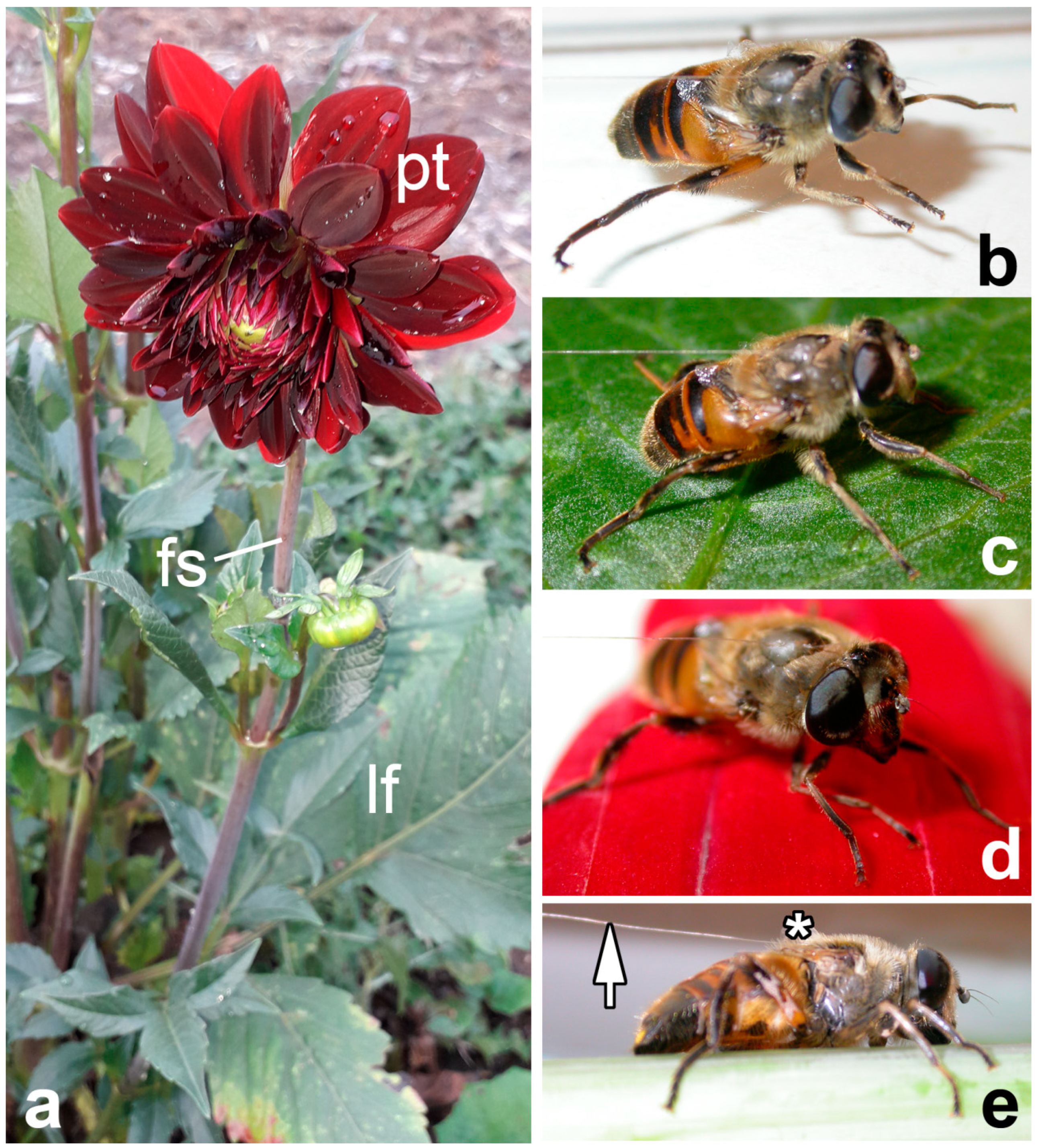
2.1. Plants and Insects
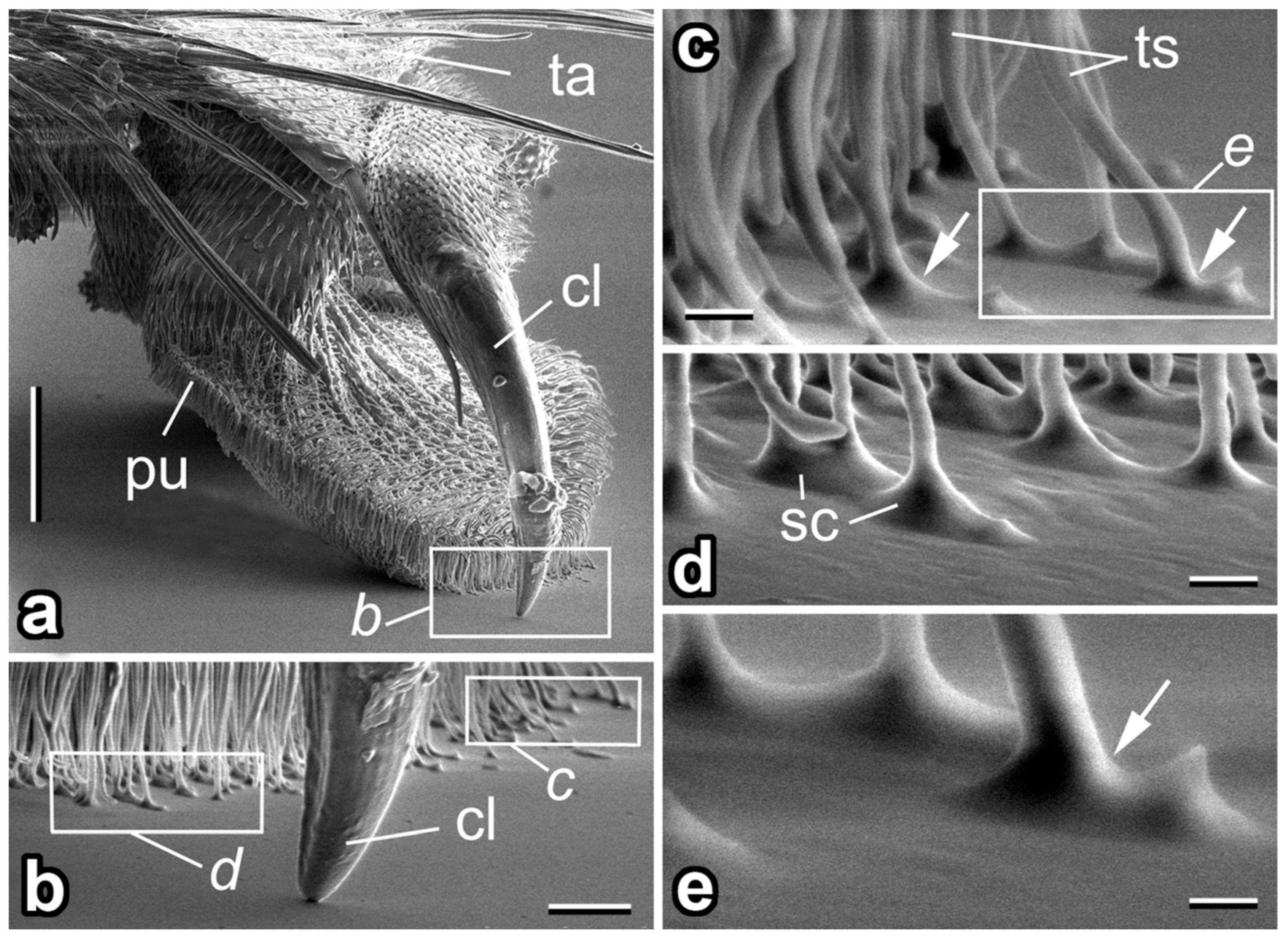
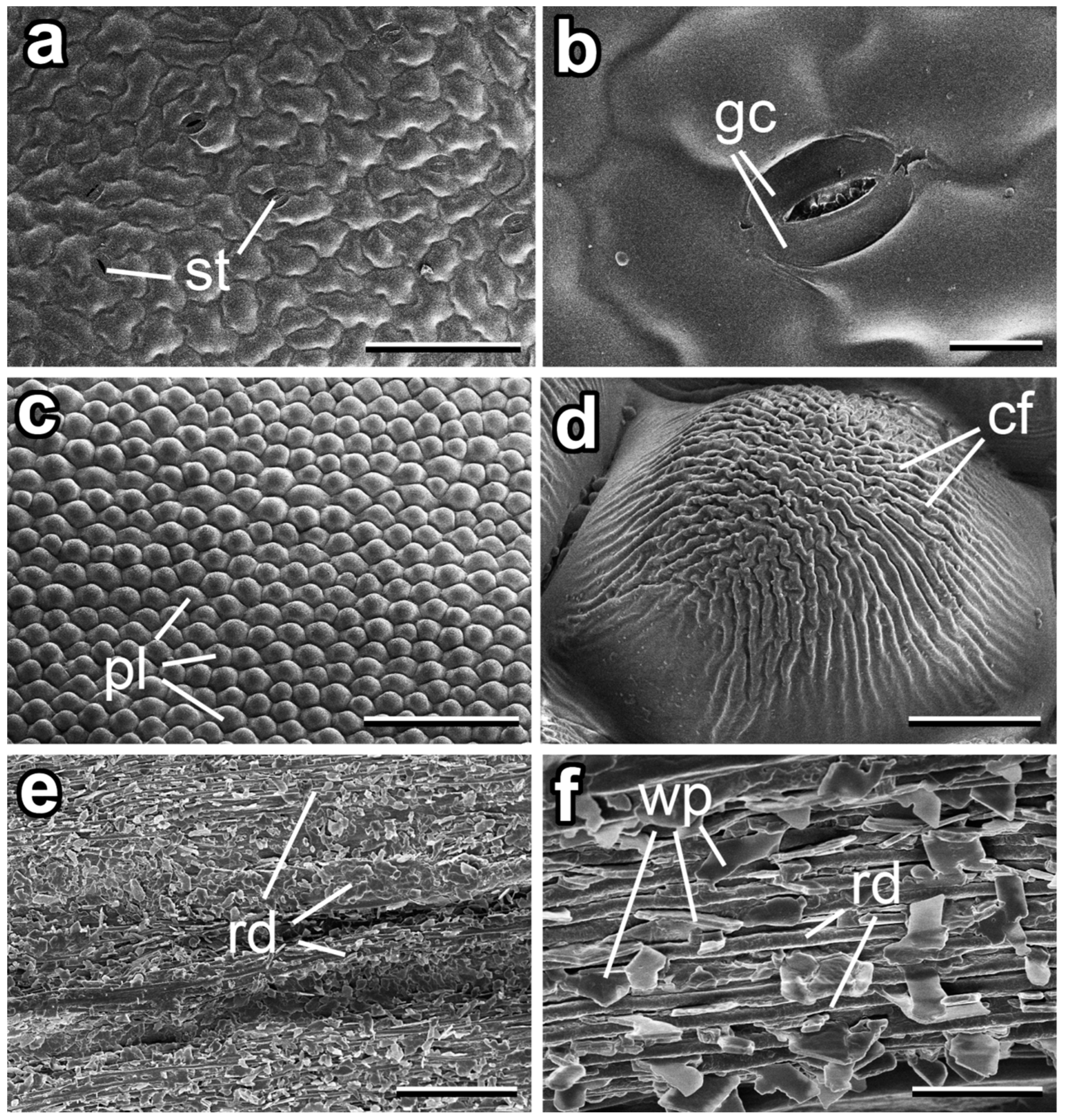
The garden dahlia D. pinnata (Figure 1a) is a 30–200 cm high perennial herbaceous plant with erect stems branching only in the inflorescence, usually simple ovate leaves, and 2–8 flower stems later called “stems” (5–15 cm long) each bearing a flower head (capitulum) with both tubular and ovate strap-shaped florets (ray flowers or ligulate flowers). As a ligula represents fused petals, we called it “petal” throughout the text. Being native to Mexico, Central America and Colombia, D. pinnata is now extensively cultivated worldwide as a decorative plant [38]. Plant samples (stems, upper leaves, and petals) for cryo-SEM examination and experiments (Figure 1) were collected from garden plants (Jagotyn, Kiev District, Ukraine, 50°17’ N 31°46’ E).
The common drone fly E. tenax mimicks a stock bee [39] that feeds on the nectar and pollen of flowers [40,41]. It has a cosmopolitan distribution and is the most widely distributed syrphid species in the world [42]. Insects were captured in August 2005 near Jagotyn and used for structural examination and experiments. The attachment system of E. tenax is composed of paired claws and paired setose pulvilli on each foot (Figure 2a,b) (for details see [43]). Microscopic tenent setae covering the pulvilli have spatula-shaped terminal elements (Figure 2d) responsible for building intimate contact with a substrate (Figure 2b–e). Additionally, a pad fluid is secreted into a contact zone (Figure 2c–e).

2.2. Microscopy

Plant samples (stem, upper leaf, and petal) (Figure 1a) for cryo-SEM examination were cut off from living plants and kept for 24 h inside small plastic vials containing wet paper in order to prevent desiccation of the plant material. Next, small (1 cm × 1 cm) portions were cut out from the middle regions of the leaf and petal and from the stem, glued with polyvinyl alcohol Tissue-Teck O.C.T.TM Compound (Sakura Finetek Europe B.V., Zoeterwoude, The Netherlands) to a metal holder, and frozen in a cryo stage preparation chamber at −140 °C (Gatan ALTO 2500 cryo preparation system, Gatan Inc., Abingdon, UK). Frozen samples of the upper (adaxial) side, in both the leaf and petal, and the stem surface were sputter coated with gold–palladium (6 nm thickness) and studied in a frozen condition in a cryo-SEM Hitachi S-4800 (Hitachi High-Technologies Corporation, Tokyo, Japan) at 3 kV accelerating voltage and −120 °C temperature. Microscopy was performed at the Max Planck Institute for Metals Research (Stuttgart, Germany).
Types of wax projections were identified according to [44]. Morphometrical variables of plant surface features were measured from digital images using the software SigmaScan Pro 5 (SPSS Inc., Chicago, IL, USA). These data are given in the text as mean ± SD for n = 10.

2.3. Experiments

Force measurements were carried out using a load cell force sensor FORT-10 (10 g capacity; World Precision Instruments Inc., Sarasota, FL, USA) connected to a force transducer MP 100 (Biopac Systems Ltd., Santa Barbara, CA, USA) [45,46]. Prior to the experiments, test insects were made incapable of flying by cutting off their wings. After a certain recovery time, the fly was attached to the force sensor by means of a 10–15 cm long hair glued to the dorsal side of its thorax with a droplet of molten beeswax (Figure 1e). The force produced by the insect walking horizontally on the test substrates and pulling the hair (Figure 1b–e) for approximately 5–30 s was measured. Force–time curves were used to estimate the maximal friction (traction) force produced by insects. Since the flies were constrained to pulling parallel (not at an angle) to the measurement axis of the sensor, the registered force corresponded to the total traction force. Three types of fresh plant samples were used as substrates: (i) the adaxial side of the upper leaf; (ii) the adaxial side of the petal; and (iii) the stem, where insects move towards the apical stem direction (Figure 1c–e). A smooth hydrophilic glass surface served as a control (Figure 1b). For each insect individual, the test substrates were used in the following sequence: (1) glass as a reference, (2 or 3) leaf, (3 or 2) petal, and (4) stem. Leaf and petal samples were randomized in the consecutive test series. Taking into account that the stem surface is covered with three-dimensional epicuticular wax projections [47], we performed experiments on the stem surface at the end of a test series, in order to avoid the effect of a possible contamination of insect feet by wax particles on the experimental results.
Force tests were carried out at 22–25 °C and ca. 60–75% humidity. On each substrate type, experiments with 10 individual flies (10 repetitions per insect) were performed. In all, 400 force tests were conducted.
Force values were analysed using two-way analysis of variance (ANOVA), considering the insect individual and the substrate as factors (software SigmaPlot 11.0, SPSS Inc., Chicago, IL, USA).

3. Results

3.1. Micromorphology of Dahlia pinnata Surfaces

The adaxial leaf side was slightly uneven because of epidermal cells showing a somewhat convex shape of the outer walls and of the sparsely dispersed (abundance: ca. 0.15 mm−2) stomata, having slightly sunk guard cells (Figure 3a,b). The surface had a smooth appearance (Figure 3b).
The adaxial petal surface was strongly uneven (Figure 3c). It was characterized by the papillate, polygonal epidermal cells, which were noticeably structured with rather uniform cuticular folds (width: 1.68 ± 0.34 µm; height: 0.68 ± 0.16 µm) (Figure 3c,d). Originating at the cell periphery, at first the folds were straight and rather low, becoming more and more wavy, higher (height: 0.69 ± 0.11 µm), and densely spaced (distance between folds: 0.51 ± 0.08 µm) to the centre of the papillae (Figure 3d).
Stems of D. pinnata had a hierarchically arranged system of ridges and grooves (Figure 3e,f). The surface bore a three-dimensional epicuticular coverage consisting of separate wax plates (abundance: ca. 0.3–0.5 plates per 1 µm) (Figure 3f). The plates varied both in dimensions (length: 2.58 ± 0.67 µm; width: 1.46 ± 0.38 µm; thickness: 0.19 ± 0.06 µm) and shapes, however, being more often rhomb shaped. Their edges were usually rather distinct, but also projections with slightly irregular edges occurred. The plates showed no preferred orientation. They protruded from the cuticle surface at various angles and were attached to the latter either by their narrow or larger sides. Such a variation in plate connections to the underlying surface was possibly caused by the particular (hierarchical ridges/grooves) geometry of the stem.

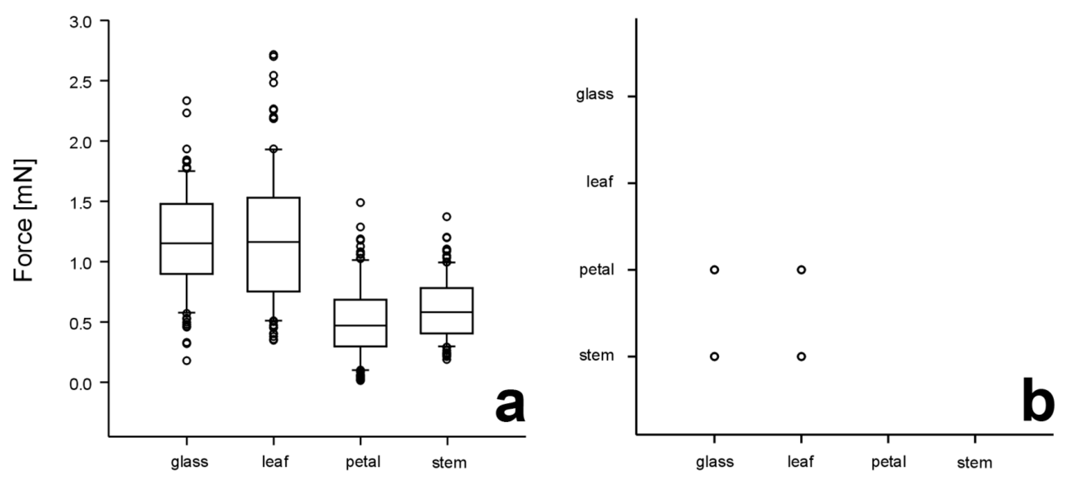
3.2. Traction Forces on Different Test Substrates

Traction force values of 10 individuals of E. tenax obtained on glass and different plant surfaces are presented in Figure 4a. Since the force generated by different insect individuals varied greatly even on glass (1.18 ± 0.38 mN, n = 100), we analysed the obtained data using two-way ANOVA, considering both the insect individual and the substrate as factors. The flies showed significantly different forces (p < 0.001, two-way ANOVA). Both factors, insect individuals and substrates, significantly influenced the force values (individuals: F9,391 = 37.565, substrates: F3,397 = 213.416, both p < 0.001). The similarly higher forces were registered on reference glass and the leaf surface (Figure 4). These force values significantly differed from much lower ones obtained on petals and stems, which did not differ significantly from each other (Figure 4).

4. Discussion

Our experimental results clearly distinguished two groups among tested surfaces. Reference glass and the upper leaf surface ensured rather high attachment force of the E. tenax fly, whereas both the flower stem and petal significantly reduced it. Strong attachment on smooth, both artificial and natural, surfaces has been observed in many insect species for a rather long time [16,46,48,49,50,51]. However, the attachment force reduction on flower stems and petals is caused by different structural effects. In the case of the flower stem, it is the combination of ridged topography and three-dimensional wax projections [47], whereas the papillate petal surface is supplemented by cuticular folds. The effect of the three-dimensional wax coverage on insect attachment has been repeatedly reported in numerous studies (reviewed in [15]) and several contributing mechanisms, such as surface roughness, insect pad contamination, pad fluid absorption, hydroplaning, and separation layer, have been proposed [16,17] and, in part, experimentally proved [21,46,52,53,54]. The role of cuticular folds in a decrease in insect attachment force has also been recently revealed by several experimental studies [12,13,14].
The biological reason of insect attachment force reduction on the flower stem is rather clear and is associated with a greasy pole syndrome, i.e., a plant defence mechanism preventing ants from visiting flowers and robbing nectar [55,56,57]. It has been described in Salix (Salicaceae), Hypenia, and Eriope (both Lamiaceae) plant species and is based on the combined effect of several stem features, such as slender elongate erect stems, rigid spreading trichomes on lower internodes, three-dimensional wax coverage, and, often, swellings in upper internodes, which hamper access of ants to apically located plant reproductive organs [57]. Recently, this syndrome was experimentally studied in several other plants [47,58,59], D. pinnata among them [47], where main attention was given to contribution of the wax coverage on flower stems to impeding the locomotion of the generalist ant species Lasius niger (Hymenoptera: Formicidae).
However, the reason behind insect attachment force reduction on the petals is less evident. It is plausible to suggest that optimal conditions for attachment of insect pollinators on petals would be advantageous from a plant perspective. That is why the obtained (opposite) result was rather surprising. One possible explanation is related to a need of plants, relying on generalist insect pollinators, to attract latter from a certain distance to their “cafeteria”-type flowers. These individual flowers are usually rather large or, if small, then combined into a rather large inflorescence, as it is the case in the plant species studied here. Many such complex inflorescences (anthodiums) in plants from the family Asteraceae have brightly coloured petals of marginal (ray) flowers. The colour intensity is usually enhanced due to conical or papilla-shaped epidermal cells often supplemented by micro- and nanostructures, such as cuticular folds. It seems to be a result of evolutionary optimization that the enhancement of one function (optical in this particular case), relying on the surface microstructuring, leads to the reduction in the other (mechanical) one. It can be further hypothesized that the function of pollinator attraction is more dominant for “cafeteria”-flowers than providing optimal attachment conditions for pollinators. Since the petals were not particularly large in comparison to the leg span of the syrphid fly studied here, and also to other typical pollinators, such as bees or calliphorid flies, these insects can presumably secure good attachment to the petals by using mechanical interlocking of their tarsal claws to the petal margins without contacting the petal surface with their adhesive pads. Another way, how the insects may avoid problems of low adhesion on D. pinnata petals is their tendency to land in the central area of the inflorescence, where the tubular flowers lacking prominent petals are situated.
Additionally, the combination of conical surface structures with cuticular folds found in a number of plant species may be responsible for so called “rose petal effect”, which is based on the ability of certain rough surfaces (with hierarchically organized surface micro- and nanostructures) to have a high contact angle with water, simultaneously with strong adhesion of water (high contact angle hysteresis) [60,61]. This is well-known for petals of plants from, for example, the Rosaceae and Asteraceae families (see Figure 1a) and was even biomimetically implemented in certain technical surfaces [62,63,64]. In plants, this kind of surfaces prevents water from wetting the petals and from rolling drops off the surface into the direction of the flower/inflorescence centre. The surfaces having low wetting ability by water, but stably holding water drops, provides optimal conditions for a quick evaporation of the fluid. Although a thin water film has a high evaporation rate due to its large surface area, breaking such a film in numerous small round droplets, having punctual contacts with the substrate, leads to an even larger overall area that can enhance evaporation rate [60,61,62,63,64]. Moreover, in contrast to the thin water film, this effect supports water evaporation in an almost non-contact state with the petal surface.
Thus, based on the combination of cryo-SEM examination of different surfaces (leaf, flower stem, petal) in the plant D. pinnata and the force measurements of the fly E. tenax attachment to these surfaces, we showed that the petal surface is not adapted to stronger attachment of generalist insect pollinators. In our opinion, these “cafeteria”-type flowers have the petals, where the colour intensity is enhanced due to papillate epidermal cells covered by cuticular folds at the micro- and nanoscale, and exactly these latter structures mainly contribute to adhesion reduction in insect pollinators. Obtained results may be potentially interesting for fabrication of technical coatings with anti-adhesive properties for insects [52,65,66,67,68,69].

Author Contributions

All authors have equally contributed to the work reported (conceptualization, methodology, investigation, data curation, writing, and visualization). All authors have read and agreed to the published version of the manuscript.

Funding

This research received no external funding.

Data Availability Statement

The data presented in this study are available on request from the corresponding author.

Acknowledgments

We are grateful to the Max Planck Institute for Metals Research (Stuttgart, Germany) for providing microscopy equipment for this study and the four anonymous reviewers for their useful comments on the initial version of this paper. Mohsen Jafarpour kindly provided linguistic corrections.

Conflicts of Interest

The authors declare no conflict of interest.

References

  1. Southwood, S. Plant Surfaces and Insects—An Overview. In Insects and the Plant Surface; Juniper, B., Southwood, R., Eds.; Edward Arnold Publishers: London, UK, 1986; pp. 1–22. [Google Scholar]
  2. Jeffree, C.F. The Cuticle, Epicuticular Waxes and Trichomes of Plants, with Reference to Their Structure, Function and Evolution. In Insects and the Plant Surface; Juniper, B., Southwood, R., Eds.; Edward Arnold Publishers: London, UK, 1986; pp. 23–63. [Google Scholar]
  3. Gorb, E.V.; Gorb, S.N. Anti-Adhesive Surfaces in Plants and Their Biomimetic Potential. In Materials Design Inspired by Nature: Function Through Inner Architecture; Fratzl, P., Dunlop, J.W.C., Weinkamer, R., Eds.; RSC Publishing: Cambridge, UK, 2013; pp. 282–309. [Google Scholar]
  4. Levin, D.A. The role of trichomes in plant defense. Q. Rev. Biol. 1973, 48, 3–15. [Google Scholar] [CrossRef]
  5. Johnson, B. The injurious effects of the hooked epidermal hairs of French beans (Phaseolus vulgaris L.) on Aphis craccivora Koch. Bull. Entomol. Res. 1953, 44, 779–788. [Google Scholar] [CrossRef]
  6. Rebora, M.; Salerno, G.; Piersanti, S.; Gorb, E.; Gorb, S.N. Entrapment of Bradysia paupera (Diptera: Sciaridae) by Phaseolus vulgaris (Fabaceae) plant leaf. Arthropod. Plant. Interact. 2020, 14, 499–509. [Google Scholar] [CrossRef]
  7. Hare, J.; Elle, E. Variable impact of diverse insect herbivores on dimorphic Datura wrightii. Ecology 2002, 83, 2711–2720. [Google Scholar] [CrossRef]
  8. Andres, M.R.; Connor, E.F. The community-wide and guild-specific effects of pubescence on the folivorous insects of manzanitas Arctostaphylos spp. Ecol. Entomol. 2003, 28, 383–396. [Google Scholar] [CrossRef] [Green Version]
  9. Gibson, R.W. Glandular hairs providing resistance to aphids in certain wild potato species. Ann. Appl. Bio. 1971, 68, 113–119. [Google Scholar] [CrossRef]
  10. Dufey, S.S. Plant Glandular Trichomes: Their Partial Role in Defense Against Insects. In Insects and the Plant Surface; Juniper, B., Southwood, R., Eds.; Edward Arnold Publishers: London, UK, 1986; pp. 151–172. [Google Scholar]
  11. Voigt, D.; Gorb, E.; Gorb, S. Plant surface–bug interactions: Dicyphus errans stalking along trichomes. Arthropod Plant Interact. 2007, 1, 221–243. [Google Scholar] [CrossRef]
  12. Prüm, B.; Seidel, R.; Bohn, H.F.; Speck, T. Plant surfaces with cuticular folds are slippery for beetles. J. R. Soc. Interface 2011, 9, 127–135. [Google Scholar] [CrossRef] [Green Version]
  13. Prüm, B.; Bohn, H.F.; Seidel, R.; Rubach, S.; Speck, T. Plant surfaces with cuticular folds and their replicas: Influence of microstructuring and surface chemistry on the attachment of a leaf beetle. Acta Biomater. 2013, 9, 6360–6368. [Google Scholar] [CrossRef]
  14. Surapaneni, V.A.; Aust, T.; Speck, T.; Thielen, M. Polarity in cuticular ridge development and insect attachment on leaf surfaces of Schismatoglottis calyptrata (Araceae). Beilstein J. Nanotechnol. 2021, 12, 1326–1338. [Google Scholar] [CrossRef]
  15. Gorb, E.V.; Gorb, S.N. Anti-adhesive effects of plant wax coverage on insect attachment. J. Exp. Bot. 2017, 68, 5323–5337. [Google Scholar] [CrossRef] [PubMed] [Green Version]
  16. Gorb, E.V.; Gorb, S.N. Attachment ability of the beetle Chrysolina fastuosa on various plant surfaces. Entomol. Exp. Appl. 2002, 105, 13–28. [Google Scholar] [CrossRef]
  17. Gorb, E.V.; Purtov, J.; Gorb, S.N. Adhesion force measurements on the two wax layers of the waxy zone in Nepenthes alata pitchers. Sci. Rep. 2014, 4, 5154. [Google Scholar] [CrossRef] [PubMed] [Green Version]
  18. Juniper, B.E.; Robins, R.J.; Joel, D.M. The Carnivorous Plants; Academic Press: London, UK, 1989; 353p. [Google Scholar]
  19. Gaume, L.; Gorb, S.; Rowe, N. Function of epidermal surfaces in the trapping efficiency of Nepenthes alata pitchers. New Phytol. 2002, 156, 479–489. [Google Scholar] [CrossRef]
  20. Gorb, E.; Haas, K.; Henrich, A.; Enders, S.; Barbakadze, N.; Gorb, S. Composite structure of the crystalline epicuticular wax layer of the slippery zone in the pitchers of the carnivorous plant Nepenthes alata and its effect on insect attachment. J. Exp. Biol. 2005, 208, 4651–4662. [Google Scholar] [CrossRef] [PubMed] [Green Version]
  21. Scholz, I.; Bückins, M.; Dolge, L.; Erlinghagen, T.; Weth, A.; Hischen, F.; Mayer, J.; Hoffmann, S.; Riederer, M.; Riedel, M.; et al. Slippery surfaces of pitcher plants: Nepenthes wax crystals minimize insect attachment via microscopic surface roughness. J. Exp. Biol. 2010, 213, 1115–1125. [Google Scholar] [CrossRef] [Green Version]
  22. Daumer, K. Blumenfarben, wie sie die Bienen sehen. Z. Vergl. Physiol. 1958, 41, 49–110. [Google Scholar] [CrossRef]
  23. Kugler, H. Blütenokologie; Gustav Fischer Verlag: Stuttgart, Germany, 1970; 345p. [Google Scholar]
  24. Willmer, P. Pollination and Floral Ecology; Princeton University Press: Princeton, NJ, USA, 2011; 832p. [Google Scholar]
  25. Oelschlägel, B.; Gorb, S.; Wanke, S.; Neinhuis, C. Structure and biomechanics of trapping flower trichomes and their role in the pollination biology of Aristolochia plants (Aristolochiaceae). New Phytol. 2009, 184, 988–1002. [Google Scholar] [CrossRef]
  26. Müller, L. Zur biologischen Anatomie der Blüte von Ceropegia woodii Schlechter. Biol. Gen. 1926, 2, 799–814. [Google Scholar]
  27. Vogel, S. Die Bestäubung der Kesselfallen-Blüten von Ceropegia. Beiträge Zur Biol. Der Pflanz. 1961, 36, 159–237. [Google Scholar]
  28. Vogel, S.; Martens, J. A survey of the lethal kettle traps of Arisaema (Araceae), with records of pollinating fungus gnats from Nepal. Bot. J. Linnean Soc. 2000, 133, 61–100. [Google Scholar] [CrossRef]
  29. Poppinga, S.; Koch, K.; Bohn, H.F.; Barthlott, W. Comparative and functional morphology of hierarchically structured anti-adhesive surfaces in carnivorous plants and kettle trap flowers. Funct. Plant Biol. 2010, 37, 952–961. [Google Scholar] [CrossRef]
  30. Barthlott, W.; Ehler, N. Raster-Elektronenmikroskopie der Epidermis-Oberflächen von Spermatophyten (Tropische und Subtropische Pflanzenwelt); Franz Steiner Verlag: Stuttgart, Germany, 1977; 105p. [Google Scholar]
  31. Christensen, K.; Hansen, H. SEM-studies of epidermal patterns of petals in the angiosperms. Opera Bot. 1998, 135, 1–91. [Google Scholar]
  32. Whitney, H.M.; Chittka, L.; Glover, B.J. Conical epidermal cells allow bees to grip flowers and increase foraging efficiency. Curr. Biol. 2009, 19, 948–953. [Google Scholar] [CrossRef] [Green Version]
  33. Whitney, H.M.; Bennett, K.M.; Dorling, M.; Sandbach, L.; Prince, D.; Chittka, L.; Glover, B.J. Why do so many petals have conical epidermal cells. Ann. Bot. 2011, 108, 609–616. [Google Scholar] [CrossRef]
  34. Prüm, B.; Seidel, R.; Bohn, H.F.; Speck, T. Impact of cell shape in hierarchically structured plant surfaces on the attachment of male Colorado potato beetles (Leptinotarsa decemlineata). Beilstein J. Nanotechnol. 2012, 3, 57–64. [Google Scholar] [CrossRef] [PubMed] [Green Version]
  35. Papiorek, S.; Junker, R.R.; Lunau, K. Gloss, colour and grip: Multifunctional epidermal cell shapes in bee- and bird-pollinated flowers. PLoS ONE 2014, 9, e112013. [Google Scholar] [CrossRef] [Green Version]
  36. Knoll, F. Insekten und Blumen: Experimentelle Arbeiten zur Vertiefung unserer Kenntnisse über die Wechselbeziehungen zwischen Pflanzen und Tieren. Abbandl. D Zool Botan. Gesellsch. Wien 1921, 12, 1–646. [Google Scholar] [CrossRef] [Green Version]
  37. Bräuer, P.; Neinhuis, C.; Voigt, D. Attachment of honeybees and greenbottle flies to petal surfaces. Arthropod Plant Interact 2017, 11, 171–192. [Google Scholar] [CrossRef]
  38. Jäger, E.J.; Ebel, F.; Hanelt, P.; Müller, G.K. Rothmaler Exkursionsflora von Deutschland. 5: Krautige Zier- und Nutzpflanzen; Spektrum: Heidelberg, Germany, 2008; 879p. [Google Scholar]
  39. Heal, J.R. Colour patterns of syrphidae: IV. Mimicry and variation in natural populations of Eristalis tenax. Heredity 1982, 49, 95–109. [Google Scholar] [CrossRef]
  40. Buckton, G.B. The Natural History of Eristalis Tenax or the Drone-Fly; Macmillan: London, UK; New York, NY, USA, 1895; 140p. [Google Scholar]
  41. Holloway, B.A. Pollen-feeding in hover-flies (Diptera: Syrphidae). N. Z. J. Zool. 1979, 3, 339–350. [Google Scholar] [CrossRef]
  42. Skevington, J.H.; Locke, M.M.; Young, A.D.; Moran, K.; Crins, W.J.; Marshall, S.A. Field Guide to the Flower Flies of Northeastern North America; Princeton University Press: Princeton, NJ, USA, 2019; 512p. [Google Scholar]
  43. Gorb, S.; Gorb, E.; Kastner, V. Scale effects on the attachment pads and friction forces in syrphid flies (Diptera, Syrphydae). J. Exp. Biol. 2001, 204, 1421–1431. [Google Scholar] [CrossRef]
  44. Barthlott, W.; Neinhuis, C.; Cutler, D.; Ditsch, F.; Meusel, I.; Theisen, I.; Wilhelmi, H. Classification and terminology of plant epicuticular waxes. Bot. J. Linn. Soc. 1998, 126, 237–260. [Google Scholar] [CrossRef]
  45. Gorb, S.N.; Popov, V.L. Probabilistic fasteners with parabolic elements: Biological system artificial model and theoretical considerations. Phil. Trans. R. Soc. Lond. A 2002, 360, 211–225. [Google Scholar] [CrossRef]
  46. Gorb, E.; Hosoda, N.; Miksch, C.; Gorb, S. Slippery pores: Anti-adhesive effect of nanoporous substrates on the beetle attachment system. J. R. Soc. Interface 2010, 7, 1571–1579. [Google Scholar] [CrossRef] [Green Version]
  47. Gorb, E.; Gorb, S. How a lack of choice can force ants to climb up waxy plant stems. Arthropod Plant Interact 2011, 5, 297–306. [Google Scholar] [CrossRef]
  48. Stork, N.E. Experimental analysis of adhesion of Chrysolina polita (Chrysomelidae, Coleoptera) on a variety of surfaces. J. Exp. Biol. 1980, 88, 91–107. [Google Scholar] [CrossRef]
  49. Edwards, P.B. Do waxes of juvenile Eucalyptus leaves provide protection from grazing insects. Aust. J. Ecol. 1982, 7, 347–352. [Google Scholar] [CrossRef]
  50. Federle, W.; Maschwitz, U.; Fiala, B.; Riederer, M.; Hölldobler, B. Slippery ant-plants and skilful climbers: Selection and protection of specific ant partners by epicuticular wax blooms in Macaranga (Euphorbiaceae). Oecologia 1997, 112, 217–224. [Google Scholar] [CrossRef]
  51. Eigenbrode, S.D.; White, C.; Rohde, M.; Simon, C.J. Behavior and effectiveness of adult Hyppodamia convergens (Coleoptera: Coccinellidae) as a predator of Acyrthosiphon pisum on a glossy-wax mutant of Pisum sativum. Environ. Entomol. 1998, 91, 902–909. [Google Scholar] [CrossRef] [Green Version]
  52. Gorb, E.; Gorb, S. Effects of surface topography and chemistry of Rumex obtusifolius leaves on the attachment of the beetle Gastrophysa viridula. Entomol. Exp. Appl. 2009, 130, 222–228. [Google Scholar] [CrossRef]
  53. Gorb, E.V.; Gorb, S.N. 2006. Do Plant Waxes Make Insect Attachment Structures Dirty? Experimental Evidence for the Contamination Hypothesis. In Ecology and Biomechanics: A Mechanical Approach to the Ecology of Animals and Plants; Herrel, A., Speck, T., Rowe, N.P., Eds.; CRC Press: Boca Raton, FL, USA, 2006; pp. 147–162. [Google Scholar]
  54. Gorb, E.V.; Lemke, W.; Gorb, S.N. Porous substrate affects a subsequent attachment ability of the beetle Harmonia axyridis (Coleoptera, Coccinellidae). J. R. Soc. Interface 2019, 16, 20180696. [Google Scholar] [CrossRef] [Green Version]
  55. Kerner von Marilaun, A. Flowers and Their Unbidden Guests; C. K. Paul & Co.: London, UK, 1878. [Google Scholar]
  56. Harley, R.M. Evolution and Distribution of Eriope (Labiatae) and its Relatives in Brazil. In Proceedings of a Workshop on Neotropical Distributions; Academia Brasileira de Ciencias: Rio de Janeiro, Brazil, 1988; pp. 71–120. [Google Scholar]
  57. Harley, R. The Greasy Pole Syndrome. In Ant-Plant Interactions; Huxley, C.R., Cutler, D.E., Eds.; Oxford University Press: Oxford, UK, 1991; pp. 430–433. [Google Scholar]
  58. Gorb, S.N.; Gorb, E.V. Frequency of plant visits by the generalist ant Lasius niger depends on the surface microstructure of plant stems. Arthropod Plant Interact 2019, 13, 311–320. [Google Scholar] [CrossRef]
  59. Gorb, S.N.; Gorb, E.V. Combined effect of different flower stem features on the visiting frequency of the generalist ant Lasius niger: An experimental study. Insects 2021, 12, 1026. [Google Scholar] [CrossRef]
  60. Feng, L.; Zhang, Y.; Xi, J.; Zhu, Y.; Wang, N.; Xia, F.; Jiang, L. Petal effect: A superhydrophobic state with high adhesive force. Langmuir 2008, 24, 4114–4119. [Google Scholar] [CrossRef]
  61. Bhushan, B.; Nosonovsky, M. The rose petal effect and the modes of superhydrophobicity. Philos. Trans. Royal. Soc. A 2010, 368, 4713–4728. [Google Scholar] [CrossRef]
  62. Jin, M.H.; Feng, X.L.; Feng, L.; Sun, T.L.; Zhai, J.; Li, T.J.; Jiang, L. Superhydrophobic aligned polystyrene nanotube films with high adhesive force. Adv. Mater. 2005, 17, 1977–1981. [Google Scholar] [CrossRef]
  63. Bormashenko, E.; Stein, T.; Pogreb, R.; Aurbach, D. “Petal Effect” on surfaces based on lycopodium: High-stick surfaces demonstrating high apparent contact angles. J. Phys. Chem. C 2009, 113, 5568–5572. [Google Scholar] [CrossRef]
  64. Park, Y.M.; Gang, M.; Seo, Y.H.; Kim, B.H. Artificial petal surface based on hierarchical micro- and nanostructures. Thin Solid Films 2011, 520, 362–367. [Google Scholar] [CrossRef]
  65. Féat, A.; Federle, W.; Kamperman, M.; van der Gucht, J. Coatings preventing insect adhesion: An overview. Progr. Org. Coat. 2019, 134, 349–359. [Google Scholar] [CrossRef]
  66. Zhou, Y.; Robinson, A.; Steiner, U.; Federle, W. 2014 Insect adhesion on rough surfaces: Analysis of adhesive contact of smooth and hairy pads on transparent microstructured substrates. J. R. Soc. Interface 2014, 11, 20140499. [Google Scholar] [CrossRef] [Green Version]
  67. Gorb, E.; Böhm, S.; Jacky, N.; Maier, L.P.; Dening, K.; Pechook, S.; Pokroy, B.; Gorb, S. Insect attachment on crystalline bioinspired wax surfaces formed by alkanes of varying chain lengths. Beilstein J. Nanotechnol. 2014, 5, 1031–1041. [Google Scholar] [CrossRef] [PubMed] [Green Version]
  68. Eichler-Volf, A.; Xue, L.; Dornberg, G.; Chen, H.; Kovalev, A.; Enke, D.; Wang, Y.; Gorb, E.V.; Gorb, S.N.; Steinhart, M. The influence of surface topography and surface chemistry on the anti-adhesive performance of nanoporous monoliths. ACS Appl. Mater. Interfaces 2016, 8, 22593–22604. [Google Scholar] [CrossRef] [PubMed] [Green Version]
  69. Eichler-Volf, A.; Kovalev, A.; Wedeking, T.; Gorb, E.V.; Xue, L.; You, C.; Piehler, J.; Gorb, S.N.; Steinhart, M. Bioinspired monolithic polymer microsphere arrays as generically anti-adhesive surfaces. Bioinspir. Biomim. 2016, 11, 025002. [Google Scholar] [CrossRef] [PubMed] [Green Version]
Figure 1. The plant Dahlia pinnata (a) and the fly Eristalis tenax in the experiments on different test substrates (be): glass (b), the adaxial (upper) side of the leaf (c), the adaxial side of the petal (d), and the stem (e). In (e), arrow points to a hair attached dorsally to the fly thorax using a droplet of molten beewax (asterisk). Abbreviations: fs, flower stem; lf, upper leaf; pt, petal (actually, ray flower).
Insects 14 00285 g001
Figure 2. The attachment system of the fly Eristalis tenax (cryo-SEM). (a) Dorso-lateral view of the distal part of the tarsus (ta) with pretarsus bearing claws (cl) and pulvilli (pu). (b) The claw (cl) and distal portion of the pulvillus in contact with a smooth substrate. (ce) Tenent setae (ts) covering the pulvillus in contact with the substrate. Note the pad secretion (sc) delivered into a contact zone. Arrows in (c,e) show examples of the setal terminal elements, spatulae, in contact. Scale bars: 100 µm (a), 10 µm (b), 2 µm (c,d), 1 µm (e).
Insects 14 00285 g002
Figure 3. Surfaces of different organs in the plant Dahlia pinnata (cryo-SEM). (a,b) Adaxial leaf side. (c,d) Adaxial petal side. (e,f). Stem. Abbreviations: cf, cuticular folds; gc, guard cells of the stoma; pl, papillae; rd, ridges; st, stomata; wp, epicuticular wax plates. Scale bars: 200 µm (a,c), 20 µm (b,e), 10 µm (d), 5 µm (f).
Insects 14 00285 g003
Figure 4. Traction forces produced by flies Eristalis tenax on different substrates (glass, adaxial sides of the upper leaf and the petal, and the stem in the plant Dahlia pinnata) (a) and results of the statistical comparison of the force values obtained on different substrates (b). In (a), boxplots show the interquartile range and the medians, whiskers indicate the 1.5 × interquartile range, and “°” are outliers. In (b), circles mean significant differences in force values between substrates at p < 0.05 (Tukey post hoc test, two-way ANOVA).
Insects 14 00285 g004
Disclaimer/Publisher’s Note: The statements, opinions and data contained in all publications are solely those of the individual author(s) and contributor(s) and not of MDPI and/or the editor(s). MDPI and/or the editor(s) disclaim responsibility for any injury to people or property resulting from any ideas, methods, instructions or products referred to in the content.

