Article

Insulin-like Peptides of the Western Flower Thrips Frankliniella occidentalis and Their Mediation of Immature Development

Department of Plant Medicals, College of Life Sciences, Andong National University, Andong 36729, Republic of Korea
*
Author to whom correspondence should be addressed.
Insects 2023, 14(1), 47; https://doi.org/10.3390/insects14010047
Submission received: 13 December 2022 / Revised: 31 December 2022 / Accepted: 1 January 2023 / Published: 3 January 2023
(This article belongs to the Section Insect Physiology, Reproduction and Development)

Abstract

:

Simple Summary

The western flower thrips, Frankliniella occidentalis, is an invasive and polyphagous insect pest. Its regional and host diversity suggests variation in its developmental rate. In addition, a previous study indicated that insulin signaling is related to thrips development. However, no insulin-like peptide (ILP) was identified in F. occidentalis. This study identified two ILP genes (Fo-ILP1 and Fo-ILP2) and their expression profiles were investigated. Further, their expressions were dependent on feeding activity, and the starvation treatment greatly reduced the expression levels of both ILP genes. The RNA interference of the ILP gene expressions had adverse effects on the immature development of the thrips. However, different host plants causing variation in developmental rates also led to variation in the ILP gene expression levels. These results report the first ILP genes in thysanopteran insects and their functional relationship with the thrips’ development.

Abstract

Insulin-like peptides (ILPs) mediate various physiological processes in insects. Specifically, ILP expression is required for immature development in different insects. The western flower thrips, Frankliniella occidentalis, is polyphagous, but its occurrence and population density vary among different hosts. This study assesses the developmental variations in the thrips through quantitative analysis of their ILP expressions. Two types of ILPs (Fo-ILP1 and Fo-ILP2) were identified from the genome of F. occidentalis, and both ILPs were predicted to have the characteristics of signal peptides and B-C-A chains linked by cysteines. A phylogenetic analysis indicates that these two ILPs in the thrips are clustered with the ILP1 of Drosophila melanogaster, suggesting their physiological roles in growth. In addition, the two ILP genes were relatively highly expressed at all feeding stages, but their expression was reduced during the nonfeeding prepupal and pupal stages. Furthermore, RNA interference of each ILP expression led to significant developmental retardation. In validating the ILP expression in the thrips’ development, five different varieties of host hot peppers were assessed in a choice test, along with the immature development of F. occidentalis. The expression levels of the two ILP genes were highly correlated with variations in the immature developmental rates of different hot pepper varieties. These suggest that Fo-ILP1 and Fo-ILP2 mediate the immature development of F. occidentalis by sensing different nutritional values of hot peppers. This study is the first report on ILPs in thysanopteran insects.

1. Introduction

Insulin signaling is required for insect development, similar to other metazoans. Insulin-like peptides (ILPs) and their signaling mechanisms have been extensively assessed in various insects. The first insect ILP was discovered unexpectedly during the identification of the prothoracicotropic hormone (PTTH) of Bombyx mori, known as bombyxin [1]. Later, 38 bombyxins were identified from its genome analysis [2]. Drosophila melanogaster encodes seven ILP genes [3]. Thus, most insects are likely to have multiple ILP genes [4]. Similar to mammalian insulin, insect ILPs consisting of three chains undergo post-translational modifications by proteolytic cleavage to produce mature heterodimeric peptides consisting of A-B chains linked by disulfide bonds [5].
Insulin signaling controls the blood sugar level, growth, and reproduction in vertebrates [6]. However, the insulin-mediated physiological processes are triggered by insulin synthesized in the β-cells of the pancreas, insulin-like growth factors synthesized by the liver in response to stimulatory signals of the growth hormone, or relaxin produced by the reproductive organs. The insect ILPs are produced in the brain, specifically in the insulin-producing cells (IPCs) in the pars intercerebralis of the brain. After food ingestion, the fat body can sense nutrients and send a nutritional signal called the “fat-body-derived signal” [7] to the IPCs in Drosophila. The secreted ILPs reach the target cells and trigger the insulin signaling pathway by binding to a membrane receptor called the insulin receptor (InR).
The western flower thrips Frankliniella occidentalis feeds on a large number of host plants and transmits a serious plant pathogen, the tomato spotted wilt virus (TSWV) [8]. Although the thrips feed on different hosts, they show host preference and subsequent developmental variations with different hosts [9,10,11]. Based on insulin signaling in thrips, an insulin receptor, Fo-InR, was identified in a previous study [12]. This study showed that Fo-InR encodes a sequence of 1645 amino acids, which is highly homologous (>70% homologies) to known insect InRs [12]. Fo-InR was expressed in most developmental stages, while starvation upregulated the expression levels. Furthermore, expression analysis in thrips reared on different diets indicates that the InR expression levels are negatively correlated with the immature developmental rate of F. occidentalis. This suggests that insulin signaling mediates the growth and development of F. occidentalis. However, no ILPs have been identified in thysanopteran insects, including F. occidentalis.
This study predicted two ILP genes in the F. occidentalis genome and analyzed the expression patterns to confirm their association with the development of the thrips. Additionally, using RNA interference, the physiological functions during immature growth were assessed. Furthermore, the expressional variations of the two ILP genes for different host plant varieties were explained in accordance with the host preference and subsequent developmental rates.

2. Materials and Methods

2.1. Insect Rearing and Host Plant Culture

A laboratory population of F. occidentalis was obtained from the Department of Crop Protection, National Institute of Agricultural Sciences (Jeonju, Republic of Korea), and cultured under laboratory conditions of 25 ± 2 °C, 16:8 h (L:D), and 60 ± 5% relative humidity. The germinated beans (Phaseolus coccineus L.) were supplied for feeding and oviposition. The newly laid eggs on the beans in adult colonies were transferred to a breeding dish (SPL Life Sciences, Pochon, Republic of Korea). After hatching, beans were supplied every day to the breeding dish. Under the laboratory conditions, the larvae underwent two instars (L1–L2) that were distinct from prepupae or pupae with wing pads.
The hot pepper (Capsicum annuum) seeds were purchased from different companies (Table S1) and germinated in a nursery plate. The resulting seedlings after 2 weeks were transferred to pots (5 cm in diameter and 8 cm in height) and maintained under the thrips rearing conditions. The bioassay leaves for feeding were obtained from hot pepper plants with a height of 30 cm.

2.2. Bioinformatics to Predict ILP Genes in F. occidentalis

The amino acid and DNA sequences of two ILP genes (Fo-ILP1 and Fo-ILP2) of F. occidentalis were obtained from the National Center for Biotechnology Information (NCBI; https://blast.ncbi.nlm.nih.gov) (accessed on 31 December 2022) with accession numbers as shown in Table S2. MEGA6.0 was used for the phylogenetic tree by maximum likelihood, where the evolutionary distances were computed using the Poisson correction method. Bootstrapped values were obtained with 1000 replications to support branching and clustering. The N-terminal signal peptides of Fo-ILP1 and Fo-ILP2 were determined using the SignalP 5.0 server (https://services.healthtech.dtu.dk/service.SignalP-5.0/) (accessed on 31 December 2022). Subsequently, three different chains of ILPs were predicted from the ILP templates of D. melanogaster [13]. These chains were confirmed by prediction of the putative cleavage sites using a program developed by the Neuroproteomics and Neurometabolomics Center on Cell–Cell Signaling (http://stagbeetle.animal.uiuc.edu/cgi-bin/neuropred.py) (accessed on 31 December 2022).

2.3. RNA Extraction and cDNA Preparation

The RNAs of F. occidentalis were extracted from different developmental stages using Trizol reagent (Invitrogen, Carlsbad, CA, USA) according to the manufacturer’s instructions. Each extracted RNA was then resuspended in 30 μL of diethyl pyrocarbonate and quantified using a spectrophotometer (Nanodrop, Thermo Fisher Scientific, Wilmington, DE, USA). In addition, about 400 ng of the RNA were used for cDNA synthesis using an RT premix (Intron Biotechnology, Seoul, Republic of Korea) containing the oligo dT primer according to the manufacturer’s instructions.

2.4. RT-PCR and RT-qPCR

The prepared cDNAs were used for RT-PCR with Taq polymerase (GeneALL, Seoul, Republic of Korea) and gene-specific primers (Table S3). After initial denaturation at 95 °C for 5 min, PCR was performed using a thermocycler (Step One Plus Real-Time PCR System; Applied Biosystems) with 35 amplification cycles (95 °C for 1 min, 50–55 °C for 30 s, and 72 °C for 1 min) and finalized with an additional extension step at 72 °C for 10 min. The PCR products were assessed by 1% agarose gel electrophoresis. RT-qPCR was performed as per the procedures noted by Bustin et al. [14] using Power SYBR Green PCR Master Mix (Toyobo, Osaka, Japan) with gene-specific primers (Table S3). In addition, after initial heat treatment at 95 °C for 2 min, qPCR was performed with 40 cycles of denaturation at 95 °C for 30 s, annealing at 50−55 °C for 30 s, and extension at 72 °C for 30 s. The elongation factor 1 (EF1, Table S3) was used as a reference gene to normalize the expression level of each qPCR sample. Further, quantitative analyses were performed using the comparative CT (2−ΔΔCT) method [15]. All experiments were independently replicated three times.

2.5. RNA Interference (RNAi) of ILP Genes Using Gene-Specific Double-Stranded RNA (dsRNA)

The template DNAs of the two ILP genes were amplified into 316 base pairs (bp) for Fo-ILP1 and 201 bp for Fo-ILP2 using cDNA with gene-specific primers (Table S3) containing the T7 promoter sequence (5′-TAATACGACTCACTATAGGGAGA-3′) at the 5′ end. The PCR products were used for the in vitro synthesis of the dsRNAs with the NTP mixture at 37 °C for 3 h using T7 RNA polymerase in the MEGAscript RNAi kit (Ambion, Austin, TX, USA). The resulting dsRNAs were mixed with a transfection reagent, Metafectene PRO (Biontex, Plannegg, Germany), at a 1:1 (v/v) ratio, and incubated at 25 °C for 30 min to form liposomes that protect the dsRNA from attack by the digestive enzymes. This dsRNA suspension (200 ng/µL) was used to evenly cover a bean diet with 10 μL volume, to which 25 test thrips at the L1 larval stage were applied and allowed to feed for 12 h. Thereafter, the fresh and untreated diets were replaced. A control dsRNA (dsCON) specific to enhanced green fluorescence protein (EGFP) was prepared according to the method of Vatanparast and Kim [16].

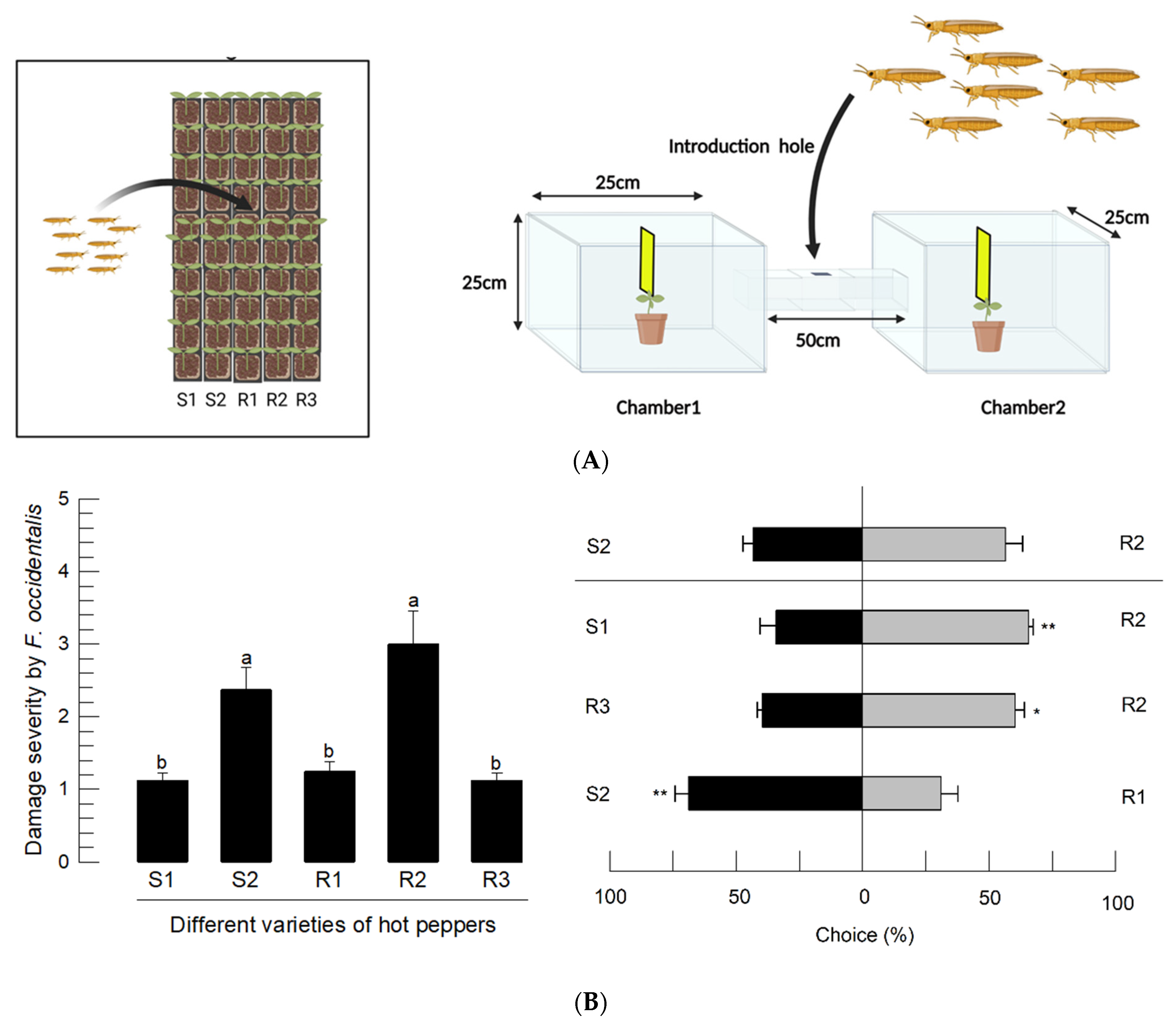
2.6. Screening Hot Pepper Varieties by Assessment of Feeding Damage

Five different hot pepper varieties (S1, S2, and R1–R3) (Table S1) were cultured in pots until the four-leaf stage. These were commercially procured and had different susceptibilities to TSWV; S1 and S2 were susceptible, whereas R1–R3 were resistant to TSWV infection. A total of 50 hot peppers, with 10 plants per variety, were contained in 100 L-size screen cages (Gaonunri, Seoul, Republic of Korea). Hundred adults of F. occidentalis were released into these cages and allowed to feed on the hot peppers for 1 week under the rearing conditions described above. The damage intensity was evaluated on a scale of 6 grades (Figure S1): no damage for grade 0, small damage limited to one leaf for grade 1, two leaves damaged on the upper 1/3 for grade 2, two leaves damaged on the upper 1/2 for grade 3, more than 2 leaves damaged for grade 4, and all leaves damaged with a yellowish color for grade 5.

2.7. Choice Test Using a Two-Way Tunnel Assay

A two-way tunnel assay was used to determine the choice test for flying adults [17]. Using this method, two different hot pepper hosts were compared to determine the choices of the F. occidentalis adults. The arena consisted of two chambers of identical sizes (25 × 25 × 25 cm) for setting two hot pepper plant varieties for the choice test (illustrated in Figure 4B). The distance between the two chambers was 50 cm. In each test, 100 adults were released through an introduction hole to observe their choices. The yellow sticky traps (15 × 10 cm, Green-AgroTech, Gyeongsan, Republic of Korea) were installed above the test hot peppers to collect the thrips. The tests were performed for 24 h at 25 ± 2 °C and 70 ± 5% relative humidity under 1000 lux illumination.

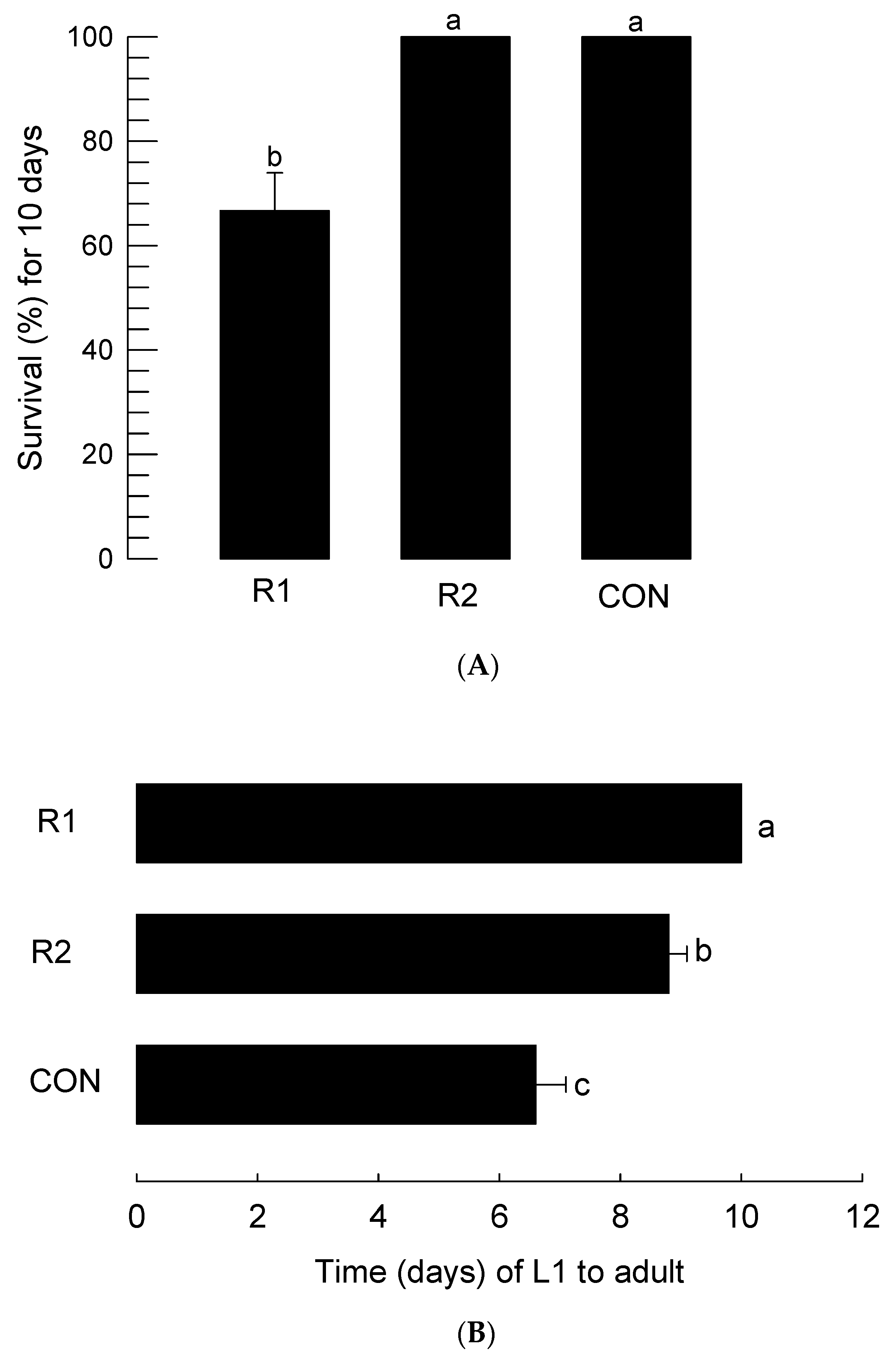
2.8. Assessment of Immature Development Period and Survival Rate for Two Hot Pepper Varieties

The thrips were reared on two different hot pepper varieties (R1 and R2) with a control diet for rearing thrips from L1 to adult under the rearing conditions described above. Newly hatched L1 larvae were placed on a disc (4 cm in diameter) of hot pepper leaves or a control diet in the insect breeding dish. In order to prevent desiccation, a wet filter paper was placed in the dish. The developmental stages or survival were observed under a microscope (EZ4, Leica, Wetzlar, Germany) every day at 10 p.m. Each treatment used 20 thrips with three replications. The developmental rate was estimated as the inverse of the developmental period from L1 to adult emergence.

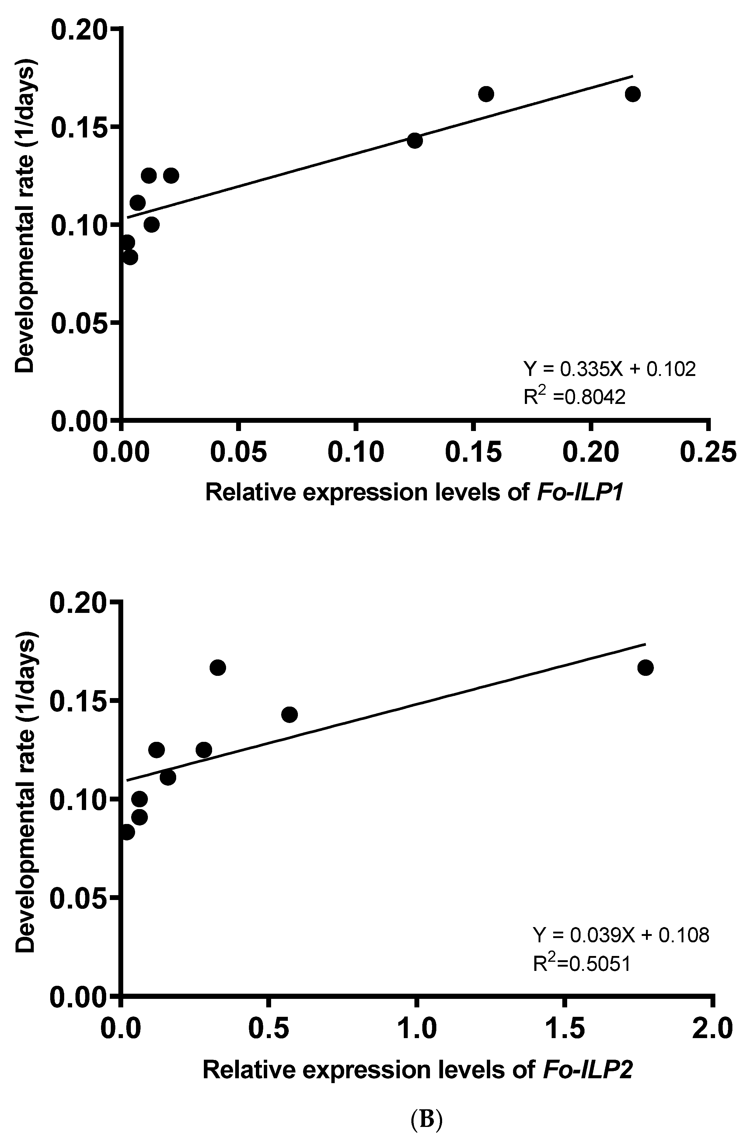
2.9. Correlation Analysis between Developmental Rate and ILP Expression

The developmental rates under three different diet conditions (R1, R2, and control diet) were compared with the expression levels of the two ILP genes (Fo-ILP1 and Fo-ILP2). The gene expression levels were estimated by RT-qPCR, as described above. However, for the RT-qPCR, the RNAs were extracted on the first days of the L2 larvae, pupae, and adults. Each RNA sample used 25 individuals, and each treatment was replicated three times. Additionally, in the correlation analysis, the expression levels at the pupal stage were compared with the developmental rates.

2.10. Statistical Analysis

The percentage data were arcsine-transformed to be normalized, and data obtained from the feeding damage or tunnel tests were subjected to a one-way analysis of variance using PROC GLM in SAS [18]. The means were compared using the least significant difference (LSD) test with a type I error of 0.05. The frequency data were analyzed by the mean difference test using PROC FREQ at a type I error of 0.05 (*) and 0.01 (**).

3. Results

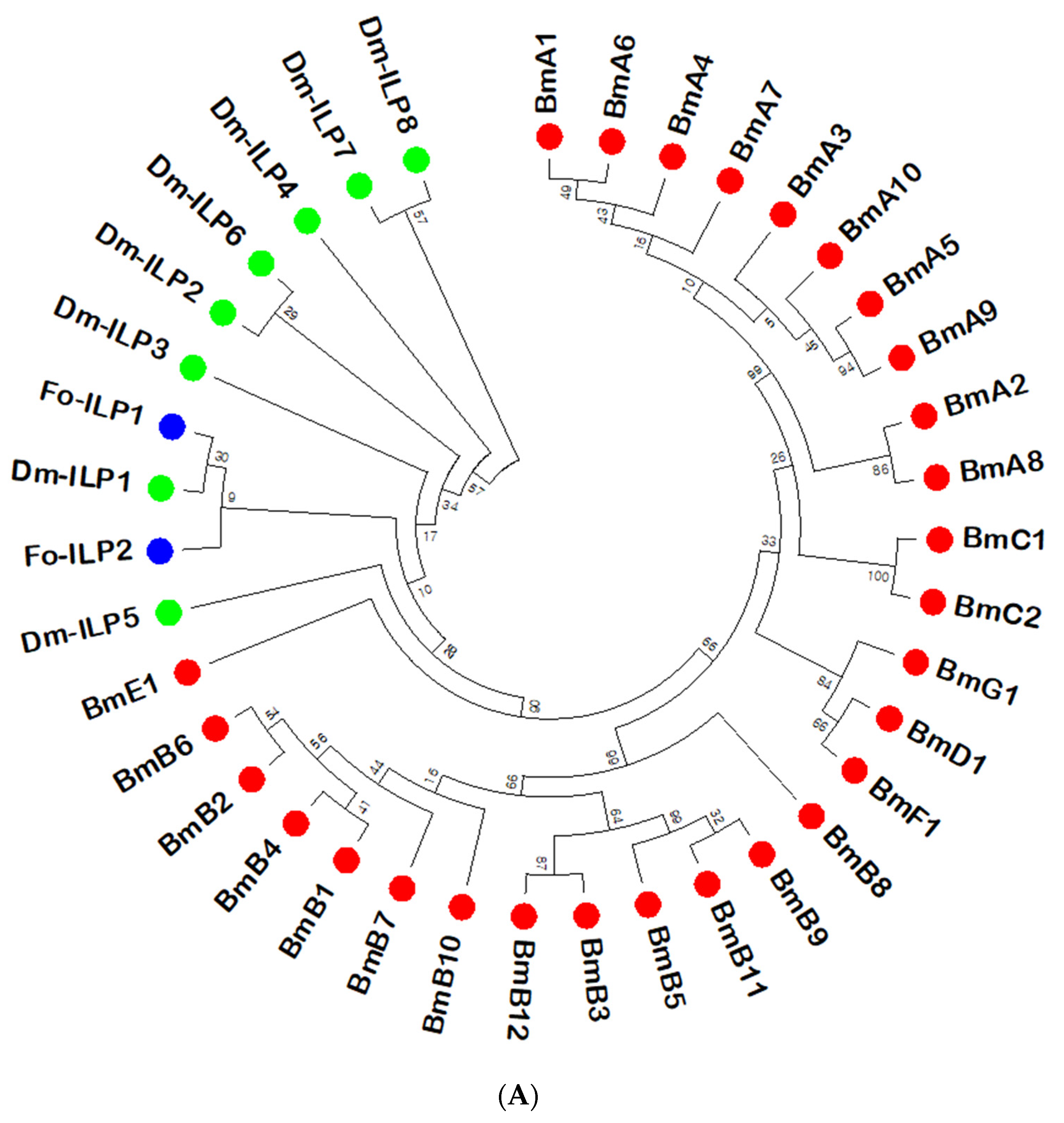
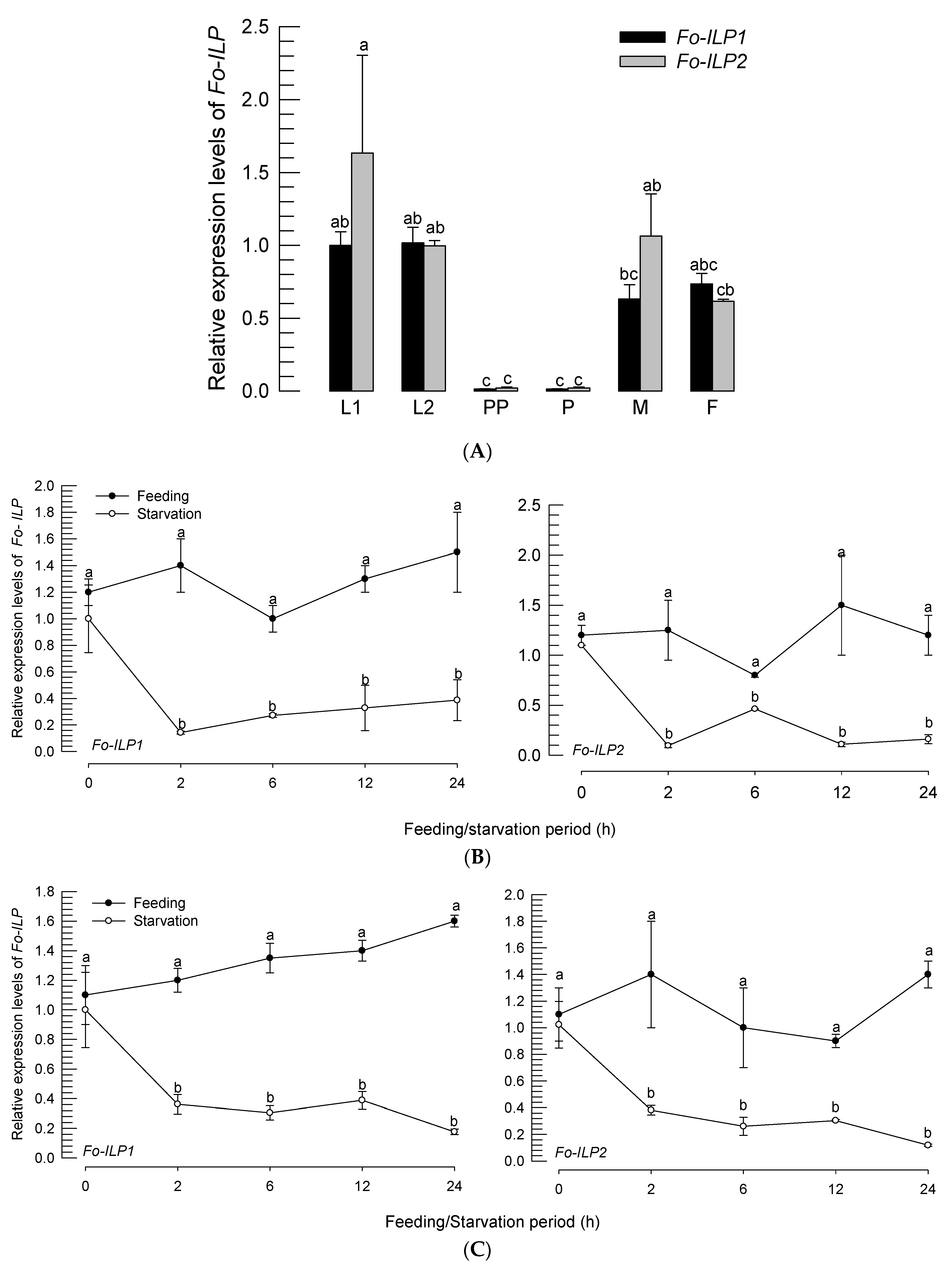
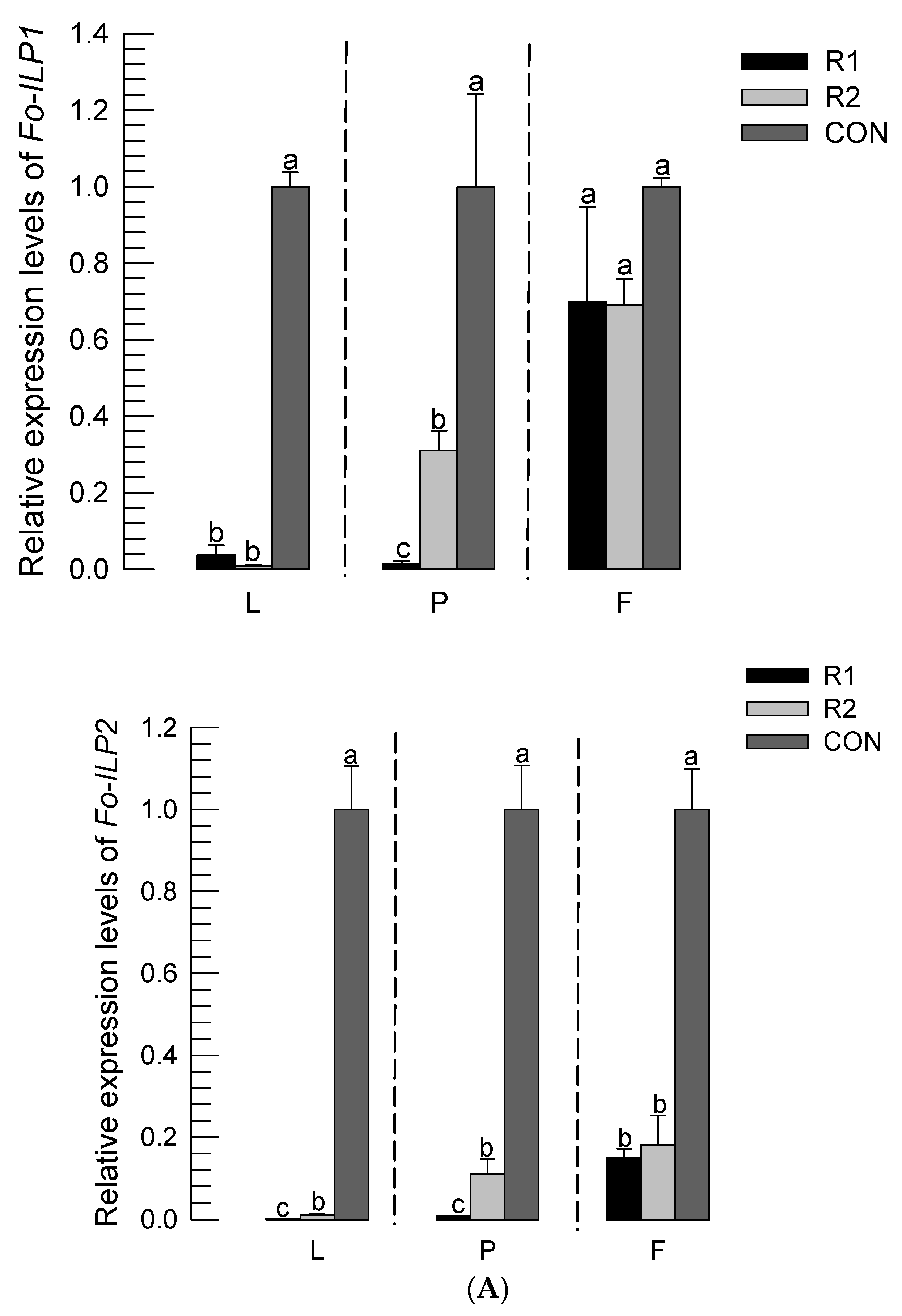
3.1. ILPs of F. occidentalis (Fo-ILPs) and Their Expression Profiles

The two ILP genes (Fo-ILP1 and Fo-ILP2) were obtained from the genome of F. occidentalis (Figure 1A). The phylogenetic analysis indicated that these two ILPs are different from lepidopteran ILPs and closely related to those of D. melanogaster (Dm-ILPs), in which they were clustered with Dm-ILP1. In comparison to Dm-ILPs, the functional domains of the two thrips ILPs were predicted (Figure 1B). The cysteines and cleavage sites were conserved among the ILPs, which allowed the ILP-characteristic three chains (B-C-A) and signal peptides to be predicted.
The two ILP genes were expressed at all developmental stages, from larvae to adults (Figure 2A). With the exception of the prepupal and pupal stages, the ILP genes were expressed at relatively stable levels at the larval and adult stages. In investigating the low level of ILP expression during the nonfeeding stages, the larvae (Figure 2B) and adults (Figure 2C) were subjected to starvation. The starvation treatment significantly (p < 0.05) suppressed the gene expressions of the ILP genes.

3.2. RNAi of ILP Gene Expression Led to Developmental Retardation

In the feeding process, with gene-specific dsRNA, the expression levels of Fo-ILP1 and Fo-ILP2 (Figure 3A) were suppressed. Additionally, in both genes, significant suppression began to occur 12 h after RNAi treatment, and the suppressed levels were maintained for at least another 12 h. Under this RNAi condition, the immature period (=larva + pupal period) was estimated (Figure 3B). Further, both RNAi treatments led to significant (p < 0.05) delays in immature development in adults.

3.3. Developmental Variations of F. occidentalis for Different Varieties of Hot Pepper

Five different varieties of hot peppers were assessed for the thrips’ preference and development. These varieties were different in terms of TSWV infection susceptibility, with two susceptible (S1 and S2) and three resistant (R1–R3) varieties. To estimate the feeding preferences, the thrips adults were released into the five varieties and assessed for feeding damage (Figure 4A). However, a TSWV-resistant strain (R2) was highly susceptible to damage from thrips feeding, while a TSWV-susceptible strain (S1) was highly resistant to feeding damage. The preferences were further supported by the choice test (Figure 4B). The two varieties (S2 and R2) that were susceptible to feeding damage were preferentially favored by the thrips.
The two TSWV-resistant strains (R1 and R2) were compared in the assessment of thrips development (Figure 5). In this assay, we used a soybean-based diet as the control. Compared to control or R2 (favored by thrips in the above assays), thrips feeding on R1 (less favored by thrips) suffered from high immature mortality (Figure 5A). Furthermore, the thrips feeding on R1 exhibited significant developmental retardation compared to those feeding on R2 or the control diet (Figure 5B).

3.4. Expression Levels of Fo-ILPs Are Correlated with Immature Development of F. occidentalis

The developmental retardation of the thrips feeding on the R1 hot pepper variety was analyzed by quantifying their ILP gene expression levels (Figure 6A). In comparison to R2 and the control diet, thrips feeding on R1 exhibited significantly lower (p < 0.05) expression levels of Fo-ILP1 and Fo-ILP2 during the larval and pupal stages. Interestingly, the thrips fed on the soybean diet showed rapid development and the highest expression levels of the two ILP genes. To determine the relationship between ILP expression and thrips development, the expression levels at the non-feeding pupal stage were compared with the developmental rates (Figure 6B). In both ILP genes, there were highly positive correlations between immature development and ILP expression.

4. Discussion

This study is a pioneering work on ILP genes and their physiological functions in Thysanoptera using F. occidentalis. In addition to a previous study on the InR of F. occidentalis [12], the identification of ILPs suggests that insulin signaling plays a crucial role in mediating the physiological processes of thrips. Specifically, the current study tested the roles of two ILPs in mediating the immature growth of F. occidentalis.
The two ILPs were expressed at stable levels at all developmental stages, from larvae to adults, except at the non-feeding prepupal and pupal stages. In investigating the low expression levels in the non-feeding stages, starvation treatment was applied to the larvae and adults, which led to suppression of the gene expressions. These observations regarding the reduction of the expression levels suggest that the nutrient signals derived from the feed diet induce expression of the ILP genes in F. occidentalis. Although the source of the ILPs was not investigated in F. occidentalis, it has been well documented that insect ILPs are produced in the brain, specifically at the insulin-producing cells (IPCs) in the pars intercerebralis [19]. The feed diet and subsequent digestion by the insect gut release nutrients into the hemolymph. These nutrients are sensed by the fat body, which sends a nutritional signal called the fat-body-derived signal to the brain in Drosophila [7]. Indeed, an insect leptin called Unpaired 2 is released from the fat body in response to feeding and activates ILP secretion from the IPCs through GABAergic neurons [20]. Furthermore, the fat body can directly sense glucose and release a nutrient signal called CCHamide-2 to activate the IPCs to secrete ILPs [21]. In F. occidentalis, the secreted ILPs may bind to the transmembrane protein InR to trigger intracellular insulin signals, which are presumably conserved in metazoans [22].
The suppression of the ILP gene expressions led to developmental retardation in F. occidentalis even under ad libitum feeding conditions. This suggested the roles of the two ILPs in mediating the immature development of F. occidentalis. The insulin signaling mediates various physiological processes, including immature growth and metabolism, in both invertebrates and vertebrates [5,23]. Additionally, the insulin signaling pathway is highly conserved in animals [22]. Further, once the ILPs bind to the InR, the downstream insulin signaling pathways are triggered by activating the insulin receptor substrates, which recruit phosphoinositide-3-kinase to the membranes to phosphorylate phosphatidylinositol-4,5-bisphosphate into phosphatidylinositol-3,4,5-trisphosphate (PIP3). The accumulated PIP3 levels then recruit phosphoinositide-dependent kinase, which activates serine-threonine protein kinase (Akt). Akt then phosphorylates Forkhead Box O (FOXO) to prevent its translocation into the nucleus while it phosphorylates the tuberous sclerosis complex (TSC) to upregulate protein translation by releasing the eukaryotic initiation factor 4E from its inhibitor 4E-binding protein. The amino-acid-mediated target of rapamycin (TOR) pathway can activate the TSC and share its downstream signal with the insulin signal. Thus, the induction of ILP expression in F. occidentalis would trigger insulin signaling in the target cells by the functional binding of ILP to InR, which activates a sequence of highly conserved downstream proteins to facilitate cell growth [19,24]. Our previous study on insulin signaling showed that InR expression is negatively correlated with the developmental rate of F. occidentalis [12]. It is well documented that InR expression is associated with immature development and adult reproduction in insects of the lepidopteran [25,26,27] and hemipteran species [28]. As predicted in the induction of the InR expression under starvation, the transcriptional factor FOXO could be phosphorylated and localized in the nucleus to survive under the harsh conditions [29]. Once insulin signaling is resumed, Akt phosphorylates FOXO to maintain the protein in the cytosol and activates the protein translation initiation factor for cell growth [30], which would lead to the immature development of F. occidentalis. This is further supported by the co-clustering of the two ILP genes with Dm-ILP1, which is known to mediate fly growth [31].
The developmental rates of F. occidentalis vary depending on its host plant varieties. The susceptibility of the host plants to thrips appeared to not be directly associated with resistance to TSWV infection. The insect resistance of the hot pepper to thrips was not clearly understood at the molecular level. However, it may be explained by inhibition of the thrips feeding behaviors because the varieties resistant to thrips exhibited much less feeding damage compared to the susceptible varieties. The hot peppers are resistant to another sucking insect, Myzus persicae, and deposit callose in the sieve elements of the phloem to prevent insect feeding behaviors [32]. It was an interesting observation that the expression levels of the ILP genes fluctuated with different host variety treatments. The plant variety facilitating larval development in F. occidentalis showed high ILP expression levels, whereas the variety retarding thrips development suppressed ILP expressions in F. occidentalis. These findings support the positive correlation between ILP expression and immature growth rate. The nutritional values of the different hot pepper varieties of F. occidentalis remain unknown. However, the severity of damage due to thrips feeding was highly associated with the preferences of the thrips for the host plants. Thus, the variety susceptible to thrips must have been consumed more than the resistant variety. The increased feeding amounts may be interpreted using the elevated ILP expression levels, which promote immature development through insulin signaling. However, further experiments would be necessary to elucidate the cause for differential ILP expression by assessing the composition of different nutrients as well as secondary metabolites in the different hot pepper varieties.
The positive correlation between ILP expression and immature growth suggests a regression to predict the growth rate of F. occidentalis. Our previous study on the negative correlation between InR expression and growth rate provides a regression formula: Developmental rate (1/day) = −0.035 × (InR expression) + 0.26. In ILP expression, ILP1 shows a higher regression coefficient than ILP2 in F. occidentalis, whose regression formula is as follows: Developmental rate (1/day) = 0.335 × (ILP1 expression) + 0.102. Owing to the direct physiological interactions between insulin signaling and growth rate, several mathematical models have been developed to explain the functional interactions [33]. In addition, a comprehensive mathematical model of insect growth rate has been devised as a qualitative model using the insulin signaling pathway components [34]. The two components ILP and InR can thus be used to establish a novel insect growth model for F. occidentalis using the functional parameters. In considering multiple ILPs in insects, additional ILPs were predicted in F. occidentalis. For example, an ILP dissimilar to Fo-ILP1 and Fo-ILP2 was reported in the adult stage and inducible by ovarian development [35]. This suggests additional ILPs may be involved in the growth and development of F. occidentalis.

Supplementary Materials

The following supporting information can be downloaded at: https://www.mdpi.com/article/10.3390/insects14010047/s1, Table S1: Hot pepper varieties used in the present study, where ‘R’ and ‘S’ indicate resistant and susceptible varieties to the tomato spotted wilt virus (TSWV), respectively; Table S2: GenBank accession numbers of the genes assessed in the present study; Table S3: Primer sequences used in the present study; Figure S1: Evaluation of the damage intensity on a scale of 0 (no damage) to 5 (severe damage) due to thrips feeding. Damage was classified into 6 grades: no damage ① for grade 0, small damage limited to one leaf ② for grade 1, two leaves damage on the upper 1/3 ③ for grade 2, two leaves damage on the upper 1/2 ④ for grade 3, more than 2 leaves damage ⑤ for grade 4, and all leaves damaged with yellowish color ⑥ for grade 5.

Author Contributions

Conceptualization, Y.-G.K.; methodology, C.-Y.K. and Y.-G.K.; software, C.-Y.K. and Y.-G.K.; validation, C.-Y.K. and Y.-G.K.; formal analysis, C.-Y.K.; investigation, C.-Y.K. and Y.-G.K.; resources, Y.-G.K.; data curation, C.-Y.K.; writing—original draft preparation, C.-Y.K. and Y.-G.K.; writing—review and editing, Y.-G.K.; visualization, C.-Y.K.; funding acquisition, Y.-G.K. All authors have read and agreed to the published version of the manuscript.

Funding

This work was financially supported by a grant from the Cooperative Research Program for Agriculture Science & Technology Development (Project No. PJ01578901) funded by the Rural Development Administration, Republic of Korea.

Institutional Review Board Statement

This study did not involve humans or animals.

Data Availability Statement

DNA and protein sequences are found in GenBank under the accession numbers given in the Methods section.

Conflicts of Interest

The authors declare no conflict of interest. The funders had no role in the design of the study, in the collection, analysis, or interpretation of data, in the writing of the manuscript, or in the decision to publish the results.

References

  1. Nagasawa, H.; Kataoka, H.; Isogai, A.; Tamura, S.; Suzuki, A.; Ishizaki, H. Amino-terminal amino acid sequence of the silkworm prothoracicotropic hormone: Homology with insulin. Science 1984, 226, 1344–1345. [Google Scholar] [CrossRef] [PubMed]
  2. Aslam, A.F.; Kiya, T.; Mita, K.; Iwami, M. Identification of novel bombyxin genes from the genome of the silkmoth Bombyx mori and analysis of their expression. Zool. Soc. 2011, 28, 609–616. [Google Scholar] [CrossRef] [Green Version]
  3. Liao, S.; Nässel, D.R. Drosophila insulin-like peptide 8 (DILP8) in ovarian follicle cells regulates ovulation and metabolism. Front. Endocrinol. 2020, 11, 461. [Google Scholar] [CrossRef] [PubMed]
  4. Al Baki, M.A.; Jung, J.K.; Kim, Y. Physiological alterations in mutants of two insulin-like peptides encoded in Maruca vitrata using CRISPR/Cas9. Front. Physiol. 2021, 12, 701616. [Google Scholar] [CrossRef] [PubMed]
  5. Antonova, Y.; Arik, A.J.; Moore, W.; Riehle, M.A.; Brown, M.R. Insulin-like peptides: Structure, signaling, and function. In Insect Endocrinology; Gilbert, L.I., Ed.; Academic Press Deletion: New York, NY, USA, 2012; pp. 63–92. [Google Scholar]
  6. Mathieu, C.; Gillard, P.; Benhalima, K. Insulin analogues in type 1 diabetes mellitus: Getting better all the time. Nat. Rev. Endocrinol. 2017, 13, 385–399. [Google Scholar] [CrossRef]
  7. Géminard, C.; Rullifson, E.J.; Leopold, P. Remote control of insulin secretion by fat body cells in Drosophila. Cell Metab. 2009, 10, 199–207. [Google Scholar] [CrossRef] [Green Version]
  8. Whitfield, A.E.; Ullman, D.E.; German, T.L. Tospovirus-thrips interactions. Annu. Rev. Phytopathol. 2005, 43, 459–489. [Google Scholar] [CrossRef]
  9. Morse, J.G.; Hoddle, M.S. Invasion biology of thrips. Annu. Rev. Entomol. 2005, 1, 67–89. [Google Scholar] [CrossRef] [Green Version]
  10. Pearsall, I.A. Flower preference behaviour of western flower thrips in the Similkameen valley, British Columbia, Canada. Entomol. Exp. Appl. 2000, 95, 303–313. [Google Scholar] [CrossRef]
  11. Mainali, B.P.; Lim, U.T. Behavioral response of western flower thrips to visual and olfactory cues. J. Insect Behav. 2011, 24, 436–446. [Google Scholar] [CrossRef]
  12. Kim, Y.; Lee, D.; Ahmed, S. First report of insulin receptor in Thysanoptera and its expression variation with development of western flower thrips, Frankliniella occidentalis. J. Asia Pac. Entomol. 2021, 24, 1004–1009. [Google Scholar] [CrossRef]
  13. Post, S.; Karashchuk, G.; Wade, J.D.; Sajid, W.; De Meyts, P.; Tatar, M. Drosophila insulin-like peptides DILP2 and DILP5 differentially stimulate cell signaling and glycogen phosphorylase to regulate longevity. Front. Endocrinol. 2018, 9, 245. [Google Scholar] [CrossRef] [PubMed] [Green Version]
  14. Bustin, S.A.; Benes, V.; Garson, J.A.; Hellemans, J.; Huggett, J.; Kubista, M.; Mueller, R.; Nolan, T.; Pfaffl, M.W.; Shipley, G.L.; et al. The MIQE guidelines: Minimum information for publication of quantitative real-time PCR experiments. Clin. Chem. 2009, 55, 4. [Google Scholar] [CrossRef] [Green Version]
  15. Livak, K.J.; Schmittgen, T.D. Analysis of relative gene expression data using real-time quantitative PCR and the 2−ΔΔCT Method. Methods 2001, 25, 402–408. [Google Scholar] [CrossRef] [PubMed]
  16. Vatanparast, M.; Kim, Y. Optimization of recombinant bacteria expressing dsRNA to enhance insecticidal activity against a lepidopteran insect, Spodoptera exigua. PLoS ONE 2017, 8, e0183054. [Google Scholar] [CrossRef] [PubMed] [Green Version]
  17. Ahmed, S.; Roy, M.C.; Al Baki, M.A.; Jung, J.K.; Lee, D.; Kim, Y. CRISPR/Cas9 mutagenesis against sex pheromone biosynthesis leads to loss of female attractiveness in Spodoptera exigua, an insect pest. PLoS ONE 2021, 16, e0259322. [Google Scholar] [CrossRef]
  18. SAS Institute, Inc. SAS/STAT User’s Guide; SAS Institute, Inc.: Cary, NC, USA, 1989. [Google Scholar]
  19. Wu, Q.; Brown, M.R. Signaling and function of insulin-like peptides in insects. Annu. Rev. Entomol. 2006, 51, 1–24. [Google Scholar] [CrossRef]
  20. Rajan, A.; Perrimon, N. Drosophila cytokine unpaired 2 regulates physiological homeostasis by remotely controlling insulin secretion. Cell 2012, 151, 123–137. [Google Scholar] [CrossRef] [Green Version]
  21. Sano, H.; Nakamura, A.; Texada, M.J.; Truman, J.W.; Ishimoto, H.; Kamikouchi, A.; Nibu, Y.; Kume, K.; Ida, T.; Kojima, M. The nutrient-responsive hormone CCHamide-2 controls growth by regulating insulin-like peptides in the brain of Drosophila melanogaster. PLoS Genet. 2015, 11, e1105481. [Google Scholar]
  22. Das, D.; Arur, S. Conserved insulin signaling in the regulation of oocyte growth, development, and maturation. Mol. Reprod. Dev. 2017, 84, 444–459. [Google Scholar] [CrossRef] [Green Version]
  23. Garofalo, R.S. Genetic analysis of insulin signaling in Drosophila. Trends Endocrinol. Metab. 2002, 13, 156–162. [Google Scholar] [CrossRef] [PubMed]
  24. Wen, Z.; Gulia, M.; Clark, K.D.; Dhara, A.; Crim, J.W. Two insulin-like peptide family members from the mosquito Aedes aegypti exhibit differential biological and receptor binding activities. Mol. Cell. Endocrinol. 2010, 328, 47–55. [Google Scholar] [CrossRef] [PubMed] [Green Version]
  25. Iga, M.; Smagghe, G. Relationship between larval-pupal metamorphosis and transcript expression of insulin-like peptide and insulin receptor in Spodoptera littoralis. Peptides 2011, 32, 531–538. [Google Scholar] [CrossRef]
  26. Al Baki, M.A.; Jung, J.K.; Kim, Y. Regulation of hemolymph trehalose titers by insulin signaling in the legume pod borer, Maruca vitrata (Lepidoptera: Crambidae). Peptides 2018, 106, 28–36. [Google Scholar] [CrossRef] [PubMed]
  27. Al Baki, M.A.; Lee, D.; Jung, J.K.; Kim, Y. Insulin signaling mediates previtellogenic development and enhances juvenile hormone-mediated vitellogenesis in a lepidopteran insect, Maruca vitrata. BMC Dev. Biol. 2019, 19, 14. [Google Scholar] [CrossRef] [Green Version]
  28. Ding, B.Y.; Shang, F.; Zhang, Q.; Xiong, Y.; Yang, Q.; Niu, J.Z.; Smagghe, G.; Wang, J.J. Silencing of two insulin receptor genes disrupts nymph-adult transition of alate brown citrus aphid. Int. J. Mol. Sci. 2017, 18, 357. [Google Scholar] [CrossRef]
  29. Puig, O.; Tjian, R. Transcriptional feedback control of insulin receptor by dFOXO/FOXO1. Genes Dev. 2005, 19, 2435–2446. [Google Scholar] [CrossRef] [Green Version]
  30. Puig, O.; Marr, M.T.; Ruhf, M.L.; Tjian, R. Control of cell number by Drosophila FOXO: Downstream and feedback regulation of the insulin receptor pathway. Genes Dev. 2003, 17, 2006–2020. [Google Scholar] [CrossRef] [Green Version]
  31. Zhang, H.; Liu, J.; Li, C.R.; Momen, B.; Kohanski, R.A.; Pick, L. Deletion of Drosophila insulin-like peptides causes growth defects and metabolic abnormalities. Proc. Natl. Acad. Sci. USA 2009, 106, 19617–19622. [Google Scholar] [CrossRef] [Green Version]
  32. Sun, M.; Voorrips, R.E.; Steenhuis-Broers, G.; Van’t Westende, W.; Vosman, B. Reduced phloem uptake of Myzus persicae on an aphid resistant pepper accession. BMC Plant Biol. 2018, 18, 138. [Google Scholar] [CrossRef] [Green Version]
  33. Sedaghat, A.R.; Sherman, A.; Quon, M.J. A mathematical model of metabolic insulin signaling pathways. Am. J. Physiol. Endocrinol. Metab. 2002, 283, E1084–E1101. [Google Scholar] [CrossRef] [PubMed]
  34. Nijhout, H.F.; Callier, V. A new mathematical approach for qualitative modeling of the insulin-TOR-MAPK network. Front. Physiol. 2013, 4, 245. [Google Scholar] [CrossRef] [PubMed] [Green Version]
  35. Choi, D.Y.; Kim, Y. Transcriptome analysis of female western flower thrips, Frankliniella occidentalis, exhibiting neo-panoistic ovarian development. PLoS ONE 2022, 17, e0272399. [Google Scholar] [CrossRef] [PubMed]
Figure 1. Two insulin-like peptides (ILPs; Fo-ILP1 and Fo-ILP2) of Frankliniella occidentalis. (A) Phylogenetic analysis of the two ILPs with the eight ILPs of Drosophila melanogaster (DmILP1~Dm-ILP8) and 28 ILPs of Bombyx mori (BmA1~BmA10, BmB1~Bm12, BmC1, BmC2, BmD1, BmE1, BmF1, and BmG1). The tree was generated by the neighbor-joining method using MEGA 6.0. Bootstrapped values were obtained with 1000 repetitions to support branching and clustering. Amino acid sequences were retrieved from GenBank, and the accession numbers of the genes are shown in Table S2. (B) Domain analyses of Fo-ILP1 and Fo-ILP2 compared to the known domains of Dm-ILPs [13]. ‘*’ indicates conserved cysteine residues at the B chain (marked with yellow squares) and the A chain (marked with green squares). Putative proteolytic cleavage sites are marked with blue squares. Signal peptides (marked with red squares) were predicted using the SignalP 5.0 server (https://services.healthtech.dtu.dk/service.SignalP-5.0/ (accessed on 31 December 2022)). Putative cleavage sites were predicted using the Neuroproteomics and Neurometabolomics Center on Cell–Cell Signaling (http://stagbeetle.animal.uiuc.edu/cgi-bin/neuropred.py (accessed on 31 December 2022)).
Insects 14 00047 g001aInsects 14 00047 g001b
Figure 2. Expression profiles of the two ILPs (Fo-ILP1 and Fo-ILP2) in F. occidentalis. (A) Expression patterns at different developmental stages: first and second instars (‘L1’ and ‘L2’), prepupa (‘PP’), pupa (‘P’), male adult (‘M’), and female adult (‘F’). (B) Effect of starvation on ILP expression in L2 larvae. (C) Effects of starvation on ILP expressions in female adults. Each treatment used 25 individuals for cDNA preparation and was independently replicated three times. The letters above the standard deviation bars in each gene indicate significant differences among the means at Type I error = 0.05 (LSD test).
Insects 14 00047 g002
Figure 3. Effects of RNA interference (RNAi) of ILP expression on the immature development of F. occidentalis. RNAi was performed by feeding dsRNA specific to each ILP gene of F. occidentalis. (A) RNAi efficacy measured by mRNA levels using RT-qPCR. Each treatment used 25 individuals for cDNA preparation and was independently replicated three times. The asterisks above the standard deviation bars indicate significant differences between two treatments at type I error = 0.05 (LSD test). ‘NS’ stands for no significant difference. (B) Change in the immature developmental period from egg hatching (L1) to adult emergence by the RNAi treatments. ‘dsCON’ represents dsRNA specific to EGFP, which is a nontarget gene. Each treatment used 30 individuals to measure their developmental periods. The letters above the standard deviation bars indicate significant differences among the means at type I error = 0.05 (LSD test).
Insects 14 00047 g003
Figure 4. Variations among different hot pepper varieties for susceptibility to F. occidentalis. ‘S’ and ‘R’ stand for susceptibility or resistance to TSWV infection. (A) Feeding damage analysis of the hot pepper varieties to F. occidentalis. After 100 adults were released into the plastic bag (100 L), they were allowed to choose and feed on the hosts. After 7 days, feeding damage severity was graded using the criterion described in Figure S1. Each treatment was replicated three times. The letters above the standard deviation bars indicate significant differences among the means at type I error = 0.05 (LSD test). (B) Choice test of F. occidentalis adults between two hot pepper varieties via a two-armed tunnel assay. Different varieties of hot peppers were placed in each of the chambers with a yellow sticky trap to capture the thrips. A hundred adults were released at the entrance hole. Each treatment was replicated four times by exchanging the left and right positions. The asterisks above the standard deviation bars indicate significant differences between the means at Type I error = 0.05 (*) or 0.01 (**) (LSD test).
Insects 14 00047 g004
Figure 5. Influences of different varieties of hot peppers on the immature development of F. occidentalis. ‘R1’ and ‘R2’ are varieties resistant to TSWV infection. ‘CON’ represents the control based on the soybean diet. Leaf disks of the hot peppers were used for the feeding treatment of the thrips larvae. (A) Survival rate after feeding treatment for 10 days. Each treatment used 30 larvae and was replicated three times. (B) Changes in the immature developmental period from egg hatching (L1) to adult emergence by the feeding treatments. Each treatment used 20 individuals to measure their developmental periods. The letters above the standard deviation bars indicate significant differences among the means at type I error = 0.05 (LSD test).
Insects 14 00047 g005
Figure 6. Influences of different varieties of hot peppers on the expressions of the two ILP genes of F. occidentalis. ‘R1’ and ‘R2’ are varieties resistant to TSWV infection. ‘CON’ represents control based on the soybean diet. Leaf disks of the two hot peppers were used for the feeding treatment of the thrips larvae. (A) Changes in the expression levels of the two ILPs (Fo-ILP1 and Fo-ILP2) in F. occidentalis. RT-qPCR was performed on the first days of the second instar larvae (‘L’), pupae (‘P’), and adult females (‘F’). The letters above the standard deviation bars indicate significant differences among the means at type I error = 0.05 (LSD test). (B) Regression analysis between the developmental rate and ILP expression levels. The developmental rate was estimated by the inverse of the developmental period. ILP expression levels were obtained from the pupal stage. Each treatment used 20 larvae to measure the developmental periods.
Insects 14 00047 g006aInsects 14 00047 g006b
Disclaimer/Publisher’s Note: The statements, opinions and data contained in all publications are solely those of the individual author(s) and contributor(s) and not of MDPI and/or the editor(s). MDPI and/or the editor(s) disclaim responsibility for any injury to people or property resulting from any ideas, methods, instructions or products referred to in the content.

