Article

Reactive Oxygen Species Initiate Defence Responses of Potato Photosystem II to Sap-Sucking Insect Feeding

by
Ilektra Sperdouli
1,
Stefanos S. Andreadis
1,*,
Ioannis-Dimosthenis S. Adamakis
2,
Julietta Moustaka
3,†,
Eleni I. Koutsogeorgiou
1 and
Michael Moustakas
3,*
1
Institute of Plant Breeding and Genetic Resources, Hellenic Agricultural Organization–Demeter (ELGO-Dimitra), 57001 Thermi, Greece
2
Section of Botany, Department of Biology, National and Kapodistrian University of Athens, 15784 Athens, Greece
3
Department of Botany, Aristotle University of Thessaloniki, 54124 Thessaloniki, Greece
*
Authors to whom correspondence should be addressed.
Present address: Department of Plant and Environmental Sciences, University of Copenhagen, Thorvaldsensvej 40, 1871 Frederiksberg C, Denmark.
Insects 2022, 13(5), 409; https://doi.org/10.3390/insects13050409
Submission received: 2 April 2022 / Revised: 15 April 2022 / Accepted: 20 April 2022 / Published: 24 April 2022
(This article belongs to the Collection Plant Responses to Insect Herbivores)

Abstract

:

Simple Summary

Potato is one of the most universally cultivated horticultural crops and is vulnerable to a range of herbivorous insects. One of them is the brown marmorated stink bug, an invasive polyphagous sap-sucking agricultural insect pest that penetrates the phloem to sieve elements and removes sap via a specialized mouthpart, the stylet. By using the chlorophyll fluorescence imaging methodology, we examined potato photosystem II (PSII) photochemistry responses in the area of feeding on the whole leaf area. Highly increased reactive oxygen species (ROS) generation was observed as rapidly as 3 min after feeding to initiate defence responses and can be considered the primary plant defence response mechanism against herbivores. Our experimental results confirmed that chlorophyll fluorescence imaging methodology can detect spatial heterogeneity of PSII efficiency at the whole leaf surface and is a promising tool for investigating plant response mechanisms of sap-sucking insect herbivores. We suggest that PSII responses to insect feeding underlie ROS-dependent signalling. We conclude that the potato PSII response mechanism to sap-sucking insect herbivores is described by the induction of the defence response to reduce herbivory damage, instead of induction of tolerance, through a compensatory photosynthetic response mechanism that is observed after chewing insect feeding.

Abstract

Potato, Solanum tuberosum L., one of the most commonly cultivated horticultural crops throughout the world, is susceptible to a variety of herbivory insects. In the present study, we evaluated the consequence of feeding by the sap-sucking insect Halyomorpha halys on potato leaf photosynthetic efficiency. By using chlorophyll fluorescence imaging methodology, we examined photosystem II (PSII) photochemistry in terms of feeding and at the whole leaf area. The role of reactive oxygen species (ROS) in potato’s defence response mechanism immediately after feeding was also assessed. Even 3 min after feeding, increased ROS generation was observed to diffuse through the leaf central vein, probably to act as a long-distance signalling molecule. The proportion of absorbed energy being used in photochemistry (ΦPSII) at the whole leaf level, after 20 min of feeding, was reduced by 8% compared to before feeding due to the decreased number of open PSII reaction centres (qp). After 90 min of feeding, ΦPSII decreased by 46% at the whole leaf level. Meanwhile, at the feeding zones, which were located mainly in the proximity of the leaf midrib, ΦPSII was lower than 85%, with a concurrent increase in singlet-excited oxygen (1O2) generation, which is considered to be harmful. However, the photoprotective mechanism (ΦNPQ), which was highly induced 90 min after feeding, was efficient to compensate for the decrease in the quantum yield of PSII photochemistry (ΦPSII). Therefore, the quantum yield of non-regulated energy loss in PSII (ΦNO), which represents 1O2 generation, remained unaffected at the whole leaf level. We suggest that the potato PSII response to sap-sucking insect feeding underlies the ROS-dependent signalling that occurs immediately and initiates a photoprotective PSII defence response to reduce herbivory damage. A controlled ROS burst can be considered the primary plant defence response mechanism to herbivores.

1. Introduction

Worldwide crop production damages caused by foliage-feeding insects every year are estimated to be 5–30%, and the damage can be as high as 50% in the absence of insecticide application [1]. Two main herbivorous plant-dwelling insects can be recognized: those that chew and those that suck sap [2]. Chewing insects such as Colorado potato beetles (Leptinotarsa decemlineata) [2], potato pinworm (Tuta absoluta) [3], or tomato beet armyworm (Spodoptera exigua) [4], and grasshopper species induce severe plant tissue damage, while the sap-sucking insects instead cause indirect tissue destruction [2].
The plant vascular system is built up by the phloem and xylem tissues. The phloem is composed of sieve elements, companion cells, and phloem parenchyma cells [5]. The sieve elements are a pressure system that contains the phloem sap in which carbohydrates, proteins, and amino acids are diluted, thus, making it a favourite target of phloem-feeding insects [6]. The sap-sucking insects locate phloem sieve elements, penetrate cell walls, and extract sap via a specialized mouthpart, the stylet [2]. During stylet penetration, two types of saliva are secreted: the sheath saliva that gels soon after it is secreted (also called gelling saliva), and the watery saliva, which does not gel and is secreted into the sieve element [7]. The phloem sap-feeders are capable of retaining the penetrated sieve element and preventing or reversing the sieve element sealing response that stops the flow of sap to the pierced sieve element [7]. To protect the content and integrity of the sieve tubes against sap-sucking insects, plants retain miscellaneous chemical and physical defence mechanisms to impede the flow of sap by accumulating callose and phloem proteins [6,7], while herbivores, in response, have evolved adaptations to overcome these mechanisms [4,8]. In many circumstances, plant defence mechanisms encompass toxic or unpleasant compounds [7].
Plants emit volatile organic compounds (VOCs), and the profiles of emissions depend on the plant species [9]. Thus, when plants emit a particular compound, it is clearly detectable to particular insects that utilize the plant [9,10]. The aim of the released VOC by plants in response to an insect attack includes directly preventing herbivores, indirectly attracting natural enemies of attackers, and priming defences of intact organs on the same plant [11,12,13]. Miscellaneous molecular mechanisms regulate the interactions between plants and herbivore insects and the concomitant compensatory processes in the plants [4,14,15]. Most chewing insects feed on leaves or shoots, while others feed exclusively on roots or seeds [2]. Heavy infestations of sap-sucking insects cause long-lasting shortages of photosynthates and lead to a severe reduction in plant growth [2,16]. In order to understand the extensive range of plant responses to insects, an evaluation of how feeding affects the plant’s physiology [17], especially photosynthesis, is needed [16]. Sap-sucking insects impose a more severe negative impact on plant physiology than the chewing insects do, mostly due to the lower capabilities of plants to counterbalance sap-feeder damage in terms of growth and photosynthesis [18].
Together with several other biotic stresses, insect herbivory is known to alter photosynthesis activity (mostly negatively [3]), although compensatory responses are not exceptional [4]. Photosynthesis is allocated into two distinct parts, the electron transfer process and the carbon dioxide fixation. The absorbed light energy by the light-harvesting chlorophyll-protein complexes (LHCII) of photosystem II (PSII) is transferred into the reaction centre (RC), where water oxidation is taking place. Electrons from the water oxidation are transmitted from plastoquinone, via cytochrome b6f complex (Cyt b6f), to plastocyanin (PC), reaching through photosystem I (PSI), to ferredoxin (Fd), to reduce NADP+ and form NADPH [19]. Meanwhile, the proton gradient that is established across the thylakoid membranes is employed for ATP synthesis. NADPH and ATP are then used by the plant to maintain growth and development. Under almost all kinds of stresses, the light energy absorbed by PSII and PSI overdoes the amount that can be used for photochemistry, resulting in an increased formation of reactive oxygen species (ROS), such as singlet oxygen (1O2), superoxide anion radical (O2•−), and hydrogen peroxide (H2O2) [20,21,22,23,24,25,26]. In order to avoid photoinhibition, mainly of PSII [27,28,29,30,31,32,33], PSI photoinhibition is, to a minor level, likely to occur [33,34]. Plants have developed various mechanisms to control the absorbed light energy [35,36]. Non-photochemical quenching (NPQ) is one of the fundamental mechanisms that prevent photoinhibition in plants [35,36,37,38].
Plant cells are continuously producing ROS at basal levels, which are unable to cause harm, as they are being scavenged by different antioxidant mechanisms [23,39,40,41,42,43]. ROS–antioxidant interactions provide fundamental information for the redox state that impacts gene expression associated with abiotic and biotic stress responses regulating the induction of photosynthetic acclimation or cell death to maximize defences [39,44,45,46,47]. Despite their damaging activity, ROSs are defined as second messengers in a diversity of developmental and cellular processes [40,41], including biotic stress defences [3,4,42]. The role of chloroplast antioxidants, which often have overlying or interconnecting functions, is not to completely remove O2•−, H2O2, and 1O2. However, to accomplish a proper equilibrium between creation and removal so that to pair with the process of photosynthesis and warranty an effective spread of signals to the nucleus [39,44,45,46,47].
Biotic stresses primarily diminish the photosynthetic efficiency of plants having negative effects on photosystem II photochemistry and electron transport mechanisms [1,3]. A better understanding of plant primary photochemistry under biotic stress can help in managing the stress. Chlorophyll fluorescence imaging offers a quick, high-resolution, non-destructive technique to observe the spatial variation in PSII activity following herbivore harm [3,4,48,49,50,51].
Potato, Solanum tuberosum L., is one of the most commonly cultivated horticultural crops throughout the world, being the world’s fourth-highest important food crop, after maize, wheat, and rice, and is susceptible to a variety of herbivory insects [3]. One of these is the brown marmorated stink bug, Halyomorpha halys Stål (Hemiptera: Pentatomidae), an invasive polyphagous agricultural insect with the potential to become a serious economic threat, infesting more than 100 species besides potato, including many crops, where it causes severe economic losses [17]. Halyomorpha halys is a sap-sucking insect that penetrates the phloem sieve elements and removes the sap via a specialized mouthpart, the stylet (Figure S1). Heavy infestations of sapsucking insects cause long-lasting shortages of photosynthates and lead to a severe decrease in plant growth [18].
In the present study, we evaluated the outcome of short feeding periods of H. halys on potato leaf photosynthetic efficiency and examined if photosystem II (PSII) photochemistry of the whole leaf is influenced differentially by the feeding zone. We also assessed if any defence response mechanism is activated and whether ROSs are implicated in the response mechanism and are induced immediately after feeding to act as second messengers and activate the potato’s resistance or tolerance mechanism.

2. Materials and Methods

2.1. Plant Material and Growth Conditions

Solanum tuberosum L. cv Spunta (potato) plants were grown-up in pots containing peat moss (Terrahum) and perlite (Geoflor) (1:1 v/v), in an insect-proof greenhouse, under 22 ± 2 °C day temperature, 18 ± 2 °C night temperature, 68 ± 3% relative humidity, and natural light.

2.2. Insect Colony

Halyomorpha halys adults were obtained from a laboratory colony maintained at the Entomology Lab of the Institute of Plant Breeding and Genetic Resources (Thermi, Greece). This colony was established in the fall of 2019, originating from mixed-sex adults that were collected from households and fields in central Macedonia (Greece). Both adults and nymphs were placed in mesh cages (30 × 30 × 30 cm) with vinyl windows and zip closures (Raising Butterflies, Salt Lake City, UT, USA) and maintained at 26 °C, 60% relative humidity, and under a 16:8 h day/night photoperiod. Insects were supplied with water in a glass shell vial with a cotton wick and fed with green beans and tomatoes. Water was replenished as needed, and food was replaced thrice weekly. Adults used in this study were starved for 5 h prior to the experiments.

2.3. Experimental Design

In each potato plant, the terminal leaflet of the 4th leaf was used for the measurements [3]. The leaflet was enclosed in the measurement chamber of the fluorometer, and the chlorophyll fluorescence parameters were measured [3]. After the initial measurement, a single H. halys adult was placed on the leaflet within the measurement chamber, and a cap placed over it for enclosure [4,51]. After 20 or 90 min of feeding, the insect was removed, and the leaflet was measured immediately. In each treatment, 5 biological replicates (leaflets of different plants) were measured. In each leaflet, areas of interest (AOI) were selected before feeding, and new AOIs were added after feeding, with a total of 19–21 AOIs. ROS localization was evaluated before insect feeding, and immediately after the 3-, 5-, 10-, 20-, 30-, 60-, and 90-min feeding period.

2.4. Chlorophyll Fluorescence Imaging Analysis

In vivo chlorophyll fluorescence measurements were performed using an Imaging PAM M-Series system (Heinz Walz GmbH, Effeltrich, Germany), as described in detail before [4]. Dark-adapted (20 min) potato plants were measured before feeding (control), as well as 20 min and 90 min after feeding by H. halys. The minimum chlorophyll a fluorescence in the dark (Fo) was measured with 0.5 μmol photons m−2 s1 measuring light, and the maximum chlorophyll a fluorescence in the dark (Fm) was obtained with a saturating pulse (SP) of 6000 μmol photons m−2 s−1 (470 nm, 800 ms) followed by application of blue LED (470 nm) actinic light (AL) of 640 μmol photons m2 s1 in order to match the growth light of potato plants. The chlorophyll fluorescence parameters that were calculated by the Imaging Win V2.41a software (Heinz Walz GmbH, Effeltrich, Germany) involved the effective quantum yield of PSII photochemistry (ΦPSII), the quantum yield of regulated non-photochemical energy loss (ΦNPQ), and the quantum yield of non-regulated energy (ΦNO), according to Krammer et al. [52], as described in detail before [3,4]. In addition, we measured the efficiency of excitation energy capture by open PSII centres (Fv′/Fm′) [53]; the redox state of quinone A (QA), representing the fraction of open PSII reaction centres (qp) = (Fm′ − Fs)/(Fm′ − Fo′) [53]; the non-photochemical quenching (NPQ), reflecting the dissipation of excitation energy as heat, calculated as (Fm − Fm′)/Fm′ [54]; the electron transport rate calculated as ΦPSII × PAR × c × abs, where PAR is the photosynthetically active radiation, c is 0.5, and abs is the total light absorption of the leaf taken as 0.84 [55]; the excitation pressure (1−qp) [56]; and the excess excitation energy (EXC), calculated as (Fv/Fm − ΦPSII)/Fv/Fm [57].
Representative results such as colour-coded images of ΦPSII, ΦNPQ, ΦNO, and qp are also shown to reveal the whole leaf response to insect feeding and the spatial PSII heterogeneity before and after feeding.

2.5. Reactive Oxygen Species Imaging

We performed ROS detection in potato leaves before feeding and after 3-, 5-,10-, 20-, 30-, 60-, and 90-min of feeding, as described earlier [23]. In short, leaflets were excised and transferred to a 25 μΜ 2′,7′-dichlorofluorescein diacetate (DCF-DA, Sigma Aldrich, Chemie GmbH, Schnelldorf, Germany) aquatic solution followed by a 30 min incubation at about 25 °C in the dark for uptake in a rocking platform for equal fluorochrome distribution. When DCF-DA probe permeates membranes, it is oxidized by ROS, and the green fluorescence develops [23]. ROS production and distribution were visualized with a Zeiss AxioImager Z2 epi-fluorescence microscope equipped with an AxioCam MRc5 digital camera [3]. The relative DCF fluorescence was measured with excitation and emission wavelength set at 488 nm and 525 nm, respectively [3,23].

2.6. Statistics

Mean values were calculated for the 5 biological replicates of the two independent treatments (before and after feeding). The assumption of normality of data distribution was checked using the Shapiro–Wilk test, and the homogeneity of variance using Levene’s test, which showed unequal variances. Statistically significant differences among the means were determined using Welch’s ANOVA test. Means (±SD) were considered statistically different with p < 0.05 after a Game–Howell post-hoc test by using IBM SPSS Statistics for Windows version 28.

3. Results

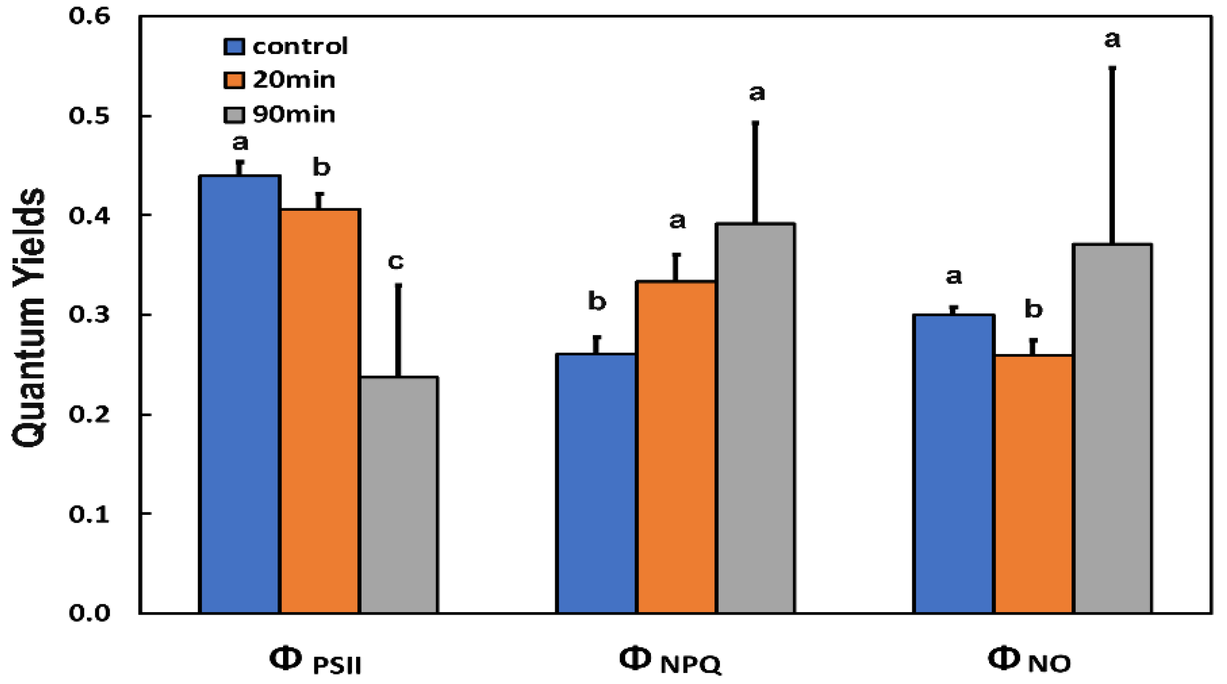
3.1. The Light Energy Distribution at Photosystem II of Potato Leaf before and after Feeding

After twenty min of feeding, the amount of energy that was used for photochemistry (ΦPSII) decreased significantly, while the 90 min feeding by H. halys decreased it further (Figure 1).
The allocation of absorbed light energy at PSII can be estimated by measuring the effective quantum yield of PSII photochemistry (ΦPSII), the quantum yield of regulated non-photochemical energy loss in PSII (ΦNPQ), and the quantum yield of non-regulated energy loss in PSII (ΦNO), the sum of all three to be equal to 1 [52]. The increased yield of regulated non-photochemical energy loss (ΦNPQ) at the whole leaf level after 20 min feeding was capable of overcompensating the decreased ΦPSII resulting in a decreased quantum yield of non-regulated energy (ΦNO) compared to before feeding (Figure 1), while after 90 min feeding, the increased ΦNPQ was sufficient enough to keep ΦNO at the same level as before feeding.

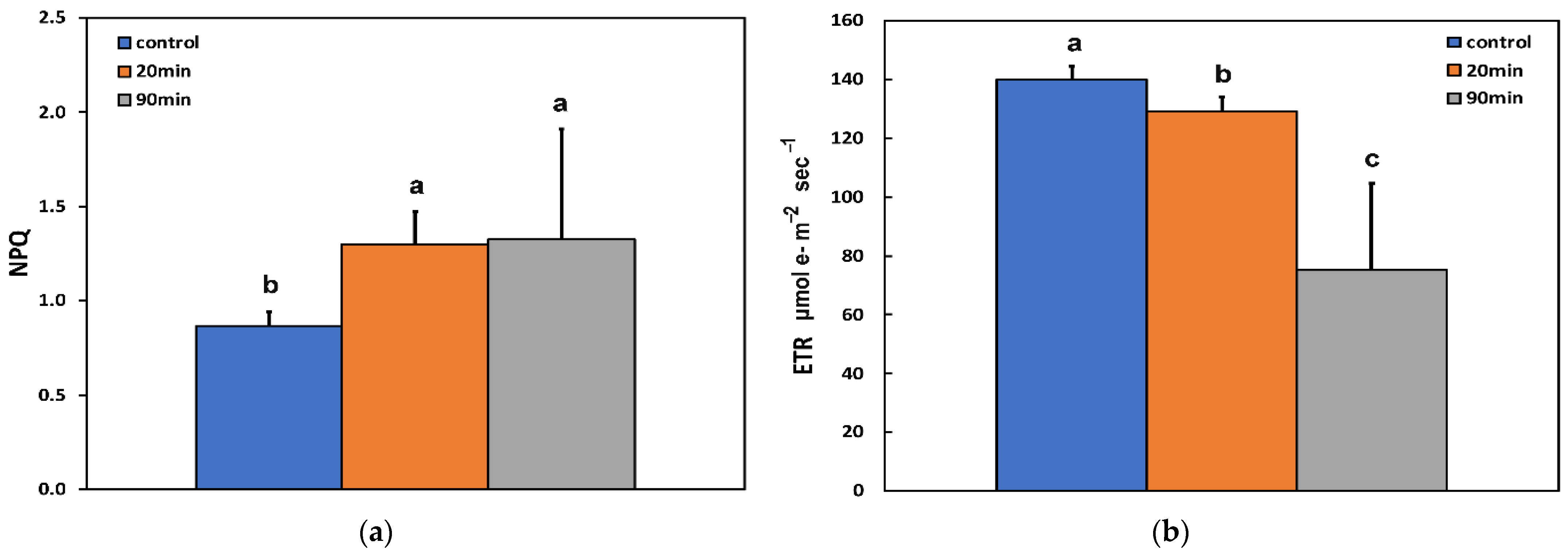
3.2. Changes in the Photoprotective Heat Dissipation, Electron Transport Rate, and the Redox State of the Plastoquinone Pool before and after Feeding

Non-photochemical quenching (NPQ), which reflects the dissipation of excitation energy as heat, increased after 20 min feeding compared to before feeding, but did not increase further at longer durations of feeding (90 min) (Figure 2a). The electron transport rate (ETR) decreased with increasing feeding time compared to before feeding (Figure 2b).
The redox state of quinone A (QA) at the whole leaf level, representing the fraction of open PSII reaction centres (qp), decreased after 20 min feeding compared to before feeding, but remained the same at longer durations of feeding (90 min) (Figure 3a).
The efficiency of excitation energy capture by open PSII centres (Fv′/Fm′) at the whole leaf level after 20 min feeding remained the same as before feeding but decreased after 90 min feeding (Figure 3b).

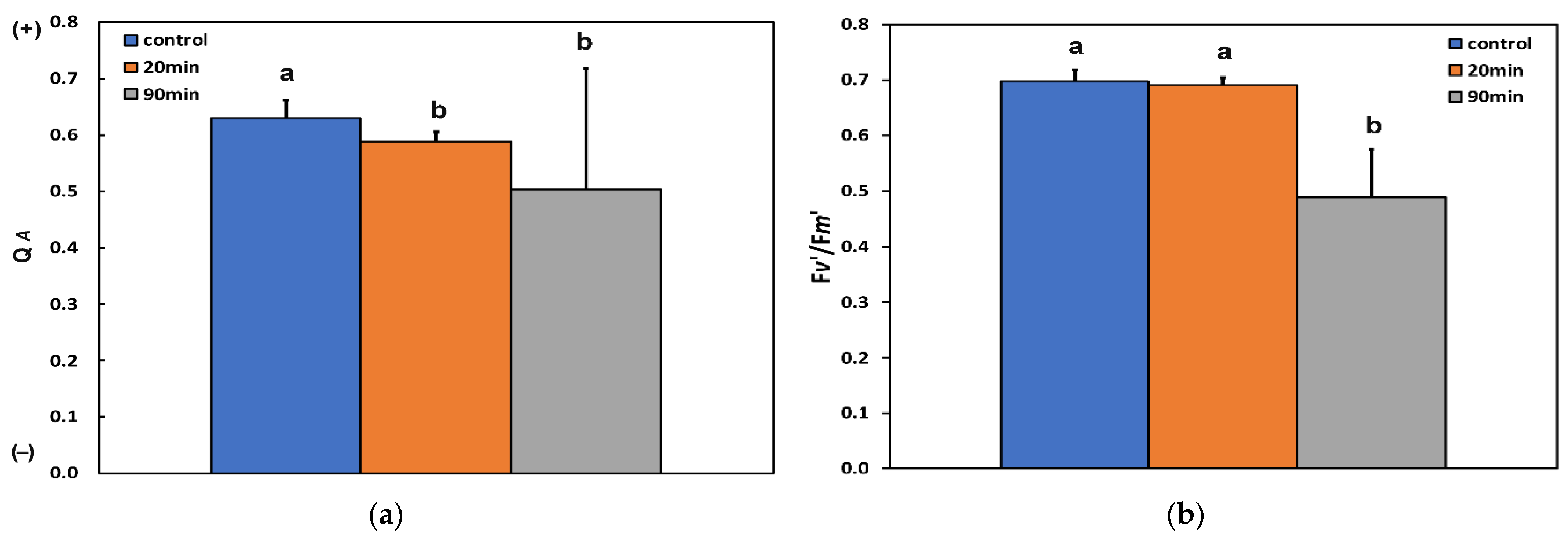
3.3. Changes in the Efficiency of Open Photosystem II Reaction Centers, the Excitation Pressure, and the Excess Excitation Energy in Photosystem II before and after Feeding

The excitation pressure at PSII (1−qp) at the whole leaf level increased after 20 min feeding compared to before feeding, but did not increase further at the longer duration of feeding (90 min) (Figure 4a).
The excess excitation energy (EXC), calculated as (Fv/Fm − ΦPSII)/Fv/Fm [52], increased with increasing feeding time compared to before feeding (Figure 4b).

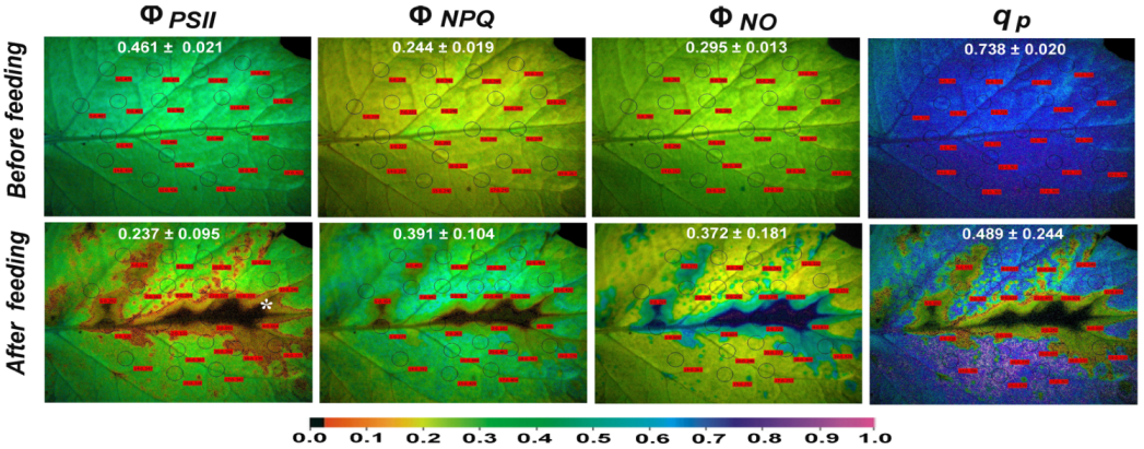
3.4. The Spatial Pattern of Photosystem II Activity of Potato before and after Feeding

After 90 min feeding, an increased spatial PSII heterogeneity was observed in potato leaves (Figure 5). ΦPSII decreased significantly at the feeding zones that were located almost exclusively at the main vein and especially at the sieve elements and the neighbouring area (Figure 5) with the feeding spot.
The area that was detrimentally affected by the insect feeding (marked by an asterisk in Figure 5) had a ΦPSII value of 0.064. Yet, ΦPSII decreased at the whole leaf level (Figure 1, Figure 5, and Figure S2). At the same time, ΦNPQ increased significantly at the whole leaf level (Figure 1, Figure 5, and Figure S3), but decreased significantly at the feeding zones compared to before feeding (Figure 5 and Figure S3).
After 90 min feeding, the decreased ΦPSII and ΦNPQ at the feeding zones resulted in a significant increase of ΦNO at these zones, especially at the main vein (Figure 5 and Figure S4). In the neighbouring area to the feeding zone, ΦNPQ increased more than in the rest of the leaf area to compensate for the higher decrease of ΦPSII in the same zone (Figure 5, Figure S2, and Figure S3). Still, at the whole leaf level, due to the significant increase of ΦNPQ, there was no significant change in ΦNO (Figure 1). The number of open PSII reaction centres decreased significantly at the whole leaf level after feeding (Figure 1 and Figure 5), with the decrease being more intense in the feeding zones (Figure 5). However, a little far from the feeding area, there was a significant increase compared to before feeding in the number of open reaction centres of PSII (qp) (Figure 5 and Figure S5).
Twenty minutes after feeding, a decreased ΦPSII was also noticed (Figure 1), but with much less spatial heterogeneity compared to the 90 min feeding (Figures S2 and S6). The same was also true for the chlorophyll fluorescence parameters ΦNPQ and ΦNO compared to the 90 min feeding (Figure S3, Figure S4, and Figure S6).

3.5. Reactive Oxygen Species Localisation before and after Feeding

ROS imaging, before feeding and 3-, 5-,10-, 20-, 30-, 60-, and 90-min after feeding, performed by staining the potato leaves with 25 µM 2′,7′-dichlorofluorescein diacetate in the dark, revealed an intense increase of ROS generation that was visible as green fluorescence as soon as 3 min after feeding (Figure 6). Reactive oxygen species before feeding were vaguely detected in leaf trichomes. Meanwhile, with the 3 min feeding, an increased generation was detected almost exclusively in the main leaf veins and the leaf trichomes (Figure 6). The ROS real-time staining pattern revealed a decreased generation with further elapsed time feeding, while after 90 min feeding time, ROS visualization was undetectable (Figure 6).

4. Discussion

Herbivory is one of the most important factors affecting plant fitness [58], and plant primary photochemistry is the principal plant trait shaping plant–herbivore interaction. The response mechanism of photosystem II photochemistry to insect herbivory and its ability to induce a compensatory mechanism that increases PSII photochemistry in response to herbivore feeding is fundamental in plant resistance to herbivores [4]. The light reactions of photosynthesis involve a set of redox reactions that are the source of energy for producing organic compounds [59]. According to the growth-differentiation hypothesis, plants have to choose between allocating their resources to defence or growth [60]; greater allocation of resources to defence comes at the expense of tolerance [61]. Induction of plant response to herbivores is explained by two traits, resistance and tolerance. Induction of resistance results in reduced herbivore damage, while induction of tolerance through the increase in growth and photosynthesis compensates for herbivore damage, reducing the negative fitness impact of injury [8,62].
The decrease in the quantum yield of PSII photochemistry (ΦPSII) that was observed after 20 min feeding (Figure 1), according to Genty et al. [53], can be attributed either to a decreased fraction of open PSII reaction centres (qp) (a measure of the redox state of quinone A (QA)) or to a decrease in the efficiency of these centres (Fv′/Fm′) (the supply of energy reaching the PSII reaction centres) [53,63]. In our case, considering that there was no significant change in the efficiency of open PSII reaction centres after 20 min feeding (Figure 3b), it is concluded that the decreased ΦPSII was due to a decreased fraction of open PSII reaction centres (Figure 3a). However, after 90 min feeding, the decreased ΦPSII was due to both a decreased efficiency of the open PSII reaction centres (Fv′/Fm′) (Figure 3b) and a more reduced state of QA (Figure 3a) compared to before feeding. Non-photochemical quenching (NPQ) mechanism can reduce the energy transfer to reaction centres, thus reducing the efficiency of PSII centres [63]. The NPQ parameter represents mainly heat dissipation from the light-harvesting complex II (LHCII) via the zeaxanthin quencher [64,65]. This heat dissipation decreases the efficiency of photochemical reactions of photosynthesis (down-regulation of PSII) [22,23,66,67].
The increased NPQ, 90 min after feeding, decreased ΦPSII due to reduced efficiency of PSII centres (Fv′/Fm′) and decreased the electron-transport rate (ETR) in order to prevent ROS formation [68]. The increased ROS generated as soon as 3 min after feeding (Figure 6) was shown to diffuse through the leaf veins to act as a long-distance signalling molecule [23,69,70,71]. ROS signalling pathways are induced by the redox state of QA, also comprising a mechanism of plant acclimation by regulating photosynthetic gene expression [23,72,73,74]. The redox state of QA is of rare significance for antioxidant defence and signalling [75]. For example, the reduced state of QA is suggested to mediate stomatal closure, probably by ROS generation, conferring acclimation to Cd exposure [76,77]. Reactive oxygen species (ROS) are now recognized as signalling molecules, with an essential part in numerous cellular processes having a tight control exerted by the antioxidant machinery and triggering signalling mechanisms governing normal growth and development and in response to external abiotic or biotic stresses [40,63,78,79,80,81].
The amount of absorbed light energy used in photochemistry (ΦPSII) after 20 min feeding, at the whole leaf level, was lower by 8% compared to before feeding (Figure 1) and by 46% after 90 min feeding. The decrease in ΦPSII after 20 min feeding was overcompensated by the increase in the photoprotective energy dissipation (ΦNPQ) that resulted in a lower ΦNO (Figure 1). The non-regulated energy loss in PSII (ΦNO) encompasses the energy of the triplet-state chlorophylls (3Chl*), which is generated through the intersystem crossing of the singlet excited chlorophyll state (1Chl*) and is transferred to molecular oxygen (O2), to generate singlet-excited oxygen (1O2) [79,82,83,84,85,86,87]. Reactive oxygen species, estimated as 1O2, after 20 min feeding decreased (Figure 1), but total ROS generation, as observed with 2′,7′-dichlorofluorescein diacetate (DCF-DA) staining, increased compared to before feeding (Figure 6). As ROS formed either by energy transfer (1O2) and/or electron transport (O2•−, H2O2) are created concurrently, it appears likely that their action of signalling pathways can occasionally interfere or antagonize each other [23,40,45,74,79,83].
After 90 min feeding by the sap-sucking insect H. halys, ΦPSII at the whole leaf level decreased by 46%. Meanwhile, at the feeding zones, which were located in the proximity of the leaf’s midrib, ΦPSII at the spot-like area of the feeding zone was lower by 85%, as judged by the ΦPSII value of 0.064 (Figure 5). However, despite a significant increase of ΦNPQ at the whole leaf level (Figure 1 and Figure 5), at the feeding zones, it decreased significantly compared to before feeding (Figure 5). Consequently, the decreased ΦPSII and ΦNPQ at the feeding zones resulted in a significant increase of ΦNO at these zones (Figure 5), indicating high 1O2 generation, mostly at the main vein and the neighbouring area (Figure 5). Singlet-excited oxygen (1O2) is considered a harmful ROS produced by PSII that inhibits the repair of PSII reaction centres and/or contributes directly to PSII damage [70,71,79], while increased 1O2 generation can trigger programmed cell death [3,88]. Singlet-excited oxygen (1O2) is a powerful oxidant that reacts rapidly in the area where it is created, resulting in oxidation that is regularly denoted as “damage” [20]. When the rate of damage exceeds the rate of D1 protein repair, the consequence is an overall decrease in ETR [69]. In our case, after a 90 min feeding, the destructive role of ROS was due to 1O2 generation (increased ΦNO) predominantly at the main vein area (Figure 5).
The highly increased ROS generation at the feeding zones located in the proximity of the leaf’s midrib, as soon as 3 min after feeding (Figure 6), triggers defence responses through leaf vein diffusion since ROS (and especially H2O2) act as long-distance signalling molecules [23,70,71,73]. Hydrogen peroxide is the most stable ROS that can mediate plant responses to stress [89]. A controlled ROS burst has been considered the primary plant defence responses mechanism against herbivores [90]. A quick activation of plant defence signals in the cells surrounding the stylet wound by an aphid in Arabidopsis thaliana was followed by an induction spread along the veins to the whole leaf [91]. In the neighbouring area to the feeding zone, 90 min after feeding, the photoprotective mechanism ΦNPQ increased more than in the rest of the leaf area to compensate for the higher decrease of ΦPSII in the same zone. At the whole leaf level, due to the significant increase of ΦNPQ after 90 min feeding, there was no significant change of ΦNO, which represents 1O2 generation, which remains the same as before feeding.
Reactive oxygen species generation can inhibit the repair of PSII reaction centres or contribute directly to PSII damage [42,63]. ROSs act primarily by inhibiting the repair of photodamaged PSII and not by damaging PSII directly [92]. However, ROS production in chloroplasts not only generates oxidative stress, but also presents essential biological functions in plant growth, development, and redox signalling [40,93,94]. A proper equilibrium between the creation and removal of ROS by chloroplast antioxidants is achieved so as to match the process of photosynthesis, allowing an effective scattering of ROS signals [3,45,79,95]. Consequently, ROS not only compromises cells with tools to monitor electron transport and, thus, avoid over-reduction or over-oxidation, but also creates redox regulatory networks that enable plants to sense and react to biotic and abiotic stress conditions [39,70,79,95,96]. A high level of ROS is considered to be useful for initiating defense responses [45,80,95]. Photosystem II responses under biotic or abiotic stress are activated by the NPQ mechanism that is a strategy to protect the chloroplasts from photo-oxidative damage by heat dissipation [80,97,98], regulating ETR [71,95], and avoiding detrimental ROS generation [66,80]. A basal level of ROS is required for optimum plant growth [40,45], with a controlled, increased ROS level to be favourable in activating defence responses, while a high level of ROS out of the limits to be destructive to plants [3,40,45,80].
Our experimental results confirmed that chlorophyll fluorescence imaging methodology can detect spatial heterogeneity of PSII efficiency at the whole leaf surface and is a promising tool for investigating plant response mechanisms to sap-sucking insect herbivores. As it has been previously suggested, it can distinguish leaf photosynthetic spatiotemporal heterogeneity that cannot be identified through conventional chlorophyll fluorescence analysis, and it can be used to explore plant response mechanisms to biotic or abiotic stresses [50,80,99,100,101,102,103]. Plant-insect interactions are getting more defined and comprehensive with the development of novel visualization methods that permit leaf photosynthetic efficiency evaluation after herbivore attack [4,91].

5. Conclusions

We suggest that PSII responses after feeding by H. halys underlie ROS-dependent signalling that occurs immediately (<3 min) after feeding. It is concluded that the potato PSII response mechanism to sap-sucking insect herbivores is a defence response that reduces damage to herbivory instead of the induction of tolerance through a compensatory photosynthetic response mechanism that was observed in chewing insects [4,51].

Supplementary Materials

The following supporting information can be downloaded at: https://www.mdpi.com/article/10.3390/insects13050409/s1. Figure S1: Dorsal view of the brown marmorated stink bug (Halyomorpha halys). Figure S2: Histogram of all data points of the effective quantum yield of PSII photochemistry (ΦPSII) per time point. Figure S3: Histogram of all data points of the quantum yield of regulated non-photochemical energy loss (ΦNPQ) per time point. Figure S4: Histogram of all data points of the quantum yield of non-regulated energy (ΦNO) per time point. Figure S5: Histogram of all data points of the fraction of open PSII reaction centres (qp) per time point. Figure S6: Typical colour-coded images of ΦPSΙΙ, ΦNPQ, and ΦNO, before insect feeding and immediately after 20 min feeding.

Author Contributions

Conceptualization, I.S., S.S.A., J.M. and M.M.; methodology, I.S., S.S.A., I.-D.S.A., J.M. and M.M.; validation, M.M.; formal analysis, I.S. and M.M.; investigation, I.S., I.-D.S.A., J.M. and E.I.K.; resources, M.M.; data curation, I.S., S.S.A., I.-D.S.A., J.M., E.I.K. and M.M.; writing—original draft preparation, I.S. and M.M.; writing—review and editing, I.S., S.S.A., I.-D.S.A., J.M., E.I.K. and M.M.; visualization, I.S., J.M. and M.M.; supervision, M.M.; project administration, M.M. All authors have read and agreed to the published version of the manuscript.

Funding

This research received no external funding.

Institutional Review Board Statement

Not applicable.

Informed Consent Statement

Not applicable.

Data Availability Statement

The data presented in this study are available in this article.

Conflicts of Interest

The authors declare no conflict of interest.

References

  1. Frier, D.J.P.; Hernández, S.C.V.; Tiessen, A. Friend or foe? Exploring the factors that determine the difference between positive and negative effects on photosynthesis in response to insect herbivory. In Artificial Photosynthesis; Najafpour, M.M., Ed.; Tech: Rijeka, Croatia, 2012; pp. 155–206. [Google Scholar]
  2. Hammond-Kosack, K.E.; Jones, J.D.G. Responses to plant pathogens. In Biochemistry & Molecular Biology of Plants, 2nd ed.; Buchanan, B.B., Gruissem, W., Jones, R.L., Eds.; John Wiley & Sons, Ltd.: Hoboken, NJ, USA, 2015; pp. 984–1050. [Google Scholar]
  3. Sperdouli, I.; Andreadis, S.; Moustaka, J.; Panteris, E.; Tsaballa, A.; Moustakas, M. Changes in light energy utilization in photosystem II and reactive oxygen species generation in potato leaves by the pinworm Tuta absoluta. Molecules 2021, 26, 2984. [Google Scholar] [CrossRef] [PubMed]
  4. Moustaka, J.; Meyling, N.V.; Hauser, T.P. Induction of a compensatory photosynthetic response mechanism in tomato leaves upon short time feeding by the chewing insect Spodoptera exigua. Insects 2021, 12, 562. [Google Scholar] [CrossRef] [PubMed]
  5. Hafke, J.B.; van Amerongen, J.K.; Kelling, F.; Furch, A.C.U.; Gaupels, F.; van Bel, A.J.E. Thermodynamic battle for photosynthate acquisition between sieve tubes and adjoining parenchyma in transport phloem. Plant Physiol. 2005, 138, 1527–1537. [Google Scholar] [CrossRef] [PubMed] [Green Version]
  6. Will, T.; Furch, A.C.U.; Zimmermann, M.R. How phloem-feeding insects face the challenge of phloem-located defenses. Front. Plant Sci. 2013, 4, 336. [Google Scholar] [CrossRef] [PubMed] [Green Version]
  7. Walker, G.P. Sieve element occlusion: Interactions with phloem sap-feeding insects. A review. J. Plant Physiol. 2021, 269, 153582. [Google Scholar] [CrossRef]
  8. Mauch-Mani, B.; Baccelli, I.; Luna, E.; Flors, V. Defense priming: An adaptive part of induced resistance. Annu. Rev. Plant Biol. 2017, 68, 485–512. [Google Scholar] [CrossRef] [Green Version]
  9. Masui, N.; Agathokleous, E.; Mochizuki, T.; Tani, A.; Matsuura, H.; Koike, T. Ozone disrupts the communication between plants and insects in urban and suburban areas: An updated insight on plant volatiles. J. For. Res. 2021, 32, 1337–1349. [Google Scholar] [CrossRef]
  10. Masui, N.; Mochizuki, T.; Tani, A.; Matsuura, H.; Agathokleous, E.; Watanabe, T.; Koike, T. Does ozone alter the attractiveness of Japanese white birch leaves to the leaf beetle Agelastica coerulea via changes in biogenic volatile organic compounds (BVOCs): An examination with the Y-tube test. Forests 2020, 11, 58. [Google Scholar] [CrossRef] [Green Version]
  11. Łyczko, J.; Twardowski, J.P.; Skalny, B.; Galek, R.; Szumny, A.; Gruss, I.; Piesik, D.; Sendel, S. Sarracenia alata (Alph.Wood) Alph.Wood microcuttings as a source of volatiles potentially responsible for insects’ respond. Molecules 2021, 26, 2406. [Google Scholar] [CrossRef]
  12. Piesik, D.; Wenda-Piesik, A. Sitophilus granarius responses to blends of five groups of cereal kernels and one group of plant volatiles. J. Stored Prod. Res. 2015, 62, 36–39. [Google Scholar] [CrossRef]
  13. Piesik, D.; Rochat, D.; Delaney, K.J.; Marion-Poll, F. Orientation of European corn borer first instar larvae to synthetic green leaf volatiles. Appl. Entomol. 2013, 137, 234–240. [Google Scholar] [CrossRef] [Green Version]
  14. Erb, M.; Reymond, P. Molecular interactions between plants and insect herbivores. Annu. Rev. Plant Biol. 2019, 29, 527–557. [Google Scholar] [CrossRef] [PubMed] [Green Version]
  15. Thomson, V.P.; Cunningham, S.A.; Ball, M.C.; Nicotra, A.B. Compensation for herbivory by Cucumis sativus through increased photosynthetic capacity and efficiency. Oecologia 2003, 134, 167–175. [Google Scholar] [CrossRef] [PubMed]
  16. Delaney, K.J. Injured and uninjured leaf photosynthetic responses after mechanical injury on Nerium oleander leaves, and Danaus plexippus herbivory on Asclepias curassavica leaves. Plant Ecol. 2008, 199, 187–200. [Google Scholar] [CrossRef]
  17. Andreadis, S.S.; Navrozidis, E.I.; Farmakis, A.; Pisalidis, A. First evidence of Halyomorpha halys (Hemiptera: Pentatomidae) infesting kiwifruit (Actinidia chinensis) in Greece. J. Entomol. Sci. 2018, 53, 402–405. [Google Scholar] [CrossRef]
  18. Zvereva, E.L.; Lanta, V.; Kozlov, M.V. Effects of sap-feeding insect herbivores on growth and reproduction of woody plants: A meta-analysis of experimental studies. Oecologia 2010, 163, 949–960. [Google Scholar] [CrossRef]
  19. Niyogi, K.K.; Wolosiuk, R.A.; Malkin, R. Photosynthesis. In Biochemistry & Molecular Biology of Plants, 2nd ed.; Buchanan, B.B., Gruissem, W., Jones, R.L., Eds.; John Wiley & Sons, Ltd.: Hoboken, NJ, USA, 2015; pp. 508–566. [Google Scholar]
  20. Apel, K.; Hirt, H. Reactive oxygen species: Metabolism, oxidative stress, and signal transduction. Annu. Rev. Plant Biol. 2004, 55, 373–399. [Google Scholar] [CrossRef] [Green Version]
  21. Gill, S.S.; Tuteja, N. Reactive oxygen species and antioxidant machinery in abiotic stress tolerance in crop plants. Plant Physiol. Biochem. 2010, 48, 909–930. [Google Scholar] [CrossRef]
  22. Moustaka, J.; Moustakas, M. Photoprotective mechanism of the non-target organism Arabidopsis thaliana to paraquat exposure. Pest. Biochem. Physiol. 2014, 111, 1–6. [Google Scholar] [CrossRef]
  23. Moustaka, J.; Tanou, G.; Adamakis, I.D.; Eleftheriou, E.P.; Moustakas, M. Leaf age dependent photoprotective and antioxidative mechanisms to paraquat-induced oxidative stress in Arabidopsis thaliana. Int. J. Mol. Sci. 2015, 16, 13989–14006. [Google Scholar] [CrossRef] [Green Version]
  24. Dietz, K.J.J.; Turkan, I.; Krieger-Liszkay, A. Redox- and reactive oxygen species dependent signaling into and out of the photosynthesizing chloroplast. Plant Physiol. 2016, 171, 1541–1550. [Google Scholar] [CrossRef] [PubMed]
  25. Takagi, D.; Takumi, S.; Hashiguchi, M.; Sejima, T.; Miyake, C. Superoxide and singlet oxygen produced within the thylakoid membranes both cause photosystem I photoinhibition. Plant. Physiol. 2016, 171, 1626–1634. [Google Scholar] [CrossRef] [PubMed] [Green Version]
  26. Moustakas, M.; Malea, P.; Zafeirakoglou, A.; Sperdouli, I. Photochemical changes and oxidative damage in the aquatic macrophyte Cymodocea nodosa exposed to paraquat-induced oxidative stress. Pest. Biochem. Physiol. 2016, 126, 28–34. [Google Scholar] [CrossRef]
  27. Ruban, A.V. Light harvesting control in plants. FEBS Lett. 2018, 592, 3030–3039. [Google Scholar] [CrossRef]
  28. Sun, H.; Shi, Q.; Zhang, S.-B.; Huang, W. Coordination of cyclic electron flow and water–water cycle facilitates photoprotection under fluctuating light and temperature stress in the epiphytic orchid Dendrobium officinale. Plants 2021, 10, 606. [Google Scholar] [CrossRef] [PubMed]
  29. Krieger-Liszkay, A. Singlet oxygen production in photosynthesis. J. Exp. Bot. 2005, 56, 337–346. [Google Scholar] [CrossRef] [PubMed] [Green Version]
  30. Moustaka, J.; Ouzounidou, G.; Sperdouli, I.; Moustakas, M. Photosystem II is more sensitive than photosystem I to Al3+ induced phytotoxicity. Materials 2018, 11, 1772. [Google Scholar] [CrossRef] [Green Version]
  31. Murata, N.; Takahashi, S.; Nishiyama, Y.; Allakhverdiev, S.I. Photoinhibition of photosystem II under environmental stress. Biochim. Biophys. Acta 2007, 1767, 414–421. [Google Scholar] [CrossRef] [Green Version]
  32. Sperdouli, I.; Moustaka, J.; Ouzounidou, G.; Moustakas, M. Leaf age-dependent photosystem II photochemistry and oxidative stress responses to drought stress in Arabidopsis thaliana are modulated by flavonoid accumulation. Molecules 2021, 26, 4157. [Google Scholar] [CrossRef]
  33. Trinh, M.D.L.; Hashimoto, A.; Kono, M.; Takaichi, S.; Nakahira, Y.; Masuda, S. Lack of plastid-encoded Ycf10, a homolog of the nuclear encoded DLDG1 and the cyanobacterial PxcA, enhances the induction of non-photochemical quenching in tobacco. Plant Direct 2021, 5, e368. [Google Scholar] [CrossRef]
  34. Krieger-Liszkay, A.; Fufezan, C.; Trebst, A. Singlet oxygen production in photosystem II and related protection mechanism. Photosynth. Res. 2008, 98, 551–564. [Google Scholar] [CrossRef] [PubMed]
  35. Müller, P.; Li, X.P.; Niyogi, K.K. Non-photochemical quenching. A response to excess light energy. Plant Physiol. 2001, 125, 1558–1566. [Google Scholar] [CrossRef] [PubMed] [Green Version]
  36. Ruban, A.V. Nonphotochemical chlorophyll fluorescence quenching: Mechanism and effectiveness in protecting plants from photodamage. Plant Physiol. 2016, 170, 1903–1916. [Google Scholar] [CrossRef] [PubMed] [Green Version]
  37. Demmig-Adams, B.; Adams, W.W., III. Photoprotection and other responses of plants to high light stress. Annu. Rev. Plant Physiol. Plant Mol. Biol. 1992, 43, 599–626. [Google Scholar] [CrossRef]
  38. Demmig-Adams, B.; Cohu, C.M.; Muller, O.; Adams, W.W. Modulation of photosynthetic energy conversion efficiency in nature: From seconds to seasons. Photosynth. Res. 2012, 113, 75–88. [Google Scholar] [CrossRef]
  39. Foyer, C.H.; Noctor, G. Redox homeostasis and antioxidant signaling: A metabolic interface between stress perception and physiological responses. Plant Cell 2005, 17, 1866–1875. [Google Scholar] [CrossRef] [Green Version]
  40. Mittler, R. ROS are good. Trends Plant Sci. 2017, 22, 11–19. [Google Scholar] [CrossRef] [Green Version]
  41. Czarnocka, W.; Karpiński, S. Friend or foe? Reactive oxygen species production, scavenging and signaling in plant response to environmental stresses. Free Radic. Biol. Med. 2018, 122, 4–20. [Google Scholar] [CrossRef]
  42. Stamelou, M.L.; Sperdouli, I.; Pyrri, I.; Adamakis, I.D.S.; Moustakas, M. Hormetic responses of photosystem II in tomato to Botrytis cinerea. Plants 2021, 10, 521. [Google Scholar] [CrossRef]
  43. Sperdouli, I.; Mellidou, I.; Moustakas, M. Harnessing chlorophyll fluorescence for phenotyping analysis of wild and cultivated tomato for high photochemical efficiency under water deficit for climate change resilience. Climate 2021, 9, 154. [Google Scholar] [CrossRef]
  44. Hossain, M.A.; Bhattacharjee, S.; Armin, S.M.; Qian, P.; Xin, W.; Li, H.Y.; Burritt, D.J.; Fujita, M.; Tran, L.S. Hydrogen peroxide priming modulates abiotic oxidative stress tolerance: Insights from ROS detoxification and scavenging. Front. Plant Sci. 2015, 16, 420. [Google Scholar] [CrossRef] [PubMed] [Green Version]
  45. Adamakis, I.D.S.; Sperdouli, I.; Eleftheriou, E.P.; Moustakas, M. Hydrogen peroxide production by the spot-like mode action of bisphenol A. Front. Plant Sci. 2020, 11, 1196. [Google Scholar] [CrossRef] [PubMed]
  46. Elena-Real, C.A.; González-Arzola, K.; Pérez-Mejías, G.; Díaz-Quintana, A.; Velázquez-Campoy, A.; Desvoyes, B.; Gutiérrez, C.; De la Rosa, M.A.; Díaz-Moreno, I. Proposed mechanism for regulation of H2O2-induced programmed cell death in plants by binding of cytochrome c to 14-3-3 proteins. Plant J. 2021, 106, 74–85. [Google Scholar] [CrossRef] [PubMed]
  47. Adamakis, I.D.S.; Malea, P.; Sperdouli, I.; Panteris, E.; Kokkinidi, D.; Moustakas, M. Evaluation of the spatiotemporal effects of bisphenol A on the leaves of the seagrass Cymodocea nodosa. J. Hazard. Mater. 2021, 404, 124001. [Google Scholar] [CrossRef]
  48. Tang, J.Y.; Zielinski, R.E.; Zangerl, A.R.; Crofts, A.R.; Berenbaum, M.R.; DeLucia, E.H. The differential effects of herbivory by first and fourth instars of Trichoplusia ni (Lepidoptera: Noctuidae) on photosynthesis in Arabidopsis thaliana. J. Exp. Bot. 2006, 57, 527–536. [Google Scholar] [CrossRef] [Green Version]
  49. Pérez-Bueno, M.L.; Pineda, M.; Barón, M. Phenotyping plant responses to biotic stress by chlorophyll fluorescence imaging. Front. Plant Sci. 2019, 10, 1135. [Google Scholar] [CrossRef]
  50. Moustakas, M.; Calatayud, A.; Guidi, L. Chlorophyll fluorescence imaging analysis in biotic and abiotic stress. Front. Plant Sci. 2021, 12, 658500. [Google Scholar] [CrossRef]
  51. Moustaka, J.; Meyling, V.N.; Hauser, T.P. Root-associated entomopathogenic fungi modulate host plant’ s photosystem II photochemistry and its response to herbivorous insects. Molecules 2022, 27, 207. [Google Scholar] [CrossRef]
  52. Kramer, D.M.; Johnson, G.; Kiirats, O.; Edwards, G.E. New fluorescence parameters for the determination of QA redox state and excitation energy fluxes. Photosynth. Res. 2004, 79, 209–218. [Google Scholar] [CrossRef]
  53. Genty, B.; Briantais, J.M.; Baker, N.R. The relationship between the quantum yield of photosynthetic electron transport and quenching of chlorophyll fluorescence. Biochim. Biophys. Acta 1989, 990, 87–92. [Google Scholar] [CrossRef]
  54. Bilger, W.; Björkman, O. Role of the xanthophyll cycle in photoprotection elucidated by measurements of light-induced absorbance changes, fluorescence and photosynthesis in leaves of Hedera canariensis. Photosynth. Res. 1990, 25, 173–185. [Google Scholar] [CrossRef] [PubMed]
  55. Schreiber, U.; Bilger, W.; Neubauer, C. Chlorophyll fluorescence as a non-intrusive indicator for rapid assessment of in vivo photosynthesis. In Ecophysiology of Photosynthesis; Series Ecological Studies; Schulze, E.D., Caldwell, M.M., Eds.; Springer: Berlin, Germany, 1994; Volume 100, pp. 49–70. [Google Scholar]
  56. Gray, G.R.; Savitch, L.V.; Ivanov, A.G.; Huner, N.P.A. Photosystem II excitation pressure and development of resistance to photoinhibition. II. Adjustment of photosynthetic capacity in winter wheat and winter rye. Plant Physiol. 1996, 110, 61–71. [Google Scholar] [CrossRef] [PubMed]
  57. Bilger, W.; Schreiber, U.; Bock, M. Determination of the quantum efficiency of photosystem II and of non-photochemical quenching of chlorophyll fluorescence in the field. Oecologia 1995, 102, 425–432. [Google Scholar] [CrossRef] [PubMed]
  58. Münzbergová, Z.; Skuhrovec, J. Data on herbivore performance and plant herbivore damage identify the same plant traits as the key drivers of plant–herbivore interaction. Insects 2020, 11, 865. [Google Scholar] [CrossRef] [PubMed]
  59. González, M.C.; Cejudo, F.J.; Sahrawy, M.; Serrato, A.J. Current knowledge on mechanisms preventing photosynthesis redox imbalance in plants. Antioxidants 2021, 10, 1789. [Google Scholar] [CrossRef] [PubMed]
  60. Herms, D.A.; Mattson, W.J. The dilemma of plants: To grow or defend. Q. Rev. Biol. 1992, 67, 283–335. [Google Scholar] [CrossRef] [Green Version]
  61. Schröder, R.; Forstreuter, M.; Hilker, M. A plant notices insect egg deposition and changes its rate of photosynthesis. Plant Physiol. 2005, 138, 470–477. [Google Scholar] [CrossRef] [Green Version]
  62. Mitchell, C.; Brennan, R.M.; Graham, J.; Karley, A.J. Plant defense against herbivorous pests: Exploiting resistance and tolerance traits for sustainable crop protection. Front. Plant Sci. 2016, 7, 1132. [Google Scholar] [CrossRef] [Green Version]
  63. Moustakas, M.; Bayçu, G.; Sperdouli, I.; Eroğlu, H.; Eleftheriou, E.P. Arbuscular mycorrhizal symbiosis enhances photosynthesis in the medicinal herb Salvia fruticosa by improving photosystem II photochemistry. Plants 2020, 9, 962. [Google Scholar] [CrossRef]
  64. Demmig-Adams, B.; Polutchko, S.K.; Adams, W.W., III. Structure-function-environment relationship of the isomers zeaxanthin and lutein. Photochem 2022, 2, 308–325. [Google Scholar] [CrossRef]
  65. Demmig-Adams, B. Carotenoids and photoprotection in plants: A role for the xanthophyll zeaxanthin. Biochim. Biophys. Acta 1990, 1020, 1–24. [Google Scholar] [CrossRef]
  66. Takahashi, S.; Badger, M.R. Photoprotection in plants: A new light on photosystem II damage. Trends Plant Sci. 2011, 16, 53–60. [Google Scholar] [CrossRef] [PubMed]
  67. Ruban, A.V. The mechanism of nonphotochemical quenching: The end of the ongoing debate. Plant Physiol. 2019, 181, 383–384. [Google Scholar] [CrossRef] [PubMed] [Green Version]
  68. Roach, T.; Na, C.S.; Stöggl, W.; Krieger-Liszkay, A. The non-photochemical quenching protein LHCSR3 prevents oxygen-dependent photoinhibition in Chlamydomonas reinhardtii. J. Exp. Bot. 2020, 71, 2650–2660. [Google Scholar] [CrossRef]
  69. Wilson, K.E.; Ivanov, A.G.; Öquist, G.; Grodzinski, B.; Sarhan, F.; Huner, N.P.A. Energy balance, organellar redox status, and acclimation to environmental stress. Can. J. Bot. 2006, 84, 1355–1370. [Google Scholar] [CrossRef]
  70. Mittler, R.; Vanderauwera, S.; Suzuki, N.; Miller, G.; Tognetti, V.B.; Vandepoele, K.; Gollery, M.; Shulaev, V.; Van Breusegem, F. ROS signaling: The new wave? Trends Plant Sci. 2011, 16, 300–309. [Google Scholar] [CrossRef]
  71. Baxter, A.; Mittler, R.; Suzuki, N. ROS as key players in plant stress signaling. J. Exp. Bot. 2014, 65, 1229–1240. [Google Scholar] [CrossRef]
  72. Bräutigam, K.; Dietzel, L.; Kleine, T.; Ströher, E.; Wormuth, D.; Dietz, K.J.; Radke, D.; Wirtz, M.; Hell, R.; Dörmann, P.; et al. Dynamic plastid redox signals integrate gene expression and metabolism to induce distinct metabolic states in photosynthetic acclimation in Arabidopsis. Plant Cell 2009, 21, 2715–2732. [Google Scholar] [CrossRef] [Green Version]
  73. Dietz, K.J.; Pfannschmidt, T. Novel regulators in photosynthetic redox control of plant metabolism and gene expression. Plant Physiol. 2011, 155, 1477–1485. [Google Scholar] [CrossRef] [Green Version]
  74. Sperdouli, I.; Moustaka, J.; Antonoglou, O.; Adamakis, I.D.S.; Dendrinou-Samara, C.; Moustakas, M. Leaf age dependent effects of foliar-sprayed CuZn nanoparticles on photosynthetic efficiency and ROS generation in Arabidopsis thaliana. Materials 2019, 12, 2498. [Google Scholar] [CrossRef] [Green Version]
  75. Borisova-Mubarakshina, M.M.; Vetoshkina, D.V.; Ivanov, B.N. Antioxidant and signaling functions of the plastoquinone pool in higher plants. Physiol. Plant. 2019, 166, 181–198. [Google Scholar] [CrossRef] [PubMed]
  76. Bayçu, G.; Moustaka, J.; Gevrek, N.; Moustakas, M. Chlorophyll fluorescence imaging analysis for elucidating the mechanism of photosystem II acclimation to cadmium exposure in the hyperaccumulating plant Noccaea caerulescens. Materials 2018, 11, 2580. [Google Scholar] [CrossRef] [PubMed] [Green Version]
  77. Moustakas, M. The role of metal ions in biology, biochemistry and medicine. Materials 2021, 14, 549. [Google Scholar] [CrossRef] [PubMed]
  78. Antonoglou, O.; Moustaka, J.; Adamakis, I.D.; Sperdouli, I.; Pantazaki, A.; Moustakas, M.; Dendrinou-Samara, C. Nanobrass CuZn nanoparticles as foliar spray non phytotoxic fungicides. ACS Appl. Mater. Interfaces 2018, 10, 4450–4461. [Google Scholar] [CrossRef]
  79. Moustakas, M. Plant photochemistry, reactive oxygen species, and photoprotection. Photochem 2022, 2, 5–8. [Google Scholar] [CrossRef]
  80. Moustakas, M.; Moustaka, J.; Sperdouli, I. Hormesis in photosystem II: A mechanistic approach. Curr. Opin. Toxicol. 2022, 29, 57–64. [Google Scholar] [CrossRef]
  81. Kerchev, P.I.; Van Breusegem, F. Improving oxidative stress resilience in plants. Plant J. 2022, 109, 359–372. [Google Scholar] [CrossRef]
  82. Kasajima, I.; Ebana, K.; Yamamoto, T.; Takahara, K.; Yano, M.; Kawai-Yamada, M.; Uchimiya, H. Molecular distinction in genetic regulation of nonphotochemical quenching in rice. Proc. Natl. Acad. Sci. USA 2011, 108, 13835–13840. [Google Scholar] [CrossRef] [Green Version]
  83. Moustaka, J.; Tanou, G.; Giannakoula, A.; Panteris, E.; Eleftheriou, E.P.; Moustakas, M. Anthocyanin accumulation in poinsettia leaves and its functional role in photo-oxidative stress. Environ. Exp. Bot. 2020, 175, 104065. [Google Scholar] [CrossRef]
  84. Hideg, É.; Spetea, C.; Vass, I. Singlet oxygen production in thylakoid membranes during photoinhibition as detected by EPR spectroscopy. Photosynth. Res. 1994, 39, 191–199. [Google Scholar] [CrossRef]
  85. Krieger-Liszkay, A.; Trebst, A. Tocopherol is the scavenger of singlet oxygen produced by the triplet states of chlorophyll in the PSII reaction centre. J. Exp. Bot. 2006, 57, 1677–1684. [Google Scholar] [CrossRef] [PubMed]
  86. Triantaphylidès, C.; Havaux, M. Singlet oxygen in plants: Production, detoxification and signaling. Trends Plant Sci. 2009, 14, 219–228. [Google Scholar] [CrossRef] [PubMed]
  87. Telfer, A. Singlet oxygen production by PSII under light stress: Mechanism, detection and the protective role of beta-carotene. Plant Cell Physiol. 2014, 55, 1216–1223. [Google Scholar] [CrossRef] [Green Version]
  88. Laloi, C.; Havaux, M. Key players of singlet oxygen-induced cell death in plants. Front. Plant Sci. 2015, 6, 39. [Google Scholar] [CrossRef] [PubMed] [Green Version]
  89. Li, H.; Jiang, X.; Lv, X.; Ahammed, G.J.; Guo, Z.; Qi, Z.; Yu, J.; Zhou, Y. Tomato GLR3.3 and GLR3.5 mediate cold acclimation-induced chilling tolerance by regulating apoplastic H2O2 production and redox homeostasis. Plant Cell Environ. 2019, 42, 3326–3339. [Google Scholar] [CrossRef]
  90. Ali, M.; Cheng, Z.; Ahmad, H.; Hayat, S. Reactive oxygen species (ROS) as defenses against a broad range of plant fungal infections and case study on ROS employed by crops against Verticillium dahliae wilts. J. Plant Interact. 2018, 13, 353–363. [Google Scholar] [CrossRef] [Green Version]
  91. Rubil, N.; Kalachova, T.; Hauser, T.P.; Burketová, L. Specialist aphid feeding causes local activation of salicylic and jasmonic acid signaling in Arabidopsis veins. Mol. Plant Microbe Interact. 2022, 35, 119–124. [Google Scholar] [CrossRef]
  92. Nishiyama, Y.; Allakhverdiev, S.I.; Murata, N. A new paradigm for the action of reactive oxygen species in the photoinhibition of photosystem II. Biochim. Biophys. Acta 2006, 1757, 742–749. [Google Scholar] [CrossRef] [Green Version]
  93. Foyer, C.H.; Noctor, G. Redox signaling in plants. Antioxid. Redox Signal. 2013, 18, 2087–2090. [Google Scholar] [CrossRef]
  94. Romero-Puertas, M.C.; Terrón-Camero, L.C.; Peláez-Vico, M.Á.; Molina-Moya, E.; Sandalio, L.M. An update on redox signals in plant responses to biotic and abiotic stress crosstalk: Insights from cadmium and fungal pathogen interactions. J. Exp. Bot. 2021, 72, 5857–5875. [Google Scholar] [CrossRef]
  95. Foyer, C.H. Reactive oxygen species, oxidative signaling and the regulation of photosynthesis. Environ. Exp. Bot. 2018, 154, 134–142. [Google Scholar] [CrossRef] [PubMed]
  96. Miller, G.; Suzuki, N.; Ciftci-Yilmaz, S.; Mittler, R. Reactive oxygen species homeostasis and signalling during drought and salinity stresses. Plant Cell Environ. 2010, 33, 453–467. [Google Scholar] [CrossRef] [PubMed]
  97. Niyogi, K.K. Safety valves for photosynthesis. Curr. Opin. Plant Biol. 2000, 3, 455–460. [Google Scholar] [CrossRef]
  98. Li, X.P.; Müller-Moulé, P.; Gilmore, A.M.; Niyogi, K.K. PsbS-dependent enhancement of feedback de-excitation protects photosystem II from photoinhibition. Proc. Natl. Acad. Sci. USA 2002, 99, 15222–15227. [Google Scholar] [CrossRef] [PubMed] [Green Version]
  99. Sperdouli, I.; Moustakas, M. Spatio-temporal heterogeneity in Arabidopsis thaliana leaves under drought stress. Plant Biol. 2012, 14, 118–128. [Google Scholar] [CrossRef] [PubMed]
  100. Gorbe, E.; Calatayud, A. Applications of chlorophyll fluorescence imaging technique in horticultural research: A review. Sci. Hortic. 2012, 138, 24–35. [Google Scholar] [CrossRef]
  101. Guidi, L.; Calatayud, A. Non-invasive tools to estimate stress-induced changes in photosynthetic performance in plants inhabiting Mediterranean areas. Environ. Exp. Bot. 2014, 103, 42–52. [Google Scholar] [CrossRef]
  102. Moustakas, M.; Bayçu, G.; Gevrek-Kürüm, N.; Moustaka, J.; Csatári, I.; Rognes, S.E. Spatiotemporal heterogeneity of photosystem II function during acclimation to zinc exposure and mineral nutrition changes in the hyperaccumulator Noccaea caerulescens. Environ. Sci. Pollut. Res. Int. 2019, 26, 6613–6624. [Google Scholar] [CrossRef]
  103. Moustakas, M.; Hanć, A.; Dobrikova, A.; Sperdouli, I.; Adamakis, I.D.S.; Apostolova, E. Spatial heterogeneity of cadmium effects on Salvia sclarea leaves revealed by chlorophyll fluorescence imaging analysis and laser ablation inductively coupled plasma mass spectrometry. Materials 2019, 12, 2953. [Google Scholar] [CrossRef] [Green Version]
Figure 1. Changes in the allocation of absorbed light energy at photosystem II (ΦPSΙΙ, ΦNPQ and ΦNO) of potato leaves at the whole leaf level, before (control), and after 20 and 90 min feeding by the sap-sucking insect Halyomorpha halys. Error bars ± SD (n = 5). In each parameter, the columns with different lowercase letters are statistically different (p < 0.05).
Insects 13 00409 g001
Figure 2. Changes in (a) NPQ; and (b) ETR (in μmol electrons m−2 s−1); of potato leaves, at the whole leaf level, before (control) and after 20 and 90 min feeding by Halyomorpha halys. Error bars ± SD (n = 5). In each parameter, the columns with different lowercase letters are statistically different (p < 0.05).
Insects 13 00409 g002
Figure 3. Changes in (a) the QA; and (b) the Fv′/Fm′; of potato leaves, at the whole leaf level, before (control) and after 20 and 90 min feeding by Halyomorpha halys. Error bars ± SD (n = 5). In each parameter, the columns with different lowercase letters are statistically different (p < 0.05).
Insects 13 00409 g003
Figure 4. Changes in (a) the 1−qp (data of Figure 3a) and (b) the EXC of potato leaves, at the whole leaf level, before (control) and after 20 and 90 min feeding by Halyomorpha halys. Error bars ± SD (n = 5). In each parameter, the columns with different lowercase letters are statistically different (p < 0.05).
Insects 13 00409 g004
Figure 5. Typical colour-coded images of ΦPSΙΙ, ΦNPQ, ΦNO, and qp, of a potato leaflet before insect feeding (upper row) and immediately after 90 min feeding (second row) by Halyomorpha halys. The areas of interest (AOIs) that were measured are shown in circles with values in red labels. The whole leaflet values (average ± SD) are given with the white numbers. In the image of ΦPSΙΙ after 90 min feeding; asterisk indicates ΦPSII value 0.064. The colour code at the bottom ranges from pixel values 0.0 to 1.0.
Insects 13 00409 g005
Figure 6. Reactive oxygen species (ROS) localization by staining of potato leaflet with 2′,7′-dichlorofluorescein diacetate (DCF-DA) before and after 3-, 5-,10-, 20-, 30-, 60-, and 90-min of feeding by Halyomorpha halys. Increased ROS generation visible by light green colour.
Insects 13 00409 g006
Publisher’s Note: MDPI stays neutral with regard to jurisdictional claims in published maps and institutional affiliations.

