

The Role of Myocardial Perfusion Imaging in the Prediction of Major Adverse Cardiovascular Events at 1 Year Follow-Up: A Single Center’s Experience
 , , , , and
Abstract
:1. Introduction
2. Patients and Methods
2.1. Study Population
2.2. Study Protocol
2.3. Myocardial Perfusion Imaging Protocol
2.4. Visual Analysis of Myocardial Perfusion
2.5. Statistical Analysis
3. Results
4. Discussion
5. Limitations
6. Conclusions
Author Contributions
Funding
Institutional Review Board Statement
Informed Consent Statement
Data Availability Statement
Conflicts of Interest
References
- Townsend, N.; Wilson, L.; Bhatnagar, P.; Wickramasinghe, K.; Rayner, M.; Nichols, M. Cardiovascular disease in Europe: Epidemiological update 2016. Eur. Heart J. 2016, 37, 3232–3245. [Google Scholar] [CrossRef]
- Knuuti, J.; Wijns, W.; Saraste, A.; Capodanno, D.; Barbato, E.; Funck-Brentano, C.; Prescott, E.; Storey, R.F.; Deaton, C.; Cuisset, T.; et al. 2019 ESC Guidelines for the diagnosis and management of chronic coronary syndromes. Eur. Heart J. 2020, 41, 407–477. [Google Scholar] [CrossRef] [PubMed]
- Shah, B.N.; Khattar, R.S.; Senior, R. The hibernating myocardium: Current concepts, diagnostic dilemmas, and clinical challenges in the post-STICH era. Eur. Heart J. 2013, 34, 1323–1336. [Google Scholar] [CrossRef] [PubMed]
- Medical Advisory Secretariat. Single photon emission computed tomography for the diagnosis of coronary artery disease: An evidence-based analysis. Ont. Health Technol. Assess Ser. 2010, 10, 1–64. [Google Scholar]
- Abdel Fattah, A.; Kamal, A.M.; Pancholy, S.; Ghods, M.; Russell, J.; Cassel, D.; Wasserleben, V.; Heo, J.; Iskandrian, A.S. Prognostic implications of normal exercise tomographic thallium images in patients with angiographic evidence of significant coronary artery disease. Am. J. Cardiol. 1994, 74, 769–771. [Google Scholar] [CrossRef] [PubMed]
- Verberne, H.J.; Acampa, W.; Anagnostopoulos, C.; Ballinger, J.; Bengel, F.; De Bondt, P.; Buechel, R.R.; Cuocolo, A.; van Eck-Smit, B.L.F.; Flotats, A.; et al. EANM procedural guidelines for radionuclide myocardial perfusion imaging with SPECT and SPECT/CT: 2015 revision. Eur. J. Nucl. Med. Mol. Imaging 2015, 42, 1929–1940. [Google Scholar] [CrossRef]
- Tilkemeier, P.L.; Bourque, J.; Doukky, R.; Sanghani, R.; Weinberg, R.L. ASNC imaging guidelines for nuclear cardiology procedures: Standardized reporting of nuclear cardiology procedures. J. Nucl. Cardiol. 2017, 24, 2064–2128. [Google Scholar] [CrossRef]
- Betancur, J.; Otaki, Y.; Motwani, M.; Fish, M.B.; Lemley, M.; Dey, D.; Gransar, H.; Tamarappoo, B.; Germano, G.; Sharir, T.; et al. Prognostic Value of Combined Clinical and Myocardial Perfusion Imaging Data Using Machine Learning. JACC Cardiovasc. Imaging 2018, 11, 1000–1009. [Google Scholar] [CrossRef]
- Otaki, Y.; Betancur, J.; Sharir, T.; Hu, L.H.; Gransar, H.; Liang, J.X.; Azadani, P.N.; Einstein, A.J.; Fish, M.B.; Ruddy, T.D.; et al. 5-Year Prognostic Value of Quantitative Versus Visual MPI in Subtle Perfusion Defects: Results from REFINE SPECT. JACC Cardiovasc. Imaging 2020, 13, 774–785. [Google Scholar] [CrossRef]
- Kato, T.; Momose, M.; Uemura, Y.; Naya, M.; Matsumoto, N.; Hida, S.; Yamauchi, T.; Nakajima, T.; Suzuki, E.; Inoko, M.; et al. Association of the extent of myocardial ischemia with outcomes in patients with suspected coronary artery disease in Japan. J. Cardiol. 2022, 80, 475–481. [Google Scholar] [CrossRef]
- Sakatani, T.; Nakajima, K.; Nishimura, T. Cardiovascular event risk estimated by myocardial perfusion SPECT combined with clinical data. J. Cardiol. 2021, 80, 64–71. [Google Scholar] [CrossRef] [PubMed]
- Liu, L.; Abdu, F.A.; Yin, G.; Xu, B.; Mohammed, A.-Q.; Xu, S.; Lv, X.; Luo, Y.; Zu, L.; Yang, C.; et al. Prognostic value of myocardial perfusion imaging with D-SPECT camera in patients with ischemia and no obstructive coronary artery disease (INOCA). J. Nucl. Cardiol. 2020, 28, 3025–3037. [Google Scholar] [CrossRef] [PubMed]
- Javaid, A.; Ahmed, A.I.; Han, Y.; Al Rifai, M.; Saad, J.M.; Alfawara, M.S.; Alahdab, F.; El Nihum, L.; Jimenez, Y.; Newstorm, E.; et al. Incremental prognostic value of spect over CCTA. Int. J. Cardiol. 2022, 358, 120–127. [Google Scholar] [CrossRef] [PubMed]
- Caobelli, F.; Haaf, P.; Haenny, G.; Pfisterer, M.; Zellweger, M.J. Prognostic value of myocardial perfusion scintigraphy in asymptomatic patients with diabetes mellitus at high cardiovascular risk: 5-year follow-up of the prospective multicenter BARDOT trial. Eur. J. Nucl. Med. Mol. Imaging 2021, 48, 3512–3521. [Google Scholar] [CrossRef] [PubMed]
- Cantoni, V.; Green, R.; Acampa, W.; Assante, R.; Zampella, E.; Nappi, C.; Gaudieri, V.; Mannarino, T.; D’Antonio, A.; Petretta, M.; et al. Prognostic value of myocardial perfusion imaging in patients with chronic kidney disease: A systematic review and meta-analysis. J. Nucl. Cardiol. 2022, 29, 141–154. [Google Scholar] [CrossRef]
- Djaileb, L.; Seiller, A.; Canu, M.; De Leiris, N.; Martin, A.; Poujol, J.; Fraguas-Rubio, A.; Leenhardt, J.; Carabelli, A.; Calizzano, A.; et al. Prognostic value of SPECT myocardial perfusion entropy in high-risk type 2 diabetic patients. Eur. J. Nucl. Med. Mol. Imaging 2021, 48, 1813–1821. [Google Scholar] [CrossRef]
- Hachamovitch, R.; Hayes, S.W.; Friedman, J.D.; Cohen, I.; Berman, D.S. A prognostic score for pre-diction of cardiac mortality risk after adenosine stress myocardial perfusion scintigraphy. J. Am. Coll. Cardiol. 2005, 45, 722–729. [Google Scholar] [CrossRef]
- Chan, H.P.; Chang, C.C.; Hu, C.; Wang, W.H.; Peng, N.J.; Tyan, Y.C.; Yang, M.H. The Evaluation of Left Ventricle Ischemic Extent in Patients with Significantly Suspicious Cardiovascular Disease by (99m)Tc-Sestamibi Dynamic SPECT/CT and Myocardial Perfusion Imaging: A Head-to-Head Comparison. Diagnostics 2021, 11, 1101. [Google Scholar] [CrossRef]
- Georgiopoulos, G.; Mavraganis, G.; Aimo, A.; Giorgetti, A.; Cavaleri, S.; Fabiani, I.; Giannoni, A.; Emdin, M.; Gimelli, A. Sex-specific associations of myocardial perfusion imaging with outcomes in patients with suspected chronic coronary syndrome. Hellenic J. Cardiol. 2022. [Google Scholar] [CrossRef]
- Alama, M.; Labos, C.; Emery, H.; Iwanochko, R.M.; Freeman, M.; Husain, M.; Lee, D.S. Diagnostic and prognostic significance of transient ischemic dilation (TID) in myocardial perfusion imaging: A systematic review and meta-analysis. J. Nucl. Cardiol. 2018, 25, 724–737. [Google Scholar] [CrossRef]
- Emmett, L.; Ng, A.; Ha, L.; Russo, R.; Mansberg, R.; Zhao, W.; Chow, S.V.; Kritharides, L. Comparative assessment of rest and post-stress left ventricular volumes and left ventricular ejection fraction on gated myocardial perfusion imaging (MPI) and echocardiography in patients with transient ischaemic dilation on adenosine MPI: Myocardial stunning or subendocardial hypoperfusion? J. Nucl. Cardiol. 2012, 19, 735–742. [Google Scholar] [PubMed]
- Chen, L.; Zhang, M.; Jiang, J.; Lei, B.; Sun, X. Coronary microvascular dysfunction: An important interpretation on the clinical significance of transient ischemic dilation of the left ventricle on myocardial perfusion imaging. J. X-Ray Sci. Technol. 2021, 29, 347–360. [Google Scholar] [CrossRef] [PubMed]


| Age, years | 67 ± 10 |
| Male gender, n (%) | 340 (55) |
| Body mass index, kg/m2 | 29.1 ± 4.8 |
| Waist circumference, cm | 111 ± 12 |
| Chest Pain (CCS class I-II), n (%) | 383 (62) |
| Typical pain | 108 (17) |
| Atypical pain | 215 (35) |
| Non-angina pain | 60 (10) |
| Dyspnea, n (%) | 466 (76) |
| Low workload—NYHA III | 340 (55) |
| Moderate workload—NYHA II | 73 (12) |
| High workload—NYHA I-II | 53 (9) |
| Currently smoking, n (%) | 113 (18) |
| Hypertension, n (%) | 477 (78) |
| Dyslipidemia, n (%) | 469 (76) |
| Diabetes mellitus, n (%) | 205 (33) |
| History of coronary artery disease, n (%) | 208 (34) |
| History of myocardial infarction | 115 (19) |
| History of Percutaneous coronary interventions | 138 (23) |
| History of Coronary artery bypass surgery | 46 (8) |
| History of atrial fibrillation, n (%) | 79 (13) |
| History of heart failure, n (%) | 38 (6) |
| History of stroke, n (%) | 53 (9) |
| History of peripheral arterial disease, n (%) | 75 (12) |
| Medications, n (%) | |
| Beta blockers | 335 (55) |
| ACE-I/ATIIR blockers | 395 (64) |
| MRA | 33 (5) |
| Statins | 418 (68) |
| Nitrates | 55 (9) |
| Calcium channel blockers | 210 (34) |
| Diuretics | 220 (36) |
| Antiplatelets | 299 (49) |
| Anticoagulants | 72 (12) |
| Abnormal findings in SPECT MPI study, n (%) | 478 (78) |
| Reversible perfusion defects only | 265 (43) |
| Fixed perfusion defects only | 52 (9) |
| Mixed findings | 161 (26) |
| Extent of perfusion defects in SPECT MPI study, n (%) | |
| Small/moderate | 366 (60) |
| Large | 112 (18) |
| Left ventricular dilation, n (%) | 42 (7) |
| HR | 95% CI | p Value | |
|---|---|---|---|
| Combined endpoint | |||
| Normal study | Ref. | ||
| Reversible perfusion defects only | 1.25 | 0.52, 3.01 | 0.624 |
| Fixed perfusion defects only | 1.97 | 0.62, 6.19 | 0.249 |
| Mixed findings | 1.72 | 0.69, 4.26 | 0.242 |
| Normal study/small or moderate defects | Ref. | ||
| Large defects | 1.56 | 0.79, 3.03 | 0.204 |
| Normal study | Ref. | ||
| Left ventricular dilation | 1.41 | 0.51, 3.96 | 0.509 |
| Mortality | |||
| Normal study | Ref. | ||
| Reversible perfusion defects only | 0.68 | 0.15, 3.04 | 0.614 |
| Fixed perfusion defects only | - | - | |
| Mixed findings | 2.57 | 0.70, 9.49 | 0.157 |
| Normal study/small or moderate defects | Ref. | ||
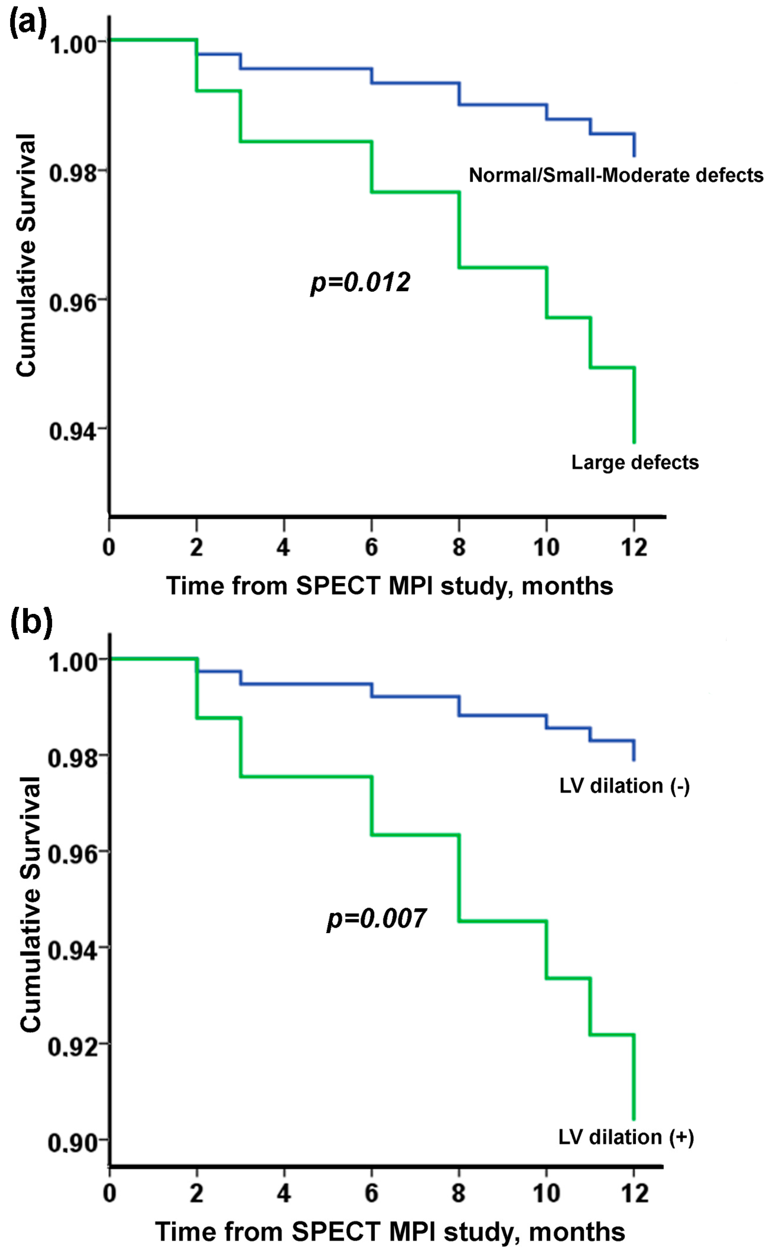
| Large defects | 3.56 | 1.32, 9.55 | 0.012 |
| Normal study | Ref. | ||
| Left ventricular dilation | 4.74 | 1.53, 14.71 | 0.007 |
| Univariate Analysis | Multivariate Analysis | |||||
|---|---|---|---|---|---|---|
| Mortality | HR | 95% CI | p Value | HR | 95% CI | p Value |
| Large defects | 3.56 | 1.32, 9.55 | 0.012 | 2.90 | 1.05, 8.06 | 0.041 |
| LV dilation | 4.74 | 1.53, 14.71 | 0.007 | |||
| Age | 1.13 | 1.05, 1.21 | 0.001 | 1.13 | 1.05, 1.22 | 0.001 |
| Male gender | 5.72 | 1.30, 25.15 | 0.021 | |||
| BMI | 0.79 | 0.69, 0.90 | 0.001 | 0.80 | 0.68, 0.93 | 0.004 |
| Waist | 0.97 | 0.93, 1.01 | 0.130 | |||
| Chest pain | 0.60 | 0.23, 1.60 | 0.310 | |||
| Dyspnea | 1.30 | 0.76, 2.22 | 0.341 | |||
| Smoking | 1.02 | 0.29, 3.57 | 0.978 | |||
| Hypertension | 0.86 | 0.28, 2.68 | 0.799 | |||
| Diabetes | 1.20 | 0.44, 3.30 | 0.725 | |||
| Dyslipidemia | 1.34 | 0.38, 4.69 | 0.651 | |||
| History of CAD | 3.28 | 1.19, 9.01 | 0.022 | |||
| Heart failure | 3.60 | 1.03, 12.62 | 0.046 | |||
| History of stroke | 1.49 | 0.34, 6.57 | 0.596 | |||
| Non-fatal MI | ||||||
| Large defects | 2.71 | 0.65, 11.35 | 0.173 | |||
| LV dilation | - | - | - | |||
| Age | 1.06 | 0.98, 1.16 | 0.150 | |||
| Male gender | 1.35 | 0.32, 5.63 | 0.684 | |||
| BMI | 1.08 | 0.95, 1.22 | 0.258 | |||
| Waist | 1.06 | 1.01, 1.12 | 0.029 | 1.06 | 1.01, 1.12 | 0.029 |
| Chest pain | 0.36 | 0.09, 1.51 | 0.163 | |||
| Dyspnea | 0.95 | 0.19, 4.70 | 0.948 | |||
| Smoking | 0.63 | 0.08, 5.12 | 0.666 | |||
| Hypertension | 0.86 | 0.17, 4.26 | 0.852 | |||
| Diabetes | 1.20 | 0.29, 5.01 | 0.805 | |||
| Dyslipidemia | 2.17 | 0.27, 17.60 | 0.470 | |||
| History of CAD | 1.97 | 0.49, 7.88 | 0.338 | |||
| Heart failure | 2.16 | 0.27, 17.57 | 0.471 | |||
| History of stroke | 1.50 | 0.19, 12.22 | 0.703 | |||
| Stroke | ||||||
| Large defects | 0.49 | 0.11, 2.12 | 0.341 | |||
| LV dilation | - | - | - | |||
| Age | 1.08 | 1.02, 1.14 | 0.010 | 1.08 | 1.02, 1.14 | 0.010 |
| Male gender | 0.99 | 0.41, 2.38 | 0.975 | |||
| BMI | 0.98 | 0.89, 1.08 | 0.674 | |||
| Waist | 0.97 | 0.94, 1.01 | 0.134 | |||
| Chest pain | 0.60 | 0.25, 1.43 | 0.247 | |||
| Dyspnea | 2.87 | 0.67, 12.38 | 0.157 | |||
| Smoking | 1.50 | 0.54, 4.12 | 0.435 | |||
| Hypertension | 1.64 | 0.48, 5.59 | 0.430 | |||
| Diabetes | 0.50 | 0.17, 1.48 | 0.208 | |||
| Dyslipidemia | 1.25 | 0.42, 3.72 | 0.695 | |||
| History of CAD | 1.61 | 0.67, 3.88 | 0.291 | |||
| Heart failure | 1.72 | 0.40, 7.41 | 0.467 | |||
| History of stroke | 2.74 | 0.91, 8.18 | 0.072 | |||
Disclaimer/Publisher’s Note: The statements, opinions and data contained in all publications are solely those of the individual author(s) and contributor(s) and not of MDPI and/or the editor(s). MDPI and/or the editor(s) disclaim responsibility for any injury to people or property resulting from any ideas, methods, instructions or products referred to in the content. |
© 2023 by the authors. Licensee MDPI, Basel, Switzerland. This article is an open access article distributed under the terms and conditions of the Creative Commons Attribution (CC BY) license (https://creativecommons.org/licenses/by/4.0/).
Share and Cite
Zotou, P.; Bechlioulis, A.; Tsiouris, S.; Naka, K.K.; Xourgia, X.; Pappas, K.; Lakkas, L.; Rammos, A.; Kalef-Ezra, J.; Michalis, L.K.; et al. The Role of Myocardial Perfusion Imaging in the Prediction of Major Adverse Cardiovascular Events at 1 Year Follow-Up: A Single Center’s Experience. J. Pers. Med. 2023, 13, 871. https://doi.org/10.3390/jpm13050871
Zotou P, Bechlioulis A, Tsiouris S, Naka KK, Xourgia X, Pappas K, Lakkas L, Rammos A, Kalef-Ezra J, Michalis LK, et al. The Role of Myocardial Perfusion Imaging in the Prediction of Major Adverse Cardiovascular Events at 1 Year Follow-Up: A Single Center’s Experience. Journal of Personalized Medicine. 2023; 13(5):871. https://doi.org/10.3390/jpm13050871
Chicago/Turabian StyleZotou, Paraskevi, Aris Bechlioulis, Spyridon Tsiouris, Katerina K. Naka, Xanthi Xourgia, Konstantinos Pappas, Lampros Lakkas, Aidonis Rammos, John Kalef-Ezra, Lampros K. Michalis, and et al. 2023. "The Role of Myocardial Perfusion Imaging in the Prediction of Major Adverse Cardiovascular Events at 1 Year Follow-Up: A Single Center’s Experience" Journal of Personalized Medicine 13, no. 5: 871. https://doi.org/10.3390/jpm13050871