
Dipeptidyl Amino-Peptidase 3 (DPP3) as an Early Marker of Severity in a Patient Population with Cardiogenic Shock
 ,
,  , , ,
, , ,
Abstract
:1. Introduction
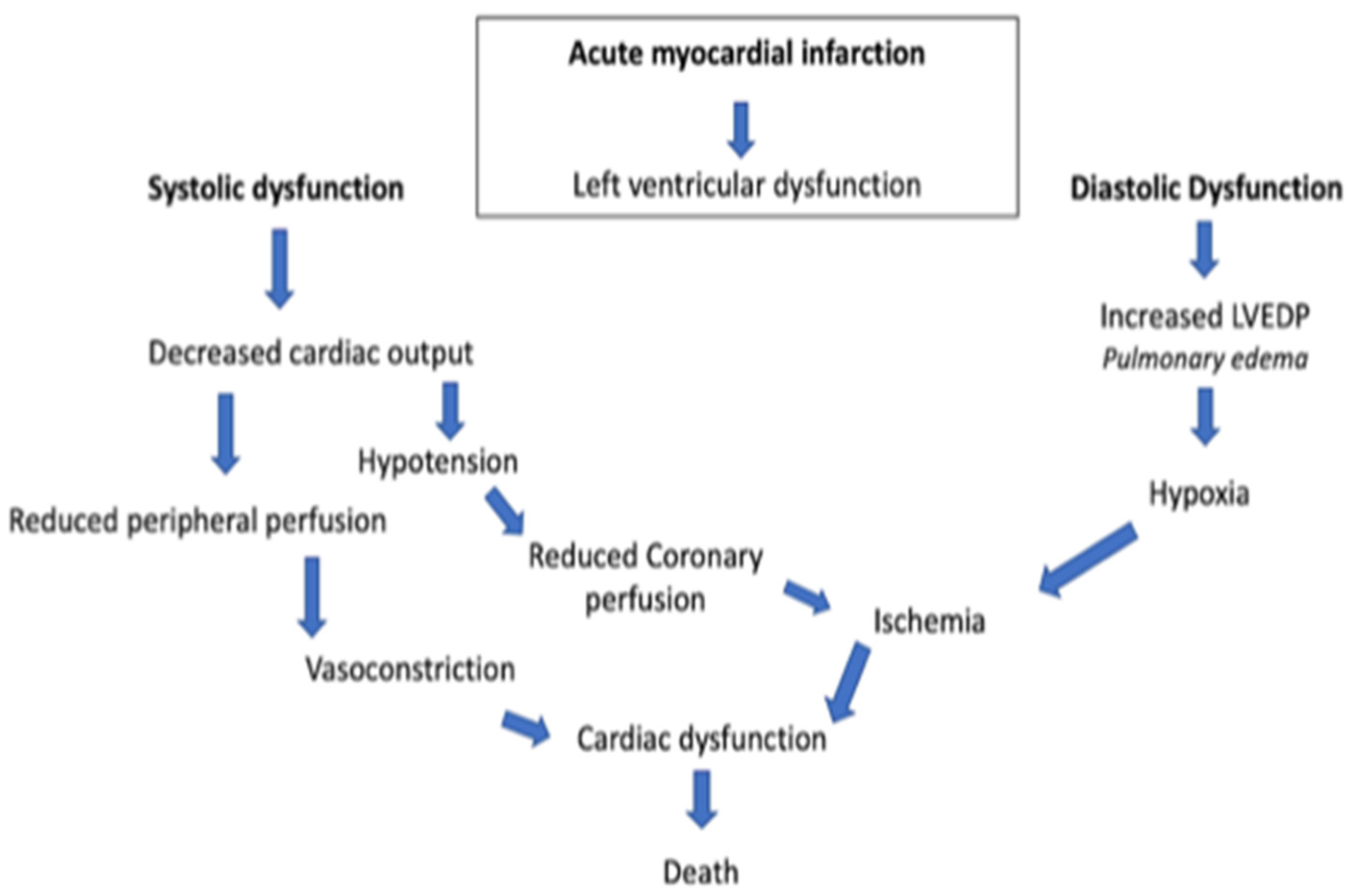
1.1. Diseases of the Cardiovascular System
1.2. Cardiogenic Shock
1.3. Dipeptidyl Amino-Peptidase 3 in the Context of Cardiovascular Diseases
1.4. Study Purpose
2. Materials and Methods
2.1. Study Design and Population
2.2. Measurements
2.3. Echocardiographic Measurements
- (a)
- Diameter of the aortic annulus: This measurement is made in the parasternal long- axis view during systole, when the diameter is greatest (usually halfway through systole). Zoom in LVOT to improve the accuracy of the measurement was performed.
- (b)
- Flow velocity in LVOT: Velocity is measured in apical four-chamber view (4C) or five-chamber view (5C) using pulsed-wave doppler with sample volume in the valve orifice. The VTI (Velocity Time Integral) is automatically calculated.
2.4. Statistical Analyses
3. Results
4. Discussion
5. Conclusions
Strengths and Limitations
Author Contributions
Funding
Institutional Review Board Statement
Informed Consent Statement
Data Availability Statement
Conflicts of Interest
References
- Palacios Ordonez, C.; Garan, A.R. The landscape of cardiogenic shock: Epidemiology and current definitions. Curr. Opin. Cardiol. 2022, 37, 236–240. [Google Scholar] [CrossRef] [PubMed]
- Hollenberg, S.M.; Singer, M. Pathophysiology of sepsis-induced cardiomyopathy. Nat. Rev. Cardiol. 2021, 18, 424–434. [Google Scholar] [CrossRef] [PubMed]
- Heidenreich, P.A.; Bozkurt, B.; Aguilar, D.; Allen, L.A.; Byun, J.J.; Colvin, M.M.; Deswal, A.; Drazner, M.H.; Dunlay, S.M.; Evers, L.R.; et al. 2022 AHA/ACC/HFSA Guideline for the Management of Heart Failure: Executive Summary: A Report of the American College of Cardiology/American Heart Association Joint Committee on Clinical Practice Guidelines. Circulation 2022, 145, E876–E894. [Google Scholar] [CrossRef] [PubMed]
- Thiele, H.; Ohman, E.M.; Desch, S.; Eitel, I.; De Waha, S. Management of cardiogenic shock. Eur. Heart J. 2015, 36, 1223–1230. [Google Scholar] [CrossRef] [Green Version]
- Brusselaers, N.; Monstrey, S.; Vogelaers, D.; Hoste, E.; Blot, S. Severe burn injury in europe: A systematic review of the incidence, etiology, morbidity, and mortality. Crit. Care 2010, 14, 1–12. [Google Scholar] [CrossRef] [Green Version]
- Krittanawong, C.; Rivera, M.R.; Shaikh, P.; Kumar, A.; May, A.; Mahtta, D.; Jentzer, J.; Civitello, A.; Katz, J.; Naidu, S.S.; et al. Key Concepts Surrounding Cardiogenic Shock. Curr. Probl. Cardiol. 2022, 47, 101303. [Google Scholar] [CrossRef]
- Aissaoui, N.; Puymirat, E.; Delmas, C.; Ortuno, S.; Durand, E.; Bataille, V.; Drouet, E.; Bonello, L.; Bonnefoy-Cudraz, E.; Lesmeles, G.; et al. Trends in cardiogenic shock complicating acute myocardial infarction. Eur. J. Heart Fail. 2020, 22, 664–672. [Google Scholar] [CrossRef] [PubMed]
- Biswas, S.; Malik, A.H.; Bandyopadhyay, D.; Gupta, R.; Goel, A.; Briasoulis, A.; Fonarow, G.C.; Lanier, G.M.; Naidu, S.S. Matanalysis comparing the efficacy of dobutamine versus Milrinone in acute decompensated heart failure. Curr. Probl. Cardiol. 2022, 8, 101245. [Google Scholar] [CrossRef]
- Paternoster, G.; Bertini, P.; Innelli, P.; Trambaiolo, P.; Landoni, G.; Franchi, F.; Scolletta, S.; Guarracino, F. Right Ventricular Dysfunction in Patients with COVID-19: A Systematic Review and Meta-analysis. J. Cardiothorac. Vasc. Anesth. 2021, 35, 3319–3324. [Google Scholar] [CrossRef]
- Ashish, A.H.; Puri, R.; Kalra, A. Management of cardiogenixc shock complicating acute myocardila infarction: A review. Clin. Cardiol. 2019, 42, 484–493. [Google Scholar]
- Prajapati, S.C.; Chauhan, S.S. Dipeptidyl peptidase III: A multifaceted oligopeptide N-end cutter. FEBS J. 2011, 278, 3256–3276. [Google Scholar] [CrossRef]
- Ohkubo, I.; Li, Y.H.; Maeda, T.; Yamamoto, Y.; Yamane, T.; Du, P.G.; Nishi, K. Molecular cloning and immunohistochemical localization of rat dipeptidyl peptidase III. Forensic Sci. Int. 2000, 113, 147–151. [Google Scholar] [CrossRef]
- Hashimoto, J.I.; Yamamoto, Y.; Kurosawa, H.; Nishimura, K.; Hazato, T. Identification of dipeptidyl peptidase III in human neutrophils. Biochem. Biophys. Res. Commun. 2000, 273, 393–397. [Google Scholar] [CrossRef] [PubMed]
- Wattiaux, R.; Wattiaux-de coninck, S.; Thirion, J.; Gasingirwa, M.-C.; Jadot, M. Lysosomes and Fas-mediated liver cell death. Biochem. J. 2007, 403, 89–95. [Google Scholar] [CrossRef] [PubMed]
- Rehfeld, L.; Funk, E.; Jha, S.; Macheroux, P.; Melander, O.; Bergmann, A. Novel Methods for the Quantification of Dipeptidyl Peptidase 3 (DPP3) Concentration and Activity in Human Blood Samples. J. Appl. Lab. Med. 2019, 3, 943–953. [Google Scholar] [CrossRef] [PubMed]
- Jha, S.; Taschler, U.; Domenig, O.; Poglitsch, M.; Bourgeois, B.; Pollheimer, M.; Pusch, L.M.; Malovan, G.; Frank, S.; Madl, T.; et al. Dipeptidyl peptidase 3 modulates the renin–Angiotensin system in mice. J. Biol. Chem. 2020, 295, 13711–13723. [Google Scholar] [CrossRef]
- Lee, C.; Snyder, S.H. Dipeptidyl-aminopeptidase III of Rat Brain. J. Biol. Chem. 1982, 257, 12043–12050. [Google Scholar] [CrossRef]
- Bezerra, G.A.; Dobrovetsky, E.; Viertlmayr, R.; Dong, A.; Binter, A.; Abramić, M.; Macheroux, P.; Dhe-Paganon, S.; Gruber, K. Entropy-driven binding of opioid peptides induces a large domain motion in human dipeptidyl peptidase III. Proc. Natl. Acad. Sci. USA 2012, 109, 6525–6530. [Google Scholar] [CrossRef] [Green Version]
- Baršun, M.; Jajčanin, N.; Vukelić, B.; Špoljarić, J.; Abramić, M. Human dipeptidyl peptidase III acts as a post-proline-cleaving enzyme on endomorphins. Biol. Chem. 2007, 388, 343–348. [Google Scholar] [CrossRef] [PubMed]
- Pang, X.; Shimizu, A.; Kurita, S.; Zankov, D.P.; Takeuchi, K.; Yasuda-Yamahara, M.; Kume, S.; Ishida, T.; Ogita, H. Novel therapeutic role for dipeptidyl peptidase III in the treatment of hypertension. Hypertension 2016, 68, 630–641. [Google Scholar] [CrossRef]
- Malovan, G.; Hierzberger, B.; Suraci, S.; Schaefer, M.; Santos, K.; Jha, S.; Macheroux, P. The emerging role of dipeptidyl peptidase 3 in pathophysiology. FEBS J. 2022, 1–17. [Google Scholar] [CrossRef]
- Takagi, K.; Blet, A.; Levy, B.; Deniau, B.; Azibani, F.; Feliot, E.; Bergmann, A.; Santos, K.; Hartmann, O.; Gayat, E.; et al. Circulating dipeptidyl peptidase 3 and alteration in haemodynamics in cardiogenic shock: Results from the OptimaCC trial. Eur. J. Heart Fail. 2020, 22, 279–286. [Google Scholar] [CrossRef]
- Frigyesi, A.; Lengquist, M.; Spångfors, M.; Annborn, M.; Cronberg, T.; Nielsen, N.; Levin, H.; Friberg, H. Circulating dipeptidyl peptidase 3 on intensive care unit admission is a predictor of organ dysfunction and mortality. J. Intensive Care 2021, 9, 52. [Google Scholar] [CrossRef] [PubMed]
- Deniau, B.; Rehfeld, L.; Santos, K.; Dienelt, A.; Azibani, F.; Sadoune, M.; Kounde, P.R.; Samuel, J.L.; Tolpannen, H.; Lassus, J.; et al. Circulating dipeptidyl peptidase 3 is a myocardial depressant factor: Dipeptidyl peptidase 3 inhibition rapidly and sustainably improves haemodynamics. Eur. J. Heart Fail. 2020, 22, 290–299. [Google Scholar] [CrossRef] [Green Version]
- Chioncel, O.; Parissis, J.; Mebazaa, A.; Thiele, H.; Desch, S.; Bauersachs, J.; Harjola, V.P.; Antohi, E.L.; Arrigo, M.; Gal, T.B.; et al. Epidemiology, pathophysiology and contemporary management of cardiogenic shock—A position statement from the Heart Failure Association of the European Society of Cardiology. Eur. J. Heart Fail. 2020, 22, 1315–1341. [Google Scholar] [CrossRef]
- McDonagh, T.A.; Metra, M.; Adamo, M.; Gardner, R.S.; Baumbach, A.; Böhm, M.; Burri, H.; Butler, J.; Celutkiene, J.; Chioncel, O.; et al. 2021 ESC Guidelines for the diagnosis and treatment of acute and chronic heart failure. Eur. Heart J. 2021, 42, 3599–3726. [Google Scholar] [CrossRef] [PubMed]
- Reynolds, H.R.; Hochman, J.S. Cardiogenic shock current concepts and improving outcomes. Circulation 2008, 117, 686–697. [Google Scholar] [CrossRef]
- Blet, A.; Deniau, B.; Santos, K.; Van Lier, D.P.T.; Azibani, F.; Wittebole, X.; Chousterman, B.G.; Gayat, E.; Hartmann, O.; Struck, J.; et al. Monitoring Circulating dipeptidyl peptidase 3 (DPP3) predicts improvement of organ failure and survival in sepsis: A prospective observational multinational study. Crit. Care 2021, 3, 1–10. [Google Scholar] [CrossRef]
- Deniau, B.; Picod, A.; Van Lier, D.; Vaittinada Ayar, P.; Santos, K.; Hartmann, O.; Gayat, E.; Mebazaa, A.; Blet, A.; Azibani, F. High plasma dipeptidyl peptidase 3 levels are associated with mortality and organ failure in shock: Results from the international, prospective and observational FROG-ICU cohort. Br. J. Anaesth. 2022, 128, e54–e57. [Google Scholar] [CrossRef] [PubMed]
- Deniau, B.; Blet, A.; Santos, K.; Ayar, P.V.; Genest, M.; Kästorf, M.; Sadoune, M.; de Sousa Jorge, A.; Samuel, J.L.; Vodovar, N.; et al. Inhibition of circulating dipeptidyl-peptidase 3 restores cardiac function in a sepsis-induced model in rats: A proof of concept study. PLoS ONE 2020, 15, e0238039. [Google Scholar] [CrossRef]
- Van Diepen, S.; Katz, J.N.; Albert, N.M.; Henry, T.D.; Jacobs, A.K.; Kapur, N.K.; Kilic, A.; Menon, V.; Ohman, E.M.; Sweitzer, N.K.; et al. Contemporary Management of Cardiogenic Shock: A Scientific Statement from the American Heart Association. Circulation 2017, 136, e232–e268. [Google Scholar] [CrossRef] [PubMed]
- Kolte, D.; Khera, S.; Aronow, W.S.; Mujib, M.; Palaniswamy, C.; Sule, S.; Jain, D.; Gotsis, W.; Ahmed, A.; Frishman, W.H.; et al. Trends in incidence, management, and outcomes of cardiogenic shock complicating ST-elevation myocardial infarction in the United States. J. Am. Heart Assoc. 2014, 3, e000590. [Google Scholar] [CrossRef] [Green Version]
- Miller, P.E.; Gimenez, M.R.; Thiele, H. Mechanical respiratory support in cardiogenic shock. Eur. J. Heart Fail. 2020, 22, 168. [Google Scholar] [CrossRef] [PubMed]
- Helgestad, O.K.L.; Josiassen, J.; Hassager, C.; Jensen, L.O.; Holmvang, L.; Sørensen, A.; Frydland, M.; Lassen, A.T.; Udesen, N.L.J.; Schmidt, H.; et al. Temporal trends in incidence and patient characteristics in cardiogenic shock following acute myocardial infarction from 2010 to 2017: A Danish cohort study. Eur. J. Heart Fail. 2019, 21, 1370–1378. [Google Scholar] [CrossRef] [PubMed]
- Lang, R.M.; Badano, L.P.; Victor, M.A.; Afilalo, J.; Armstrong, A.; Ernande, L.; Flachskampf, F.A.; Foster, E.; Goldstein, S.A.; Kuznetsova, T.; et al. Recommendations for cardiac chamber quantification by echocardiography in adults: An update from the American Society of Echocardiography and the European Association of Cardiovascular Imaging. J. Am. Soc. Echocardiogr. 2015, 28, 1–39.e14. [Google Scholar] [CrossRef] [PubMed] [Green Version]
- Hu, K.; Mathew, R. Inotrope and vasopressor use in cardiogenic shock: What, when and why? Curr. Opin. Crit. Care 2022, 28, 419–425. [Google Scholar] [CrossRef] [PubMed]
- Mebazaa, A.; Combes, A.; van Diepen, S.; Hollinger, A.; Katz, J.N.; Landoni, G.; Hajjar, L.A.; Lassus, J.; Lebreton, G.; Montalescot, G.; et al. Management of cardiogenic shock complicating myocardial infarction. Intensive Care Med. 2018, 44, 760–773. [Google Scholar] [CrossRef]
- Magliocca, A.; Omland, T.; Latini, R. Dipeptidyl peptidase 3, a biomarker in cardiogenic shock and hopefully much more. Eur. J. Heart Fail. 2020, 22, 300–302. [Google Scholar] [CrossRef]


| Baseline Variables | All Patients (n = 15) (%) | Ventilated (n = 6 (40%)) | Non-Ventilated (n = 9 (60%)) | p-Value |
|---|---|---|---|---|
| Gender (F) | 7 (46.7%) | 2 (33%) | 5 (55.6%) | 0.002 |
| Age (years) | 69.0 ± 18.6 | 61.0 ± 8.5 | 67.4 ± 18.7 | 0.329 |
| SBP (mmHg) | 79.5± 8.5 | 70.1 ± 9.4 | 87 ± 9.1 | 0.038 |
| Hb (g/dL) | 12.0 ± 2.0 | 10.5 ± 1.8 | 12.1 ± 2.1 | 0.128 |
| BNP (mmol/L) | 971,6 | 989.0 ± 217.0 | 1036.7 ± 325.0 | 0.750 |
| HS Trop (mmol/L) | 37,692.9 ± 86,529.8 | 57,867.0 ± 109,964.0 | 4334.0 ± 7993.0 | 0.659 |
| CRP (mmol/L) | 31.8 ± 60.1 | 99.5 ± 79.8 | 27.5 ± 53.3 | 0.047 |
| STEMI, n (%) | 6 (40%) | 2 (33.3%) | 4 (44.4%) | 0.0008 |
| CHF, n (%) | 5 (33%) | 2 (33.3%) | 3 (33.3%) | 0.0004 |
| Other, n (%) | 4 (26.7%) | 2 (33.3%) | 2 (22%) | 0.0004 |
| Diabetes, n (%) | 4 (26.7%) | 3 (50%) | 1 (11%) | 0.177 |
| Hypertension, n (%) | 7 (46.7%) | 4 (66.7%) | 3 (33.3%) | 0.061 |
| Hypercholesterolemia, n (%) | 10 (66.7%) | 6 (100%) | 4 (44.4%) | 0.218 |
| Chronic Kidney Disease, n (%) | 3 (20%) | 0 | 3 (33.3%) | <0.000001 |
| Valvular Regurgitation, n (%) | 6 (40%) | 1 (16.7%) | 5 (55.6%) | 0.00008 |
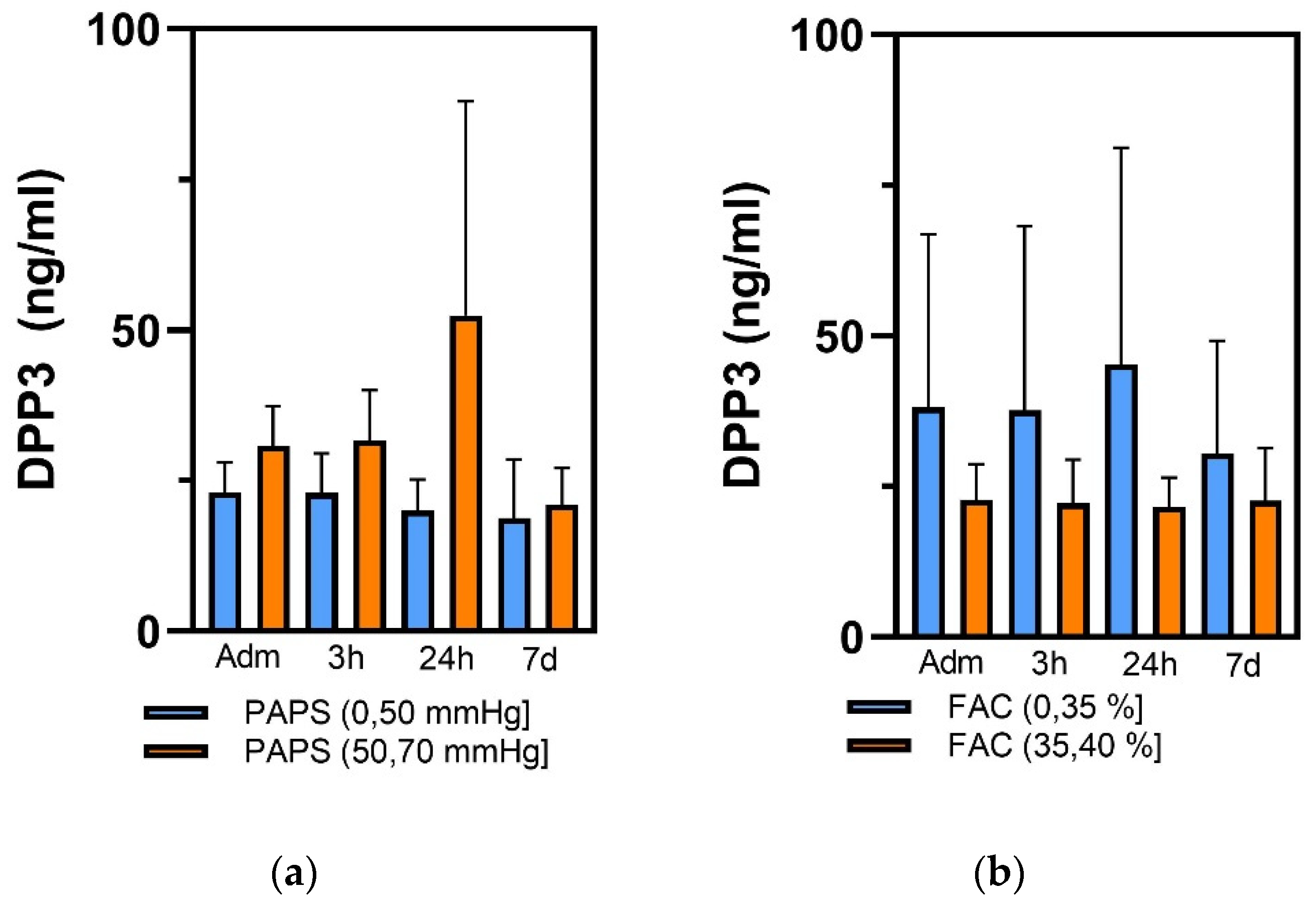
| Patients | Admission | 3 h | 24 h | 7 d |
|---|---|---|---|---|
| Ventilated (n = 6) | 132.4 ± 73.0 | 130.1 ± 67.1 | 109.4 ± 73.7 | 86.0 ± 71.1 |
| Non-ventilated (n = 9) | 93.6 ± 93.1 | 93.5 ± 93.3 | 60.0 ± 52.9 | 108.7 ± 111 |
| ECHO Parameters | Entire Cohort (n = 15) (%) | Ventilated (n = 6 (40%)) | Not-Ventilated (n = 9 (60%)) | p-Value |
|---|---|---|---|---|
| EDV (mL) | 91.7 ± 37.1 | 92.2 ± 36.5 | 91.2 ± 43.3 | 0.978 |
| EDD (mL) | 48.5 ± 6.5 | 45.7 ± 4.3 | 51.2 ± 7.8 | 0.346 |
| EF (%) | 36.1 ± 12.3 | 29.6 ± 7.7 | 42.5 ± 12.1 | 0.247 |
| SV (mL/m2) | 30.2 ± 14.3 | 22.7 ± 4.2 | 37.7 ± 17.6 | 0.228 |
| LAV (mL) | 60.4 ± 22.8 | 47.5 ± 18.1 | 77.6 ± 17.1 | 0.233 |
| RVD1 (mm) | 36.7 ± 11.1 | 38.7 ± 10.8 | 35.6 ± 12.1 | 0.711 |
| TAPSE (mm) | 13.5 ± 5.7 | 15.2 ± 6.1 | 19.1 ± 12.7 | 0.552 |
| FAC (%) | 29.9 ± 7.5 | 30.7 ± 10.7 | 36.2 ± 2.6 | 0.847 |
| PAPS (mmHg) | 42.8 ± 13.5 | 51.0 ± 17.3 | 52.0 ± 11.3 | 0.259 |
Disclaimer/Publisher’s Note: The statements, opinions and data contained in all publications are solely those of the individual author(s) and contributor(s) and not of MDPI and/or the editor(s). MDPI and/or the editor(s) disclaim responsibility for any injury to people or property resulting from any ideas, methods, instructions or products referred to in the content. |
© 2023 by the authors. Licensee MDPI, Basel, Switzerland. This article is an open access article distributed under the terms and conditions of the Creative Commons Attribution (CC BY) license (https://creativecommons.org/licenses/by/4.0/).
Share and Cite
Innelli, P.; Lopizzo, T.; Paternò, G.; Bruno, N.; Radice, R.P.; Bertini, P.; Marabotti, A.; Luzi, G.; Stabile, E.; Di Fazio, A.; et al. Dipeptidyl Amino-Peptidase 3 (DPP3) as an Early Marker of Severity in a Patient Population with Cardiogenic Shock. Diagnostics 2023, 13, 1350. https://doi.org/10.3390/diagnostics13071350
Innelli P, Lopizzo T, Paternò G, Bruno N, Radice RP, Bertini P, Marabotti A, Luzi G, Stabile E, Di Fazio A, et al. Dipeptidyl Amino-Peptidase 3 (DPP3) as an Early Marker of Severity in a Patient Population with Cardiogenic Shock. Diagnostics. 2023; 13(7):1350. https://doi.org/10.3390/diagnostics13071350
Chicago/Turabian StyleInnelli, Pasquale, Teresa Lopizzo, Giovanni Paternò, Noemi Bruno, Rosa Paola Radice, Pietro Bertini, Alberto Marabotti, Giampaolo Luzi, Eugenio Stabile, Aldo Di Fazio, and et al. 2023. "Dipeptidyl Amino-Peptidase 3 (DPP3) as an Early Marker of Severity in a Patient Population with Cardiogenic Shock" Diagnostics 13, no. 7: 1350. https://doi.org/10.3390/diagnostics13071350