1. Introduction
Neurofibromatosis type 1 is an autosomal dominant disorder involving aberrant proliferation of multiple tissues of neural crest origin. It is principally associated with cutaneous, neurologic, and orthopedic manifestations. Most epidemiological studies have reported a prevalence between 1/3000 and 1/6000. It is a very heterogeneous disorder, with multisystem involvement in most cases, affecting individuals variably. NF1 reduces life expectancy due to an increased risk of malignancy [
1,
2].
The diagnosis of NF1 is based on the clinical diagnostic criteria established by the National Institutes of Health (NIH) and/or a molecularly confirmed mutation in the NF1 gene (complete sequencing of the coding region of the most relevant genes for clinical practice in solid adult tumors was tested when patients presented with an unusual phenotype or an incomplete clinical picture).
The NIH clinical diagnostic criteria for NF1 [
3,
4] have high specificity and sensitivity for most patients. Ophthalmological manifestations of NF1 that are included in the established diagnostic criteria are Lisch nodules, optic glioma, and a distinctive osseous lesion (sphenoid wing dysplasia) [
5]. Legius et al. [
6] revised the diagnostic criteria for NF1 in 2021, incorporating significant developments and creating new criteria for Legius syndrome (LGSS) to differentiate the two conditions. They proposed an initiative to update the diagnostic criteria for NF1, including choroidal abnormalities as an ophthalmologic criterion because of its high specificity, sensitivity, and ability to differentiate from LGSS.
Few reports have shown the presence of choroidal hyperreflective nodules [
1,
2,
3,
5,
6,
7,
8,
9,
10,
11,
12,
13,
14,
15,
16,
17,
18,
19,
20,
21,
22,
23]. The reason is that they are undetectable with conventional ophthalmoscopic fundus examination [
5]. The retinal pigment epithelium (RPE) contains melanin that blocks the passage of visible light to the choroid. Infrared light penetrates the RPE better than visible light [
7] and allows observing choroidal abnormalities.
In a sample of NF1 patients, the study aimed to investigate whether the presence of choroidal nodules detected by NIR (near-infrared reflectance) is a valuable criterion for diagnosis in patients with and without high myopia.
2. Materials and Methods
All patients signed the informed consent. The protocol of this study was approved by the Research Ethics Committee of the Autonomous Community of Aragón (Spain).
Between September 2021 and May 2022, 60 eyes of 30 white Caucasian adult patients (eighteen female, twelve male; mean age 57 years) diagnosed with NF1 using the National Institutes of Health (NIH) criteria were studied. So far, the diagnosis relies on clinical diagnosis, and genetic testing is optional when the diagnosis has already been established [
24]. We also examined 60 eyes of 30 healthy control subjects. Each subject underwent a general ophthalmologic examination, including an accurate slit lamp examination searching for Lisch nodules. Patients with NF1 were referred from the department of Internal Medicine (where only patients over 18 are checked) to the department of Ophthalmology of the Lozano Blesa University Hospital in Zaragoza (Spain) as a part of a screening program to detect ocular pathology in NF1 patients. Patients under eighteen years old were excluded from the study since it has been found that choroidal abnormalities tend to increase with patient age [
8], and it was intended to find a homogeneous sample, as well as those with the presence of media opacities and retinal pathology.
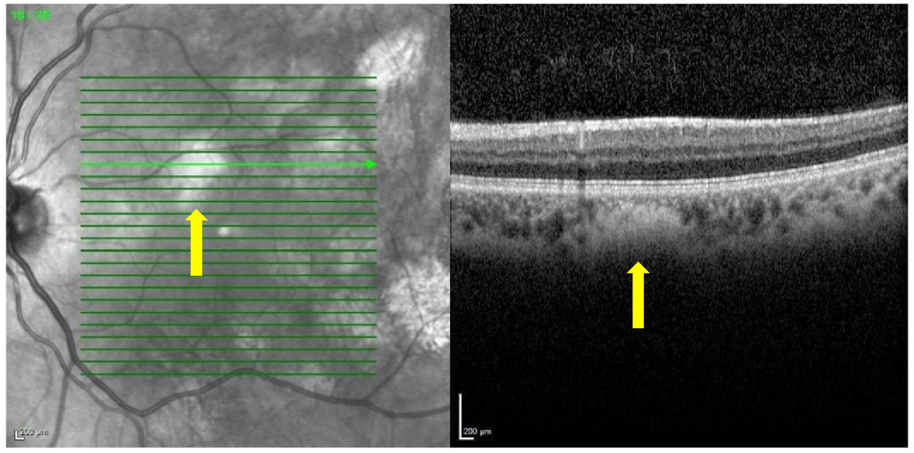
Near-infrared reflectance at 815 nm and red-free (RF) 488 nm images of the posterior pole and mid-periphery of the retina were taken of each patient and monitored with a confocal scanning laser ophthalmoscope (Spectralis HRA+OCT, Heidelberg Engineering, Heidelberg, Germany) (
Figure 1) [
9]. The combined spectral domain optical coherence tomography (SD-OCT) system allows for simultaneous recording with precise alignment of OCT and topographic images (NIR, RF). Using this technology, which is non-invasive, fast, and abundantly used in routine exams, a good correlation of the topographic image with the morphological changes in the retina and choroid of the macular area was obtained [
9]. No dilating eye drops were used before the examination.
Red-free images were analyzed in a masked way by the same investigator, who searched for choroidal hyperreflective nodules that were counted, objectifying more than 10 in most patients diagnosed with NF1, and any other unusual findings. A different independent investigator repeated the examination to obtain another analysis for an agreement evaluation.
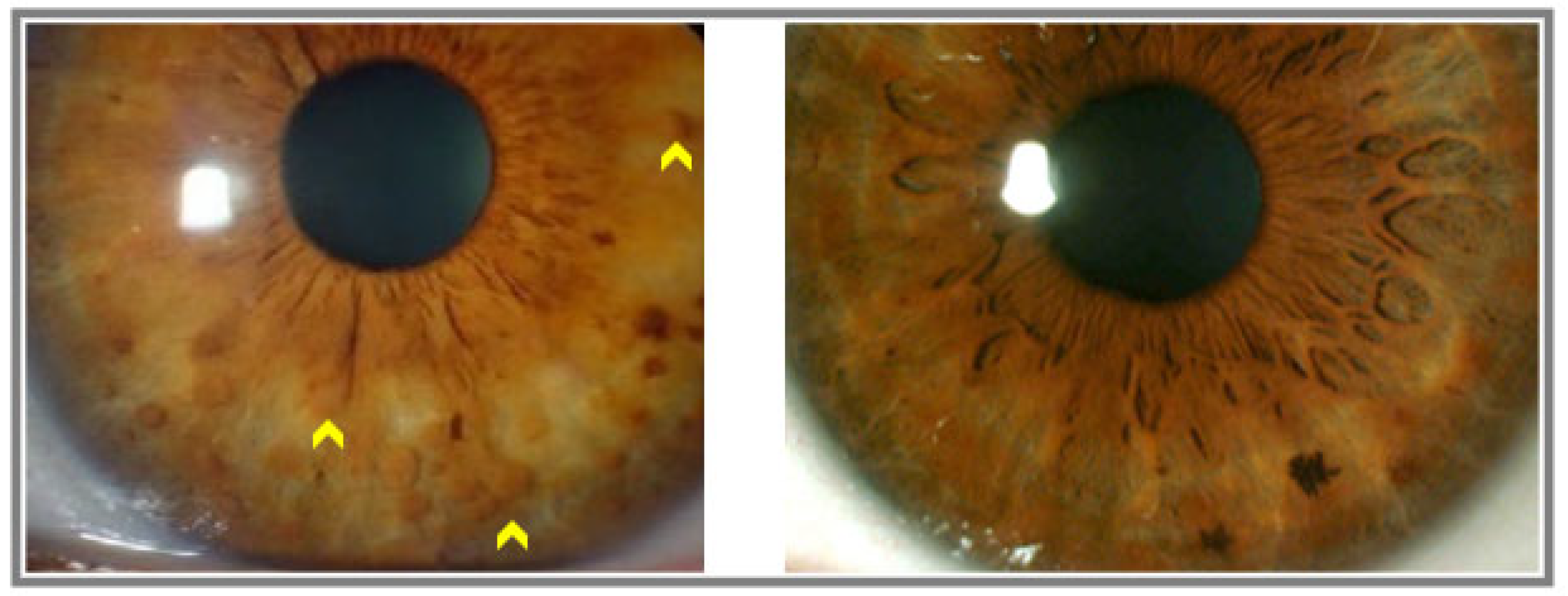
A similar procedure occurred with slit lamp examination where the principal investigator noted the presence or absence of iridian Lisch nodules (
Figure 2) and took anterior segment photographs with the Topcon slit lamp imaging system (SL-D701, Topcon Healthcare). They were also counted, objectifying an average of 11 nodules in the patients diagnosed with NF1. Another independent investigator reviewed the images and repeated the analysis to obtain data for the consensus of the final results.
Data regarding refractive errors, which could affect the retinal anatomy, and consequently the detection of bright, patchy nodules by NIR also was noted. High myopia was defined as the spherical equivalent refraction of −5.0 D or more.
3. Results
In the interest of verifying the homogeneity of the two experimental groups (including 60 eyes of 30 Caucasian adult patients in each group) in terms of certain sociodemographic variables, the age and sex of the patients were compared. It was observed that there were no significant differences between both groups.
The frequency of choroidal hyperreflective nodules, detected by NIR, and Lisch nodules, detected by slit lamp examination, were compared between patients and control subjects by the Chi-square test. It was observed that, in the group diagnosed with NF1, 80% showed iridian Lisch nodules, while in the control group, there were no cases (
Table 1).
The numbers in
Table 1 and
Table 2 represent the number of patients, considering that Lisch and choroidal nodules were always bilateral.
In the analysis of the comparison in the diagnosis of choroidal patches between the two experimental groups employing the Chi-square test, it was observed that 83.3% of patients diagnosed with NF1 showed the alteration, whereas, in the control group, none of them did so (
Table 2).
When comparing whether there are significant differences in the prevalence of choroidal patches and Lisch nodules among patients with NF1, considering those with high myopia, it is observed that there are no significant differences between the prevalence (
Table 3).
When analyzing the sample of patients with NF1, patients with high myopia were not considered (five patients), it was observed that 84% of patients showed Lisch nodules compared to 100% of patients that presented choroidal nodules. The differences between prevalence were statistically significant, confirming the higher prevalence of choroidal patches in this new group of patients close to emmetropia (
Table 4).
4. Discussion
The prevalence of choroidal hyper-reflective nodules may be higher than believed because asymptomatic patients without eye discomfort or blurred vision tend not to undergo ocular examinations. Moreover, patients with good vision that perform a thorough eye exam usually are subjected to an eye fundus (that does not reveal the existence of this particular finding) and are not tested with either NIR imaging or OCT.
Previous research shows that choroidal nodules in the setting of NF1 can be seen in up to 82% of NF1 patients [
10]. Moramarco et al. [
11] reported a prevalence of 97% of choroidal abnormalities in patients with NF1. This study reports that 83.3% of patients diagnosed with NF1 present choroidal hyperreflective nodules. The study’s limitations include the small number of patients, and the OCT with NIR, although present in many ophthalmology departments, might be absent. What is new in our study and what we want to emphasize are that our research also studies the prevalence of choroidal nodules in patients diagnosed with NF1, excluding patients with high myopia, resulting in 100% prevalence. This is a new finding that has not been previously described before in the literature. We speculate that the choroid of highly myopic patients is significantly thinner, and choroidal vascularization is considerably altered in these patients; This characteristic may have to do with the lower presence of choroidal nodules [
12,
13]. Considering that most of the population does not suffer from high myopia, we believe that more studies should be carried out, including this new exclusion criterion, since it has been shown to significantly increase the prevalence of choroidal nodules in patients with NF1.
The bright lesions on NIR imaging correspond to the hyperflow areas of deep choroids on OCT angiography (OCTA), indicating a rich vascular supply of these nodules. When studying the choroid in these nodules, an alteration of the morphology and thickness has been reported. The mean choroidal thickness is reduced with generalized thinning of the neuroepithelium, retinal pigmentary epithelium, and outer nuclear layer [
14].
Considering the literature, the presence of choroidal nodules in NF1 does not produce a visual loss or other ophthalmological symptomatology, so it continues to be a diagnostic challenge to detect them.
Although choroidal abnormalities have already been previously included in the revised diagnostic criteria as described by Legius et al., only a consensus recommendation has been achieved; we believe that this study can provide more support to the literature to consolidate choroidal nodules as a new diagnostic criterion and can also offer a new exclusion criterion to support the evidence further already raised.
5. Conclusions
NIR OCT represents a non-invasive, easy-to-perform reproducible exam to detect choroidal nodules in NF1 patients. Although more studies are needed to support the evidence, the current research suggests that the frequency of appearance of these choroidal changes leads to the possibility of adding choroidal hyperreflective nodules as an additional diagnostic criterion for NF1 but only in patients without high myopia.
Author Contributions
M.O.d.R.: writing—review and editing, investigation, software, conceptualization, project administration. J.M.G.: project administration, supervision, writing—review, and editing, investigation, methodology. M.Á.T.C.: data curation, resources. O.E.F.: formal analysis. R.H.L.: investigation. E.N.M.: visualization. J.A.C.: software. G.P.R.: visualization. J.A.P.: funding acquisition, writing—review and editing, validation. All authors have read and agreed to the published version of the manuscript.
Funding
This research received no external funding.
Institutional Review Board Statement
The study was conducted following the Declaration of Helsinki and approved by the Institutional Review Board (or Ethics Committee) of the Research Ethics Committee of the community of Aragón (CEICA) (PI22-035, 20 February 2022).
Informed Consent Statement
Informed consent was obtained from all subjects involved in the study. Written informed consent has been obtained from the patients to publish this paper.
Data Availability Statement
The data presented in this study are available on request from the Department of Internal Medicine, Lozano Blesa University Hospital.
Conflicts of Interest
The authors declare no conflict of interest.
References
- Cimino, P.J.; Gutmann, D.H. Neurofibromatosis Type 1. En: Neurogenetics, Part II; Elsevier: Amsterdam, The Netherlands, 2018; pp. 799–811. [Google Scholar]
- Ly, K.I.; Blakeley, J.O. The diagnosis and management of neurofibromatosis type 1. Med. Clin. N. Am. 2019, 103, 1035–1054. [Google Scholar] [CrossRef] [PubMed]
- Gutmann, D.H.; Aylsworth, A.; Carey, J.C.; Korf, B.; Marks, J.; Pyeritz, R.E.; Rubenstein, A.; Viskochil, D. The diagnostic evaluation and multidisciplinary management of neurofibromatosis 1 and neurofibromatosis 2. JAMA 1997, 278, 51–57. [Google Scholar] [CrossRef] [PubMed]
- National institutes of health consensus development conference statement: Neurofibromatosis, Bethesda, MD, USA, July 13–15 1987. Neurofibromatosis 1988, 1, 172–178.
- Cassiman, C.; Casteels, I.; Stalmans, P.; Legius, E.; Jacob, J. Optical coherence tomography angiography of retinal microvascular changes overlying choroidal nodules in neurofibromatosis type 1. Case Rep. Ophthalmol. 2017, 8, 214–220. [Google Scholar] [CrossRef]
- Legius, E.; Messiaen, L.; Wolkenstein, P.; Pancza, P.; Avery, R.A.; Berman, Y.; Blakeley, J.; Babovic-Vuksanovic, D.; Cunha, K.S.; Ferner, R.; et al. Revised diagnostic criteria for neurofibromatosis type 1 and Legius syndrome: An international consensus recommendation. Genet Med. 2021, 23, 1506–1513. [Google Scholar] [CrossRef]
- Yasunari, T.; Shiraki, K.; Hattori, H.; Miki, T. Frequency of choroidal abnormalities in neurofibromatosis type 1. Lancet 2000, 356, 988–992. [Google Scholar] [CrossRef] [PubMed]
- Nakakura, S.; Shiraki, K.; Yasunari, T.; Hayashi, Y.; Ataka, S.; Kohno, T. Quantification and anatomic distribution of choroidal abnormalities in patients with type 1 neurofibromatosis. Graefes Arch. Clin. Exp. Ophthalmol. 2005, 243, 980–984. [Google Scholar] [CrossRef]
- Helb, H.M.; Issa, C.; Fleckenstein, P. Clinical evaluation of simultaneous confocal scanning laser ophthalmoscopy imaging combined with high-resolution, spectraldomain optical coherence tomography. Acta Ophthalmol. 2010, 88, 842–849. [Google Scholar] [CrossRef]
- Viola, F.; Villani, E.; Natacci, F.; Selicorni, A.; Melloni, G.; Vezzola, D.; Barteselli, G.; Mapelli, C.; Pirondini, C.; Ratiglia, R. Choroidal abnormalities detected by near-infrared reflectance imaging as a new diagnostic criterion for neurofibromatosis 1. Ophthalmology 2012, 119, 369–375. [Google Scholar] [CrossRef] [Green Version]
- Moramarco, A.; Giustini, S.; Nofroni, I.; Mallone, F.; Miraglia, E.; Iacovino, C.; Calvieri, S.; Lambiase, A. Near-infrared imaging: An in vivo, non-invasive diagnostic tool in neurofibromatosis type 1. Arbeitsphysiologie 2018, 256, 307–311. [Google Scholar] [CrossRef]
- Alshareef, R.A.; Khuthaila, M.K.; Januwada, M.; Goud, A.; Ferrara, D.; Chhablani, J. Choroidal vascular analysis in myopic eyes: Evidence of foveal medium vessel layer thinning. Int. J. Retin. Vitr. 2017, 3, 28. [Google Scholar] [CrossRef]
- Moriyama, M.; Ohno-Matsui, K.; Futagami, S.; Yoshida, T.; Hayashi, K.; Shimada, N.; Kojima, A.; Tokoro, T.; Mochizuki, M. Morphology and long-term changes of choroidal vascular structure in highly myopic eyes with and without posterior staphyloma. Ophthalmology 2007, 114, 1755–1762. [Google Scholar] [CrossRef] [PubMed]
- Kumar, V.; Singh, S. Multimodal imaging of choroidal nodules in neurofibromatosis type-1. Indian J. Ophthalmol. 2018, 66, 586. [Google Scholar] [CrossRef] [PubMed]
- Abdolrahimzadeh, S.; Felli, L.; Plateroti, R.; Plateroti, A.M.; Giustini, S.; Calvieri, S.; Recupero, S.M. Morphologic and vasculature features of the choroid and associated choroid–retinal thickness alterations in neurofibromatosis type 1. Br. J. Ophthalmol. 2015, 99, 789–793. [Google Scholar] [CrossRef]
- Byun, Y.S.; Park, Y.H. Indocyanine green angiographic findings of obscure choroidal abnormalities in neurofibromatosis. Korean J. Ophthalmol. 2012, 26, 230–234. [Google Scholar] [CrossRef] [Green Version]
- Vagge, A.; Camicione, P.; Capris, C.; Sburlati, C.; Panarello, S.; Calevo, M.G.; Traverso, C.E.; Capris, P. Choroidal abnormalities in neurofibromatosis type 1 detected by near-infrared reflectance imaging in paediatric population. Acta Ophthalmol. 2015, 93, e667–e671. [Google Scholar] [CrossRef] [PubMed]
- Parrozzani, R.; Clementi, M.; Frizziero, L.; Miglionico, G.; Perrini, P.; Cavarzeran, F.; Kotsafti, O.; Comacchio, F.; Trevisson, E.; Convento, E.; et al. In vivo detection of choroidal abnormalities related to NF1: Feasibility and comparison with standard NIH diagnostic criteria in pediatric patients. Investig. Ophthalmol. Vis. Sci. 2015, 56, 6036–6042. [Google Scholar] [CrossRef]
- Cruciani, F.; Piraino, D.C.; Albanese, G.; Rahimi, S.; Abdolrahimzadeh, B. Neurofibromatosis: An update of ophthalmic characteristics and applications of optical coherence tomography. Clin. Ophthalmol. 2016, 10, 851–860. [Google Scholar] [CrossRef] [Green Version]
- Makino, S.; Tampo, H.; Arai, Y.; Obata, H. Correlations between choroidal abnormalities, Lisch nodules, and age in patients with neurofibromatosis type 1. Clin. Ophthalmol. 2014, 8, 165–168. [Google Scholar] [CrossRef] [Green Version]
- Rao, R.C.; Choudhry, N. Enhanced depth imaging spectral-domain optical coherence tomography findings in choroidal neurofibromatosis. Ophthalmic Surg. Lasers Imaging Retin. 2014, 45, 466–468. [Google Scholar] [CrossRef] [Green Version]
- Moreno-Morillo, F.J.; Fernández-Vigo, J.I.; Burgos-Blasco, B.; Orden, C.L.-L.; Vidal-Villegas, B.; Santos-Bueso, E. Optical coherence tomography angiography of choroidal nodules in neurofibromatosis type-1: A case series. Eur. J. Ophthalmol. 2022, 32, NP91–NP94. [Google Scholar] [CrossRef] [PubMed]
- Pimentel, M.F.; Heath, A.; Wan, M.J.; Hussein, R.; Leahy, K.E.; MacDonald, H.; Tavares, E.; VandenHoven, C.; MacNeill, K.; Kannu, P.; et al. Prevalence of choroidal abnormalities and Lisch nodules in children meeting clinical and molecular diagnosis of neurofibromatosis type 1. Transl. Vis. Sci. Technol. 2022, 11, 10. [Google Scholar] [CrossRef] [PubMed]
- Bergqvist, C.; Network, N.F.; Servy, A.; Valeyrie-Allanore, L.; Ferkal, S.; Combemale, P.; Wolkenstein, P. Neurofibromatosis 1 French national guidelines based on an extensive literature review since 1966. Orphanet J. Rare Dis. 2020, 15, 37. [Google Scholar] [CrossRef] [PubMed] [Green Version]
| Disclaimer/Publisher’s Note: The statements, opinions and data contained in all publications are solely those of the individual author(s) and contributor(s) and not of MDPI and/or the editor(s). MDPI and/or the editor(s) disclaim responsibility for any injury to people or property resulting from any ideas, methods, instructions or products referred to in the content. |
© 2023 by the authors. Licensee MDPI, Basel, Switzerland. This article is an open access article distributed under the terms and conditions of the Creative Commons Attribution (CC BY) license (https://creativecommons.org/licenses/by/4.0/).

 ,
, 


