Interesting Images

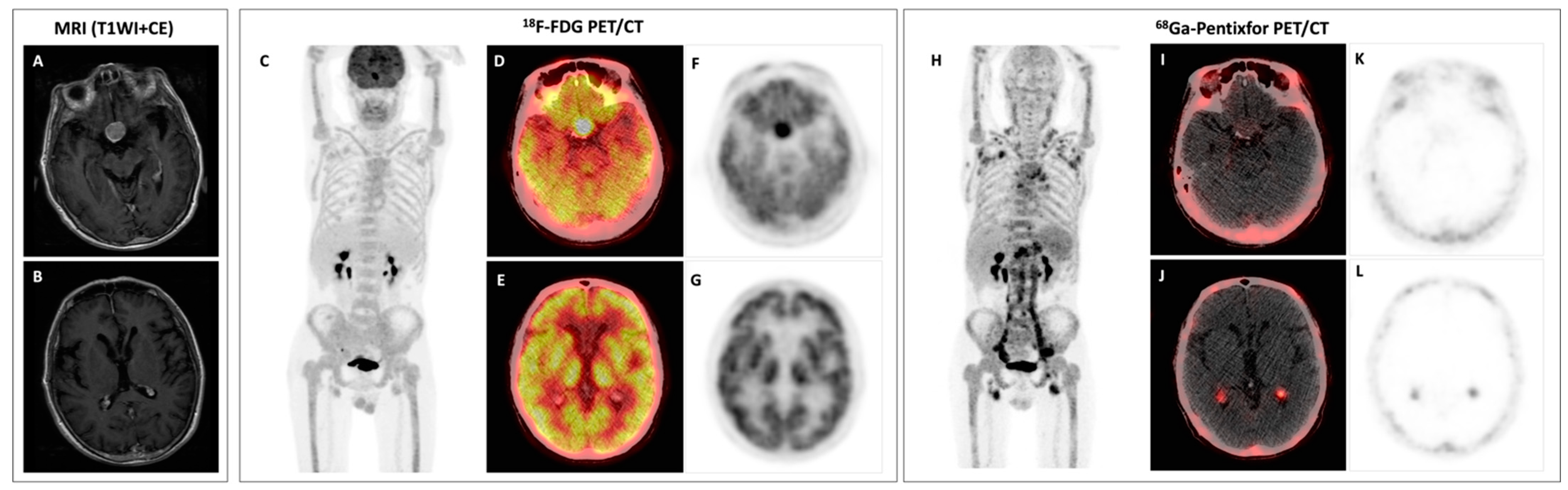
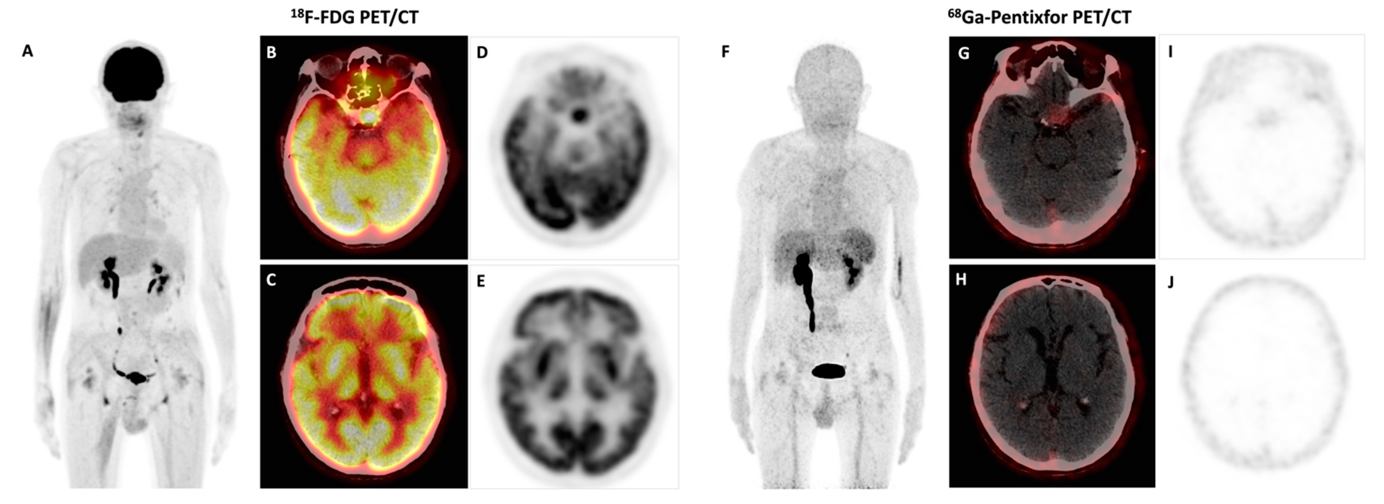
Bing–Neel Syndrome and Coexisting Pituitary Macroadenoma in a Patient with Waldenström Macroglobulinemia Revealed by 18F-FDG and 68Ga-Pentixafor PET/CT

1
Department of Nuclear Medicine, Chinese Academy of Medical Sciences and Peking Union Medical College Hospital, Beijing 100730, China
2
Beijing Key Laboratory of Molecular Targeted Diagnosis and Therapy in Nuclear Medicine, Beijing 100730, China
3
Department of Hematology, Chinese Academy of Medical Sciences and Peking Union Medical College Hospital, Beijing 100730, China
*
Author to whom correspondence should be addressed.
Diagnostics 2023, 13(7), 1334; https://doi.org/10.3390/diagnostics13071334
Submission received: 30 January 2023 / Revised: 29 March 2023 / Accepted: 29 March 2023 / Published: 3 April 2023
(This article belongs to the Special Issue Advances in PET/CT Imaging)

Abstract

:
A 63-year-old man presenting with peripheral neuropathies was diagnosed of Waldenström’s macroglobulinemia, and Bing–Neel syndrome was subsequently confirmed via cerebrospinal fluid examinations. Besides involvement in bone marrow, lymph nodes, as well as the thoracic and sacral nerve root, 68Ga-Pentixafor PET/CT detected active tracer uptake in bilateral choroid plexus, which was negative in 18F-FDG PET/CT, possibly suggesting the involvement of Bing–Neel syndrome. The coexisting pituitary macroadenoma was FDG-avid but negative in 68Ga-Pentixafor PET/CT. After six cycles of chemotherapy, the follow-up PET/CT showed complete remission of the previous disease, including the high uptake of 68Ga-Pentixafor in choroid plexus. However, the hypermetabolic pituitary macroadenoma remained unchanged.

Figure 1. Baseline evaluation, axial contrast-enhanced T1-weighted MRI (A,B), MIP image of the 18F-FDG PET (C), axial fusion 18F-FDG PET/CT (D,E), axial 18F-FDG PET (F,G), MIP image of the 68Ga-Pentixafor PET (H), axial fusion 68Ga-Pentixafor PET/CT (I,J), and axial 68Ga-Pentixafor PET (K,L). This patient (male, 63 years old) had a numbness and amyosthenia in the right foot 2 months prior, and his symptom progressively developed to the bilateral lower and upper extremities. Electromyography showed demyelination and axonal injury of sensorimotor peripheral nerves in the lower extremities. Laboratory tests showed anemia (hemoglobin 94 g/L) and elevated serum IgM (30.49 g/L; reference: 0.4–2.3 g/L). Serum protein and immunofixation electrophoresis were positive for monoclonal protein (18.5 g/L, IgM-κ). Bone marrow biopsy showed infiltration with lymphoplasmacytoid cells accounting for 10% of the cells in bone marrow. Therefore, the diagnosis of Waldenström’s macroglobulinemia (WM), a low-grade lymphoma, was established. Since the patient had a chief complaint of peripheral neuropathies, Bing–Neel syndrome (central nervous system involvement in WM) was considered, as other common causes of peripheral neuropathies in WM—paraprotein-related peripheral neuropathies, cryoglobulinemia, light chain amyloidosis—were excluded by sural nerve biopsy and negative serum cryoglobulin. The cerebrospinal fluid examinations revealed an M-spike of 0.19 g/L (IgM-κ restricted) and positive mutation of MYD88L265P (an important marker for diagnosing WM [1,2]), supporting the diagnosis of Bing–Neel syndrome. The patient then underwent a brain MRI, which was unremarkable except for a pituitary macroadenoma (A). In 18F-FDG PET/CT, the pituitary macroadenoma was hypermetabolic ((D,F), SUVmax 10.4); additionally, diffusely and homogenously increased uptake in bone marrow and involvement in lymph nodes were also depicted (C). The patient then underwent 68Ga-Pentixafor PET/CT, a chemokine receptor CXCR4-targeted imaging, which we previously reported as being superior to 18F-FDG in patients with WM [3]. MIP image of the 68Ga-Pentixafor PET showed markedly increased radioactivity throughout the axial and appendicular skeleton, as well as multiple lymph nodes in the neck, axilla, para-aortic, pelvic, and inguinal areas with increased uptake (H).Surprisingly, 68Ga-Pentixafor PET/CT showed active tracer uptake in bilateral choroid plexus ((J,L), SUVmax 4.0) that was negative with 18F-FDG (E,G) and also with normal signal intensity and contrast-enhancement in MRI (B); the FDG-avid pituitary macroadenoma was negative with 68Ga-Pentixafor (I,K). Furthermore, involvements in the thoracic nerve root and sacral nerve root were also detected.
Diagnostics 13 01334 g001
Figure 2. After six cycles of chemotherapy, the patient underwent follow-up PET/CT with both 18F-FDG and 68Ga-Pentixafor. MIP image of the 18F-FDG PET (A), axial fusion 18F-FDG PET/CT (B,C), axial 18F-FDG PET (D,E), MIP image of the 68Ga-Pentixafor PET (F), axial fusion 68Ga-Pentixafor PET/CT (G,H), and axial 68Ga-Pentixafor PET (I,J). The previous high uptake of 68Ga-Pentixafor in the choroid plexus returned to normal. There was also a complete remission of bone marrow, lymph node, and nerve roots involvement in both 68Ga-Pentixafor and 18F-FDG PET/CT. However, the manifestations of the pituitary macroadenoma remained unchanged (SUVmax 11.2 in 18F-FDG PET/CT). Bing–Neel syndrome is a rare complication involving almost 1% of WM patients [4]. About one-third of patients with Bing–Neel syndrome had symptoms of peripheral neuropathies [4,5]. Leptomeningeal infiltration was most commonly seen, followed by parenchymal and dural involvement [4,6]. Involvement in the choroid plexus is rare. MRI can be used to detect central nervous system involvement in Bing–Neel syndrome, but it lacks specificity. In a previous case report of Bing–Neel syndrome, the lesions shown in MRI were hypometabolic in 18F-FDG PET/CT [7], which was consistent with our case. Two other cases reported FDG-avid central nervous system involvement of Bing–Neel syndrome [8,9]. 18F-FDG showed pronounced physiological accumulations in the brain, which may omit the pathological findings. However, the physiologic uptake of 68Ga-Pentixafor is very low in the head, resulting in excellent tumor-to-background contrast and a higher detection rate of cerebral lesions. Thus, as there is a high level of CXCR4 expression in B-cells of WM [1,10], 68Ga-Pentixafor should be further assessed as a potential tracer for the evaluation of WM, including the involvement of the central nervous system.
Diagnostics 13 01334 g002

Author Contributions

Y.L. was responsible for the article’s conception, study design, and revisions. Q.P. was responsible for collecting data, as well as writing and submitting the manuscript. X.C., J.L. and J.F. contributed to this work via supporting the study with their knowledge of hematological and nuclear medicine. All authors have read and agreed to the published version of the manuscript.

Funding

This work was supported by National High Level Hospital Clinical Research Funding (2022-PUMCH-A-127, 2022-PUMCH-B-070, 2022-PUMCH-B-071), CAMS Initiative for Innovative Medicine (CAMS-I2M, 2017-I2M-3-001), and the Beijing Natural Science Foundation (7202160).

Institutional Review Board Statement

Not applicable.

Informed Consent Statement

Written informed consent was obtained from the patient to publish this paper.

Data Availability Statement

The data presented in this study are available on request from the corresponding author.

Conflicts of Interest

The authors report no conflict of interest.

References

  1. Hunter, Z.R.; Yang, G.; Xu, L.; Liu, X.; Castillo, J.J.; Treon, S.P. Genomics, Signaling, and Treatment of Waldenstrom Macroglobulinemia. J. Clin. Oncol. 2017, 35, 994–1001. [Google Scholar] [CrossRef] [PubMed]
  2. Mazzucchelli, M.; Frustaci, A.M.; Deodato, M.; Cairoli, R.; Tedeschi, A. Waldenstrom’s Macroglobulinemia: An Update. Mediterr. J. Hematol. Infect. Dis. 2018, 10, e2018004. [Google Scholar] [CrossRef] [PubMed]
  3. Luo, Y.; Cao, X.; Pan, Q.; Li, J.; Feng, J.; Li, F. 68Ga-Pentixafor PET/CT for Imaging of Chemokine Receptor 4 Expression in Waldenström Macroglobulinemia/Lymphoplasmacytic Lymphoma: Comparison to 18F-FDG PET/CT. J. Nucl. Med. 2019, 60, 1724–1729. [Google Scholar] [CrossRef] [PubMed]
  4. Minnema, M.C.; Kimby, E.; D’Sa, S.; Fornecker, L.-M.; Poulain, S.; Snijders, T.J.; Kastritis, E.; Kremer, S.; Fitsiori, A.; Simon, L.; et al. Guideline for the diagnosis, treatment and response criteria for Bing-Neel syndrome. Haematologica 2017, 102, 43–51. [Google Scholar] [CrossRef] [PubMed] [Green Version]
  5. Castillo, J.J.; D’Sa, S.; Lunn, M.; Minnema, M.C.; Tedeschi, A.; Lansigan, F.; Palomba, M.L.; Varettoni, M.; Garcia-Sanz, R.; Nayak, L.; et al. Central nervous system involvement by Waldenstrom macroglobulinaemia (Bing-Neel syndrome): A multi-institutional retrospective study. Br. J. Haematol. 2016, 172, 709–715. [Google Scholar] [CrossRef] [PubMed] [Green Version]
  6. Fitsiori, A.; Fornecker, L.M.; Simon, L.; Karentzos, A.; Galanaud, D.; Outteryck, O.; Vermersch, O.; Pruvo, P.; Gerardin, J.-P.; Lebrun-Frenay, E.; et al. Imaging spectrum of Bing-Neel syndrome: How can a radiologist recognise this rare neurological complication of Waldenstrom’s macroglobulinemia? Eur. Radiol. 2019, 29, 102–114. [Google Scholar] [CrossRef] [PubMed]
  7. Bund, C.; Lhermitte, B.; De Seze, J.; Kremer, S.; Namer, I.J. FDG PET and MRI Findings in a Case of Bing-Neel Syndrome. Clin. Nucl. Med. 2018, 43, 262–264. [Google Scholar] [CrossRef] [PubMed]
  8. Illarramendi, O.A.; Flynt, L.; Wong, F. 18F-FDG PET/CT in the Evaluation of Bing-Neel Syndrome. J. Nucl. Med. Technol. 2019, 47, 343–344. [Google Scholar] [CrossRef] [PubMed]
  9. Riaz, S.; Priftakis, D.; Arulogun, S.; Wan, S.; Bomanji, J. Before and After Treatment Characterization of Cerebrospinal Disease in Bing-Neel Syndrome Using 18F FDG PET/MRI. Clin. Nucl. Med. 2020, 45, 700–702. [Google Scholar] [CrossRef] [PubMed]
  10. Ngo, H.T.; Leleu, X.; Lee, J.; Jia, X.; Melhem, M.; Runnels, J.; Moreau, J.; Burwick, A.-S.; Azab, N.; Roccaro, A.K.; et al. SDF-1/CXCR4 and VLA-4 interaction regulates homing in Waldenstrom macroglobulinemia. Blood 2008, 112, 150–158. [Google Scholar] [CrossRef] [PubMed] [Green Version]
Disclaimer/Publisher’s Note: The statements, opinions and data contained in all publications are solely those of the individual author(s) and contributor(s) and not of MDPI and/or the editor(s). MDPI and/or the editor(s) disclaim responsibility for any injury to people or property resulting from any ideas, methods, instructions or products referred to in the content.

