Article

Pre-Analytical Evaluation of Streck Cell-Free DNA Blood Collection Tubes for Liquid Profiling in Oncology

1
Research and Development, Sysmex Inostics GmbH, 20251 Hamburg, Germany
2
Department of Hematology and Oncology, University Hospital, 53127 Bonn, Germany
3
Institute of Clinical Chemistry and Clinical Pharmacology, University Hospital, 53127 Bonn, Germany
4
Department of Pneumology, University Hospital, 53127 Bonn, Germany
5
German Heart Centre Munich, Institute for Laboratory Medicine, Technical University, 80636 Munich, Germany
*
Authors to whom correspondence should be addressed.
These authors contributed equally to this work.
Diagnostics 2023, 13(7), 1288; https://doi.org/10.3390/diagnostics13071288
Submission received: 1 March 2023 / Revised: 27 March 2023 / Accepted: 27 March 2023 / Published: 29 March 2023

Abstract

:
Excellent pre-analytical stability is an essential precondition for reliable molecular profiling of circulating tumor DNA (ctDNA) in oncological diagnostics. Therefore, in vitro degradation of ctDNA and the additional release of contaminating genomic DNA from lysed blood cells must be prevented. Streck Cell-Free DNA blood collection tubes (cfDNA BCTs) have proposed advantages over standard K2EDTA tubes, but mainly have been tested in healthy individuals. Blood was collected from cancer patients (n = 53) suffering from colorectal (n = 21), pancreatic (n = 11), and non-small-cell lung cancer (n = 21) using cfDNA BCT tubes and K2EDTA tubes that were processed immediately or after 3 days (BCTs) or 6 hours (K2EDTA) at room temperature. The cfDNA isolated from these samples was characterized in terms of yield using LINE-1 qPCR; the level of gDNA contamination; and the mutation status of KRAS, NRAS, and EGFR genes using BEAMing ddPCR. CfDNA yield and gDNA levels were comparable in both tube types and were not affected by prolonged storage of blood samples for at least 3 days in cfDNA BCTs or 6 hours in K2EDTA tubes. In addition, biospecimens collected in K2EDTA tubes and cfDNA BCTs stored for up to 3 days demonstrated highly comparable levels of mutational load across all respective cancer patient cohorts and a wide range of concentrations. Our data support the applicability of clinical oncology specimens collected and stored in cfDNA BCTs for up to 3 days for reliable cfDNA and mutation analyses.

1. Introduction

Continuous improvement of sensitive technologies for the detection and analysis of aberrations in circulating tumor DNA (ctDNA) has paved the way for clinical liquid biopsy applications [1,2]. Because ctDNA is only a marginal amount among a majority of wild-type circulating cell-free DNA (cfDNA), these applications require highly sensitive methods such as barcoded next-generation sequencing or digital PCR approaches [3,4]. Whereas the focus lies on technology development to detect very low mutant allele frequencies (MAFs) as one mutant copy per 10,000 wild-type DNA copies, the influence of pre-analytical steps on the sensitivity of detecting cfDNA remains a major issue [5,6,7,8]. However, this should be given more attention because it can directly affect the sensitivity of ctDNA detection because the release of wild-type DNA due to leukocyte lysis results in the dilution of ctDNA. Pre-analytical steps that can influence sample quality are the blood draw procedure itself (needle diameters and tourniquet), blood collection (tube type, fill volume, and mixing), shipment and storage of the biospecimens, as well as the plasma processing and DNA extraction. However, the most critical step is the storage time between blood sampling and processing. With standard EDTA blood collection tubes, the processing of plasma needs to be conducted within less than 4 h to prevent the release of genomic DNA due to cell lysis [8]. This is not always possible in oncological practices due to schedule conflicts or the lack of equipment such as high-speed centrifuges. Thus, blood samples need to be shipped to a testing laboratory, which requires specialized blood collection systems. Blood collection tubes containing a cell-stabilizing and nuclease inhibiting cocktail such as Cell-Free DNA BCT tubes (cfDNA BCTs) by Streck (La Vista, NE, USA) are suitable for shipping, as they are designed for up to 14 days whole blood storage at temperatures between 6 and 37 °C. The advantage of cfDNA BCTs over other cell stabilizing tubes on the market is the broad operating temperature range [8,9,10]. This enables an economical shipping box design, whereas users of blood collection tubes with temperature ranges between 18 and 25 °C need controlled room temperature (CRT) shipping solutions, e.g., phase exchange material, which increases shipping costs. Another advantage of the Cell-Free DNA BCT tubes (Streck tubes) is that they are known to be able to successfully maintain stable cfDNA levels at RT over time. Several studies have confirmed that RT storage prior to processing can be extended to 3 days [11], 4 days [9,10], 7 days [12,13,14,15], and even up to 14 days (in the case of fetal cfDNA levels) [16].
Aside from the wide application of cfDNA BCTs for noninvasive prenatal testing (NIPT) [17], only a few studies have reported on the utility of cfDNA BCTs for oncological applications. Promising results were already shown for colorectal cancer (CRC) [4], breast cancer [18,19,20], melanoma [21], and non-small-cell lung cancer (NSCLC) [22]. However, for all these studies, different pre-analytical conditions were chosen. The lack of harmonized pre-analytical standards [6,17,22,23,24,25,26] leads to deviating performances of cfDNA applications, which could be hindering reliable clinical data evaluation. Therefore, well defined and consolidated pre-analytical standards will ensure high-quality cfDNA samples for further downstream processes. Here, we propose a common consensus of pre-analytical steps using cfDNA BCTs for ctDNA analysis in different cancer types.

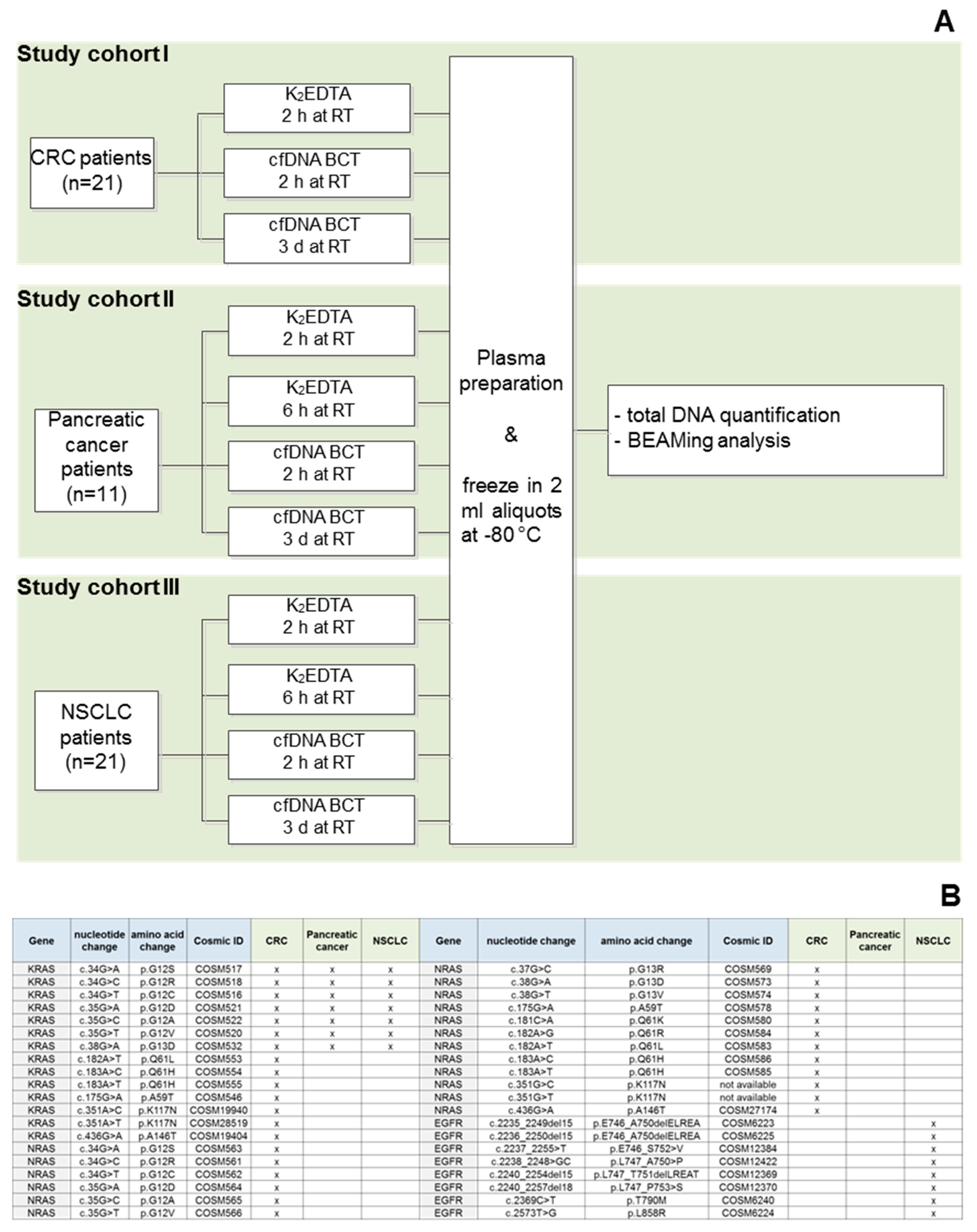
2. Materials and Methods

2.1. Ethics Statement

Samples for this study were either collected commercially from Indivumed GmbH (Hamburg, Germany), or in collaboration with University Hospital, Bonn. Indivumed GmbH obtained approval from the institutional review board of the Physicians Association of Hamburg, Germany (PV2963). The University of Bonn obtained approval from the Ethics Committee of the Faculty of Medicine of the University of Bonn. Written consent was obtained from all study subjects by the sample providers. All clinical data and samples were received by Sysmex Inostics GmbH anonymously.

2.2. Blood Collection

Venous blood from patients with CRC stage II–IV (n = 21) (study cohort I), advanced pancreatic cancer (n = 11) (study cohort II), and advanced stage NSCLC (n = 21) (study cohort III) was collected using standard phlebotomy techniques in both BD Vacutainer K2EDTA tubes (Becton Dickinson, Franklin Lakes, NJ; referred to as K2EDTA tubes) and Cell-Free I BCT tubes (Streck, La Vista, NJ, USA; referred to as cfDNA BCTs). Samples from cohort I were also used in a previous study [4], and were included in this study for a higher (n) as well as to show that the cfDNA BCTs showed comparable performance for various cancer types (i.e., CRC, NSCLC, and pancreatic cancer).
All blood collection tubes were filled to 10 mL as recommended by both manufacturers. Tubes were then inverted 10 times before room temperature (RT) transportation to the laboratory. Storage and processing conditions were performed according to the scheme in Figure 1.

2.3. Blood Storage

As shown in Figure 1A, blood tubes were stored for either 2 h (K2EDTA and cfDNA BCT) or 3 days (cfDNA BCT) before processing. In order to evaluate the nuclease activity, an additional K2EDTA tube was stored for 6 h before plasma preparation in study cohorts II and III.

2.4. Plasma Preparation

Following the indicated storage times, tubes were centrifuged at 1600× g for 10 min at RT using a swing-out rotor. To prevent contamination of the plasma with cells, a smooth braking profile was used, and the tubes were carefully removed. Without disrupting the buffy coat layer, the plasma fraction was transferred to a fresh 15 mL tube, leaving approximately 500 µL plasma in the blood collection tube. The supernatant was centrifuged a second time at 6000× g for 10 min at RT using a swing-out rotor and a smooth braking profile. The supernatant was again transferred to a fresh tube, leaving approximately 300 µL above the cell pellet, gently mixed by pipetting and aliquoted in 2.1 mL cryotubes. Plasma aliquots were frozen at −80 °C.

2.5. Cell-Free DNA Extraction

CfDNA from 2 mL plasma was extracted using the QIAamp Circulating Nucleic Acid Kit (Qiagen, Hilden, Germany). Differing from the manufacturer’s instructions, the proteinase K incubation time at 60 °C was extended from 30 to 60 min for both the cfDNA BCT and K2EDTA samples. This is recommended by Streck for plasma samples where blood was collected in cfDNA BCTs; to guarantee the comparability, this method was also applied to K2EDTA-collected samples. CfDNA was eluted in 140 µL AVE buffer and stored at 4 °C until qPCR and BEAMing analysis.

2.6. Cell-Free DNA Quantification

DNA was quantified using a qPCR assay specific for the LINE-1 sequence exactly as described in earlier publications [4,27]. Briefly, a short LINE-1 amplicon (96 bp) was used as a target for quantifying cfDNA fragments that exhibit a modal range of ~166 bp [28], while a larger LINE-1 amplicon (402 bp) was used as a target to quantify contaminating genomic DNA originating from white blood cells (WBCs) [29]. Because the short amplicon was selected from the sequence of the long amplicon, the ratio between the measured amounts of these amplicons (i.e., 402:96 bp ratio) served as a surrogate marker of genomic DNA contamination and cfDNA sample quality. Therefore, a high value indicates dilution of cfDNA with wild-type genomic DNA derived from peripheral blood cells lysed during storage or blood processing, while a low value indicates good cfDNA quality.

2.7. Mutation Analysis of cfDNA

CfDNA from CRC, pancreatic cancer, and NSCLC patients (n = 53), which was isolated from blood samples collected in both K2EDTA tubes and cfDNA BCTs, were analyzed for mutational load using BEAMing technology [30]. The BEAMing procedure was carried out as described previously [4]. As shown in Figure 1B, various common alterations in the KRAS, NRAS, and EGFR genes were tested for in the respective cohorts.

2.8. Statistical Analysis
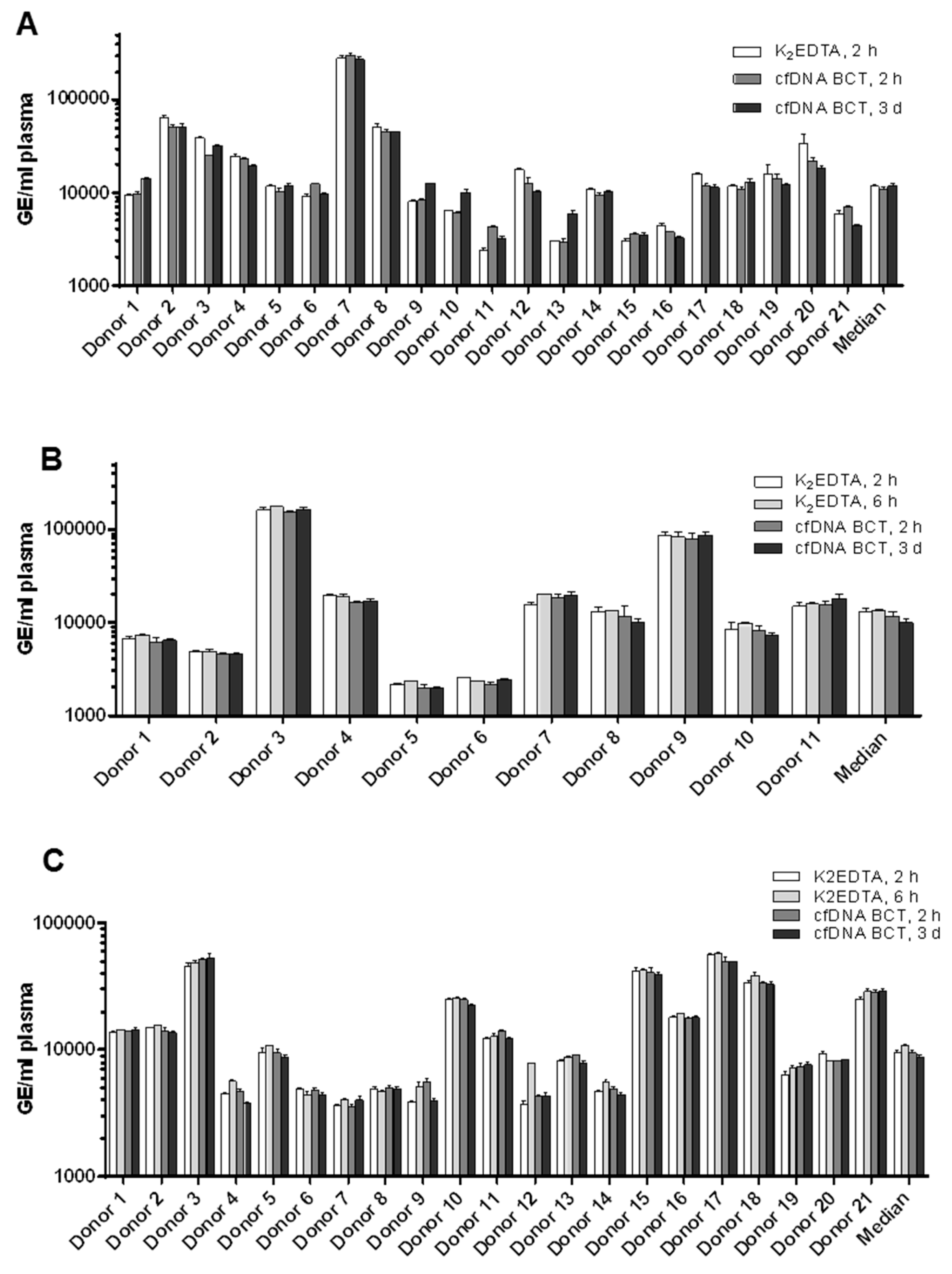
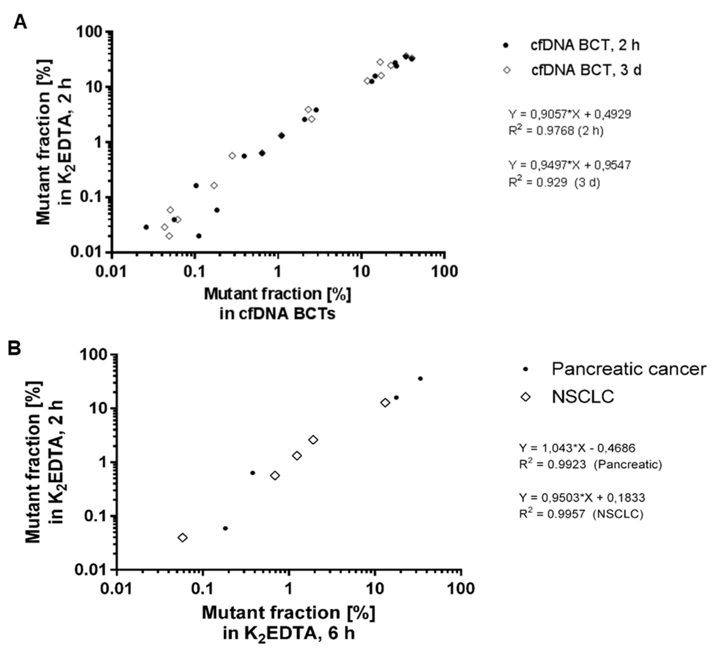
Statistical analysis using one-way ANOVA and linear regression with R2 calculation was performed in GraphPad Prism 6.07 (GraphPad Software, Inc., La Jolla, CA, USA). The p-values < 0.05 were considered to be statistically significant. Graphs were generated in GraphPad Prism.

3. Results

As shown in Figure 1A, plasma was prepared after 2 h, 6 h, or 3 days storage at RT (18–22 °C) in K2EDTA tubes and cfDNA BCTs. For the analysis of cfDNA yield, a 96 bp multicopy LINE-1 fragment was amplified with qPCR. Figure 2 shows the cfDNA yields assessed for CRC patients (A), pancreatic cancer patients (B), and NSCLC patients (C). No significant differences were detected for cfDNA yields in the three study cohorts for samples collected in K2EDTA vs. cfDNA BCTs for all tested storage conditions. This shows that cfDNA BCTs were able to stabilize cfDNA in clinical samples for 3 days at RT. In addition, the pancreatic cancer samples, which were tested for higher nuclease activity, showed no significant change in yields between the samples stored for 2 and 6 h in K2EDTA tubes. Previous studies have also found that cfDNA BCT tubes were able to deliver stable yields of cfDNA after 2 [20], 4 [31,32], and 7 [33] days of storage at RT.
To evaluate the effects of RT storage (18–22 °C) for up to 3 days in cfDNA BCTs, a ratio of a long (402 bp) and a short (96 bp) LINE-1 fragment was amplified and calculated as a gDNA/cfDNA ratio. According to Dunnett’s multiple comparisons test, no significant difference between long (402 bp) and short (96 bp) LINE-1 fragments was identified for blood stored for up to 3 days at RT in Streck cfDNA BCTs for (A) CRC patient samples, (B) pancreatic patient samples, or (C) NSCLC patient samples (p > 0.05) (Figure 3). We also did not identify significant differences between long and short LINE-1 fragments when blood was stored in BD K2EDTA for 2 h vs. 6 h for either pancreatic cancer or NSCLC samples (results not shown). Increased ratios compared with the K2EDTA reference values would have been indicative of a gDNA release or cfDNA decrease. Thus, qPCR results indicated cfDNA and WBC stability, thereby proving the ability of cfDNA BCTs to prevent gDNA contamination in clinical samples for up to 3 days’ storage at RT.
Blood samples from CRC, pancreatic cancer, and NSCLC patients (n = 53) were collected in K2EDTA tubes and cfDNA BCTs, and samples were analyzed for mutational load using BEAMing after preprocessing storage at RT (18–22 °C) for 2 h, 6 h, or 3 days (mutation analysis results are summarized in Table 1). Mutant fractions of all detectable mutation sets were plotted for cfDNA BCT conditions versus matched K2EDTA values as a reference. A total of 16 mutations were found in 15 of 53 samples (28.3%), with one patient harboring a mutation in two genes. Mutant allele frequencies in cfDNA ranged from 0.026% to 41.6% and showed very comparable results between the tube conditions even for low-positive samples (Figure 4A). Therefore, storage of clinical blood samples in cfDNA BCTs for up to 3 days did not impair the detectability of low-level mutations. These results were in accordance with previously published results, where cfDNA BCT tubes were found to successfully preserve samples for the detection of gene mutations in ctDNA in the plasma of CRC and NSCLC patients after storage at RT of 3 days [4,22] and up to 10 days for melanoma patients [21]. Additionally, blood samples collected from NSCLC (n = 21) and pancreatic cancer (n = 11) patients in K2EDTA tubes, and processed after 2 h and 6 h following phlebotomy, were analyzed for mutational load using BEAMing. Interestingly, samples stored for either 2 h or 6 h in K2EDTA tubes yielded comparable mutation testing results (Figure 4B). This suggests that the K2EDTA tubes are useful for mutation analysis of cfDNA from NSCLC and pancreatic cancer patient samples after up to 6 h of storage at RT (Figure 4B).

4. Discussion

Whether ctDNA is characterized for the purposes of basic research or clinical diagnostics, high-quality biospecimens are crucial for accurate and reliable measurements. One of the first steps that can be taken to ensure good sample quality is to select blood collection procedures that preserve the integrity of the ctDNA and prevent the disruption of peripheral blood cells, thereby limiting the release of wild-type gDNA and the subsequent dilution of the target molecules. Because the traditionally used K2EDTA tubes are not able to maintain the quality of ctDNA in blood samples when stored for extended periods prior to processing [10,14,16,19,22,31,32,34], we have in this study evaluated Streck cfDNA BCTs as an alternative method for blood storage. Proper storage conditions for longer time periods are crucial, because blood samples are often collected offsite and need to be shipped to the testing laboratory, which may be situated at a distant location.
In order to evaluate the efficacy of cfDNA BCTs in the context of clinical oncology, we characterized the cfDNA in plasma derived from blood collected from colorectal (n = 21), pancreatic (n = 11), and non-small-cell lung cancer patients (n = 21) in cfDNA BCTs. For each of the cohorts, blood was processed either immediately or 3 days following collection. Results from the cfDNA BCTs were compared with those from matching specimens collected in K2EDTA tubes. In addition, to test the effects of nuclease activity in a slight processing delay, we compared cfDNA measurements from blood samples collected from pancreatic cancer and NSCLC patients stored for 2 h vs. 6 h in K2EDTA tubes. CfDNA isolated from each of the above samples was characterized in terms of yield; level of gDNA contamination; and mutation status of KRAS, NRAS, and EGFR genes, as determined using BEAMing technology.
Results show that (i) cfDNA BCTs stabilize ctDNA for at least 3 days at RT in clinical samples (Figure 2), (ii) cfDNA BCTs prevent gDNA contamination for at least 3 days at RT in clinical samples (Figure 3), (iii) storage of blood in cfDNA BCTs for up to 3 days does not impair detectability of low-level mutations in any of the cancer patient cohorts (Figure 4A), and (iv) blood samples from pancreatic cancer and NSCLC patients stored for 2 h vs. 6 h in K2EDTA tubes yield comparable mutation testing results (Figure 4B). Taken together, the resulting data not only show the advantages of cfDNA BCTs over standard K2EDTA tubes pertaining to extended storage times, but also demonstrate the general suitability of cfDNA BCTs for specimen collection in clinical oncology. This is because cfDNA BCTs can be stored for extended time periods and can be shipped at room temperature, while at the same time conserving ctDNA sequence integrity and preventing gDNA contamination by stabilizing peripheral blood cells, which facilitates the detection of low-abundance mutations in cancer patients with various tumor types. These results are corroborated by several recent studies that have also demonstrated the utility of cfDNA BCTs [9,10,15,16,19,20,22,35]. However, some important caveats merit discussion here. First, as demonstrated previously, it is important that shipping temperatures do not fluctuate beyond the ranges recommended by the manufacturers, as only slight variations can result in a significant loss of peripheral blood cell stability and gDNA contamination [4,15]. As such, temperature-controlled shipping is required. Second, the diagnostic sensitivity and specificity of ctDNA have been shown to be increased by parallel assessment of other biomarkers and other genomic features of ctDNA [36,37,38]; however, cfDNA BCTs were not designed, optimized, and tested for the analysis of auxiliary biomarkers such as DNA methylation patterns [39], proteins, or cell-free RNA [40]. Last, in order to reverse the chemical fixations in these tubes, the DNA extraction procedure requires an extended heating step [10]. As such, ctDNA extraction methods that do not include or cannot accommodate an extended heating step may not be compatible with cfDNA BCTs. Despite these disadvantages, cfDNA BCTs significantly simplify and enhance the liquid-profiling-based mutational profiling of cancer patients, which has in the last decade proven extremely useful at various stages of the disease [41].
It is also important to note that cfDNA BCT tubes have been compared to other non-K2EDTA tubes, such as: (a) PAXgene tubes (PreAnalytiX GmbH, Hombrechtikon, Switzerland), which show similar efficacy for preserving cfDNA yield and integrity over extended storage times [32]; (b) Cell-free DNA collection tubes (Roche Diagnostics GmbH, Mannheim, Germany), which show performance similar to cfDNA BCTs in preserving cfDNA yield and integrity for up to 7 days of storage, after which samples stored in Roche cell-free DNA collection tubes demonstrated signs of gDNA contamination [35] (Another study showed that that PAXgene Blood ccfDNA Tubes (Qiagen), Cell-free DNA collection tubes (Roche), and cfDNA BCTs (Streck) performed similarly for the detection of EGFR T790M mutations with artificial spiked-in DNA fragments [42] after 7 days of storage.); (c) CellSave Preservative Tubes (Menarini Silicon Biosystems, Inc., Bryn Athyn, PA, USA), which were reported to perform similarly to cfDNA BCTs in their ability to stabilize both ctDNA and wild-type DNA after 48 [20] and 96 h [31] at RT.

5. Conclusions

The current pre-analytic study provides a firm basis for the reliable assessment of ctDNA yield as well as quantitative mutational analysis of the common genes KRAS, NRAS, and EGFR on plasma ctDNA even in very low concentration ranges. Stability in cfDNA BCT tubes was shown for at least 3 days and in conventional K2EDTA tubes for at least 6 h at RT, demonstrating excellent pre-analytical preconditions for future applications in routine oncology diagnostics.

Author Contributions

Conceptualization, I.M.D., F.D. and F.H.; sample collection: S.A.E.H., M.K., D.S. and S.H.; pre-analytical investigation: M.K. and S.H.; methodology and analysis, I.M.D., A.N. and F.H.; validation, I.M.D., A.N. and F.H.; statistical analysis, I.M.D., F.D. and F.H.; resources, F.D. and F.H.; writing—original draft preparation, I.M.D., A.J.B. and V.U.; writing—review and editing, I.M.D., A.J.B., V.U., S.H. and F.H.; visualization, I.M.D., A.N., J.F. and F.H.; supervision, F.D. and F.H.; project administration, S.H. and F.H. All authors have read and agreed to the published version of the manuscript.

Funding

The reagents and analyses of the study were provided by Sysmex Inostics GmbH.

Institutional Review Board Statement

The study was conducted in accordance with the Declaration of Helsinki. Indivumed GmbH obtained approval from the institutional review board of the Physicians Association of Hamburg, Germany (PV2963). The University of Bonn obtained approval from the Ethics Committee of the Faculty of Medicine of the University of Bonn.

Informed Consent Statement

Informed consent for sample collection was obtained from all subjects involved in the study.

Data Availability Statement

The data presented in this study are available in the article.

Acknowledgments

The authors would like to thank the team of Stefan Holdenrieder from the Institute for Clinical Chemistry and Pharmacology, University Hospital Bonn, for their contribution during pancreatic and NSCLC sample collection and processing, Claudia Stamm and the Service Laboratory team of Sysmex Inostics for their contribution during CRC sample analysis, as well as the R&D and Production teams of Sysmex Inostics for their support during execution of the study. Abel Bronkhorst would like to thank the Georg Forster Research Fellowship Program of the Alexander von Humboldt Foundation for financial assistance.

Conflicts of Interest

I.M.D., A.N., F.D. and F.H. are employees of Sysmex Inostics GmbH. S.H. has received research funding and honoraria from Sysmex Inostics GmbH.

References

  1. Wan, J.C.M.; Massie, C.; Garcia-Corbacho, J.; James, D.; Caldas, C.; Pacey, S.; Baird, R.D.; Rosenfeld, N. Liquid biopsies come of age: Clinical applications of circulating tumour DNA. Nat. Rev. Cancer 2016, 17, 223–238. [Google Scholar] [CrossRef] [PubMed]
  2. Bronkhorst, A.J.; Ungerer, V.; Oberhofer, A.; Gabriel, S.; Polatoglou, E.; Randeu, H.; Uhlig, C.; Pfister, H.; Mayer, Z.; Holdenrieder, S. New Perspectives on the Importance of Cell-Free DNA Biology. Diagnostics 2022, 12, 2147. [Google Scholar] [CrossRef] [PubMed]
  3. Tu, M.; Chia, D.; Wei, F.; Wong, D. Liquid biopsy for detection of actionable oncogenic mutations in human cancers and electric field induced release and measurement liquid biopsy (eLB). Analyst 2016, 141, 393–402. [Google Scholar] [CrossRef] [PubMed]
  4. Medina Diaz, I.; Nocon, A.; Mehnert, D.H.; Fredebohm, J.; Diehl, F.; Holtrup, F. Performance of Streck cfDNA blood collection tubes for liquid biopsy testing. PLoS ONE 2016, 11, e0166354. [Google Scholar] [CrossRef] [PubMed]
  5. Tsui, D.W.; Barnett, E.; Scher, H.I. Toward Standardization of Preanalytical Procedures for Cell-Free DNA Profiling. Clin. Chem. 2019, 66, 3–5. [Google Scholar] [CrossRef]
  6. Ungerer, V.; Bronkhorst, A.J.; Holdenrieder, S. Preanalytical variables that affect the outcome of cell-free DNA measurements. Crit. Rev. Clin. Lab. Sci. 2020, 57, 484–507. [Google Scholar] [CrossRef] [PubMed]
  7. Lampignano, R.; Neumann, M.H.D.; Weber, S.; Kloten, V.; Herdean, A.; Voss, T.; Groelz, D.; Babayan, A.; Tibbesma, M.; Schlumpberger, M.; et al. Multicenter Evaluation of Circulating Cell-Free DNA Extraction and Downstream Analyses for the Development of Standardized (Pre)analytical Work Flows. Clin. Chem. 2020, 66, 149–160. [Google Scholar] [CrossRef]
  8. Meddeb, R.; Pisareva, E.; Thierry, A.R. Guidelines for the Preanalytical Conditions for Analyzing Circulating Cell-Free DNA. Clin. Chem. 2019, 65, 623–633. [Google Scholar] [CrossRef]
  9. Norton, S.E.; Luna, K.K.; Lechner, J.M.; Qin, J.; Fernando, M.R. A new blood collection device minimizes cellular DNA release during sample storage and shipping when compared to a standard device. J. Clin. Lab. Anal. 2013, 27, 305–311. [Google Scholar] [CrossRef]
  10. Norton, S.E.; Lechner, J.M.; Williams, T.; Fernando, M.R. A stabilizing reagent prevents cell-free DNA contamination by cellular DNA in plasma during blood sample storage and shipping as determined by digital PCR. Clin. Biochem. 2013, 46, 1561–1565. [Google Scholar] [CrossRef]
  11. Barrett, A.N.; Zimmermann, B.G.; Wang, D.; Holloway, A.; Chitty, L.S. Implementing prenatal diagnosis based on cell-free fetal DNA: Accurate identification of factors affecting fetal DNA yield. PLoS ONE 2011, 6, 4–11. [Google Scholar] [CrossRef] [PubMed]
  12. Jeong, T.-D.; Kim, M.H.; Park, S.; Chung, H.-S.; Lee, J.W.; Chang, J.H.; Huh, J. Effects of Pre-analytical Variables on Cell-free DNA Extraction for Liquid Biopsy. Lab. Med. Online 2019, 9, 45–56. [Google Scholar] [CrossRef]
  13. Risberg, B.; Tsui, D.W.Y.; Biggs, H.; Ruiz-Valdepenas Martin de Almagro, A.; Dawson, S.J.; Hodgkin, C.; Jones, L.; Parkinson, C.; Piskorz, A.; Marass, F.; et al. Effects of Collection and Processing Procedures on Plasma Circulating Cell-Free DNA from Cancer Patients. J. Mol. Diagn. 2018, 20, 883–892. [Google Scholar] [CrossRef]
  14. Parpart-Li, S.; Bartlett, B.; Popoli, M.; Adleff, V.; Tucker, L.; Steinberg, R.; Georgiadis, A.; Phallen, J.; Brahmer, J.; Azad, N.; et al. The effect of preservative and temperature on the analysis of circulating tumor DNA. Clin. Cancer Res. 2017, 23, 2471–2477. [Google Scholar] [CrossRef] [PubMed]
  15. Wong, D.; Moturi, S.; Angkachatchai, V.; Mueller, R.; DeSantis, G.; van den Boom, D.; Ehrich, M. Optimizing blood collection, transport and storage conditions for cell free DNA increases access to prenatal testing. Clin. Biochem. 2013, 46, 1099–1104. [Google Scholar] [CrossRef]
  16. Fernando, M.R.; Chen, K.; Norton, S.; Krzyzanowski, G.; Bourne, D.; Hunsley, B.; Ryan, W.L.; Bassett, C. A new methodology to preserve the original proportion and integrity of cell-free fetal DNA in maternal plasma during sample processing and storage. Prenat. Diagn. Publ. Affil. Int. Soc. Prenat. Diagn. 2010, 30, 418–424. [Google Scholar] [CrossRef] [PubMed]
  17. El Messaoudi, S.; Rolet, F.; Mouliere, F.; Thierry, A.R. Circulating cell free DNA: Preanalytical considerations. Clin. Chim. Acta. 2013, 424, 222–230. [Google Scholar] [CrossRef] [PubMed]
  18. Hrebien, S.; O’Leary, B.; Beaney, M.; Schiavon, G.; Fribbens, C.; Bhambra, A.; Johnson, R.; Garcia-Murillas, I.; Turner, N. Reproducibility of digital PCR assays for circulating tumor DNA analysis in advanced breast cancer. PLoS ONE 2016, 11, e0165023. [Google Scholar] [CrossRef]
  19. Toro, P.V.; Erlanger, B.; Beaver, J.A.; Cochran, R.L.; VanDenBerg, D.A.; Yakim, E.; Cravero, K.; Chu, D.; Zabransky, D.J.; Wong, H.Y.; et al. Comparison of cell stabilizing blood collection tubes for circulating plasma tumor DNA. Clin. Biochem. 2015, 48, 993–998. [Google Scholar] [CrossRef]
  20. Kang, Q.; Henry, N.L.; Paoletti, C.; Jiang, H.; Vats, P.; Chinnaiyan, A.M.; Hayes, D.F.; Merajver, S.D.; Rae, J.M.; Tewari, M. Comparative analysis of circulating tumor DNA stability In K3EDTA, Streck, and CellSave blood collection tubes. Clin. Biochem. 2016, 49, 1354–1360. [Google Scholar] [CrossRef]
  21. Denis, M.G.; Knol, A.-C.; Théoleyre, S.; Vallée, A.; Dréno, B. Efficient detection of BRAF mutation in plasma of patients after long-term storage of blood in cell-free DNA blood collection tubes. Clin. Chem. 2015, 61, 886–888. [Google Scholar] [CrossRef] [PubMed]
  22. Sherwood, J.L.; Corcoran, C.; Brown, H.; Sharpe, A.D.; Musilova, M.; Kohlmann, A. Optimised pre-analytical methods improve KRAS mutation detection in circulating tumour DNA (ctDNA) from patients with non-small cell lung cancer (NSCLC). PLoS ONE 2016, 11, e0160197. [Google Scholar] [CrossRef] [PubMed]
  23. Malentacchi, F.; Pizzamiglio, S.; Verderio, P.; Pazzagli, M.; Orlando, C.; Ciniselli, C.M.; Gunther, K.; Gelmini, S. Influence of storage conditions and extraction methods on the quantity and quality of circulating cell-free DNA (ccfDNA): The SPIDIA-DNAplas External Quality Assessment experience. Clin. Chem. Lab. Med. 2015, 53, 1935–1942. [Google Scholar] [CrossRef] [PubMed]
  24. Fleischhacker, M.; Schmidt, B.; Weickmann, S.; Fersching, D.M.I.; Leszinski, G.S.; Siegele, B.; Stötzer, O.J.; Nagel, D.; Holdenrieder, S. Methods for isolation of cell-free plasma DNA strongly affect DNA yield. Clin. Chim. Acta 2011, 412, 2085–2088. [Google Scholar] [CrossRef] [PubMed]
  25. Bronkhorst, A.J.; Aucamp, J.; Pretorius, P.J. Cell-free DNA: Preanalytical variables. Clin. Chim. Acta 2015, 450, 243–253. [Google Scholar] [CrossRef]
  26. Greytak, S.R.; Engel, K.B.; Parpart-Li, S.; Murtaza, M.; Bronkhorst, A.J.; Pertile, M.D.; Moore, H.M. Harmonizing cell-free DNA collection and processing practices through evidence-based guidance. Clin. Cancer Res. 2020, 26, 3104–3109. [Google Scholar] [CrossRef]
  27. Rago, C.; Huso, D.L.; Diehl, F.; Karim, B.; Liu, G.; Papadopoulos, N.; Samuels, Y.; Velculescu, V.E.; Vogelstein, B.; Kinzler, K.W.; et al. Serial assessment of human tumor burdens in mice by the analysis of circulating DNA. Cancer Res. 2007, 67, 9364–9370. [Google Scholar] [CrossRef]
  28. Jiang, P.; Lo, Y.M.D. The Long and Short of Circulating Cell-Free DNA and the Ins and Outs of Molecular Diagnostics. Trends Genet. 2016, 32, 360–371. [Google Scholar] [CrossRef]
  29. Mouliere, F.; Robert, B.; Arnau Peyrotte, E.; Del Rio, M.; Ychou, M.; Molina, F.; Gongora, C.; Thierry, A.R. High fragmentation characterizes tumour-derived circulating DNA. PLoS ONE 2011, 6, e23418. [Google Scholar] [CrossRef]
  30. Diehl, F.; Li, M.; He, Y.; Kinzler, K.W.; Vogelstein, B.; Dressman, D. BEAMing: Single-molecule PCR on microparticles in water-in-oil emulsions. Nat. Methods 2006, 3, 551–559. [Google Scholar] [CrossRef]
  31. Van Dessel, L.F.; Beije, N.; Helmijr, J.C.A.; Vitale, S.R.; Kraan, J.; Look, M.P.; de Wit, R.; Sleijfer, S.; Jansen, M.P.H.M.; Martens, J.W.M.; et al. Application of circulating tumor DNA in prospective clinical oncology trials—Standardization of preanalytical conditions. Mol. Oncol. 2017, 11, 295–304. [Google Scholar] [CrossRef] [PubMed]
  32. Warton, K.; Yuwono, N.L.; Cowley, M.J.; McCabe, M.J.; So, A.; Ford, C.E. Evaluation of Streck BCT and PAXgene Stabilised Blood Collection Tubes for Cell-Free Circulating DNA Studies in Plasma. Mol. Diagn. Ther. 2017, 21, 563–570. [Google Scholar] [CrossRef] [PubMed]
  33. Schmidt, B.; Reinicke, D.; Reindl, I.; Bork, I.; Wollschläger, B.; Lambrecht, N.; Fleischhacker, M. Liquid biopsy—Performance of the PAXgene® Blood ccfDNA Tubes for the isolation and characterization of cell-free plasma DNA from tumor patients. Clin. Chim. Acta 2017, 469, 94–98. [Google Scholar] [CrossRef]
  34. Rothwell, D.G.; Smith, N.; Morris, D.; Leong, H.S.; Li, Y.; Hollebecque, A.; Ayub, M.; Carter, L.; Antonello, J.; Franklin, L.; et al. Genetic profiling of tumours using both circulating free DNA and circulating tumour cells isolated from the same preserved whole blood sample. Mol. Oncol. 2016, 10, 566–574. [Google Scholar] [CrossRef]
  35. Parackal, S.; Zou, D.; Day, R.; Black, M.; Guilford, P. Comparison of Roche Cell-Free DNA collection Tubes® to Streck Cell-Free DNA BCT®s for sample stability using healthy volunteers. Pract. Lab. Med. 2019, 16, e00125. [Google Scholar] [CrossRef] [PubMed]
  36. Bronkhorst, A.J.; Ungerer, V.; Holdenrieder, S. The emerging role of cell-free DNA as a molecular marker for cancer management. Biomol. Detect. Quantif. 2019, 17, 100087. [Google Scholar] [CrossRef]
  37. Van der Pol, Y.; Mouliere, F. Toward the Early Detection of Cancer by Decoding the Epigenetic and Environmental Fingerprints of Cell-Free DNA. Cancer Cell 2019, 36, 350–368. [Google Scholar] [CrossRef]
  38. Oberhofer, A.; Bronkhorst, A.J.; Uhlig, C.; Ungerer, V.; Holdenrieder, S. Tracing the Origin of Cell-Free DNA Molecules through Tissue-Specific Epigenetic Signatures. Diagnostics 2022, 12, 1834. [Google Scholar] [CrossRef]
  39. Distler, J.; Tetzner, R.; Weiss, G.; Knig, T.; Schlegel, A.; Bagrowski, M. Evaluation of Different Blood Collection Tubes and Blood Storage Conditions for the Preservation and Stability of Cell-Free Circulating DNA for the Analysis of the Methylated mSEPT9 Colorectal Cancer Screening Marker. In Circulating Nucleic Acids in Serum and Plasma–CNAPS IX; Springer International Publishing: New York, NY, USA, 2016; pp. 175–178. [Google Scholar]
  40. Gahlawat, A.W.; Lenhardt, J.; Witte, T.; Keitel, D.; Kaufhold, A.; Maass, K.K.; Pajtler, K.W.; Sohn, C.; Schott, S. Evaluation of storage tubes for combined analysis of circulating nucleic acids in liquid biopsies. Int. J. Mol. Sci. 2019, 20, 704. [Google Scholar] [CrossRef]
  41. Heitzer, E.; Haque, I.S.; Roberts, C.E.S.; Speicher, M.R. Current and future perspectives of liquid biopsies in genomics-driven oncology. Nat. Rev. Genet. 2019, 20, 71–88. [Google Scholar] [CrossRef]
  42. Alidousty, C.; Brandes, D.; Heydt, C.; Wagener, S.; Wittersheim, M.; Schäfer, S.C.; Holz, B.; Merkelbach-Bruse, S.; Büttner, R.; Fassunke, J.; et al. Comparison of Blood Collection Tubes from Three Different Manufacturers for the Collection of Cell-Free DNA for Liquid Biopsy Mutation Testing. J. Mol. Diagn. 2017, 19, 801–804. [Google Scholar] [CrossRef] [PubMed]
Figure 1. Experiment layout and study cohorts. (A) Experimental setup for evaluating the performance of cfDNA BCTs and K2EDTA tubes. Plasma samples from all patients were evaluated for cfDNA yield and mutational load using qPCR and BEAMing, respectively. Cohorts II and III included an additional storage condition (K2EDTA, 2 h at RT) to access nuclease activity with DNA quantification. (B) Analyzed mutations for CRC, pancreatic cancer, and NSCLC cohorts.
Diagnostics 13 01288 g001
Figure 2. Evaluation of cfDNA yield. Plasma from CRC patients (A), pancreatic cancer patients (B), and NSCLC patients (C) was prepared after different whole-blood storage times at room temperature (18–22 °C) in K2EDTA tubes and cfDNA BCTs. CfDNA concentration in each plasma sample was determined with qPCR amplification using a 96 bp multi-copy LINE-1 fragment. Error bars indicate standard deviation.
Diagnostics 13 01288 g002
Figure 3. Effect of storage time on cfDNA sample quality. The level of genomic DNA contamination in plasma samples from (A) CRC patients, (B) pancreatic cancer patients, and (C) NSCLC patients following 2 h of RT blood storage in K2EDTA tubes, 2 h of RT storage in cfDNA BCTs, and 3 days of RT storage in cfDNA BCTs, was assessed by calculating the ratio between the measured amounts of long (402 bp) and short (96 bp) LINE-1 cfDNA fragments (y-axis). CfDNA BCTs were able to stabilize cfDNA and WBCs over 3 days of storage at RT. Statistical analysis was performed using Dunnett’s multiple comparisons test, and the Tukey method was used to create whiskers and outliers (black dots). No statistically significant differences were observed for all tested conditions (p > 0.05). Error bars indicate standard deviation.
Diagnostics 13 01288 g003
Figure 4. Correlation of BEAMing mutation test results between collection tubes. (A) Blood from CRC, pancreatic cancer, and NSCLC patients (n = 53) was collected in K2EDTA tubes and cfDNA BCTs, and samples were analyzed for mutational load using BEAMing after the indicated storage time at RT. Mutant fractions of all detectable mutation sets are plotted for both cfDNA BCT conditions (x-axis) and matched K2EDTA reference (y-axis). A total of 16 mutations were found in 15 of the 53 samples (28.3%), with one patient harboring a mutation in two genes. Mutant allele frequencies in cfDNA ranged from 0.026% to 41.6% and showed very comparable results between the tube conditions, even for low-positive samples. (B) Blood samples from NSCLC (n = 21) and pancreatic cancer patients (n = 11) were collected in K2EDTA tubes and processed at 2 h and 6 h following phlebotomy. BEAMing analysis revealed very comparable results, suggesting that K2EDTA tubes are sufficient for storage of samples for up to 6 h after collection. Good correlations were obtained for both pancreatic cancer and NSCLC samples, with R2 values of 0.9923 and 0.9957, respectively.
Diagnostics 13 01288 g004
Table 1. Mutation analysis results from CRC, pancreatic cancer, and NSCLC samples collected in K2EDTA and cfDNA BCTs. Total DNA amount and mutant molecules refer to 2 mL plasma volume aliquots.
Cancer TypeSample ID StageMutationMutant Fraction (%)Total DNA Amount (GE)Mutant Molecules
ColorectalDonor 2, K2EDTA, 2h, RTIVBKRAS_g38a32.8311755338593
Donor 2, cfDNA BCT, 2h, RT41.639397539119
Donor 2, cfDNA BCT, 3d, RT40.849417238457
Donor 3, K2EDTA, 2h, RTIVAKRAS_g34t27.867094719768
Donor 3, cfDNA BCT, 2h, RT26.244530611887
Donor 3, cfDNA BCT, 3d, RT16.87585509877
Donor 5, K2EDTA, 2h, RTIIAKRAS_g38a0.020211974,2
Donor 5, cfDNA BCT, 2h, RT0.1121887321
Donor 5, cfDNA BCT, 3d, RT0.0492150511
Donor 7, K2EDTA, 2h, RTIVKRAS_g35a24.36518814126362
Donor 7, cfDNA BCT, 2h, RT27.25554853151175
Donor 7, cfDNA BCT, 3d, RT22.70501963113966
Donor 7, K2EDTA, 2h, RTIVNRAS_g34a0.029518814151
Donor 7, cfDNA BCT, 2h, RT0.026554853144
Donor 7, cfDNA BCT, 3d, RT0.043501963216
Donor 10, K2EDTA, 2h, RTIII BKRAS_a183t0.1641451424
Donor 10, cfDNA BCT, 2h, RT0.1041488315
Donor 10, cfDNA BCT, 3d, RT0.1702300139
Donor 18, K2EDTA, 2h, RTIVBKRAS_g35t3.883288161119
Donor 18, cfDNA BCT, 2h, RT2.92321746636
Donor 18, cfDNA BCT, 3d, RT2.30620766479
PancreaticDonor 18, K2EDTA, 2h, RTn/aKRAS_1_g35t0.6321155072.9
Donor 18, K2EDTA, 6h, RT0.3811333850.8
Donor 18, cfDNA BCT, 2h, RT0.6471071669.4
Donor 18, cfDNA BCT, 3d, RT0.6411105570.9
Donor 06, K2EDTA, 2h, RTn/aKRAS_1_g35t0.05946292.7
Donor 06, K2EDTA, 6h, RT0.18337256.8
Donor 06, cfDNA BCT, 2h, RT0.18532696.1
Donor 06, cfDNA BCT, 3d, RT0.05139522.0
Donor 07, K2EDTA, 2h, RTn/aKRAS_1_g35a35.992800110078.7
Donor 07, K2EDTA, 6h, RT33.873026910252.7
Donor 07, cfDNA BCT, 2h, RT35.323378311930.7
Donor 07, cfDNA BCT, 3d, RT34.503633712536.1
Donor 09, K2EDTA, 2h, RTn/aKRAS_1_g35a15.9415364524497.9
Donor 09, K2EDTA, 6h, RT17.8214587325994.9
Donor 09, cfDNA BCT, 2h, RT15.0414071021155.8
Donor 09, cfDNA BCT, 3d, RT17.3315791427367.4
NSCLCDonor 03, K2EDTA, 2h, RTn/aKRAS_1_g35a0.0406961927.6
Donor 03, K2EDTA, 6h, RT0.0588944652.2
Donor 03, cfDNA BCT, 2h, RT0.0579390153.1
Donor 03, cfDNA BCT, 3d, RT0.0629666459.8
Donor 06, K2EDTA, 2h, RTn/aKRAS_1_g34t0.564881649.7
Donor 06, K2EDTA, 6h, RT0.690790154.5
Donor 06, cfDNA BCT, 2h, RT0.396865634.3
Donor 06, cfDNA BCT, 3d, RT0.280793622.2
Donor 08, K2EDTA, 2h, RTn/aKRAS_1_g35c1.3129025118.4
Donor 08, K2EDTA, 6h, RT1.2438654107.6
Donor 08, cfDNA BCT, 2h, RT1.1209162102.6
Donor 08, cfDNA BCT, 3d, RT1.091900598.2
Donor 20, K2EDTA, 2h, RTn/aKRAS_1_g35t12.71154151958.9
Donor 20, K2EDTA, 6h, RT13.25142941893.8
Donor 20, cfDNA BCT, 2h, RT13.70145701996.6
Donor 20, cfDNA BCT, 3d, RT11.87149141770.7
Donor 21, K2EDTA, 2h, RTn/aEGFR_19_2236-50D2.602458391192.7
Donor 21, K2EDTA, 6h, RT1.921527311013.0
Donor 21, cfDNA BCT, 2h, RT2.116521901104.1
Donor 21, cfDNA BCT, 3d, RT2.533532791349.3
Disclaimer/Publisher’s Note: The statements, opinions and data contained in all publications are solely those of the individual author(s) and contributor(s) and not of MDPI and/or the editor(s). MDPI and/or the editor(s) disclaim responsibility for any injury to people or property resulting from any ideas, methods, instructions or products referred to in the content.

