
Cutaneous Sarcoidosis-like Eruption Following Second Dose of Moderna mRNA-1273 Vaccine: Case or Relationship?

 , , , , ,
  and
and 
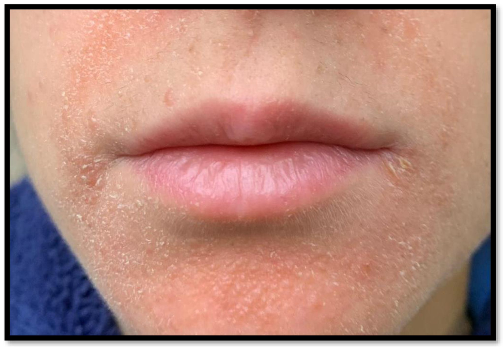
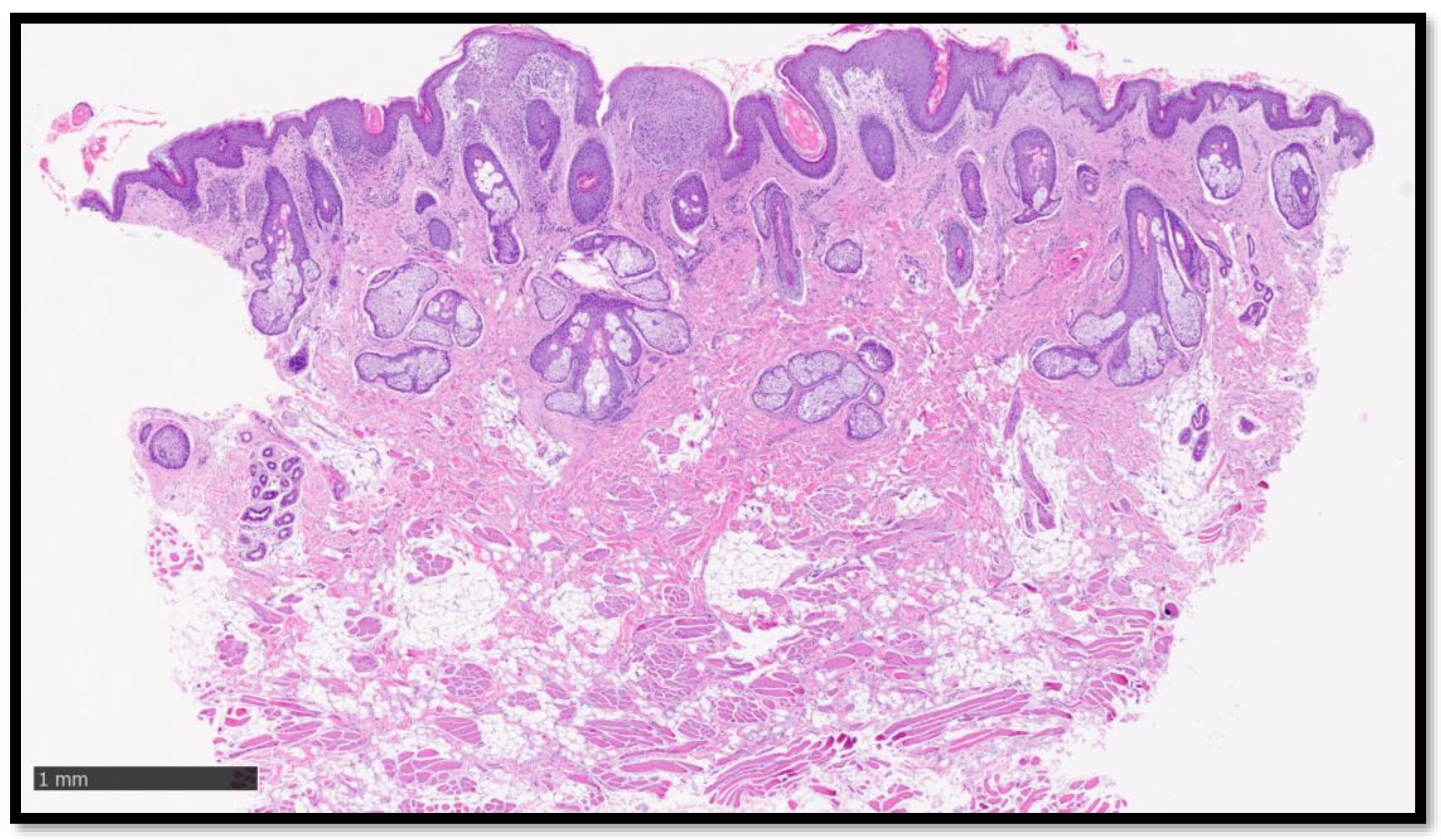
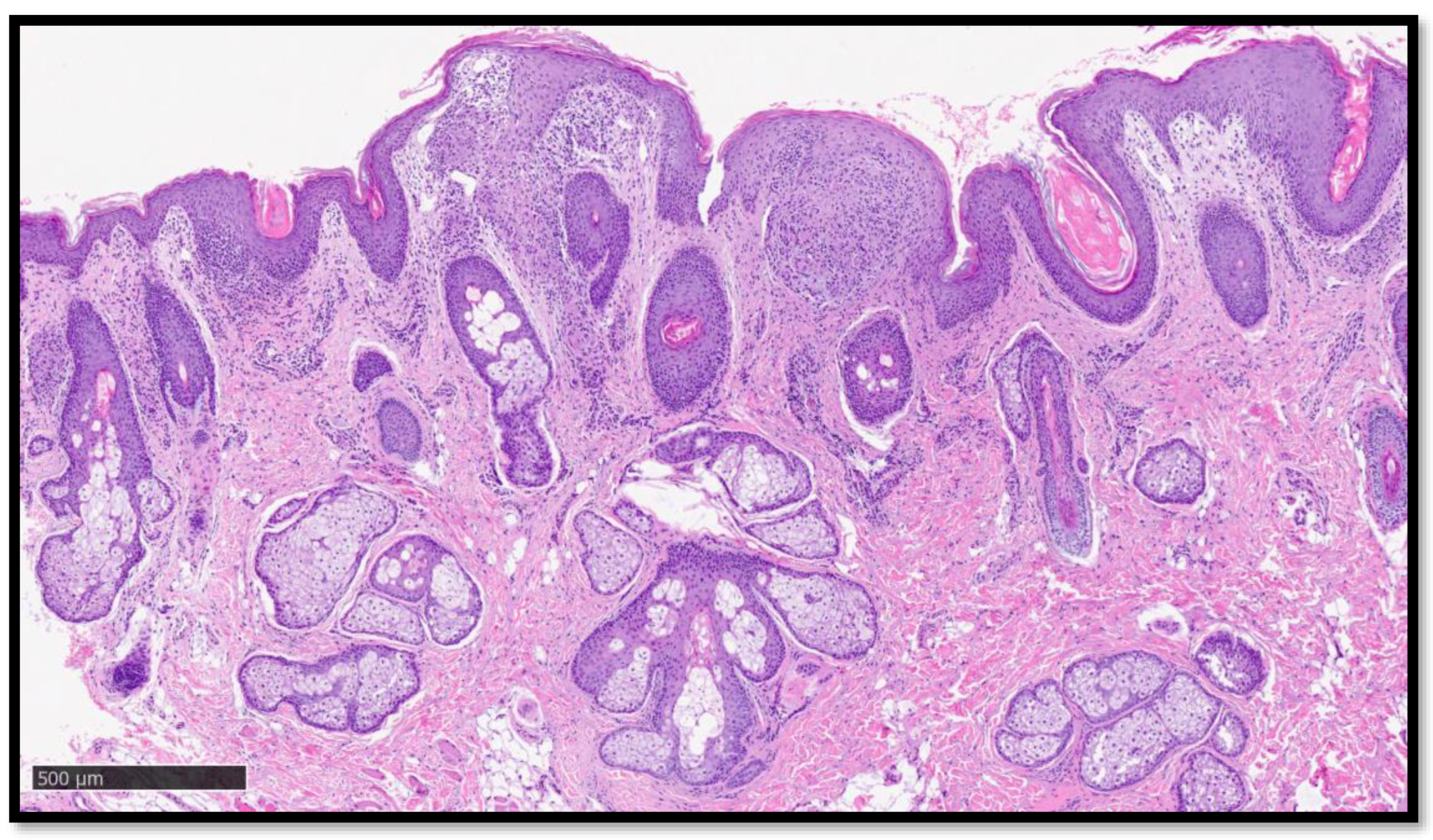
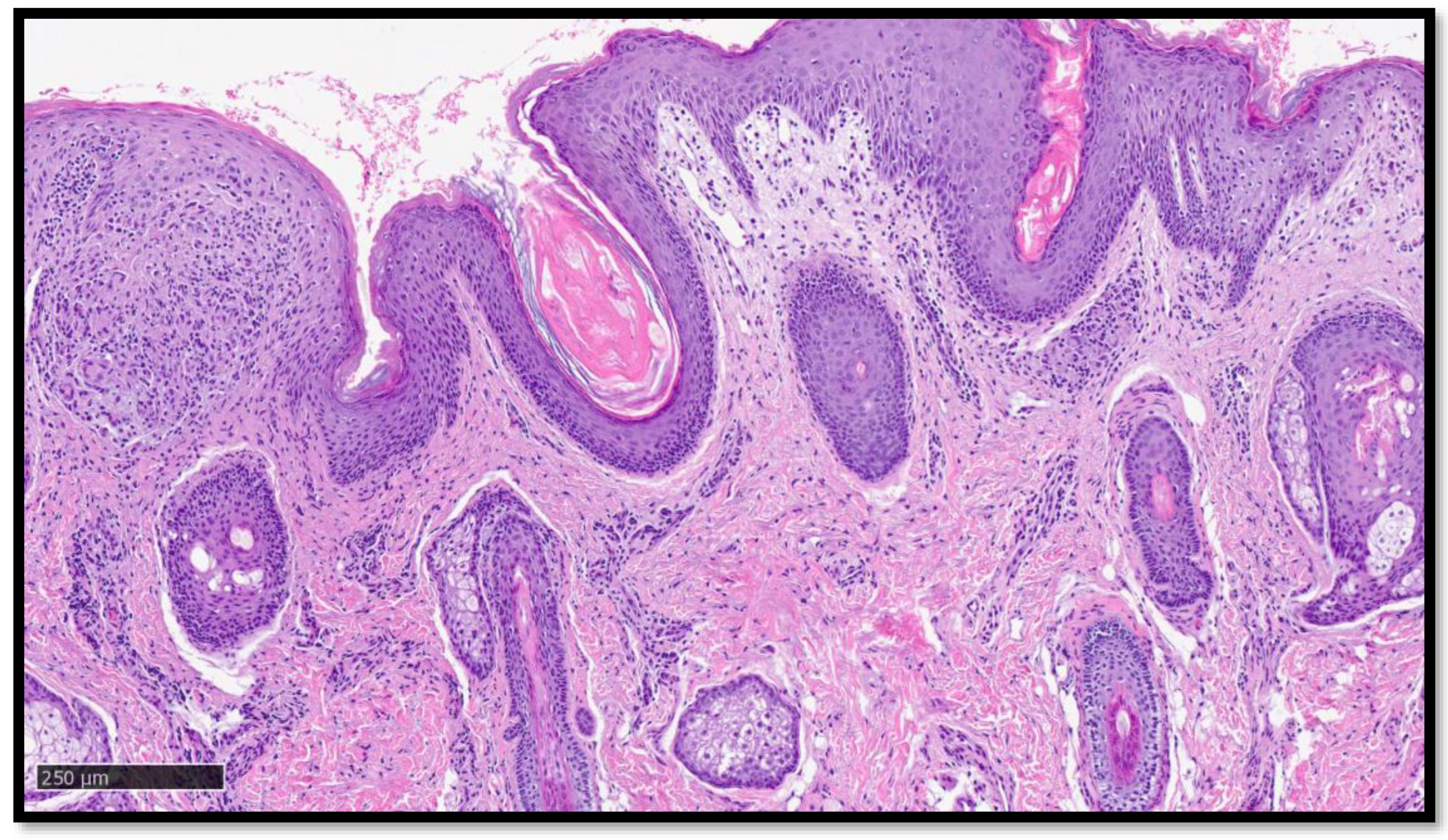
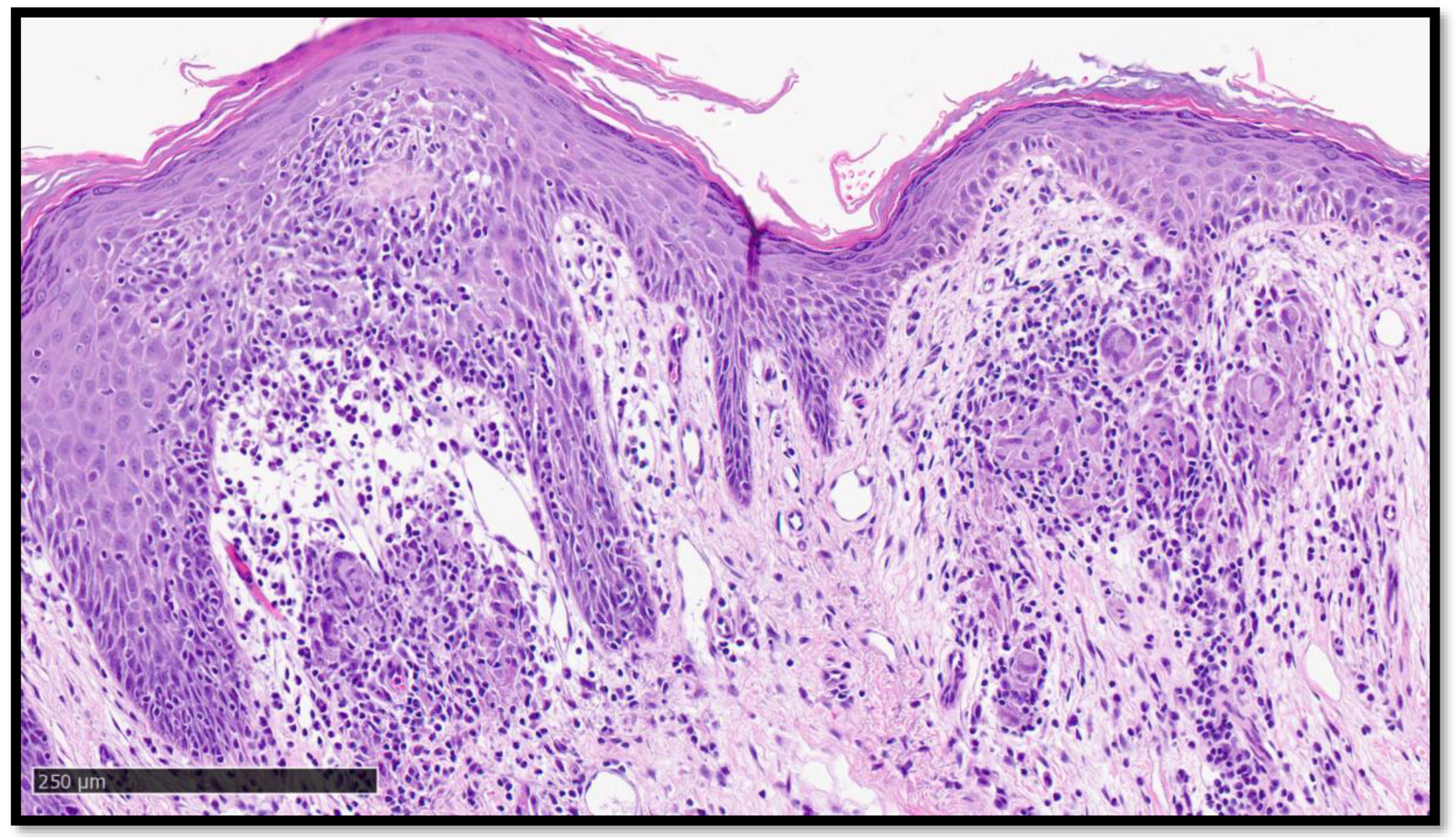
Abstract
:


Author Contributions
Funding
Institutional Review Board Statement
Informed Consent Statement
Data Availability Statement
Acknowledgments
Conflicts of Interest
References
- Golob, J.L.; Lugogo, N.; Lauring, A.S.; Lok, A.S. SARS-CoV-2 vaccines: A triumph of science and collaboration. J. Clin. Investig. 2021, 6, e149187. [Google Scholar] [CrossRef] [PubMed]
- Bellinato, F.; Maurelli, M.; Gisondi, P.; Girolomoni, G. Cutaneous Adverse Reactions Associated with SARS-CoV-2 Vaccines. J. Clin. Med. 2021, 10, 5344. [Google Scholar] [CrossRef] [PubMed]
- Avallone, G.; Quaglino, P.; Cavallo, F.; Roccuzzo, G.; Ribero, S.; Zalaudek, I.; Conforti, C. SARS-CoV-2 vaccine-related cutaneous manifestations: A systematic review. Int. J. Dermatol. 2022, 61, 1187–1204. [Google Scholar] [CrossRef] [PubMed]
- Gambichler, T.; Boms, S.; Susok, L.; Dickel, H.; Finis, C.; Abu Rached, N.; Barras, M.; Stücker, M.; Kasakovski, D. Cutaneous findings following COVID-19 vaccination: Review of world literature and own experience. J. Eur. Acad. Dermatol. Venereol. 2021, 36, 172–180. [Google Scholar] [CrossRef] [PubMed]
- Agrawal, S.; Verma, K.; Verma, I.; Gandhi, J. Reactivation of Herpes Zoster Virus After COVID-19 Vaccination: Is There Any Association? Cureus 2022, 14, e25195. [Google Scholar] [CrossRef] [PubMed]
- Khattab, E.; Christaki, E.; Pitsios, C. Pityriasis Rosea Induced by COVID-19 Vaccination. Eur. J. Case Rep. Intern. Med. 2022, 9, 003164. [Google Scholar] [CrossRef] [PubMed]
- Rademacher, J.-G.; Tampe, B.; Korsten, P. First Report of Two Cases of Löfgren’s Syndrome after SARS-CoV-2 Vaccination-Coincidence or Causality? Vaccines 2021, 9, 1313. [Google Scholar] [CrossRef] [PubMed]
- Behbahani, S.; Baltz, J.O.; Droms, R.; Deng, A.C.; Amano, S.U.; Levin, N.A.; O’Brien, M.C.; Wiss, K. Sarcoid-like reaction in a patient recovering from coronavirus disease 19 pneumonia. JAAD Case Rep. 2020, 6, 915–917. [Google Scholar] [CrossRef] [PubMed]
- Grieco, T.; Rossi, A.; Maddalena, P.; Sernicola, A.; Ambrosio, L.; Fino, P.; Gomes, V. COVID-19 infection and BNT162b2 vaccine triggering sarcoid-like lesions in the same patient. Response to: Sarcoid-like reaction in a patient recovering from COVID-19 pneumonia. JAAD Case Rep. 2022, 23, 162–163. [Google Scholar] [CrossRef] [PubMed]
- Numakura, T.; Murakami, K.; Tamada, T.; Yamaguchi, C.; Inoue, C.; Ohkouchi, S.; Tode, N.; Sano, H.; Aizawa, H.; Sato, K.; et al. A Novel Development of Sarcoidosis Following COVID-19 Vaccination and a Literature Review. Intern. Med. 2022, 61, 3101–3106. [Google Scholar] [CrossRef] [PubMed]
Disclaimer/Publisher’s Note: The statements, opinions and data contained in all publications are solely those of the individual author(s) and contributor(s) and not of MDPI and/or the editor(s). MDPI and/or the editor(s) disclaim responsibility for any injury to people or property resulting from any ideas, methods, instructions or products referred to in the content. |
© 2023 by the authors. Licensee MDPI, Basel, Switzerland. This article is an open access article distributed under the terms and conditions of the Creative Commons Attribution (CC BY) license (https://creativecommons.org/licenses/by/4.0/).
Share and Cite
Cazzato, G.; Ambrogio, F.; Foti, C.; Capuzzolo, M.; Trilli, I.; Casatta, N.; Lupo, C.; Carrieri, M.; Daini, D.; Colagrande, A.; et al. Cutaneous Sarcoidosis-like Eruption Following Second Dose of Moderna mRNA-1273 Vaccine: Case or Relationship? Diagnostics 2023, 13, 1286. https://doi.org/10.3390/diagnostics13071286
Cazzato G, Ambrogio F, Foti C, Capuzzolo M, Trilli I, Casatta N, Lupo C, Carrieri M, Daini D, Colagrande A, et al. Cutaneous Sarcoidosis-like Eruption Following Second Dose of Moderna mRNA-1273 Vaccine: Case or Relationship? Diagnostics. 2023; 13(7):1286. https://doi.org/10.3390/diagnostics13071286
Chicago/Turabian StyleCazzato, Gerardo, Francesca Ambrogio, Caterina Foti, Marialessandra Capuzzolo, Irma Trilli, Nadia Casatta, Carmelo Lupo, Marianna Carrieri, Daniele Daini, Anna Colagrande, and et al. 2023. "Cutaneous Sarcoidosis-like Eruption Following Second Dose of Moderna mRNA-1273 Vaccine: Case or Relationship?" Diagnostics 13, no. 7: 1286. https://doi.org/10.3390/diagnostics13071286