
Deep Learning Based Methods for Breast Cancer Diagnosis: A Systematic Review and Future Direction


Abstract
:1. Introduction
- Summarizing the deep learning-based methods popularly applied for breast cancer detection.
- Identifying the deep learning-based methods with the best performances for breast cancer diagnosis.
- Investigating the datasets generally applied in the deep learning-based methods for breast cancer diagnosis.
- Summarizing the evaluation metrics used for breast cancer diagnosis using deep learning-based methods.
- Analyzing the research gaps and future direction for deep learning-based breast cancer detection.
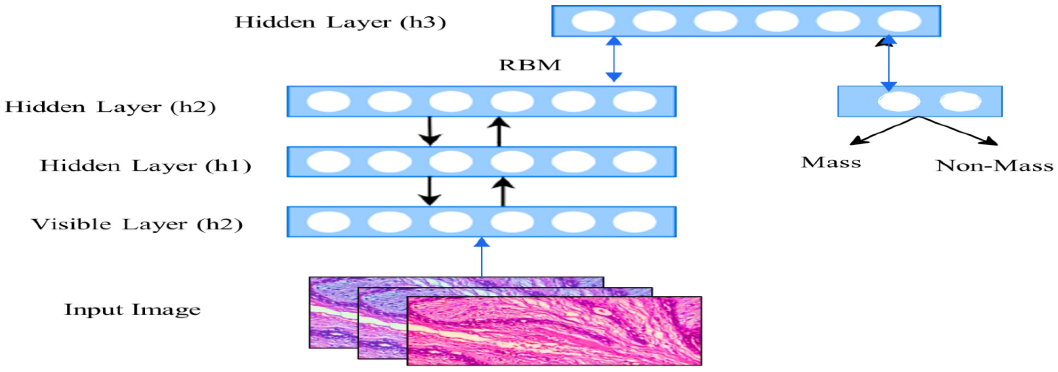
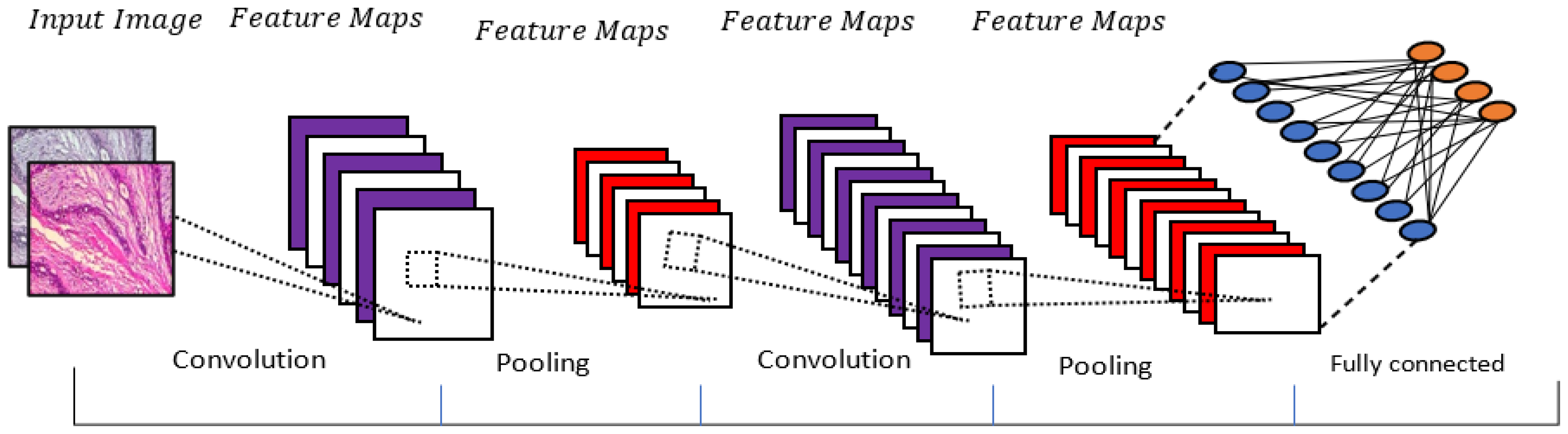
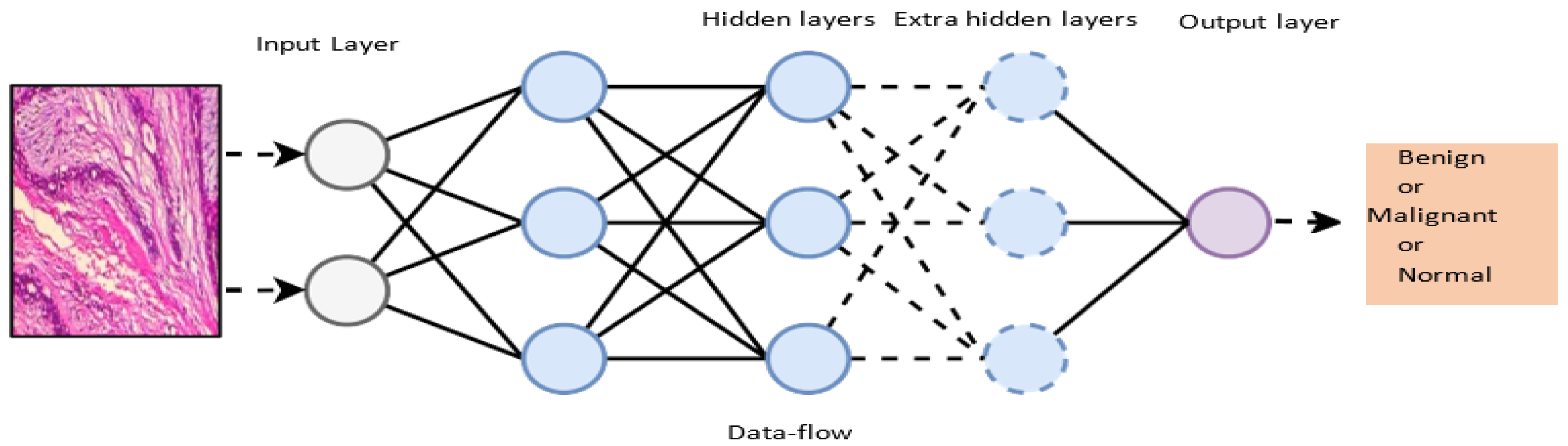
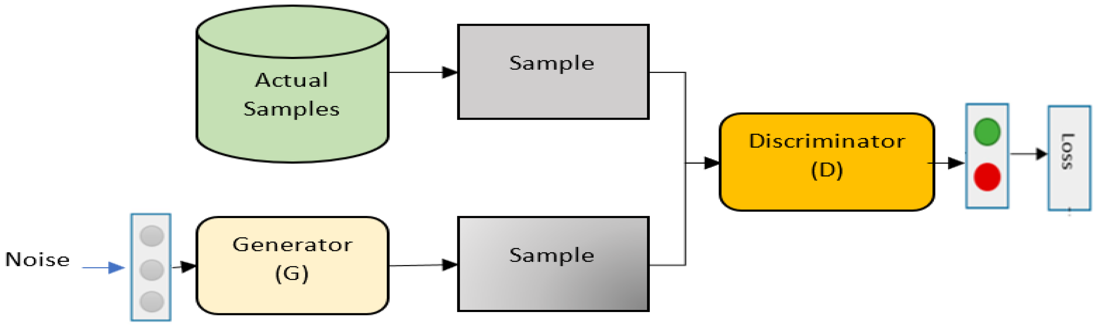
2. Background of Deep Learning Methods
3. Methods
3.1. Data Sources and Search Strategy
3.2. Selection Criteria
3.3. Quality Assessment
3.4. Data Extraction and Synthesis
4. Research Questions
- RQ1: What are the most common deep learning-based methods applied for breast cancer detection?
- RQ2: What is the most effective deep learning-based method for breast cancer detection in terms of performance?
- RQ3: What are the commonly used performance evaluation metrics for deep learning-based breast cancer detection?
- RQ4: What are the common datasets used for deep learning-based breast cancer detection?
- RQ5: What are the challenges and future directions of the deep learning-based methods for cancer detection?
5. Results and Metanalysis
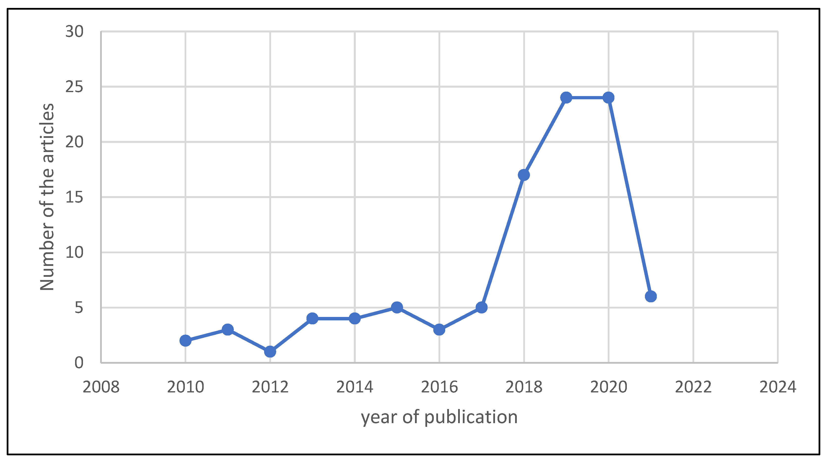
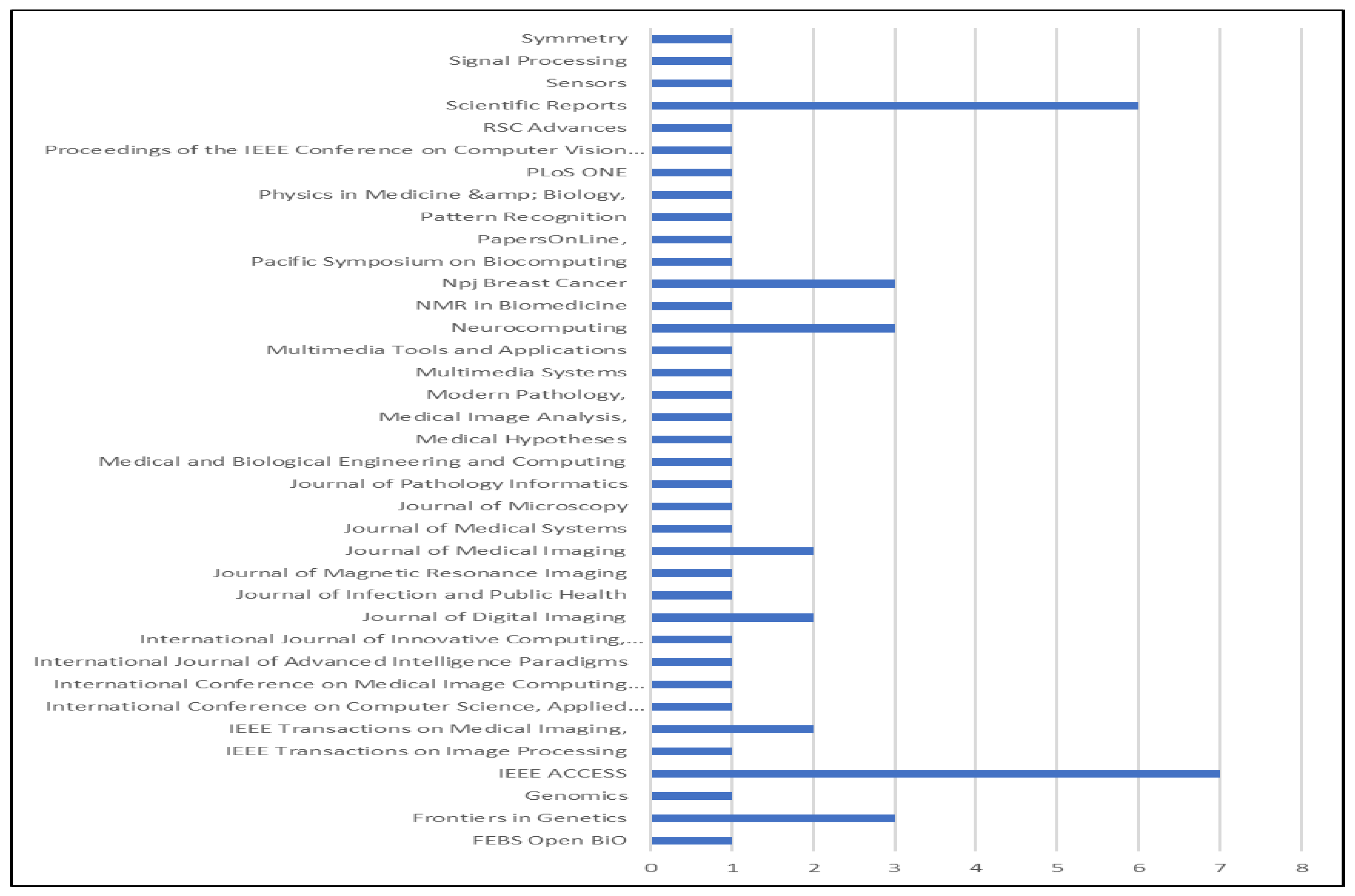
5.1. Description of the Selected Studies
5.2. RQ1: What Are the Common Deep Learning Methods for Breast Cancer?
5.3. RQ2: Which Deep Learning Models Perform Most Effectively?
5.4. RQ3: What Are the Evaluation Measures Commonly Used for Deep Learning-Based Breast Cancer Detection?
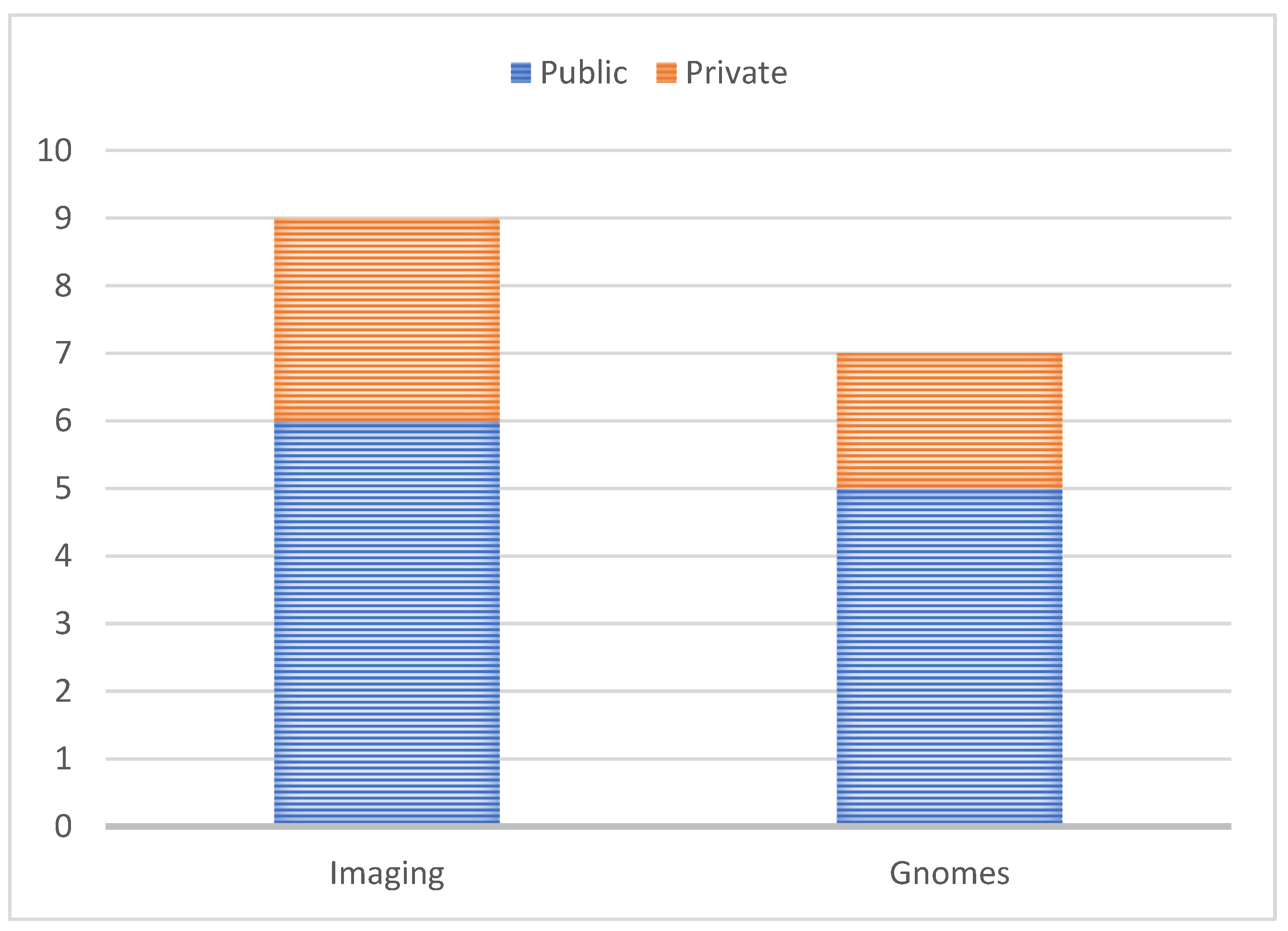
5.5. RQ4: What Datasets Are Available for Breast Cancer Diagnosis?
5.6. RQ5: What Are the Research Gaps, Challenges and Future Directions?
5.6.1. Balanced Dataset
5.6.2. Interpretable Deep Architecture
5.6.3. Clinical Application
6. Limitations of the Study
- This SLR is solely restricted to journal and conference materials that discuss breast cancer detection in DL. Several irrelevant research publications were found and eliminated from this review in the early stages of the study by using our search method. This guarantees that the chosen research papers met the requirements for the investigation. However, it is believed that incorporating other sources—such as extra sourcebooks, for instance—would have improved this review.
- We limited our search to items written in English. Due to the possibility of related publications in this area of study existing in other languages, this leads to linguistic bias. Thankfully, all of the papers gathered for this study were written in English. We are not language-biased as a result.
- Although the primary databases were considered when looking through the study articles, it is possible that other digital libraries with pertinent studies were disregarded. To overcome this limitation, we compared the search phrases and keywords to a well-known collection of research studies. However, when looking for the keywords, certain synonyms might be missed. To solve this issue, the SLR protocol has been updated to make sure no crucial phrases are omitted.
7. Conclusions
Author Contributions
Funding
Institutional Review Board Statement
Informed Consent Statement
Data Availability Statement
Acknowledgments
Conflicts of Interest
References
- Aggarwal, R.; Sounderajah, V.; Martin, G.; Ting, D.S.W.; Karthikesalingam, A.; King, D.; Ashrafian, H.; Darzi, A. Diagnostic accuracy of deep learning in medical imaging: A systematic review and meta-analysis. Npj Digit. Med. 2021, 4, 23. [Google Scholar] [CrossRef]
- Yassin, N.I.; Omran, S.; Houby, E.M.; Allam, H. Machine learning techniques for breast cancer computer aided diagnosis using different image modalities: A systematic review. Comput. Methods Programs Biomed. 2018, 156, 25–45. [Google Scholar] [CrossRef]
- El-Nabawy, A.; El-Bendary, N.; Belal, N.A. A feature-fusion framework of clinical, genomics, and histopathological data for METABRIC breast cancer subtype classification. Appl. Soft Comput. 2020, 91, 20. [Google Scholar] [CrossRef]
- Nassif, A.B.; Abu Talib, M.; Nasir, Q.; Afadar, Y.; Elgendy, O. Breast cancer detection using artificial intelligence techniques: A systematic literature review. Artif. Intell. Med. 2022, 127, 13. [Google Scholar] [CrossRef]
- Yao, H.; Zhang, X.; Zhou, X.; Liu, S. Parallel Structure Deep Neural Network Using CNN and RNN with an Attention Mechanism for Breast Cancer Histology Image Classification. Cancers 2019, 11, 1901. [Google Scholar] [CrossRef] [Green Version]
- Ha, R.; Chang, P.; Mutasa, S.; Karcich, J.; Goodman, S.; Blum, E.; Kalinsky, K.; Ms, M.Z.L.; Jambawalikar, S. Convolutional Neural Network Using a Breast MRI Tumor Dataset Can Predict Oncotype Dx Recurrence Score. J. Magn. Reson. Imaging 2019, 49, 518–524. [Google Scholar] [CrossRef]
- Bhatt, C.; Kumar, I.; Vijayakumar, V.; Singh, K.U.; Kumar, A. The state of the art of deep learning models in medical science and their challenges. Multimed. Syst. 2021, 27, 599–613. [Google Scholar] [CrossRef]
- Fujioka, T.; Mori, M.; Kubota, K.; Oyama, J.; Yamaga, E.; Yashima, Y.; Katsuta, L.; Nomura, K.; Nara, M.; Oda, G.; et al. The Utility of Deep Learning in Breast Ultrasonic Imaging: A Review. Diagnostics 2020, 10, 1055. [Google Scholar] [CrossRef]
- Mullooly, M.; Bejnordi, B.E.; Pfeiffer, R.M.; Fan, S.; Palakal, M.; Hada, M.; Vacek, P.M.; Weaver, D.L.; Shepherd, J.A.; Fan, B.; et al. Application of convolutional neural networks to breast biopsies to delineate tissue correlates of mammographic breast density. Npj Breast Cancer 2019, 5, 11. [Google Scholar] [CrossRef] [Green Version]
- Saba, T. Recent advancement in cancer detection using machine learning: Systematic survey of decades, comparisons and challenges. J. Infect. Public Health 2020, 13, 1274–1289. [Google Scholar] [CrossRef]
- Shah, S.M.; Khan, R.A.; Arif, S.; Sajid, U. Artificial intelligence for breast cancer analysis: Trends & directions. Comput. Biol. Med. 2022, 142, 15. [Google Scholar] [CrossRef]
- Houssein, E.H.; Emam, M.M.; Ali, A.A.; Suganthan, P.N. Deep and machine learning techniques for medical imaging-based breast cancer: A comprehensive review. Expert Syst. Appl. 2021, 167, 20. [Google Scholar] [CrossRef]
- Bharati, S.; Podder, P.; Mondal, M.R.H. Artificial neural network based breast cancer screening: A comprehensive review. Int. J. Comput. Inf. Syst. Ind. Manag. Appl. 2020, 12, 125–137. [Google Scholar]
- Bai, J.; Posner, R.; Wang, T.; Yang, C.; Nabavi, S. Applying deep learning in digital breast tomosynthesis for automatic breast cancer detection: A review. Med. Image Anal. 2021, 71, 19. [Google Scholar] [CrossRef]
- Rautela, K.; Kumar, D.; Kumar, V. A Systematic Review on Breast Cancer Detection Using Deep Learning Techniques. Arch. Comput. Methods Eng. 2022, 31, 4599–4629. [Google Scholar] [CrossRef]
- Nemade, V.; Pathak, S.; Dubey, A.K. A Systematic Literature Review of Breast Cancer Diagnosis Using Machine Intelligence Techniques. Arch. Comput. Methods Eng. 2022, 29, 4401–4430. [Google Scholar] [CrossRef]
- Huang, X.W.; Qian, S.S.; Fang, Q.; Sang, J.T.; Xu, C.S. CSAN: Contextual Self-Attention Network for User Sequential Recommendation. In Proceedings of the 26th ACM Multimedia Conference (MM), Seoul, Reuplic of Korea, 22–26 October 2018; pp. 447–455. [Google Scholar]
- Zhou, X.; Li, C.; Rahaman, M.; Yao, Y.; Ai, S.; Sun, C.; Wang, Q.; Zhang, Y.; Li, M.; Li, X.; et al. A Comprehensive Review for Breast Histopathology Image Analysis Using Classical and Deep Neural Networks. IEEE Access 2020, 8, 90931–90956. [Google Scholar] [CrossRef]
- Pang, T.; Wong, J.H.D.; Ng, W.L.; Chan, C.S. Deep learning radiomics in breast cancer with different modalities: Overview and future. Expert Syst. Appl. 2020, 158, 113501. [Google Scholar] [CrossRef]
- Fatima, N.; Liu, L.; Hong, S.; Ahmed, H. Prediction of Breast Cancer, Comparative Review of Machine Learning Techniques, and Their Analysis. IEEE Access 2020, 8, 150360–150376. [Google Scholar] [CrossRef]
- Lu, Y.; Li, J.Y.; Su, Y.T.; Liu, A.A. A Review of Breast Cancer Detection in Medical Images. In Proceedings of the 33rd IEEE International Conference on Visual Communications and Image Processing (IEEE VCIP), Taichung, Taiwan, 9–12 December 2018. [Google Scholar]
- Gupta, N.P.; Malik, P.K.; Ram, B.S. A Review on Methods and Systems for Early Breast Cancer Detection. In Proceedings of the International Conference on Computation, Automation and Knowledge Management, ICCAKM 2020, Dubai, United Arab Emirates, 9–10 January 2020; pp. 42–46. [Google Scholar] [CrossRef]
- Oyelade, O.N.; Ezugwu, A.E.-S. A State-of-the-Art Survey on Deep Learning Methods for Detection of Architectural Distortion From Digital Mammography. IEEE Access 2020, 8, 148644–148676. [Google Scholar] [CrossRef]
- Roslidar, R.; Rahman, A.; Muharar, R.; Syahputra, M.R.; Arnia, F.; Syukri, M.; Pradhan, B.; Munadi, K. A Review on Recent Progress in Thermal Imaging and Deep Learning Approaches for Breast Cancer Detection. IEEE Access 2020, 8, 116176–116194. [Google Scholar] [CrossRef]
- Schmidhuber, J. Deep Learning in Neural Networks: An Overview. Neural Netw. 2015, 61, 85–117. [Google Scholar] [CrossRef] [Green Version]
- Hatcher, W.G.; Yu, W. A Survey of Deep Learning: Platforms, Applications and Emerging Research Trends. IEEE Access 2018, 6, 24411–24432. [Google Scholar] [CrossRef]
- Da’U, A.; Salim, N. Aspect extraction on user textual reviews using multi-channel convolutional neural network. PeerJ Comput. Sci. 2019, 16, e191. [Google Scholar] [CrossRef] [Green Version]
- Da′U, A.; Salim, N. Sentiment-Aware Deep Recommender System with Neural Attention Networks. IEEE Access 2019, 7, 45472–45484. [Google Scholar] [CrossRef]
- Deng, L. Deep Learning: Methods and Applications. Found. Trends Signal Process. 2013, 7, 197–387. [Google Scholar] [CrossRef] [Green Version]
- Xue, P.; Wang, J.; Qin, D.; Yan, H.; Qu, Y.; Seery, S.; Jiang, Y.; Qiao, Y. Deep learning in image-based breast and cervical cancer detection: A systematic review and meta-analysis. Npj Digit. Med. 2022, 5, 19. [Google Scholar] [CrossRef]
- Shibata, H.; Takama, Y.; Ieee. Behavior Analysis of RBM for Estimating Latent Factor Vectors from Rating Matrix. In Proceedings of the 6th International Conference on Informatics, Electronics and Vision (ICIEV)/7th International Symposium in Computational Medical and Health Technology (ISCMHT), Univ Hyogo, Himeji Engn, Himeji, Japan, 1–3 September 2017. [Google Scholar]
- Deng, L. Three classes of deep learning architectures and their applications: A tutorial survey. PSIPA Trans. Signal Inf. Process. 2012, 57, 58. [Google Scholar]
- Smolander, J.; Dehmer, M.; Emmert-Streib, F. Comparing deep belief networks with support vector machines for classifying gene expression data from complex disorders. FEBS Open Bio 2019, 9, 1232–1248. [Google Scholar] [CrossRef] [Green Version]
- Salas, J.; Vidal, F.D.B.; Martinez-Trinidad, F. Deep Learning: Current State. IEEE Lat. Am. Trans. 2019, 17, 1925–1945. [Google Scholar] [CrossRef]
- Pouyanfar, S.; Sadiq, S.; Yan, Y.; Tian, H.; Tao, Y.; Reyes, M.P.; Shyu, M.-L.; Chen, S.-C.; Iyengar, S.S. A Survey on Deep Learning: Algorithms, Techniques, and Applications. ACM Comput. Surv. 2019, 51, 36. [Google Scholar] [CrossRef]
- Hossain, Z.; Sohel, F.; Shiratuddin, M.F.; Laga, H. A Comprehensive Survey of Deep Learning for Image Captioning. ACM Comput. Surv. 2019, 51, 36. [Google Scholar] [CrossRef] [Green Version]
- Alom, M.Z.; Taha, T.M.; Yakopcic, C.; Westberg, S.; Sidike, P.; Nasrin, M.S.; Hasan, M.; Van Essen, B.C.; Awwal, A.A.S.; Asari, V.K. A State-of-the-Art Survey on Deep Learning Theory and Architectures. Electronics 2019, 8, 292. [Google Scholar] [CrossRef] [Green Version]
- Yu, X.; Zhou, Q.; Wang, S.; Zhang, Y. A systematic survey of deep learning in breast cancer. Int. J. Intell. Syst. 2022, 37, 152–216. [Google Scholar] [CrossRef]
- Zhao, Y.; Hu, B.; Wang, Y.; Yin, X.; Jiang, Y.; Zhu, X. Identification of gastric cancer with convolutional neural networks: A systematic review. Multimed. Tools Appl. 2022, 81, 11717–11736. [Google Scholar] [CrossRef]
- Binder, A.; Bach, S.; Montavon, G.; Müller, K.-R.; Samek, W. Layer-Wise Relevance Propagation for Deep Neural Network Architectures. In Lecture Notes in Electrical Engineering; Springer: Singapore, 2016; Volume 376, pp. 913–922. [Google Scholar] [CrossRef]
- Mambou, S.J.; Maresova, P.; Krejcar, O.; Selamat, A.; Kuca, K. Breast Cancer Detection Using Infrared Thermal Imaging and a Deep Learning Model. Sensors 2018, 18, 2799. [Google Scholar] [CrossRef] [Green Version]
- Egger, J.; Gsaxner, C.; Pepe, A.; Pomykala, K.L.; Jonske, F.; Kurz, M.; Li, J.; Kleesiek, J. Medical deep learning—A systematic meta-review. Comput. Methods Programs Biomed. 2022, 221, 22. [Google Scholar] [CrossRef]
- Miotto, R.; Wang, F.; Wang, S.; Jiang, X.; Dudley, J.T. Deep learning for healthcare: Review, opportunities and challenges. Brief. Bioinform. 2018, 19, 1236–1246. [Google Scholar] [CrossRef]
- Shreir, L.L. (Ed.) Front Matter. In Corrosion, 2nd ed.; Newnes: London, UK, 1976; p. iii. [Google Scholar]
- Bentur, Y.; Layish, I.; Krivoy, A.; Berkovitch, M.; Rotman, E.; Bar Haim, S.; Yehezkelli, Y.; Kozer, E. Civilian Adult Self Injections of Atropine—Trimedoxime (TMB4) Auto-Injectors. Clin. Toxicol. 2006, 44, 301–306. [Google Scholar] [CrossRef]
- Wang, Q.Y.; Yin, H.Z.; Hu, Z.T.; Lian, D.F.; Wang, H.; Huang, Z. Neural Memory Streaming Recommender Networks with Adversarial Training. In Proceedings of the 24th ACM SIGKDD Conference on Knowledge Discovery and Data Mining (KDD), London, UK, 19–23 August 2018; pp. 2467–2475. [Google Scholar]
- Page, M.J.; McKenzie, J.E.; Bossuyt, P.M.; Boutron, I.; Hoffmann, T.C.; Mulrow, C.D.; Shamseer, L.; Tetzlaff, J.M.; Akl, E.A.; Brennan, S.E.; et al. The PRISMA 2020 Statement: An Updated Guideline for Reporting Systematic Reviews. BMJ Br. Med. J. 2021, 372, n71. [Google Scholar] [CrossRef]
- Keele, S. Guidelines for Performing Systematic Literature Reviews in Software Engineering, Technical Report, version 2.3 ESBE Technical Report; ESBE.; Keele University: Keele, UK, 2007. [Google Scholar]
- Genc-Nayebi, N.; Abran, A. A systematic literature review: Opinion mining studies from mobile app store user reviews. J. Syst. Softw. 2017, 125, 207–219. [Google Scholar] [CrossRef]
- Kitchenham, B.; Brereton, O.P.; Budgen, D.; Turner, M.; Bailey, J.; Linkman, S. Systematic literature reviews in software engineering—A systematic literature review. Inf. Softw. Technol. 2009, 51, 7–15. [Google Scholar] [CrossRef]
- Mridha, M.F.; Hamid, A.; Monowar, M.M.; Keya, A.J.; Ohi, A.Q.; Islam, R.; Kim, J.-M. A Comprehensive Survey on Deep-Learning-Based Breast Cancer Diagnosis. Cancers 2021, 13, 6116. [Google Scholar] [CrossRef]
- Mostavi, M.; Chiu, Y.-C.; Huang, Y.; Chen, Y. Convolutional neural network models for cancer type prediction based on gene expression. BMC Med. Genom. 2020, 13, 44. [Google Scholar] [CrossRef]
- Lancashire, L.J.; Powe, D.G.; Reis-Filho, J.S.; Rakha, E.; Lemetre, C.; Weigelt, B.; Abdel-Fatah, T.M.; Green, A.; Mukta, R.; Blamey, R.; et al. A validated gene expression profile for detecting clinical outcome in breast cancer using artificial neural networks. Breast Cancer Res. Treat. 2010, 120, 83–93. [Google Scholar] [CrossRef] [Green Version]
- Paul, A.; Mukherjee, D.P. Mitosis Detection for Invasive Breast Cancer Grading in Histopathological Images. IEEE Trans. Image Process. 2015, 24, 4041–4054. [Google Scholar] [CrossRef]
- Bejnordi, B.E.; Mullooly, M.; Pfeiffer, R.M.; Fan, S.; Vacek, P.M.; Weaver, D.L.; Herschorn, S.; Brinton, L.A.; Van Ginneken, B.; Karssemeijer, N.; et al. Using deep convolutional neural networks to identify and classify tumor-associated stroma in diagnostic breast biopsies. Mod. Pathol. 2018, 31, 1502–1512. [Google Scholar] [CrossRef]
- Simidjievski, N.; Bodnar, C.; Tariq, I.; Scherer, P.; Andres-Terre, H.; Shams, Z.; Jamnik, M.; Liò, P. Variational Autoencoders for Cancer Data Integration: Design Principles and Computational Practice. Front. Genet. 2019, 10, 14. [Google Scholar] [CrossRef] [Green Version]
- Ha, R.; Chin, C.; Karcich, J.; Liu, M.Z.; Chang, P.; Mutasa, S.; Pascual Van Sant, E.; Wynn, R.T.; Connolly, E.; Jambawalikar, S. Prior to initiation of chemotherapy, can we predict breast tumor response? Deep learning convolutional neural networks ap-proach using a breast MRI tumor dataset. J. Digit. Imaging 2019, 32, 693–701. [Google Scholar] [CrossRef]
- Turkki, R.; Byckhov, D.; Lundin, M.; Isola, J.; Nordling, S.; Kovanen, P.E.; Verrill, C.; von Smitten, K.; Joensuu, H.; Lundin, J.; et al. Breast cancer outcome prediction with tumour tissue images and machine learning. Breast Cancer Res. Treat. 2019, 177, 41–52. [Google Scholar] [CrossRef]
- Toğaçar, M.; Ergen, B.; Cömert, Z. Application of breast cancer diagnosis based on a combination of convolutional neural networks, ridge regression and linear discriminant analysis using invasive breast cancer images processed with autoencoders. Med. Hypotheses 2020, 135, 10. [Google Scholar] [CrossRef]
- Chen, H.; Gao, M.; Zhang, Y.; Liang, W.; Zou, X. Attention-Based Multi-NMF Deep Neural Network with Multimodality Data for Breast Cancer Prognosis Model. BioMed Res. Int. 2019, 2019, 11. [Google Scholar] [CrossRef]
- Lu, Z.; Xu, S.; Shao, W.; Wu, Y.; Zhang, J.; Han, Z.; Feng, Q.; Huang, K. Deep-Learning–Based Characterization of Tumor-Infiltrating Lymphocytes in Breast Cancers from Histopathology Images and Multiomics Data. JCO Clin. Cancer Inform. 2020, 4, 480–490. [Google Scholar] [CrossRef]
- Guo, Y.; Shang, X.; Li, Z. Identification of cancer subtypes by integrating multiple types of transcriptomics data with deep learning in breast cancer. Neurocomputing 2019, 324, 20–30. [Google Scholar] [CrossRef]
- Lim, J.; Bang, S.; Kim, J.; Park, C.; Cho, J.; Kim, S. Integrative Deep Learning for Identifying Differentially Expressed (DE) Biomarkers. Comput. Math. Methods Med. 2019, 2019, 10. [Google Scholar] [CrossRef]
- Sur, C. GSIAR: Gene-subcategory interaction-based improved deep representation learning for breast cancer subcategorical analysis using gene expression, applicable for precision medicine. Med. Biol. Eng. Comput. 2019, 57, 2483–2515. [Google Scholar] [CrossRef]
- Zhang, D.; Zou, L.; Zhou, X.; He, F. Integrating Feature Selection and Feature Extraction Methods with Deep Learning to Predict Clinical Outcome of Breast Cancer. IEEE Access 2018, 6, 28936–28944. [Google Scholar] [CrossRef]
- Xu, J.; Xiang, L.; Liu, Q.; Gilmore, H.; Wu, J.; Tang, J.; Madabhushi, A. Stacked Sparse Autoencoder (SSAE) for Nuclei Detection on Breast Cancer Histopathology Images. IEEE Trans. Med. Imaging 2016, 35, 119–130. [Google Scholar] [CrossRef] [Green Version]
- Xu, J.; Xiang, L.; Hang, R.L.; Wu, J.Z. Stacked Sparse Autoencoder (SSAE) Based Framework for Nuclei Patch Classifi-cation on Breast Cancer Histopathology. In Proceedings of the 11th IEEE International Symposium on Biomedical Imaging (ISBI), Beijing, China, 29 April–2 May 2014; pp. 999–1002. [Google Scholar]
- Shams, S.; Platania, R.; Zhang, J.; Kim, J.; Lee, K.; Park, S.J. Deep Generative Breast Cancer Screening and Diagnosis. In Proceedings of the 21st International Conference on Medical Image Computing and Computer-Assisted Intervention (MICCAI)/8th Eurographics Workshop on Visual Computing for Biology and Medicine (VCBM)/International Workshop on Computational Diffusion MRI (CDMRI), Granada, Spain, 16–21 September 2018; pp. 859–867. [Google Scholar]
- Singh, V.K.; Rashwan, H.A.; Romani, S.; Akram, F.; Pandey, N.; Sarker, M.K.; Saleh, A.; Arenas, M.; Arquez, M.; Puig, D.; et al. Breast tumor segmentation and shape classification in mammograms using generative adversarial and convolutional neural network. Expert Syst. Appl. 2020, 139, 14. [Google Scholar] [CrossRef]
- Fan, M.; Liu, Z.; Xu, M.; Wang, S.; Zeng, T.; Gao, X.; Li, L. Generative adversarial network-based super-resolution of diffusion-weighted imaging: Application to tumour radiomics in breast cancer. NMR Biomed. 2020, 33, 12. [Google Scholar] [CrossRef]
- Guan, S.Y.; Loew, M. Breast cancer detection using synthetic mammograms from generative adversarial networks in convolutional neural networks. J. Med. Imaging 2019, 6, 10. [Google Scholar] [CrossRef]
- Rawat, R.R.; Ruderman, D.; Macklin, P.; Rimm, D.L.; Agus, D.B. Correlating nuclear morphometric patterns with estrogen receptor status in breast cancer pathologic specimens. Npj Breast Cancer 2018, 4, 7. [Google Scholar] [CrossRef] [Green Version]
- Cruz-Roa, A.; Gilmore, H.; Basavanhally, A.; Feldman, M.; Ganesan, S.; Shih, N.; Tomaszewski, J.; Madabhushi, A.; González, F. High-throughput adaptive sampling for whole-slide histopathology image analysis (HASHI) via convolutional neural networks: Application to invasive breast cancer detection. PLoS ONE 2018, 13, 23. [Google Scholar] [CrossRef]
- Turkki, R.; Linder, N.; Kovanen, P.E.; Pellinen, T.; Lundin, J. Antibody-supervised deep learning for quantification of tumor-infiltrating immune cells in hematoxylin and eosin stained breast cancer samples. J. Pathol. Inform. 2016, 7, 38. [Google Scholar] [CrossRef]
- Khameneh, F.D.; Razavi, S.; Kamasak, M. Automated segmentation of cell membranes to evaluate HER2 status in whole slide images using a modified deep learning network. Comput. Biol. Med. 2019, 110, 164–174. [Google Scholar] [CrossRef]
- Murtaza, G.; Shuib, L.; Mujtaba, G.; Raza, G. Breast Cancer Multi-classification through Deep Neural Network and Hierarchical Classification Approach. Multimed. Tools Appl. 2020, 79, 15481–15511. [Google Scholar] [CrossRef]
- Bardou, D.; Zhang, K.; Ahmad, S.M. Classification of Breast Cancer Based on Histology Images Using Convolutional Neural Networks. IEEE Access 2018, 6, 24680–24693. [Google Scholar] [CrossRef]
- Tellez, D.; Balkenhol, M.; Otte-Holler, I.; van de Loo, R.; Vogels, R.; Bult, P.; Wauters, C.; Vreuls, W.; Mol, S.; Karssemeijer, N.; et al. Whole-Slide Mitosis Detection in H&E Breast Histology Using PHH3 as a Reference to Train Distilled Stain-Invariant Convolutional Networks. IEEE Trans. Med. Imaging 2018, 37, 2126–2136. [Google Scholar] [CrossRef] [Green Version]
- Luo, J.; Ning, Z.; Zhang, S.; Feng, Q.; Zhang, Y. Bag of deep features for preoperative prediction of sentinel lymph node metastasis in breast cancer. Phys. Med. Biol. 2018, 63, 11. [Google Scholar] [CrossRef]
- Elbashir, M.K.; Ezz, M.; Mohammed, M.; Saloum, S.S. Lightweight Convolutional Neural Network for Breast Cancer Classification Using RNA-Seq Gene Expression Data. IEEE Access 2019, 7, 185338–185348. [Google Scholar] [CrossRef]
- Saha, M.; Arun, I.; Ahmed, R.; Chatterjee, S.; Chakraborty, C. HscoreNet: A Deep network for estrogen and progesterone scoring using breast IHC images. Pattern Recognit. 2020, 102, 11. [Google Scholar] [CrossRef]
- Ha, R.; Mutasa, S.; Karcich, J.; Gupta, N.; Van Sant, E.P.; Nemer, J.; Sun, M.; Chang, P.; Liu, M.Z.; Jambawalikar, S. Predicting Breast Cancer Molecular Subtype with MRI Dataset Utilizing Convolutional Neural Network Algorithm. J. Digit. Imaging 2019, 32, 276–282. [Google Scholar] [CrossRef] [PubMed]
- Zheng, J.; Lin, D.; Gao, Z.; Wang, S.; He, M.; Fan, J. Deep Learning Assisted Efficient AdaBoost Algorithm for Breast Cancer Detection and Early Diagnosis. IEEE Access 2020, 8, 96946–96954. [Google Scholar] [CrossRef]
- Tewary, S.; Arun, I.; Ahmed, R.; Chatterjee, S.; Chakraborty, C. AutoIHC-scoring: A machine learning framework for automated Allred scoring of molecular expression in ER- and PR-stained breast cancer tissue. J. Microsc. 2017, 268, 172–185. [Google Scholar] [CrossRef]
- Fang, Y.; Zhao, J.; Hu, L.; Ying, X.; Pan, Y.; Wang, X. Image classification toward breast cancer using deeply-learned quality features. J. Vis. Commun. Image Represent. 2019, 64, 7. [Google Scholar] [CrossRef]
- Danaee, P.; Ghaeini, R.; Hendrix, D.A. A Deep Learning Approach for Cancer Detection and Relevant Gene Identification. In Proceedings of the 22nd Pacific Symposium on Biocomputing (PSB), Kohala Coast, 4–8 January 2017; pp. 219–229. [Google Scholar]
- Punitha, S.; Amuthan, A.; Joseph, K.S. Enhanced Monarchy Butterfly Optimization Technique for effective breast cancer diagnosis. J. Med. Syst. 2019, 43, 14. [Google Scholar] [CrossRef]
- Romo-Bucheli, D.; Janowczyk, A.; Gilmore, H.; Romero, E.; Madabhushi, A. Automated Tubule Nuclei Quantification and Correlation with Oncotype DX risk categories in ER+ Breast Cancer Whole Slide Images. Sci. Rep. 2016, 6, 9. [Google Scholar] [CrossRef] [Green Version]
- Zemouri, R.; Omri, N.; Morello, B.; Devalland, C.; Arnould, L.; Zerhouni, N.; Fnaiech, F. Constructive Deep Neural Network for Breast Cancer Diagnosis. IFAC-Pap. 2018, 51, 98–103. [Google Scholar] [CrossRef]
- Salma, M.U.; Doreswamy, N. Hybrid BATGSA: A metaheuristic model for classification of breast cancer data. Int. J. Adv. Intell. Paradig. 2020, 15, 207–227. [Google Scholar] [CrossRef]
- Karakış, R.; Tez, M.; Kılıç, Y.; Kuru, Y.; Güler, I. A genetic algorithm model based on artificial neural network for prediction of the axillary lymph node status in breastcancer. Eng. Appl. Artif. Intell. 2013, 26, 945–950. [Google Scholar] [CrossRef]
- Shimizu, H.; Nakayama, K.I. A 23 gene–based molecular prognostic score precisely predicts overall survival of breast cancer patients. EBioMedicine 2019, 46, 150–159. [Google Scholar] [CrossRef] [PubMed] [Green Version]
- Jafari-Marandi, R.; Davarzani, S.; Gharibdousti, M.S.; Smith, B. An optimum ANN-based breast cancer diagnosis: Bridging gaps between ANN learning and decision-making goals. Appl. Soft Comput. 2018, 72, 108–120. [Google Scholar] [CrossRef]
- Tripathy, R.K.; Mahanta, S.; Paul, S. Artificial intelligence-based classification of breast cancer using cellular images. RSC Adv. 2014, 4, 9349–9355. [Google Scholar] [CrossRef]
- Giordano, A.; Giuliano, M.; De Laurentiis, M.; Eleuteri, A.; Iorio, F.; Tagliaferri, R.; Hortobagyi, G.N.; Pusztai, L.; De Placido, S.; Hess, K.; et al. Artificial neural network analysis of circulating tumor cells in metastatic breast cancer patients. Breast Cancer Res. Treat. 2011, 129, 451–458. [Google Scholar] [CrossRef]
- Mert, A.; Kılıç, N.; Bilgili, E.; Akan, A. Breast Cancer Detection with Reduced Feature Set. Comput. Math. Methods Med. 2015, 2015, 11. [Google Scholar] [CrossRef] [PubMed] [Green Version]
- Hajiabadi, H.; Babaiyan, V.; Zabihzadeh, D.; Hajiabadi, M. Combination of loss functions for robust breast cancer prediction. Comput. Electr. Eng. 2020, 84, 12. [Google Scholar] [CrossRef]
- Agrawal, U.; Soria, D.; Wagner, C.; Garibaldi, J.; Ellis, I.O.; Bartlett, J.M.; Cameron, D.; Rakha, E.A.; Green, A.R. Combining clustering and classification ensembles: A novel pipeline to identify breast cancer profiles. Artif. Intell. Med. 2019, 97, 27–37. [Google Scholar] [CrossRef]
- Denkiewicz, M.; Saha, I.; Rakshit, S.; Sarkar, J.P.; Plewczynski, D. Identification of Breast Cancer Subtype Specific MicroRNAs Using Survival Analysis to Find Their Role in Transcriptomic Regulation. Front. Genet. 2019, 10, 19. [Google Scholar] [CrossRef] [Green Version]
- Rawat, R.R.; Ortega, I.; Roy, P.; Sha, F.; Shibata, D.; Ruderman, D.; Agus, D.B. Deep learned tissue “fingerprints” classify breast cancers by ER/PR/Her2 status from H&E images. Sci. Rep. 2020, 10, 7275. [Google Scholar] [CrossRef]
- Rouhi, R.; Jafari, M.; Kasaei, S.; Keshavarzian, P. Benign and malignant breast tumors classification based on region growing and CNN segmentation. Expert Syst. Appl. 2015, 42, 990–1002. [Google Scholar] [CrossRef]
- Cheng, J.-Z.; Ni, D.; Chou, Y.-H.; Qin, J.; Tiu, C.-M.; Chang, Y.-C.; Huang, C.-S.; Shen, D.; Chen, C.-M. Computer-Aided Diagnosis with Deep Learning Architecture: Applications to Breast Lesions in US Images and Pulmonary Nodules in CT Scans. Sci. Rep. 2016, 6, 13. [Google Scholar] [CrossRef] [PubMed] [Green Version]
- Thuy, M.B.H.; Hoang, V.T. Fusing of Deep Learning, Transfer Learning and GAN for Breast Cancer Histopathological Image Classification. In Proceedings of the 6th International Conference on Computer Science, Applied Mathematics and Applications (ICCSAMA), Hanoi, Vietman, 19–20 December 2019; pp. 255–266. [Google Scholar]
- Swiecicki, A.; Buda, M.; Saha, A.; Li, N.; Ghate, S.V.; Walsh, R.; Mazurowski, M.A. Generative adversarial network-based image completion to identify abnormal locations in digital breast tomosynthesis images. In Proceedings of the Conference on Medical Imaging—Computer-Aided Diagnosis, Houston, TX, USA, 16–19 February 2020. [Google Scholar]
- Tien, H.-J.; Yang, H.-C.; Shueng, P.-W.; Chen, J.-C. Cone-beam CT image quality improvement using Cycle-Deblur consistent adversarial networks (Cycle-Deblur GAN) for chest CT imaging in breast cancer patients. Sci. Rep. 2021, 11, 12. [Google Scholar] [CrossRef] [PubMed]
- Umamaheswari, T.S.; Sumathi, P. Enhanced firefly algorithm (EFA) based gene selection and adaptive neuro neutrosophic inference system (ANNIS) prediction model for detection of circulating tumor cells (CTCs) in breast cancer analysis. Clust. Comput. 2019, 22, 14035–14047. [Google Scholar] [CrossRef]
- Zhang, F.; Chen, J.; Wang, M.; Drabier, R. A neural network approach to multi-biomarker panel discovery by high-throughput plasma proteomics profiling of breast cancer. BMC Proc. 2013, 7, S10. [Google Scholar] [CrossRef] [PubMed] [Green Version]
- Jaber, M.I.; Song, B.; Taylor, C.; Vaske, C.J.; Benz, S.; Rabizadeh, S.; Soon-Shiong, P.; Szeto, C.W. A deep learning image-based intrinsic molecular subtype classifier of breast tumors reveals tumor heterogeneity that may affect survival. Breast Cancer Res. 2020, 22, 10. [Google Scholar] [CrossRef] [PubMed] [Green Version]
- Inan, O.; Uzer, M.S.; Yilmaz, N. A New Hybrid Feature Selection Method Based on Association Rules and PCA for Detection of Breast Cancer. Int. J. Innov. Comp. Inf. Control 2013, 9, 727–739. [Google Scholar]
- Mert, A.; Kılıç, N.; Akan, A. An improved hybrid feature reduction for increased breast cancer diagnostic performance. Biomed. Eng. Lett. 2014, 4, 285–291. [Google Scholar] [CrossRef]
- Ronoud, S.; Asadi, S. An evolutionary deep belief network extreme learning-based for breast cancer diagnosis. Soft Comput. 2019, 23, 13139–13159. [Google Scholar] [CrossRef]
- Chen, R.; Yang, L.; Goodison, S.; Sun, Y. Deep-learning approach to identifying cancer subtypes using high-dimensional genomic data. Bioinformatics 2020, 36, 1476–1483. [Google Scholar] [CrossRef]
- Liu, Q.; Hu, P. Association Analysis of Deep Genomic Features Extracted by Denoising Autoencoders in Breast Cancer. Cancers 2019, 11, 494. [Google Scholar] [CrossRef] [Green Version]
- Jiang, P.; Huang, S.; Fu, Z.; Sun, Z.; Lakowski, T.M.; Hu, P. Deep graph embedding for prioritizing synergistic anticancer drug combinations. Comput. Struct. Biotechnol. J. 2020, 18, 427–438. [Google Scholar] [CrossRef] [PubMed]
- Cruz-Roa, A.; Gilmore, H.; Basavanhally, A.; Feldman, M.; Ganesan, S.; Shih, N.N.; Tomaszewski, J.; González, F.A.; Madabhushi, A. Accurate and reproducible invasive breast cancer detection in whole-slide images: A Deep Learning approach for quantifying tumor extent. Sci. Rep. 2017, 7, 14. [Google Scholar] [CrossRef] [PubMed] [Green Version]
- Cui, X.; Li, Z.; Zhao, Y.; Song, A.; Shi, Y.; Hai, X.; Zhu, W. Breast cancer identification via modeling of peripherally circulating miRNAs. Peerj 2018, 6, e4551. [Google Scholar] [CrossRef] [PubMed]
- Beykikhoshk, A.; Quinn, T.P.; Lee, S.C.; Tran, T.; Venkatesh, S. DeepTRIAGE: Interpretable and individualised biomarker scores using attention mechanism for the classification of breast cancer sub-types. BMC Med. Genom. 2020, 13, 20. [Google Scholar] [CrossRef] [Green Version]
- Lin, C.-Y.; Ruan, P.; Li, R.; Yang, J.-M.; See, S.; Song, J.; Akutsu, T. Deep learning with evolutionary and genomic profiles for identifying cancer subtypes. J. Bioinform. Comput. Biol. 2019, 17, 15. [Google Scholar] [CrossRef]
- Tomczak, K.; Czerwińska, P.; Wiznerowicz, M. Review the Cancer Genome Atlas (TCGA): An immeasurable source of knowledge. Contemp. Oncol. 2015, 2015, 68–77. [Google Scholar] [CrossRef]
- Luo, P.; Ding, Y.; Lei, X.; Wu, F.-X. deepDriver: Predicting Cancer Driver Genes Based on Somatic Mutations Using Deep Convolutional Neural Networks. Front. Genet. 2019, 10, 13. [Google Scholar] [CrossRef] [Green Version]
- Rahman, A.; Muniyandi, R.C. An Enhancement in Cancer Classification Accuracy Using a Two-Step Feature Selection Method Based on Artificial Neural Networks with 15 Neurons. Symmetry 2020, 12, 271. [Google Scholar] [CrossRef] [Green Version]
- Chougrad, H.; Zouaki, H.; Alheyane, O. Multi-label transfer learning for the early diagnosis of breast cancer. Neurocomputing 2020, 392, 168–180. [Google Scholar] [CrossRef]
- Clark, K.; Vendt, B.; Smith, K.; Freymann, J.; Kirby, J.; Koppel, P.; Moore, S.; Phillips, S.; Maffitt, D.; Pringle, M.; et al. The Cancer Imaging Archive (TCIA): Maintaining and Operating a Public Information Repository. J. Digit. Imaging 2013, 26, 1045–1057. [Google Scholar] [CrossRef] [Green Version]
- Hamidinekoo, A.; Denton, E.; Rampun, A.; Honnor, K.; Zwiggelaar, R. Deep learning in mammography and breast histology, an overview and future trends. Med. Image Anal. 2018, 47, 45–67. [Google Scholar] [CrossRef] [PubMed]
- Xiao, Y.; Xu, C.; Ping, Y.; Guan, J.; Fan, H.; Li, Y.; Li, X. Differential expression pattern-based prioritization of candidate genes through integrating disease-specific expression data. Genomics 2011, 98, 64–71. [Google Scholar] [CrossRef] [PubMed] [Green Version]
- Majeed, H.; Nguyen, T.H.; Kandel, M.E.; Kajdacsy-Balla, A.; Popescu, G. Label-free quantitative evaluation of breast tissue using Spatial Light Interference Microscopy (SLIM). Sci. Rep. 2018, 8, 9. [Google Scholar] [CrossRef] [PubMed] [Green Version]
- Moreira, I.C.; Amaral, I.; Domingues, I.; Cardoso, A.; Cardoso, M.J.; Cardoso, J.S. INbreast: Toward a Full-field Digital Mammographic Database. Acad. Radiol. 2012, 19, 236–248. [Google Scholar] [CrossRef] [PubMed] [Green Version]
- Zhang, D.; Zhou, L. Discovering Golden Nuggets: Data Mining in Financial Application. IEEE Trans. Syst. Man Cybern. Part C Appl. Rev. 2004, 34, 513–522. [Google Scholar] [CrossRef]
- Pejic-Bach, M. Profiling Intelligent Systems Applications in Fraud Detection and Prevention: Survey of Research Articles. In Proceedings of the UKSim/AMSS 1st International Conference on Intelligent Systems, Modelling and Simulation, Liverpool, UK, 27–29 January 2010; pp. 80–85. [Google Scholar]
- Wang, X.G.; Wu, H.; Yi, Z.C. Research on Bank Anti-fraud Model Based on K-means and Hidden Markov Model. In Proceedings of the 3rd IEEE International Conference on Image, Vision and Computing (ICIVC), Chongqing, China, 27–29 June 2018; pp. 780–784. [Google Scholar]
- Chawla, N.V.; Bowyer, K.W.; Hall, L.O.; Kegelmeyer, W.P. SMOTE: Synthetic Minority Over-sampling Technique. J. Artif. Intell. Res. 2002, 16, 321–357. [Google Scholar] [CrossRef]
- Tan, Y.H.; Chan, C.S. Phrase-based image caption generator with hierarchical LSTM network. Neurocomputing 2019, 333, 86–100. [Google Scholar] [CrossRef]
- Zhang, Z.Z.; Xie, Y.P.; Xing, F.Y.; McGough, M.; Yang, L. MDNet: A Semantically and Visually Interpretable Medical Image Diagnosis Network. In Proceedings of the 30th IEEE/CVF Conference on Computer Vision and Pattern Recognition (CVPR), Honolulu, HI, USA, 21–26 July 2017; pp. 3549–3557. [Google Scholar]
- Wang, H.; Feng, J.; Zhang, Z.; Su, H.; Cui, L.; He, H.; Liu, L. Breast mass classification via deeply integrating the contextual information from multi-view data. Pattern Recognit. 2018, 80, 42–52. [Google Scholar] [CrossRef]


| Techniques | Description | Weakness | Strength |
|---|---|---|---|
| DBN | Comprises two top-layer undirected connections and a directed connection at the lower layer. | The lengthy parameter initialization method makes it hard to train. | Able to extract hidden features from multiple data with great power. |
| DBM | Deep neural network with undirected connections between layers. | Due to the extensive parameters adjustment, the computation took a long time. | Enhances feature extraction using unsupervised training. |
| MLP | Models the data which have a simple correlation. | Delayed convergence and high complexity. | Nonlinear transformation. |
| AE | A neural network that has been trained to reconstruct the inputs under certain restrictions. | Lower ability to scale to high-dimensional data. Relies on high parameter tuning and numerical optimization. | Using an unsupervised learning strategy, it can learn more complicated feature representation. |
| CNN | Interconnected structural design motivated by the biological visual cortex. | Requires high parameterization tuning. | Highly effective for feature extraction with contextual data. |
| RBM | Undirected, bipartite graph with visible and hidden layers. | Although not tractable, the contrastive divergence can be utilized to learn the parameters. | Suitable for simple representation learning. |
| GAN | Deep neural network with a generator and descriminator. | Difficulty in convergence. Unstable learning process. | Appropriate for both supervised and unsupervised learning. |
| Inclusion | Exclusion |
|---|---|
| Studies involving experimental results only | Studies without experimental results were not considered |
| Studies published from 2010 to 2021 | Studies published before 2010 were excluded |
| Studies involving breast cancer detection only | Studies involving other cancer detections |
| Papers focusing on deep learning-based breast cancer detection | Papers focusing on other techniques used for breast cancer diagnosis |
| Studies written in English only | Studies written in other languages |
| Only journals and conferences are used | Other sources such as books, theses and magazines were excluded |
| No | Quality Question |
|---|---|
| SCQ1 | Is the report clear and coherent? |
| SCQ2 | Is the aim of the research clearly specified? |
| SCQ3 | Is the data collection method obviously described? |
| SCQ4 | Have the diversity contexts been well explored? |
| SCQ5 | Are the findings of the study reliable? |
| SCQ6 | Are there links between the data, interpretation and conclusion? |
| SCQ7 | Are the methodology and experimentation process clear? |
| SCQ8 | Are the research procedures documented adequately? |
| SCQ9 | Are they important, if credible? |
| SCQ10 | Could the research findings be replicated? |
| Model | Description | References | Frequency |
|---|---|---|---|
| CNN | It uses convolution and pooling operations for feature extraction. | [52,53,54,55,56,57,58,59,61,72,73,74,75,76,77,78,79,80,81,82,83,84,85] | 23 |
| DNN | It is trained to construct posterior distributions for all possible values, using the input distribution’s encoding as a model distribution. | [60,61,62,63,86,87,88,89] | 8 |
| DBN | Includes directed connections at the lower layers and undirected connections at the two top layers. | [33,90] | 2 |
| RNN | An RNN version that incorporates a memory block to overcome the backpropagation problem. | [64] | 1 |
| MLP | A deep feed-forward network that produces a collection of outputs from a set of inputs. | [64,77,84,91,92,93,94,95,96,97,98,99,100] | 13 |
| AE | The generative model that recreates the input data in the output layer. | [59,65,66,67,101,102] | 6 |
| GAN | Deep learning technique that is generatively semi-supervised. | [68,69,70,71,103,104,105] | 8 |
| DL Used | Brief Description of the Method | Classification | Method | Accuracy | Reference |
|---|---|---|---|---|---|
| FFNN | Uses negative and positive classes for the breast cancer diagnosis | Binary | Genomic | 95% | [87] |
| FFNN | Uses negative and positive classes for the breast cancer detection | Binary | Genomic | 92% | [106] |
| CNN | Provides breast cancer subtype classification based on the feature extraction | Multiclass | Genomic | 95.6% | [52] |
| DNN | Uses miotic and non-miotic processes for identifying the breast cancer | Binary | Genomic | 87% | [60] |
| DNN | Identifies risk categories based on the four classes | Multiclass | Genomic | 94% | [86] |
| MLP | Axillary prediction of the lymph node status in breast cancer | Multiclass | Genomic | 84% | [91] |
| FFNN | Breast cancer detection based on the cancer subtypes | Binary | Genomic | 98.3% | [107] |
| CNN | Breast cancer detection based on the presence or absence of a tumor | Binary | Genomic | 96.7% | [53] |
| CNN | Subtype identification based on the seven cancer types | Multiclass | Genomic | 84.7% | [72] |
| AE | Predicts clinical outcomes of breast cancer | Binary | Genomic | 84% | [65] |
| CNN | Breast cancer detection based on multiple categories of cancer | Multiclass | Genomic | 76.4% | [73] |
| CNN | Mitotic and non-mitotic | Binary | Genomic | 73.6% | [54] |
| CNN | Identification and classification of tumor-associated stroma in diagnostic breast biopsies. | Binary | Genomic | 92% | [55] |
| CNN | Epithelial and stromal | Binary | Genomic | 88% | [56] |
| DNN | Breast cancer molecular subtype classification | Multiclass | Genomic | 87% | [108] |
| MLP | It uses the feature selection method for the detection | Binary | Genomic | 98% | [109] |
| PNN | Breast cancer early diagnosis | Binary | Genomic | 96% | [110] |
| MLP | Breast cancer prognosis detection | Binary | Genomic | 96% | [93] |
| CNN | Breast cancer survival according to different features | Multiclass | Genomic | 90% | [74] |
| CNN | Uses the feature selection method for the detection. | Binary | Genomic | 98.7% | [59] |
| CNN | Breast cancer image classification based on epithelial and non-epithelial | Binary | Genomic | 94% | [75] |
| DNN | Uses nuclei probability for the breast cancer tumor detection | Binary | Genomic | 93% | [88] |
| MLP | Uses malignant/benign classification for the detection | Binary | Genomic | 96.5% | [96] |
| CNN | Uses malignant/benign classification to determine the breast cancer | Binary | Genomic | 94.5% | [76] |
| CNN | It determines the low risk and high risk for breast cancer | Binary | Genomic | 98% | [58] |
| MLP | Uses multiclass based on the malignant, benign and other subclasses | Binary | Genomic | 98% | [77] |
| MLP | Uses malignant/benign classification to determine the breast cancer | Binary | Genomic | 97% | [97] |
| MLP | It uses the clustering method to identify breast cancer tumors | Binary | Genomic | 96% | [98] |
| DBN | It uses multi-categories for the breast cancer gene classification | Binary | Genomic | 90% | [33] |
| DNN | It uses the clustering method to identify breast cancer tumors | Binary | Genomic | 84% | [89] |
| MLP | Identifies risk categories based on the four classes | Binary | Genomic | 95% | [89] |
| FNN | Uses malignant/benign classification to determine the breast cancer | Binary | Genomic | 94% | [90] |
| CNN | It identifies breast cancer lymph nodes | Binary | Genomic | 84% | [79] |
| MLP | It identifies breast cancer tumors | Multiclass | Genomic | 95% | [100] |
| CNN | It identifies breast cancer tumors | Binary | Genomic | 93% | [80] |
| RNN | It identifies breast cancer tumors | Multiclass | Genomic | 82% | [64] |
| CNN | Uses feature selection for the breast cancer detection | Multiclass | Genomic | 94.5% | [81] |
| CNN | Uses feature selection for the breast cancer detection | Binary | Genomic | 95.6% | [52] |
| DNN | It uses negative/positive classification to detect breast cancer tumors | Multiclass | Genomic | 92% | [61] |
| CNN | Uses multiple categories based on A Luminal B HER2 | Multiclass | Imaging | 70% | [82] |
| CNN | Predicts breast tumors and responses to chemotherapy | Multiclass | Imaging | 88% | [57] |
| CNN | Uses negative and positive to diagnose the breast cancer | Binary | Imaging | 97% | [83] |
| MLP | Breast cancer subtype classification based on different categories | Multiclass | Imaging | 90% | [84] |
| CNN | Uses negative and positive to diagnose the breast cancer | Binary | Imaging | 71% | [85] |
| DBN | Uses the unsupervised method | Binary | Imaging | 98% | [111] |
| GAN | Uses the generative unsupervised method for the breast cancer detection | Multiclass | Imaging | 80% | [105] |
| Metrics | Description | Formula | References | Frequency |
|---|---|---|---|---|
| Accuracy | This is calculated by dividing the percentage of accurate predictions by the total number of predictions made by the model. | [33,52,53,54,56,57,59,60,61,62,65,72,73,74,75,76,77,79,81,82,87,88,89,90,91,93,95,97,98,100,106,107,108,109,110,112,113,114,115,116] | 42 | |
| Precision | This is calculated by dividing the actual positive results by the true positive results. | [54,59,60,61,62,72,73,75,76,77,79,80,81,88,89,90,96,97,100,109,114] | 21 | |
| Recall/Sensibility | This is calculated as the ratio of actual positive samples that should have been discovered to true positive results. | [33,54,59,60,61,62,72,75,79,80,81,89,90,91,96,97,100,109] | 18 | |
| F1-score | This is measured as the model’s accuracy in each class. | [33,53,54,59,61,62,65,72,74,75,77,78, 80,81,88,89,90,91,96,97,100,109,117] | 23 | |
| Specificity (TNR) | This is how well the prediction model predicts the percentage of negative tuples. | [53,57,59,61,65,72,75,78,80,81,96,97,116,117] | 14 | |
| AUC-ROC | It indicates the trade-off between the false positive rate (FPr) and the true positive rate (TPr). | Area under the ROC curve | [57,60,75,79,81,84,90,97,108,113,114,116,117] | 13 |
| Datasets | Description | Links | Accessibility | Type | Instances | Reference |
|---|---|---|---|---|---|---|
| The Cancer Genome Atlas | It offers clinical data for every participant, along with some general data. | http://cancergenome.nih.gov | Public | Genomic | 11429 | [86] |
| METABRIC | Clinical characteristics, expression and SNP genotypes derived from breast cancers are included in the databases. | https://ega-archive.org/datasets/EGAD00010000268 | Public | Genomic | 543 | [3] |
| Array Express Database | A database for high-throughput functional genomics data. | NA | Private | Genomic | NA | [125] |
| Geo Database | Data from high-throughput functional genomics investigations are stored in the functional genomics collection. | https://www.ncbi.nlm.nih.gov/geo/info/download.html | Public | Genomic | 404 | [118] |
| STRING and BIOGRID | A proteomic database involving the networks and interactions of proteins in a wide array of species. | NA | Private | Genomic | NA | [114] |
| GDC | It offers many gene-related data to researchers for use in the study and analysis of cancer. | https://gdc.cancer.gov/ | Public | Genomic | 9114 | [120] |
| Spark Dataset | A public dataset which uses gene sequence data for breast cancer diagnosis. | https://drive.google.com/file/d/1yd1gwk2o | Public | Genomic | 106 | [112] |
| Datasets | Description | Links | Accessibility | Type | Instances | Reference |
|---|---|---|---|---|---|---|
| WBCD | It comprises 699 records from the FNA of human breast tissue. Each record has nine attributes. | https://archive.ics.uci.edu/ml/datasets/Breast+Cancer+Wisconsin+ | Public | Imaging | 569 | [121] |
| Helsinki University | A customized private dataset designed by the Cancer Institute and Helsinki University. | NA | Private | Imaging | NA | [56] |
| MRI Data | The datasets are collected provisionally under an HIPAA-compliant approval from the institutional board. | https://wiki.cancerimagingarchive.net/display//Public/RIDER+Breast+MRI#2251275749b786f1af5747c39abd8eda0d12e2b7 | Public | Imaging | 1500 | [6] |
| University of Vermont Medical Center | A customized private dataset designed by the University of Vermont Medical Center. | NA | Private | Imaging | NA | [9] |
| DDSM | It is a combination of digitalized benign without callback, benign, normal and cancer volumes that were carefully chosen. | https://wiki.cancerimagingarchive.net/display/Public/CBIS-DDSM#2251662 | Public | Imaging | 10239 | [122] |
| Stanford Tissue Microarray Database | A database that offers tools for tissue microarrays, design, image scoring and annotation to researchers. | NA | Private | Imaging | -NA | [126] |
| MIAS | It is available on a 2.3 GB 8 mm (Exabyte) tape and has 322 digitized films. | http://peipa.essex.ac.uk/benchmark/databases/index.html | Public | Imaging | 322 | [124] |
| IN breast | It consists of images captured between 2008 and 2010 at the Breast Center in CHSJ, Porto. | http://medicalresearch.inescporto.pt/breastresearch/index.php/Get_INbreast_Database | Public | Imaging | 410 | [127] |
| CBIS-DDSM | It provides better ROI-segmented images in addition to a readily useable dataset. | https://wiki.cancerimagingarchive.net/display/Public/CBIS-DDSM | Public | Imaging | 1644 | [123] |
Disclaimer/Publisher’s Note: The statements, opinions and data contained in all publications are solely those of the individual author(s) and contributor(s) and not of MDPI and/or the editor(s). MDPI and/or the editor(s) disclaim responsibility for any injury to people or property resulting from any ideas, methods, instructions or products referred to in the content. |
© 2023 by the authors. Licensee MDPI, Basel, Switzerland. This article is an open access article distributed under the terms and conditions of the Creative Commons Attribution (CC BY) license (https://creativecommons.org/licenses/by/4.0/).
Share and Cite
Nasser, M.; Yusof, U.K. Deep Learning Based Methods for Breast Cancer Diagnosis: A Systematic Review and Future Direction. Diagnostics 2023, 13, 161. https://doi.org/10.3390/diagnostics13010161
Nasser M, Yusof UK. Deep Learning Based Methods for Breast Cancer Diagnosis: A Systematic Review and Future Direction. Diagnostics. 2023; 13(1):161. https://doi.org/10.3390/diagnostics13010161
Chicago/Turabian StyleNasser, Maged, and Umi Kalsom Yusof. 2023. "Deep Learning Based Methods for Breast Cancer Diagnosis: A Systematic Review and Future Direction" Diagnostics 13, no. 1: 161. https://doi.org/10.3390/diagnostics13010161