Review

Vascular Complications Caused by Tibial Osteochondroma: Focus on the Literature and Presentation of a Popliteal Artery Thrombosis with Acute Lower Limb Ischemia

1
Department of Orthopedics and Orthopedic Oncology, University of Padova, 35128 Padova, Italy
2
Department of Vascular, Endovascular Surgery, University of Padova, 35128 Padova, Italy
*
Author to whom correspondence should be addressed.
Diagnostics 2022, 12(5), 1191; https://doi.org/10.3390/diagnostics12051191
Submission received: 11 April 2022 / Revised: 6 May 2022 / Accepted: 9 May 2022 / Published: 10 May 2022
(This article belongs to the Topic Diagnostic Imaging and Pathology in Cancer Research)

Abstract

:
Osteochondromas are common benign bone tumors, frequently found in adolescents or young adults. Most often asymptomatic and discovered by accidental findings, they may be diagnosed because of compression or dislocation. Vascular complications are an atypical presentation of osteochondromas, and include vessel perforation and thrombosis, arterial thromboembolic events and pseudoaneurysm formation. Popliteal artery thrombosis and acute lower limb ischemia caused by a tibial osteochondroma are rarely observed. Starting from a case of temporary lower extremity ischaemia caused by thrombosis of the subarticular popliteal artery due to an osteochondroma of the proximal tibial protruding in popliteal fossa, we focused a literature analysis on diagnostic and management aspects. A combined vascular-orthopedic approach was performed with intra-arterial locoregional thrombolytic therapy and then a surgical tangential resection of the tibial osteochondroma. The adequate approach for these patients includes clinical evaluation, plain radiographs, CT scan and MRI. The purpose of the present review article is to underline the importance of a combined vascular-orthopedic approach to correct diagnosis and prompt surgical management of vascular complications caused by tibial osteochondromas.

1. Introduction

Osteochondromas are common benign tumors of bone, most frequently found in adolescents or young adults. Osteochondromas can be sessile or pedunculated, presenting as a solitary lesion in 85% of the cases, while 15% occur in the context of hereditary multiple exostoses (HME), a genetic autosomal dominant disorder. Although often asymptomatic and discovered incidentally, symptoms may derive from compression or dislocation of near structures such as adjacent vessels or nerves, fractures, osseous deformities, bursa formation or malignant transformation [1]. Cartilage cap thickness > 2 cm in adults or >3 cm in children as well as new onset of pain or growth, or rapid growth of the lesion, especially after the closure of the growth plate, might reflect cancerous transformation [2]. The involvement of lower extremities is common, particularly metaphyseal structures of the femur and tibia around the knee joint [3,4]. Surgical resection is indicated for symptomatic lesions, complications, cosmetic reasons or malignant transformation. Local recurrence is less than 2% if complete resection is achieved.
Vascular complications are rare and include vessel perforation and thrombosis, arterial thromboembolic events and pseudoaneurysm formation [3]. From an epidemiologic point of view, pseudoaneurysms are most frequently observed compared to thromboembolic complications [5]. The pathogenesis and development of these vascular complications are unknown because the exostoses are usually not detected until the secondary symptoms appear. Only few cases with osteochondromas of the proximal tibial causing popliteal artery thrombosis and acute lower limb ischemia have been reported [6,7,8]. The purpose of the present review article is to underline the importance of a combined vascular-orthopedic approach to correct diagnosis and prompt surgical management of vascular complications caused by tibial osteochondromas.

2. Focus on the Literature and Search Strategy

A systematic search of the literature was done to identify studies reporting on patients treated for vascular complications secondary to the lower limb osteochondroma. English and non-English language literature were searched in Pubmed using the terms ‘osteochondroma [MeSH Terms]’, ‘vascular’, ‘complications’, ‘pseudoaneurysm’, ‘thromboembolic events’, ‘artery’, ‘lower limb’ and ‘lower extremities’ in different combinations and in ISI Web of Knowledge database. The search was done on literature published between 1965 and 2022, resulting in about 210 articles (mainly small series and case reports) describing over 60 cases (Table 1). Vascular complications in lower limb osteochondromas affected young people with a mean age of 20.9 years (range 9 to 51 years). Twenty-four patients (39%) had HME and 36 solitary exostosis (59%). There is a large predominance in men: 49 male patients (80%) and 12 female patients (20%). Symptoms include swelling and pain (27, 44%), swelling (10, 16%) or pain (9, 15%) only, Ischemia (10, 16%) and Claudication (2, 3.3%). Exostosis arises from femur (53), tibia (8) or fibula (1). Vascular complications consist of pseudoaneurysm, thrombosis or acute ischemia.

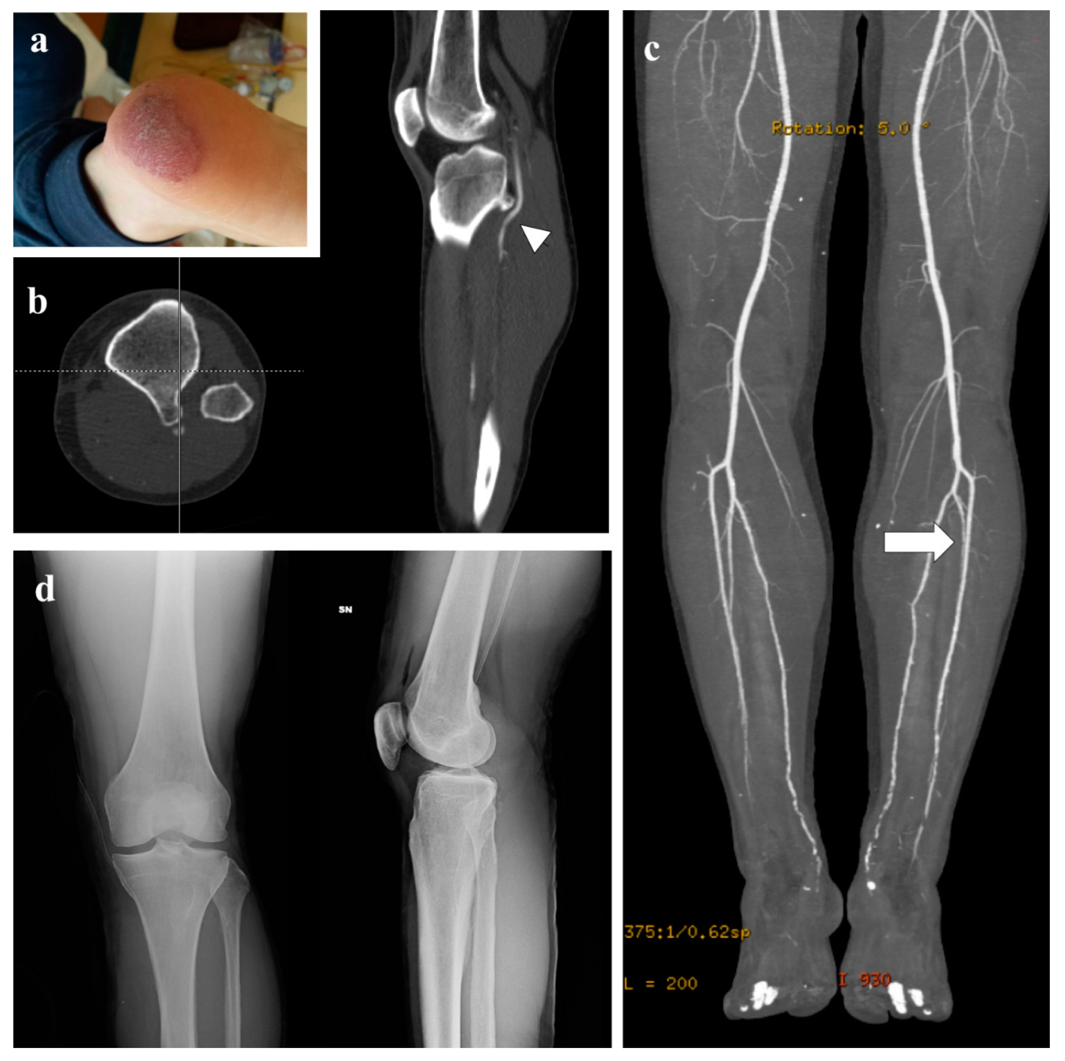
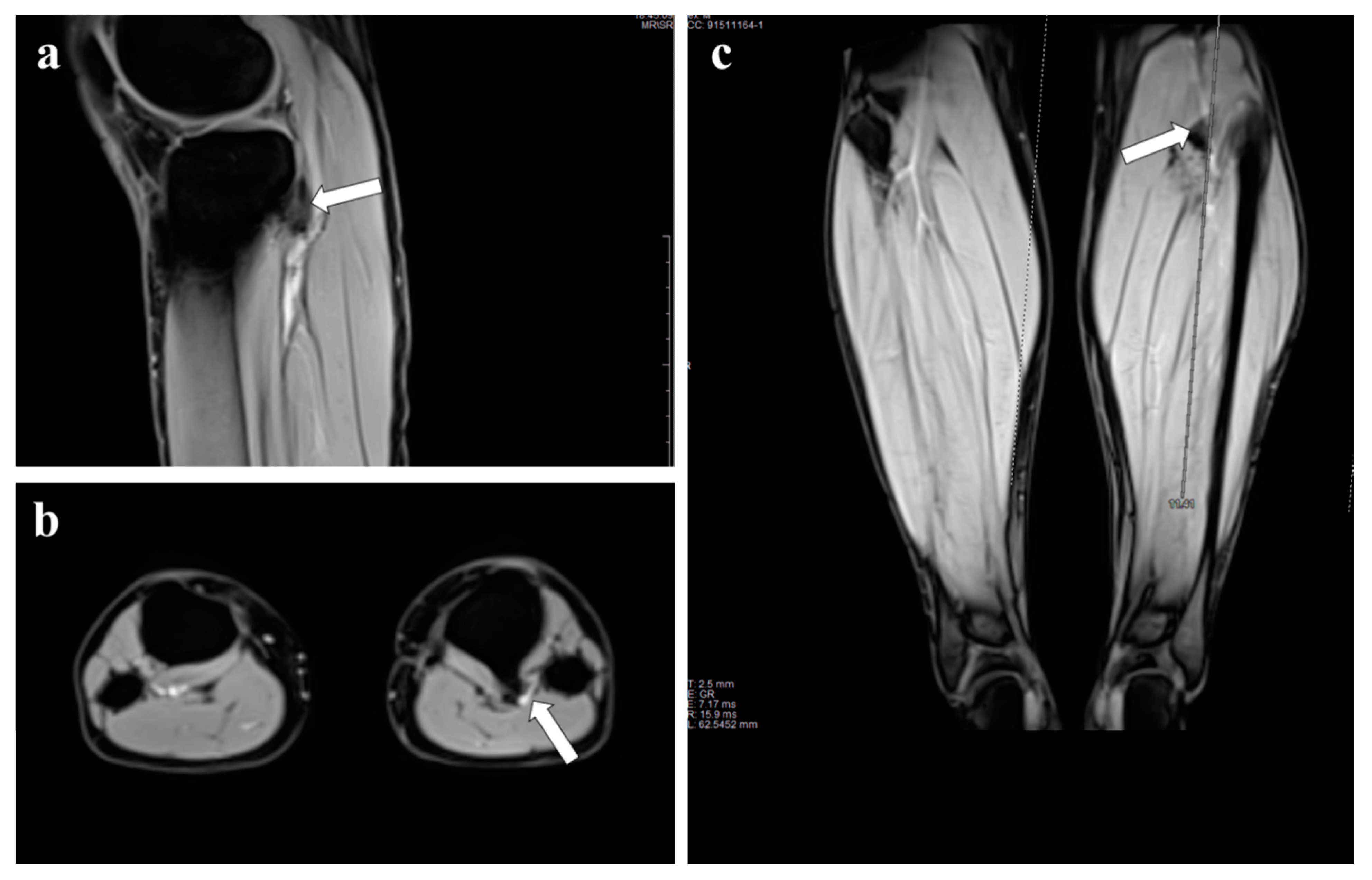
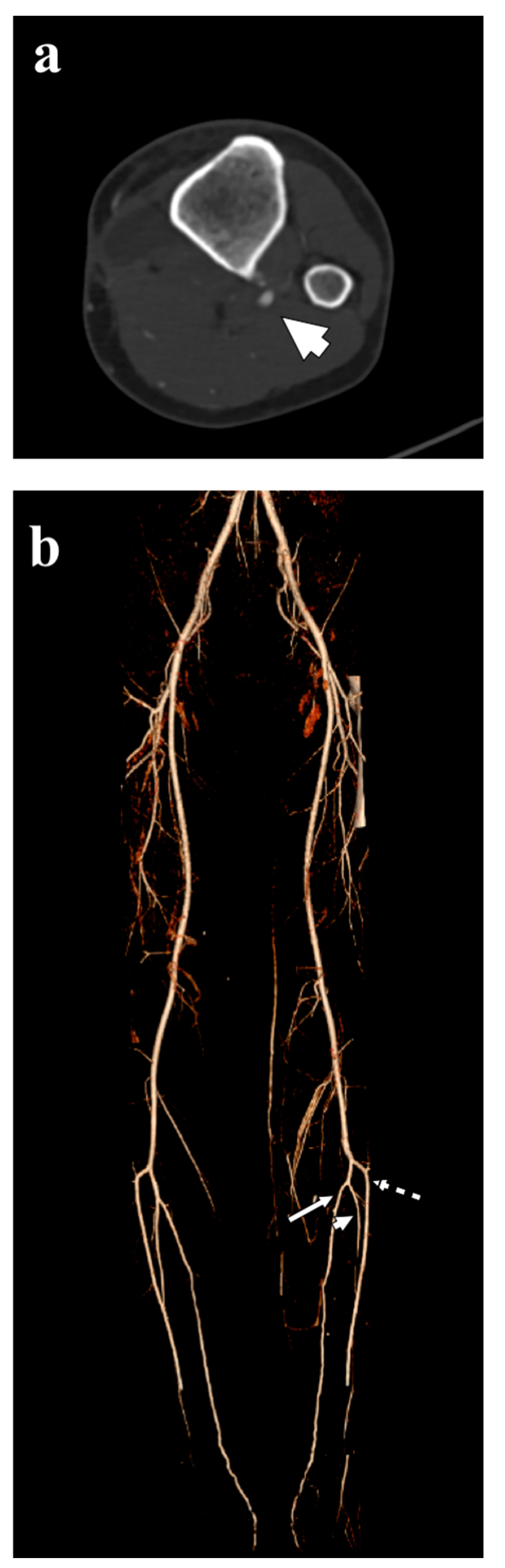
3. Representative Explanatory Case

A 34-year-old male was referred to the emergency department with acute left lower limb ischemia.
His clinical history showed a similar ischemia at the same lower left limb four years before and other temporary symptoms. Oncologic history included acute lymphoblastic leukemia treated with chemotherapy, radiotherapy and bone marrow transplantation, and thyroidectomy for papillary thyroid carcinoma. He reported a gradual decrease of walking distance with increasing pain, cold and functional impairment of lower limb. On clinical examination he presented ecchymosis at heel level (Figure 1a).
Physical examination revealed loss of popliteal and peripheral pulses whereas the ipsilateral femoral pulses were normal, with no neurological deficit of the affected limb. A CT scan revealed an osteochondroma on the posterior face of the tibial plateau, close to the vascular bundle (Figure 1b), and the CT angiogram showed thrombosis of the subarticular popliteal artery (Figure 1c). Plain radiographs (Figure 1d) and contrast-enhanced MRI showed the pedunculated tumor with a thin cartilaginous cup (Figure 2).
He was urgently treated by vascular surgeons with intra-arterial locoregional thrombolytic therapy in place for four days. At subsequent checks, occlusion remained in the upper third of the interosseous artery. Interestingly, at ultrasound doppler evaluation the blood flow disappears at complete extension of the knee and appears during flexion. The patient started oral anticoagulant therapy with enoxaparin 4000 UI twice a day at first, followed by warfarin for six months. Two months later, a surgical resection was performed in our orthopedic department: using a posteromedial approach, the bone surface was reached, the lesion was isolated and a tangential resection was performed (Figure 3a). Histo-pathology evaluation confirmed the diagnosis of osteochondroma with no malignant features. The postoperative course was uneventful with palpable distal pulses, decreasing oedema and marked pain relief. Physical rehabilitation was not required, with immediate full recovery resuming his regular activities. At two years follow up, the patient was asymptomatic and CT angiogram showed normal triphasic arterial waves in the peripheral arterial circulation (Figure 3b).

4. Discussion

Osteochondromas are the most common benign primary bone tumors in growing patients representing about 35–45% of all benign bone tumors. About 90% of these occur sporadically and are usually solitary [1,13,29,30,59]. Osteochondromas occur most often around the knee (40% of cases) and proximal tibial is affected in 15–20% of cases [59]. Symptoms are usually related with compression of close related structure: tumors can occasionally dislocate the adjacent vessels and cause various types of vascular injury such as pseudoaneurysms, arterial thrombosis or acute lower limb ischemia [4,6,13,29,30,56,59]. However, vascular complications are an atypical presentation of osteochondromas. The affected vessel is commonly the popliteal artery, which is involved in about 60% of the cases. Rarely, osteochondromas may be complicated by fracture secondary to strenuous physical exercise or trauma, resulting in sharp bone spicules that pierce the artery, causing pseudoaneurysm formation [30]. Sakata et al. reported a pseudoaneurysm of the popliteal artery in a 16-year-old boy that underwent emergency surgery. Authors concluded that all vascular surgeons should know this complication and perform early surgical intervention due to the risk of occlusion of peripheral branches, deep venous thrombosis and peripheral nerve disorders [60]. Shah at al. reported an unusual case of claudication in a young adult caused by multiple ostheocondromas [29]. Adult patients with asymptomatic osteochondromas may also present a pseudoaneurysm of the popliteal artery [29]. Pseudoaneurysms are the most common vascular complications caused by ostheocondroma, but thromboembolic complications are rare, such as lower limb ulcer [56]. The pathogenesis and development of these vascular complications are unknown because the exostoses are usually not detected until the secondary symptoms appear. Suggested mechanisms include ossification of the cartilage cap and trauma to the region either from blunt impact or vigorous exercise. Some authors reported a case of exercise-induced lower extremity claudication due to popliteal artery occlusion from a bony exostosis, in which symptoms began after the patient experienced an acute painful episode while running [61,62]. Deep venous thrombosis in association with osteochondroma is infrequent. The osteochondroma exerts mass effect on the vein inducing thrombosis [30]. In our patient, the symptoms are related to transient acute ischemia of the lower limbs and osteochondroma was occasionally found in differential diagnosis with other vascular diseases. Arterial pseudoaneurysms are the most common vascular complication observed in HME. The lesion mechanism is due to a repeated abrasion of the arterial wall by a prominent osteochondroma, which erodes the arterial surface and generates the pseudoaneurysm [58,63]. Even though these pseudoaneurysms can be presented in any artery of the body, the popliteal artery represents the most frequent affected vessel (77%) [58]. Shah at al. reported an unusual case of claudication in a young adult caused by multiple ostheocondromas [61].

5. Diagnosis

Pedunculated osteochondromas around the knee have a typical appearance, extending away from the adjacent joint, described as stalactites and stalagmites. In any bone that develops from enchondral ossification, osteochondromas have a marrow, cortical and periosteum continuity with the underlying native bone. This direct continuity clearly appreciable in CT scan is a pathognomonic finding [59]. The cartilaginous cup is the most important area that should be investigated. On MRI, the cartilaginous cap is typically hypointense on T1-weighted images and variable on T2 images (hypointense or hyperintense if densely or poorly mineralized, respectively) [59]. Angiography and other vascular studies should be considered in the presence of vascular compression or for surgical planning [7,12,19,39]. Sometimes these exams are helpful to characterize neovascularity, which may signal malignant transformation [7,19].
It is important to include bone tumors in differential diagnosis in patients with vascular symptoms. The adequate approach for these patients includes clinical evaluation, plain radiographs, CT scan and MRI. In the presence of bone tumors, CT and MRI are the most sensitive examinations to exclude malignant appearance, soft tissue extension and signs of popliteal entrapment syndrome (PES). In young adult patients presenting with intermittent claudication or vascular insufficiency mimicking atherosclerotic peripheral vascular disease, alternative causes such as trauma, tumors, thromboangioitis obliterans, fibromuscular dysplasia, vasculitis and PES should be suspected [56]. PES refers to a group of symptoms caused by mechanical compression of popliteal artery, vein or tibial nerve in the popliteal fossa. This is usually related to musculotendinous structures, but a few case reports described bony exostoses of the popliteal fossa causing PES [3,6]. Angiography and ultrasound are useful to define the precise level of vascular defects. In patients with HME, popliteal pseudoaneurysms is more frequently diagnosed in the context of a non–traumatic event (65%) than after trauma (35%), and symptoms consist of pain and swelling on the popliteal fossa with preserved distal blood flow [64]. Nevertheless, the presence of ecchymosis or pulsatile mass in the popliteal region is not frequently observed but is highly suggestive of a popliteal pseudoaneurysm [64]. Duplex ultrasound is the gold of standard for diagnosis of pseudoaneurysms because it can clearly depict the vascular lesion and its relationship with the osteochondroma [58]. However, duplex ultrasound has some limitations in patients with obesity or huge hematomas due to more thickness between the vascular structures and the transductor. In these cases, some authors have argued in favour of angiography to depict the anatomic details before surgery. [51].
With the reported case, as underlined by other Authors, we strongly support the use of doppler ultrasound because it could be used as a functional examination showing the blood flow asset during complete extension of the knee [1,3,29,56,61,62,65]. Given the benign nature of osteochondromas, surgical resection is considered the gold standard of treatment, however we suggest a combined vascular-orthopedic approach from the onset of symptoms and diagnosis [66]. When surgery is planned, a complete excision of the cartilaginous cap is critical to prevent recurrence.

6. Treatment

The main consideration about management is how to approach the vascular complication considering that treatment of osteochondroma is mainly simple surgical resection at the base of the tumor with meticulous dissection of the neurovascular structures. Most cases were managed by osteochondroma resection with or without open vascular reconstruction. Open surgical options include bypass and interposition grafting, direct repair, patch angioplasty, and aneurysm resection with direct end-to-end anastomosis. Arterial reconstruction should be considered by either closing the arterial defect in cases of pseudoaneurysm formation or using a saphenous vein graft in cases of arterial thrombosis and extensive arterial damage. From the literature review, pseudoaneurysm resection associated with venous bypass or end-to-end anastomosis represent the most frequent surgical approach, because of the high risk of recurrent pseudoaneurysm or stenosis after direct closure. Vasseur et al. reviewed 97 patients with osteochondroma associated with vascular complications, surgically repaired [19]. Among them, direct closure was performed in 25 cases with a higher risk of recurrent pseudoaneurysm if stress is applied to the suture line (as the patient ages). Moreover, revascularization is considered the gold standard surgical option for preventing tissue damage or limb loss from embolic events in the setting of acute limb ischemia [3]. Most of the surgeons suggest a complete removal of the damaged wall and the use of patch or saphenous vein for defect repair, to reduce the rate of late complications.
Endovascular management is infrequently described. In two hybrid procedures, one patient underwent thrombectomy for embolus in the tibioperoneal trunk with subsequent vein patching of the popliteal pseudoaneurysm, and another had coil embolization of the pseudoaneurysm followed by direct repair of the artery one week later [62,63]. Sometimes, treatment of vascular complication consists of emergency surgery. Banno et al. reported a pseudoaneurysm of the popliteal artery in a 16-year-old boy with HMO [55]. Duarte at al. recently reported a 15-year-old patient with a history of current massages as part of his gym routine, who arrived at the emergency department with four days of pain, and ecchymosis in the right popliteal region: duplex ultrasonography and arteriography confirming the diagnosis of popliteal pseudoaneurysm [58]. Nasr at al. described two cases of vascular complications that occurred in a solitary form and the other with HMO, for a lower limb ischemia and pseudoaneurysm of the left superficial femoral artery [56]. Chen at al. reported a large literature review on 130 vascular complications from osteochondroma in all sites. Vascular complications were due to osteochondromas of the femur (66.2%), tibia (15.4%), fibula (7.7%), humerus (5.4%), clavicle (1.5%), ribs (1.5%), pubic ramus (0.8%), scapula (0.8%) and cervical vertebra (0.8%). The popliteal artery was involved in 66% of cases, and popliteal pseudoaneurysms were the most prevalent vascular complication (49%) [30]. In the same paper the case of a 38-year-old man was reported, presenting with left calf pain and swelling due to a popliteal pseudoaneurysm and incidental peroneal vein thrombosis secondary to a fractured femoral sessile osteochondroma, treated with resection of the osteochondroma, excision of the aneurysm, and primary end-to-end anastomosis of the artery [30].
Local recurrence of osteochondroma after resection is low at 1.8% but may be higher in skeletally immature patients [26,30]. Two cases of delayed pseudoaneurysm formation after incomplete osteochondroma resection have been reported. In one case a partially resected osteochondroma resulted in pseudoaneurysm formation five years later because of ongoing arterial trauma from the residual tumor’s irregular edges [66]. Scotti et al. reported brachial pseudoaneurysm presenting with rupture secondary to a partially resected osteochondroma three years earlier [67]. In our case, combined vascular-orthopedic approach was initiated with intra-arterial locoregional thrombolytic therapy in place for four days and for the surgical tangential resection of the tibial osteochondroma.

7. Conclusions

Osteochondromas are the most common bone tumors. Most are asymptomatic and discovered incidentally. This frequent benign tumor may occasionally cause severe arterial complications requiring surgical urgent procedures. CT scan and MRI with contrast medium are adequate for diagnosis, even if simple functional doppler ultrasound is able to show clearly the correlation between vascular compression and ostheocondroma. A combined vascular-orthopedic approach is mandatory from diagnosis to surgical management, and it is crucial for limb salvage procedure in cases of vascular involvement.

Author Contributions

Conceptualization, project administration: A.A., M.C.; data curation and formal analysis: A.A., M.C.; supervision: P.R.; writing—original draft: M.C., C.M.; writing—review and editing: A.A., M.C. All authors have read and agreed to the published version of the manuscript.

Funding

There was no external funding source in support of this study.

Informed Consent Statement

Written informed consent was obtained from the patient for publication of this case report and accompanying images.

Data Availability Statement

Manuscript data are embedded in the text and fully available on specific request.

Conflicts of Interest

Ruggieri P is consultant for Stryker and Exactech (not relevant with the present manuscript). The other authors declare that there are no relationships/conditions/circumstances that present a potential conflict of interest with the present manuscript.

References

  1. Alonso-Gómez, N.; Diego, M.C.-D.; Martínez-Izquierdo, A.; Sáinz-González, F. Unusual clinical complication: Acute lower limb ischemia caused by a tibial osteochondroma. Reumatol. Clin. 2019, 15, 182–184. [Google Scholar] [CrossRef] [PubMed]
  2. Tepelenis, K.; Papathanakos, G.; Kitsouli, A.; Troupis, T.; Barbouti, A.; Vlachos, K.; Kanavaros, P.; Kitsoulis, P. Osteochondromas: An Updated Review of Epidemiology, Pathogenesis, Clinical Presentation, Radiological Features and Treatment Options. In Vivo 2021, 35, 681–691. [Google Scholar] [CrossRef] [PubMed]
  3. Argyriou, C.; Drosos, G.; Tottas, S.; Tasopoulou, K.-M.; Kougioumtzis, I.; Georgiadis, G.S. A Rare Case of Tibioperoneal Arterial Trunk Entrapment Caused by a Fibular Osteochondroma. Ann. Vasc. Surg. 2019, 55, 308.e11–308.e16. [Google Scholar] [CrossRef]
  4. Tanigawa, N.; Kariya, S.; Kojima, H.; Komemushi, A.; Fujii, H.; Sawada, S. Lower limb ischaemia caused by fractured osteochondroma of the femur. Br. J. Radiol. 2007, 80, e78–e80. [Google Scholar] [CrossRef]
  5. Rossi, G.; Mavrogenis, A.F.; Angelini, A.; Rimondi, E.; Battaglia, M.; Ruggieri, P. Vascular complications in orthopaedic surgery. J. Long-Term Eff. Med. Implants 2011, 21, 127–137. [Google Scholar] [CrossRef]
  6. Muneer, K.; Goyal, K.K.; Gupta, H.; Sajeev, C.G. Popliteal artery thrombosis caused by osteochondroma: A rare presentation. Heart India 2016, 4, 153–155. [Google Scholar]
  7. Eschelman, D.J.; Gardiner, G.A.; Deely, D.M. Osteochondroma: An Unusual Cause of Vascular Disease in Young Adults. J. Vasc. Interv. Radiol. 1995, 6, 605–613. [Google Scholar] [CrossRef]
  8. Gruber-Szydło, K.; Poręba, R.; Belowska-Bień, K.; Derkacz, A.; Badowski, R.; Andrzejak, R.; Szuba, A. Popliteal artery thrombosis secondary to a tibial osteochondroma. Vasa 2011, 40, 251–255. [Google Scholar] [CrossRef]
  9. Masson, A.F.; Pullan, J.M. Aneurysm complicating exostosis. Br. J. Surg. 1966, 53, 929–932. [Google Scholar] [CrossRef]
  10. Kover, J.H.; Schwalbe, N.; Levowitz, B.S. Popliteal aneurysm due to osteochondroma in athletic injury. N. Y. State J. Med. 1970, 70, 3001–3003. [Google Scholar]
  11. Metras, D.; Coulibaly, A.O.; Calvy, H.; Acquaviva, P.; Ouattara, K. Thrombose artérielle fémoro-poplitée un case exceptionnel de complication vasculaire par exostose [Arterial thrombosis of the femoro-popliteal axis. An exceptional case of vascular complication by exostosis (author’s transl)]. J. Mal. Vasc. 1981, 6, 289–291. (In French) [Google Scholar] [PubMed]
  12. Männer, R.; Mäkinen, E. Angiographic findings in a false popliteal aneurysm due to osteochondroma of the femur. Pediatr. Radiol. 1975, 3, 244–246. [Google Scholar] [CrossRef] [PubMed]
  13. Smits, A.; Pavoordt, H.V.; Moll, F. Unusual Arterial Complications Caused by an Osteochondroma of the Femur or Tibia in Young Patients. Ann. Vasc. Surg. 1998, 12, 370–372. [Google Scholar] [CrossRef] [PubMed]
  14. Sadeghi-Azandaryani, M.; Mendl, N.; Rademacher, A.; Hoffmann, U.; Steckmeier, B.; Heyn, J. Pseudoaneurysm of the popliteal artery due to osteochondroma of the distal femur. Vasa 2010, 39, 274–277. [Google Scholar] [CrossRef] [PubMed]
  15. Solhaugh, J.H.; Olerud, S.E. Pseudoaneurysm of the femoral artery caused by osteochondroma of the femur. A case report. J. Bone Jt. Surg. Am. 1975, 57, 867–868. [Google Scholar] [CrossRef]
  16. Leve, L.; Kalideen, J.M. Popliteal false aneurysm complicating osteochondroma. A case report. S. Afr. Med. J. 1979, 55, 1087–1088. [Google Scholar]
  17. Ennker, J.; Freyschmidt, J.; Reilmann, H.; Dimovski, D.; Refmann, H. False aneurysm of the femoral artery due to an osteochondroma. Arch. Orthop. Trauma. Surg. 1984, 102, 206–209. [Google Scholar] [CrossRef]
  18. Marcove, R.C.; Lindeque, B.G.; Silane, M.F. Pseudoaneurysm of the Popliteal Artery with an Unusual Arteriographic Presentation. A case report. Clin. Orthop. Relat. Res. 1988, 234, 142–144. [Google Scholar] [CrossRef]
  19. Vasseur, M.A.; Fabre, O. Vascular complications of osteochondromas. J. Vasc. Surg. 2000, 31, 532–538. [Google Scholar] [CrossRef]
  20. Klebuc, M.; Burrow, S.; Organek, A.; Cole, W.; Zuker, R. Osteochondroma as a Causal Agent in Popliteal Artery Pseudoaneurysms: Case Report and Literature Review. J. Reconstr. Microsurg. 2001, 17, 475–480. [Google Scholar] [CrossRef]
  21. Shah, P.J.R. Aneurysm of the popliteal artery secondary to trauma from an osteochondroma of the femur: A case report and review of the literature. Br. J. Surg. 1978, 65, 786–788. [Google Scholar] [CrossRef] [PubMed]
  22. Kieffer, E.; Maraval, M.; Tricot, J.F.; Natali, J. Anévrysme artériel compliquant une exostose ostéogénique de l’extrémité supérieure du tibia [Aneurysm due to an exostosis of the upper end of the tibia (author’s transl)]. Rev. Chir. Orthop. Reparatrice Appar. Mot. 1978, 64, 155–162. (In French) [Google Scholar] [PubMed]
  23. Ferriter, P.; Hirschy, J.; Kesseler, H.; Scott, W.N. Popliteal pseudoaneurysm. A case report. J. Bone Jt. Surg. Am. 1983, 65, 695–697. [Google Scholar] [CrossRef]
  24. Belmir, H.; Azghari, A.; Mechchat, A.; Benzirar, A.; Idrissi, R.; Lekehel, B.; Sefiani, Y.; El Mesnaoui, A.; Ammar, F.; Bensaid, Y. Rupture d’un faux anévrisme de l’artère poplitée révélant une exostose du tibia: À propos d’un cas et revue de littérature [Rupture of a popliteal artery pseudo-aneurysm revealing a tibial osteochondroma: Case report and review of the literature]. J. Mal. Vasc. 2011, 36, 50–55. [Google Scholar] [CrossRef]
  25. Greenway, G.; Resnick, D.; Bookstein, J.J. Popliteal pseudoaneurysm as a complication of an adjacent osteochondroma: Angiographic diagnosis. Am. J. Roentgenol. 1979, 132, 294–296. [Google Scholar] [CrossRef] [Green Version]
  26. Otsuka, T.; Yonezawa, M.; Kamiyama, F.; Matusita, Y.; Matui, N. Popliteal pseudoaneurysm simulating soft-tissue sarcoma: Complication of osteochondroma resection. Int. J. Clin. Oncol. 2001, 6, 105–108. [Google Scholar] [CrossRef]
  27. Israels, S.; Downs, A.R. Traumatic aneurysm of the popliteal artery due to an osteochondroma of the femur. Can. J. Surg. 1980, 23, 270–272. [Google Scholar]
  28. Zini, F.; Negri, V. Su di un caso di pseudo-aneurisma popliteo da esostosi del femore. A case of popliteal pseudoaneurysm caused by femoral exostoses. Acta Biomed. Ateneo Parmense 1984, 55, 43–47. (In Italian) [Google Scholar]
  29. Taneda, Y.; Nakamura, K.; Yano, M.; Nagahama, H.; Nakamura, E.; Niina, K.; Enomoto, Y.; Onituka, T. Popliteal Artery Pseudoaneurysm Caused by Osteochondroma. Ann. Vasc. Surg. 2004, 18, 121–123. [Google Scholar] [CrossRef]
  30. Chen, R.J.-Y.; Qi, S.D.; Vaes, R.H.; Di Bella, C.; Mayer, R. Fractured osteochondroma presenting with popliteal pseudoaneurysm: Case report and review of literature. J. Vasc. Surg. Cases Innov. Tech. 2020, 6, 96–100. [Google Scholar] [CrossRef] [Green Version]
  31. Lizama, V.; Zerbini, M.; Gagliardi, R.; Howell, L. Popliteal vein thrombosis and popliteal artery pseudoaneurysm complicating osteochondroma of the femur. Am. J. Roentgenol. 1987, 148, 783–784. [Google Scholar] [CrossRef] [PubMed] [Green Version]
  32. Harrington, I.; Campbell, V.; Valazques, R.; Williams, T. Pseudoaneurysm of the Popliteal Artery as a Complication of an Osteochondroma. A review of the literature and a case report. Clin. Orthop. Relat. Res. 1991, 270, 283–287. [Google Scholar] [CrossRef]
  33. Perez-Burkhardt, J.L.; Castilla, J.C. Postraumatic popliteal pseudoaneurysm from femoral osteochondroma: Case report and review of the literature. J. Vasc. Surg. 2003, 37, 669–671. [Google Scholar] [CrossRef] [PubMed] [Green Version]
  34. Antonio, Z.P.; Alejandro, R.M.; Luis, M.R.J.; José, G.R. Femur ostochondroma and secondary pseudoaneurysm of the popliteal artery. Arch. Orthop. Trauma Surg. 2006, 126, 127–130. [Google Scholar] [CrossRef] [PubMed]
  35. Woolson, S.T.; Maloney, W.J.; James, D.R. Superficial femoral pseudoaneurysm and arterial thromboembolism caused by an osteochondroma. J. Pediatr. Orthop. 1989, 9, 335–337. [Google Scholar] [CrossRef]
  36. Asselineau, A.; Coubret, P.; Lahoud, J.C. Faux anévrysme de l’artère fémorale compliquant une exostose [False aneurysm of the femoral artery complicating exostosis]. Rev. Chir. Orthop. Reparatrice Appar. Mot. 1993, 79, 411–414. (In French) [Google Scholar]
  37. Argin, M.; Biçeroğlu, S.; Arkun, R.; Parildar, M. Solitary osteochondroma causing popliteal pseudoaneurysm that presented as a mass lesion. Diagn. Interv. Radiol. 2007, 13, 190–192. [Google Scholar]
  38. Lieberman, J.; Mazzucco, J.; Kwasnik, E.; Loyer, R.; Knight, D. Popliteal Pseudoaneurysm as a Complication of an Adjacent Osteochondroma. Ann. Vasc. Surg. 1994, 8, 198–203. [Google Scholar] [CrossRef]
  39. Ballaro, A.; Fox, A.; Collin, J. Rupture of a popliteal artery pseudo-aneurysm secondary to a fibular osteochondroma. Eur. J. Vasc. Endovasc. Surg. 1997, 14, 151–152. [Google Scholar] [CrossRef] [Green Version]
  40. Matsushita, M.; Nishikimi, N.; Sakurai, T.; Nimura, Y. Pseudoaneurysm of the popliteal artery caused by exostosis of the femur: Case report and review of the literature. J. Vasc. Surg. 2000, 32, 201–204. [Google Scholar] [CrossRef] [Green Version]
  41. Davies, R.S.; Satti, U.; Duffield, R.G. Popliteal artery pseudo-aneurysm secondary to femoral osteochondroma: A case report and literature review. Ann. R. Coll. Surg. Engl. 2007, 89, 8–11. [Google Scholar] [CrossRef] [Green Version]
  42. Petratos, D.V.; Bakogiannis, K.S.; Anastasopoulos, J.N.; Matsinos, G.S.; Bessias, N.K. Popliteal artery pseudoaneurysm secondary to osteochondroma in children and adolescents: A case report and literature review. J. Surg. Orthop. Adv. 2009, 18, 205–210. [Google Scholar]
  43. Kalinga, M.J.; Lo, N.N.; Tan, S.K. Popliteal artery pseudoaneurysm caused by an osteochondroma—A traditional medicine massage sequelae. Singap. Med. J. 1996, 37, 443–445. [Google Scholar]
  44. Hasselgren, P.O.; Eriksson, B.; Lukes, P.; Seeman, T. False popliteal aneurysm caused by exostosis of the femur. J. Cardiovasc. Surg. 1983, 24, 540–542. [Google Scholar]
  45. Wiater, J.M.; Farley, F.A. Popliteal pseudoaneurysm caused by an adjacent osteochondroma: A case report and review of the literature. Am. J. Orthop. (Belle Mead NJ) 1999, 28, 412–416. [Google Scholar]
  46. Cardon, A.; Aillet, S.; Ledu, J.; Kerdiles, Y. Pseudo-aneurysm of the popliteal artery by femoral exostosis in a young child. J. Cardiovasc. Surg. 2001, 42, 241–244. [Google Scholar]
  47. Tóth, C.; Olvasztó, S.; Dinya, T. Femurcondylus exostosisa okozta arteria poplitea sérülés ritka esete (Esetismertetés) [Rare case of popliteal artery injury caused by distal femoral exostosis. Case report]. Magy Seb. 2001, 54, 115–117. (In Hungarian) [Google Scholar]
  48. Oxenius, A.; Knirsch, W.; Kretschmar, O.; Dodge-Khatami, A.; Lamprecht, E. Unclear Swelling of the Popliteal Fossa Due to a Giant Pseudoaneurysm Associated with Osteochondroma. J. Pediatr. 2009, 154, 147. [Google Scholar] [CrossRef]
  49. Bursztyn, M.; Stracher, M.; Sanchez, J.I.; Ramenofsky, M.; Kirwin, J.; Spero, C. Pseudoaneurysm associated with multiple osteochondromatosis. J. Pediatr. Surg. 2005, 40, 1201–1203. [Google Scholar] [CrossRef]
  50. Al-Hadidy, A.M.; Al-Smady, M.M.; Haroun, A.A.; Hamamy, H.A.; Ghoul, S.M.; Shennak, A.O. Hereditary Multiple Exostoses with Pseudoaneurysm. Cardiovasc. Interv. Radiol. 2007, 30, 537–540. [Google Scholar] [CrossRef]
  51. Thévenin, F.; Dumaine, V.; Feydy, A.; Campagna, R.; Guerini, H.; Richarme, D.; Drapé, J.; Bierry, G.; Babinet, A.; Anract, P.; et al. Faux anévrisme de l’artère fémorale compliquant une maladie des exostoses multiples [False aneurysm of the femoral artery in multiple exostosis syndrome]. J. Radiol. 2009, 90, 612–614. [Google Scholar] [CrossRef]
  52. Chaouch, N.; Alimi, F.; Kortas, C.; Limayem, F.; Braham, A.; Mlika, S.; Jerbi, S.; Ennabli, K. Complications vasculaires poplitées bilatérales d’une exostose héréditaire multiple [Bilateral popliteal artery complications of multiple hereditary exostosis]. Ann. Cardiol. Angeiol. 2011, 60, 109–112. [Google Scholar] [CrossRef]
  53. Pavić, P.; Vergles, D.; Šarlija, M.; Ajduk, M.; Cupurdija, K. Pseudoaneurysm of the Popliteal Artery in a Patient with Multiple Hereditary Exostoses. Ann. Vasc. Surg. 2011, 25, 268.e1–268.e2. [Google Scholar] [CrossRef]
  54. Vanhegan, I.S.; Shehzad, K.N.; Bhatti, T.S.; Waters, T.S. Acute popliteal pseudoaneurysm rupture secondary to distal femoral osteochondroma in a patient with hereditary multiple exostoses. Ann. R. Coll. Surg. Engl. 2012, 94, e134–e136. [Google Scholar] [CrossRef] [Green Version]
  55. Banno, H.; Houbballah, R.; Becquemin, J.-P. Acute Lower Extremity Ischemia due to the Popliteal Pseudoaneurysm in a 16-year-old Boy with Multiple Exostoses. Ann. Vasc. Dis. 2013, 6, 215–217. [Google Scholar] [CrossRef] [Green Version]
  56. Nasr, B.; Albert, B.; David, C.H.; da Fonseca, P.M.; Badra, A.; Gouny, P. Exostoses and Vascular Complications in the Lower Limbs: Two Case Reports and Review of the Literature. Ann. Vasc. Surg. 2015, 29, 1315.e7–1315.e14. [Google Scholar] [CrossRef]
  57. Orawczyk, T.; Kuczmik, W.; Kazibudzki, M.; Ludyga, T.; Cwik, P.; Ziaja, K. Popliteal Pseudoaneurysm as a Rare Complication of a Solitary Tibial Osteochondroma. EJVES Extra 2006, 12, 21–23. [Google Scholar] [CrossRef] [Green Version]
  58. Duarte, O.A.P.; Neira, J.G.A.; Herazo, V.d.C.; Lara, M.F.O.; Polanco, A.L.L.; Omaña, A.F.D.; Ortiz, A.F.H. Popliteal artery pseudoaneurysm caused by non–penetrating trauma in a patient with hereditary multiple osteochondromatosis. Radiol. Case Rep. 2021, 17, 185–189. [Google Scholar] [CrossRef]
  59. Murphey, M.D.; Choi, J.J.; Kransdorf, M.J.; Flemming, D.J.; Gannon, F.H. Imaging of Osteochondroma: Variants and Complications with Radiologic-Pathologic Correlation. RadioGraphics 2000, 20, 1407–1434. [Google Scholar] [CrossRef] [Green Version]
  60. Sakata, T.; Mogi, K.; Sakurai, M.; Nomura, A.; Fujii, M.; Takahara, Y. Popliteal Artery Pseudoaneurysm Caused by Osteochondroma. Ann. Vasc. Surg. 2017, 43, 313.e5–313.e7. [Google Scholar] [CrossRef]
  61. Shah, D.; Silver, M. Osteochondroma: An unusual case of claudication in a young adult. Vasc. Med. 2019, 25, 274–275. [Google Scholar] [CrossRef]
  62. Wang, D.; Derubertis, B.G. Popliteal Artery Occlusion Secondary to Exostosis of the Femur. Perspect. Vasc. Surg. Endovasc. Ther. 2012, 24, 217–220. [Google Scholar] [CrossRef]
  63. Blazick, E.; Keeling, W.B.; Armstrong, P.; Letson, D.; Back, M. Pseudoaneurysm of the Superficial Femoral Artery Associated with Osteochondroma: A Case Report. Vasc. Endovasc. Surg. 2005, 39, 355–358. [Google Scholar] [CrossRef]
  64. Raherinantenaina, F.; Rakoto-Ratsimba, H.N.; Rajaonanahary, T.M.A. Management of extremity arterial pseudoaneurysms associated with osteochondromas. Vascular 2016, 24, 628–637. [Google Scholar] [CrossRef]
  65. Parente, B.; Fiorucci, B.; Simonte, G.; Brambilla, D.M.; Lenti, M. Osteochondromas: An Unusual Cause of Vascular Disease in Young Patients: 2 Clinical Cases. Ann. Vasc. Surg. 2016, 32, 129.e7–129.e11. [Google Scholar] [CrossRef]
  66. Angelini, A.; Piazza, M.; Pagliarini, E.; Trovarelli, G.; Spertino, A.; Ruggieri, P. The Orthopedic-Vascular Multidisciplinary Approach Improves Patient Safety in Surgery for Musculoskeletal Tumors: A Large-Volume Center Experience. J. Pers. Med. 2021, 11, 462. [Google Scholar] [CrossRef]
  67. Scotti, C.; Marone, E.M.; Brasca, L.E.; Peretti, G.M.; Chiesa, R.; Del Maschio, A.; Fraschini, G.; Camnasio, F. Pseudoaneurysm overlying an osteochondroma: A noteworthy complication. J. Orthop. Traumatol. 2010, 11, 251–255. [Google Scholar] [CrossRef] [Green Version]
Figure 1. (a) Ecchymosis at left heel. (b) Typical imaging appearance at axial and sagittal CT scan, with pedunculated osteochondroma with posterior extension and compression of arterial bundle (arrow head). (c) CT Angiogram show thrombosis of the subarticular popliteal artery (white arrow). (d) antero-posterior and lateral radiograph appear less representative for clear diagnosis of the bony lesion.
Diagnostics 12 01191 g001
Figure 2. Magnetic resonance (a) sagittal, (b) axial and (c) coronal STIR images show the marrow and cortical continuity of the pedunculated osteochondroma and underlying tibia, and with a relatively thin cartilage cup (white arrows).
Diagnostics 12 01191 g002
Figure 3. Postoperative examination. (a) Axial CT scan with contrast show the complete removal of the tumor and the vascular bundle (arrowhead). (b) Three-dimensional volume rendered CT angiography image shows complete revascularization of the anterior tibial artery (dashed arrow), peroneal artery (arrowhead) and posterior tibial artery (flat arrow).
Diagnostics 12 01191 g003
Table 1. Summary of the published reports on osteochondroma presenting with lower extremity vascular symptoms and complications.
AuthorsPtsSymptomsBone SiteTreatment of Vascular LesionAge (Years)Exostosis
Masson et al. [9]1S + PFemurPopliteal artery ligature + VB9Multiple
Refs. [10,11]2S (1), P (1) AI (1)FemurPAR + VBMean 31.5 (15–48)Solitary
Refs. [12,13,14]4S (2), P (2),AI (2)Femurn.a.Mean 22 (13–39)Solitary
Refs. [15,16,17,18,19,20]7S (5), P (4)FemurDirect repairMean 19.7 (15–20)Multiple
Shah et al. [21]1AIFemurPAR + direct repair16Solitary
Refs. [22,23,24]3S (2), P (2), AI (1)TibiaFlattening + VBMean 22 (20–23)Solitary
Refs. [25,26]2S (1) P (2)FemurPAR + EtoEAMean 18.5 (16–18)Multiple
Refs. [17,19,27,28,29,30]6S (6), P (4)FemurPAR + EtoEAMean 34.3 (14–51)Solitary
Refs. [31,32,33,34]4S (2), P (4)FemurLigature + direct repairMean 15.3 (9–16)Solitary
Refs. [35,36,37]3S (3), P (1) AI (1)FemurFlattening + VBMean 14.3 (13–16)Solitary
Refs. [38,39,40,41,42]5S (3), P (3), Pulsatile mass (1)Femur (4)
Fibula (1)
Vein patchMean 18.8 (13–33)Solitary
Refs. [19,38,43]4S (3), P (2), AI (1)FemurLigature + VBMean 22.3 (14–37)Solitary
Hasselgren et al. [44]1S + PFemurLigature + prosthetic bypass45Multiple
Smits et al. [13]1Calf swellingTibian.a.28Solitary
Wiater et al. [45]1PFemurVB17Multiple
Cardon et al. [46]1PFemurResection + EtoEA12Solitary
Toth et al. [47]1S + PFemurFlattening + prosthetic bypass17Multiple
Oxenius et al. [48]1S + PFemurPericardial patch13Solitary
Refs. [49,50,51,52,53,54,55]8S (5), P (4), C (2), AI (1)Femur (8) Flattening + VBMean 17.5 (12–21)Multiple
Nasr et al. [56]1AIFemurVein patch + EtoEA17Multiple
1S + PFemurFlattening + EtoEA17Multiple
Refs. [57,58]2S (1), P (2), C (1) TibiaDirect repairMean 14.5 (14–15)Multiple
Present case1AITibiaThrombolytic Therapy34Solitary
VB: venous bypass; PAR: Pseudoaneurysm resection; EtoEA: end-to-end anastomosis; n.a.: not available; S: Swelling; P: pain; AI: Acute ischemia; C: Claudication.
Publisher’s Note: MDPI stays neutral with regard to jurisdictional claims in published maps and institutional affiliations.

