Next Issue
Volume 12, April
Previous Issue
Volume 12, February


Diagnostics, Volume 12, Issue 3 (March 2022) – 213 articles

Cover Story (view full-size image): 
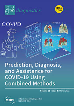
COVID-19 and its mutations, caused by the SARS-CoV-2 virus, have been reported ubiquitously worldwide, causing more infections and spreading wider than any previously known form. It infected at least 281 million individuals worldwide, causing over 5 million confirmed deaths, creating an unmatched threat to human health, food habits, financial resources, and workplaces. Currently, four key variants over 4000 SARS-CoV-2 mutations of interest are defined and require constant observations and investigations. This issue on recent COVID-19 research invites papers that aim to a) discover and classify new viral mutations and exiting ones using AI, augmented reality (AR), and virtual reality (VR), b) determine their potential consequences, and c) minimize the spreading of the novel coronavirus. View this paper
  • Issues are regarded as officially published after their release is announced to the table of contents alert mailing list.
  • You may sign up for e-mail alerts to receive table of contents of newly released issues.
  • PDF is the official format for papers published in both, html and pdf forms. To view the papers in pdf format, click on the "PDF Full-text" link, and use the free Adobe Reader to open them.
Order results
Result details
Section
Select all
Export citation of selected articles as:
11 pages, 1311 KiB  
Article
Prediction of Adverse Post-Infarction Left Ventricular Remodeling Using a Multivariate Regression Model
by Valentin Oleynikov, Lyudmila Salyamova, Olga Kvasova and Nadezhda Burko
Diagnostics 2022, 12(3), 770; https://doi.org/10.3390/diagnostics12030770 - 21 Mar 2022
Cited by 4 | Viewed by 1879
Abstract
Background. In order to provide personalized medicine and improve cardiovascular outcomes, a method for predicting adverse left ventricular remodeling (ALVR) after ST-segment elevation myocardial infarction (STEMI) is needed. Methods. A total of 125 STEMI patients, mean age 51.2 (95% CI 49.6; 52.7) years [...] Read more.
Background. In order to provide personalized medicine and improve cardiovascular outcomes, a method for predicting adverse left ventricular remodeling (ALVR) after ST-segment elevation myocardial infarction (STEMI) is needed. Methods. A total of 125 STEMI patients, mean age 51.2 (95% CI 49.6; 52.7) years were prospectively enrolled. The clinical, laboratory, and instrumental examinations were performed between the 7th and 9th day, and after 24 and 48 weeks, including plasma analysis of brain natriuretic peptide (BNP), transthoracic echocardiography, analysis of left ventricular-arterial coupling, applanation tonometry, ultrasound examination of the common carotid arteries with RF signal amplification. Results. Patients were divided into 2 groups according to echocardiography: “ALVR” (n = 63)—end-diastolic volume index (EDVI) >20% and/or end-systolic volume index (ESVI) >15% after 24 weeks compared with initial values; “non-ALVR” (n = 62)—EDVI <20% and ESVI <15%. In the ALVR group, hard endpoints (recurrent myocardial infarction, unstable angina, hospitalization for decompensated heart failure, ventricular arrhythmias, cardiac surgery, cardiovascular death) were detected in 19 people (30%). In the non-ALVR group, hard endpoints were noted in 3 patients (5%). The odds ratio of developing an adverse outcome in ALVR vs. non-ALVR group was 8.5 (95% CI 2.4–30.5) (p = 0.0004). According to the multivariate analysis, the contribution of each of the indicators to the relative risk (RR) of adverse cardiac remodeling: waist circumference, RR = 1.02 (95% CI 1.001–1.05) (p = 0.042), plasma BNP—RR = 1.81 (95% CI 1.05–3.13) (p = 0.033), arterial elastance to left ventricular end-systolic elastance (Ea/Ees)—RR = 1.96 (95% CI 1.11–3.46) (p = 0.020). Conclusion. Determining ALVR status in early stages of the disease can accurately predict and stratify the risk of adverse outcomes in STEMI patients. Full article
(This article belongs to the Special Issue Diagnosis and Management of Heart Failure)
Show Figures

Figure 1

14 pages, 2184 KiB  
Article
French Survey on Pain Perception and Management in Patients with Locked-In Syndrome
by Estelle A. C. Bonin, Zoé Delsemme, Véronique Blandin, Naji L. Alnagger, Aurore Thibaut, Marie-Elisabeth Faymonville, Steven Laureys, Audrey Vanhaudenhuyse and Olivia Gosseries
Diagnostics 2022, 12(3), 769; https://doi.org/10.3390/diagnostics12030769 - 21 Mar 2022
Cited by 3 | Viewed by 2236
Abstract
Patients with locked-in syndrome (LIS) may suffer from pain, which can significantly affect their daily life and well-being. In this study, we aim to investigate the presence and the management of pain in LIS patients. Fifty-one participants completed a survey collecting socio-demographic information [...] Read more.
Patients with locked-in syndrome (LIS) may suffer from pain, which can significantly affect their daily life and well-being. In this study, we aim to investigate the presence and the management of pain in LIS patients. Fifty-one participants completed a survey collecting socio-demographic information and detailed reports regarding pain perception and management (type and frequency of pain, daily impact of pain, treatments). Almost half of the LIS patients reported experiencing pain (49%) that affected their quality of life, sleep and cognition. The majority of these patients reported that they did not communicate their pain to clinical staff. Out of the 25 patients reporting pain, 18 (72%) received treatment (60% pharmacological, 12% non-pharmacological) and described the treatment efficacy as ‘moderate’. In addition, 14 (56%) patients were willing to try other non-pharmacological treatments, such as hypnosis or meditation. This study provides a comprehensive characterization of pain perception in LIS patients and highlights the lack of guidelines for pain detection and its management. This is especially pertinent given that pain affects diagnoses, by either inducing fatigue or by using pharmacological treatments that modulate the levels of wakefulness and concentration of such patients. Full article
(This article belongs to the Special Issue Diagnosis and Prognosis in Disorders of Consciousness)
Show Figures

Figure 1

17 pages, 13882 KiB  
Article
A Deep Learning Model for Prostate Adenocarcinoma Classification in Needle Biopsy Whole-Slide Images Using Transfer Learning
by Masayuki Tsuneki, Makoto Abe and Fahdi Kanavati
Diagnostics 2022, 12(3), 768; https://doi.org/10.3390/diagnostics12030768 - 21 Mar 2022
Cited by 13 | Viewed by 4588
Abstract
The histopathological diagnosis of prostate adenocarcinoma in needle biopsy specimens is of pivotal importance for determining optimum prostate cancer treatment. Since diagnosing a large number of cases containing 12 core biopsy specimens by pathologists using a microscope is time-consuming manual system and limited [...] Read more.
The histopathological diagnosis of prostate adenocarcinoma in needle biopsy specimens is of pivotal importance for determining optimum prostate cancer treatment. Since diagnosing a large number of cases containing 12 core biopsy specimens by pathologists using a microscope is time-consuming manual system and limited in terms of human resources, it is necessary to develop new techniques that can rapidly and accurately screen large numbers of histopathological prostate needle biopsy specimens. Computational pathology applications that can assist pathologists in detecting and classifying prostate adenocarcinoma from whole-slide images (WSIs) would be of great benefit for routine pathological practice. In this paper, we trained deep learning models capable of classifying needle biopsy WSIs into adenocarcinoma and benign (non-neoplastic) lesions. We evaluated the models on needle biopsy, transurethral resection of the prostate (TUR-P), and The Cancer Genome Atlas (TCGA) public dataset test sets, achieving an ROC-AUC up to 0.978 in needle biopsy test sets and up to 0.9873 in TCGA test sets for adenocarcinoma. Full article
(This article belongs to the Special Issue Artificial Intelligence in Pathological Image Analysis)
Show Figures

Figure 1

17 pages, 2236 KiB  
Review
Optic Nerve Sheath Diameter Ultrasound: A Non-Invasive Approach to Evaluate Increased Intracranial Pressure in Critically Ill Pediatric Patients
by Giulia Cannata, Stefano Pezzato, Susanna Esposito and Andrea Moscatelli
Diagnostics 2022, 12(3), 767; https://doi.org/10.3390/diagnostics12030767 - 21 Mar 2022
Cited by 10 | Viewed by 7969
Abstract
Early diagnosis of increased intracranial pressure (ICP) is crucial for prompt diagnosis and treatment of intracranial hypertension in critically ill pediatric patients, preventing secondary brain damage and mortality. Although the placement of an external ventricular drain coupled to an external fluid-filled transducer remains [...] Read more.
Early diagnosis of increased intracranial pressure (ICP) is crucial for prompt diagnosis and treatment of intracranial hypertension in critically ill pediatric patients, preventing secondary brain damage and mortality. Although the placement of an external ventricular drain coupled to an external fluid-filled transducer remains the gold standard for continuous ICP monitoring, other non-invasive approaches are constantly being improved and can provide reliable estimates. The use of point-of-care ultrasound (POCUS) for the assessment of ICP has recently become widespread in pediatric emergency and critical care settings, representing a valuable extension of the physical examination. The aim of this manuscript is to review and discuss the basic principles of ultra-sound measurement of the optic nerve sheath diameter (ONSD) and summarize current evidence on its diagnostic value in pediatric patients with ICP. There is increasing evidence that POCUS measurement of the ONSD correlates with ICP, thus appearing as a useful extension of the physical examination in pediatrics, especially in emergency medicine and critical care settings for the initial non-invasive assessment of patients with suspected raised ICP. Its role could be of value even to assess the response to therapy and in the follow-up of patients with diagnosed intracranial hypertension if invasive ICP monitoring is not available. Further studies on more homogeneous and extensive study populations should be performed to establish ONSD reference ranges in the different pediatric ages and to define cut-off values in predicting elevated ICP compared to invasive ICP measurement. Full article
(This article belongs to the Special Issue Point-of-Care Ultrasonography in Critical Care and Emergency Medicine)
Show Figures

Figure 1

18 pages, 3999 KiB  
Article
Intracranial Flow Volume Estimation in Patients with Internal Carotid Artery Occlusion
by Piotr Kaszczewski, Michał Elwertowski, Jerzy Leszczyński, Tomasz Ostrowski, Joanna Kaszczewska and Zbigniew Gałązka
Diagnostics 2022, 12(3), 766; https://doi.org/10.3390/diagnostics12030766 - 21 Mar 2022
Cited by 4 | Viewed by 2289
Abstract
(1) Background: Carotid artery occlusion (CAO) in population studies has a reported prevalence of about 6 per 100,000 people; however, the data may be underestimated. CAO carries a significant risk of stroke. Up to 15% of large artery infractions may be secondary to [...] Read more.
(1) Background: Carotid artery occlusion (CAO) in population studies has a reported prevalence of about 6 per 100,000 people; however, the data may be underestimated. CAO carries a significant risk of stroke. Up to 15% of large artery infractions may be secondary to the CAO, and in 27–38% of patients, ischaemic stroke is a first presentation of the disease. The presence of sufficient and well-developed collateral circulation has a protective influence, being a good prognostic factor in patients with carotid artery disease, both chronic and acute. Understanding the mechanisms and role of collateral circulation may be very important in the risk stratification of such patients. (2) Materials and Methods: This study included 46 patients (mean age: 70.5 ± 6 years old; 15 female, mean age 68.5 ± 3.8 years old and 31 male, mean age 71.5 ± 6.7 years old) with unilateral or bilateral ICA occlusion. In all patients, a Doppler ultrasound (DUS) examination, measuring blood flow volume in the internal carotid artery (ICA), external carotid artery (ECA), and vertebral artery (VA), was performed. The cerebral blood flow (CBF) was compared to the previously reported CBF values in the healthy population >65 years old. (3) Results: In comparison with CBF values in the healthy population, three subgroups with CBF changes were identified among patients with ICA occlusion: patients with significant volumetric flow compensation (CBF higher than average + standard deviation for healthy population of the same age), patients with flow similar to the healthy population (average ± standard deviation), and patients without compensation (CBF lower than the average-standard deviation for healthy population). The percentage of patients with significant volumetric flow compensation tend to rise with increasing age, while a simultaneous decline was observed in the group without compensation. The percentage of patients with flow similar to the healthy population remained relatively unchanged. ICA played the most important role in volumetric flow compensation in patients with CAO; however, the relative increase in flow in the ICA was smaller than that in the ECA and VA. Compensatory increased flow was observed in about 50% of all patent extracranial arteries and was more frequently observed in ipsilateral vessels than in contralateral ones, in both the ECA and the VA. In patients with CAO, there was no decrease in CBF, ICA, ECA, and VA flow volume with increasing age. (4) Conclusions: Volumetric flow compensation may play an important predictive role in patients with CAO. Full article
(This article belongs to the Special Issue Advances in Carotid Artery Imaging)
Show Figures

Figure 1

23 pages, 19180 KiB  
Article
WMR-DepthwiseNet: A Wavelet Multi-Resolution Depthwise Separable Convolutional Neural Network for COVID-19 Diagnosis
by Happy Nkanta Monday, Jianping Li, Grace Ugochi Nneji, Md Altab Hossin, Saifun Nahar, Jehoiada Jackson and Ijeoma Amuche Chikwendu
Diagnostics 2022, 12(3), 765; https://doi.org/10.3390/diagnostics12030765 - 21 Mar 2022
Cited by 2 | Viewed by 2664
Abstract
Timely discovery of COVID-19 could aid in formulating a suitable treatment plan for disease mitigation and containment decisions. The widely used COVID-19 test necessitates a regular method and has a low sensitivity value. Computed tomography and chest X-ray are also other methods utilized [...] Read more.
Timely discovery of COVID-19 could aid in formulating a suitable treatment plan for disease mitigation and containment decisions. The widely used COVID-19 test necessitates a regular method and has a low sensitivity value. Computed tomography and chest X-ray are also other methods utilized by numerous studies for detecting COVID-19. In this article, we propose a CNN called depthwise separable convolution network with wavelet multiresolution analysis module (WMR-DepthwiseNet) that is robust to automatically learn details from both spatialwise and channelwise for COVID-19 identification with a limited radiograph dataset, which is critical due to the rapid growth of COVID-19. This model utilizes an effective strategy to prevent loss of spatial details, which is a prevalent issue in traditional convolutional neural network, and second, the depthwise separable connectivity framework ensures reusability of feature maps by directly connecting previous layer to all subsequent layers for extracting feature representations from few datasets. We evaluate the proposed model by utilizing a public domain dataset of COVID-19 confirmed case and other pneumonia illness. The proposed method achieves 98.63% accuracy, 98.46% sensitivity, 97.99% specificity, and 98.69% precision on chest X-ray dataset, whereas using the computed tomography dataset, the model achieves 96.83% accuracy, 97.78% sensitivity, 96.22% specificity, and 97.02% precision. According to the results of our experiments, our model achieves up-to-date accuracy with only a few training cases available, which is useful for COVID-19 screening. This latest paradigm is expected to contribute significantly in the battle against COVID-19 and other life-threatening diseases. Full article
Show Figures

Figure 1

13 pages, 9671 KiB  
Review
Clinical Applications of Fetal MRI in the Brain
by Usha D. Nagaraj and Beth M. Kline-Fath
Diagnostics 2022, 12(3), 764; https://doi.org/10.3390/diagnostics12030764 - 21 Mar 2022
Cited by 7 | Viewed by 5949
Abstract
Fetal magnetic resonance imaging (MRI) has become a widely used tool in clinical practice, providing increased accuracy in prenatal diagnoses of congenital abnormalities of the brain, allowing for more accurate prenatal counseling, optimization of perinatal management, and in some cases fetal intervention. In [...] Read more.
Fetal magnetic resonance imaging (MRI) has become a widely used tool in clinical practice, providing increased accuracy in prenatal diagnoses of congenital abnormalities of the brain, allowing for more accurate prenatal counseling, optimization of perinatal management, and in some cases fetal intervention. In this article, a brief description of how fetal ultrasound (US) and fetal MRI are used in clinical practice will be followed by an overview of the most common reasons for referral for fetal MRI of the brain, including ventriculomegaly, absence of the cavum septi pellucidi (CSP) and posterior fossa anomalies. Full article
(This article belongs to the Special Issue MR Imaging of Pediatric Brain Diseases and Injury)
Show Figures

Figure 1

12 pages, 4771 KiB  
Review
Overview of Lung Ultrasound in Pediatric Cardiology
by Massimiliano Cantinotti, Pietro Marchese, Raffaele Giordano, Eliana Franchi, Nadia Assanta, Vivek Jani, Shelby Kutty and Luna Gargani
Diagnostics 2022, 12(3), 763; https://doi.org/10.3390/diagnostics12030763 - 21 Mar 2022
Cited by 8 | Viewed by 3701
Abstract
Lung ultrasound (LUS) is increasing in its popularity for the diagnosis of pulmonary complications in acute pediatric care settings. Despite the high incidence of pulmonary complications for patients with pediatric cardiovascular and congenital heart disease, especially in children undergoing cardiac surgery, the use [...] Read more.
Lung ultrasound (LUS) is increasing in its popularity for the diagnosis of pulmonary complications in acute pediatric care settings. Despite the high incidence of pulmonary complications for patients with pediatric cardiovascular and congenital heart disease, especially in children undergoing cardiac surgery, the use of LUS remains quite limited in these patients. The aim of this review is to provide a comprehensive overview and list of current potential applications for LUS in children with congenital heart disease, post-surgery. We herein describe protocols for LUS examinations in children, discuss diagnostic criteria, and introduce methods for the diagnosis and classification of pulmonary disease commonly encountered in pediatric cardiology (e.g., pleural effusion, atelectasis, interstitial edema, pneumothorax, pneumonia, and diaphragmatic motion analysis). Furthermore, applications of chest ultrasounds for the evaluation of the retrosternal area, and in particular, systematic search criteria for retrosternal clots, are illustrated. We also discussed the potential applications of LUS, including the guidance of interventional procedures, namely lung recruitment and drainage insertion. Lastly, we analyzed current gaps in knowledge, including the difficulty of the quantification of pleural effusion and atelectasis, and the need to differentiate different etiologies of B-lines. We concluded with future applications of LUS, including strain analysis and advanced analysis of diaphragmatic mechanics. In summary, US is an easy, accurate, fast, cheap, and radiation-free tool for the diagnosis and follow-up of major pulmonary complications in pediatric cardiac surgery, and we strongly encourage its use in routine practice. Full article
(This article belongs to the Special Issue Lung Ultrasound: A Leading Diagnostic Tool)
Show Figures

Figure 1

10 pages, 8129 KiB  
Case Report
Successful Full-Term Delivery via Selective Ectopic Embryo Reduction Accompanied by Uterine Cerclage in a Heterotopic Cesarean Scar Pregnancy: A Case Report and Literature Review
by Hyoeun Kim, Ji Hye Koh, Jihee Lee, Yeongeun Sim, Sang-Hun Lee, Soo-Jeong Lee, Jun-Woo Ahn, Hyun-Jin Roh and Jeong Sook Kim
Diagnostics 2022, 12(3), 762; https://doi.org/10.3390/diagnostics12030762 - 21 Mar 2022
Viewed by 4798
Abstract
Heterotopic cesarean scar pregnancy (HCSP) is a combination of cesarean scar pregnancy (CSP) and intrauterine pregnancy (IUP). Cesarean scar pregnancy is accompanied by life-threatening complications, such as uterine rupture and massive bleeding. Herein, we present a case of HCSP treated with selective potassium [...] Read more.
Heterotopic cesarean scar pregnancy (HCSP) is a combination of cesarean scar pregnancy (CSP) and intrauterine pregnancy (IUP). Cesarean scar pregnancy is accompanied by life-threatening complications, such as uterine rupture and massive bleeding. Herein, we present a case of HCSP treated with selective potassium chloride injection into the CSP under ultrasonography in association with uterine cerclage to control vaginal bleeding; this led to a successful IUP preservation and full-term delivery. Additionally, we will review several previous reports on HCSP management, including our case. Full article
(This article belongs to the Special Issue Diagnosis and Management for Obstetric and Gynecologic Diseases 2.0)
Show Figures

Graphical abstract

14 pages, 1897 KiB  
Article
Influence of the COVID-19 Pandemic on the Subjective Life Satisfaction of South Korean Adults: Bayesian Nomogram Approach
by Haewon Byeon
Diagnostics 2022, 12(3), 761; https://doi.org/10.3390/diagnostics12030761 - 21 Mar 2022
Cited by 4 | Viewed by 2157
Abstract
To understand the changes in the lives of adults living in local communities due to the COVID-19 pandemic, it is necessary to identify subjective life satisfaction and to understand key factors affecting life satisfaction. This study identified the effect on life satisfaction of [...] Read more.
To understand the changes in the lives of adults living in local communities due to the COVID-19 pandemic, it is necessary to identify subjective life satisfaction and to understand key factors affecting life satisfaction. This study identified the effect on life satisfaction of COVID-19 using epidemiological data representing adults in South Korean communities and developed a model for predicting the factors adversely affecting life satisfaction by applying a Bayesian nomogram. The subjects of this study were 227,808 adults who were 19 years old or older. Life satisfaction was measured in units of 10 points from 0 to 100: a score of 30 or less corresponding to −1 standard deviations was reclassified as dissatisfied, and a score of 40 or more was reclassified as satisfied. The nomogram developed in this study showed that “females who were between 30 and 39 years old, living in urban areas, with fewer meetings and sleeping hours, concerned about infection for themselves and the weak in the family due to the COVID-19 pandemic, concerned about death, with a mean household monthly income of KRW 3–5 million, who were non-smokers, with poor subjective health, and an education level of college graduation or above” would have a 66% chance of life dissatisfaction due to the COVID-19 pandemic. The results of this study suggest that the government needs not only to provide economic support but also to support education on infectious diseases and customized psychological counseling programs for those at high risk of life dissatisfaction after the COVID-19 pandemic. Full article
(This article belongs to the Special Issue Artificial Intelligence Approaches for Medical Diagnostics in Korea)
Show Figures

Figure 1

12 pages, 5369 KiB  
Article
Two-Photon Vision in Age-Related Macular Degeneration: A Translational Study
by Grzegorz Łabuz, Agnieszka Zielińska, Lucy J. Kessler, Asu Rayamajhi, Katarzyna Komar, Ramin Khoramnia and Gerd U. Auffarth
Diagnostics 2022, 12(3), 760; https://doi.org/10.3390/diagnostics12030760 - 21 Mar 2022
Cited by 4 | Viewed by 1890
Abstract
The recently introduced term “two-photon vision” relates to the visual perception resulting from a simultaneous absorption of two photons by photoreceptors. In this study, we determined two-photon retinal sensitivity in age-related macular degeneration (AMD) and compared it that in normal aging. Microperimetry was [...] Read more.
The recently introduced term “two-photon vision” relates to the visual perception resulting from a simultaneous absorption of two photons by photoreceptors. In this study, we determined two-photon retinal sensitivity in age-related macular degeneration (AMD) and compared it that in normal aging. Microperimetry was performed with visible (white) light and infrared (IR) light, which was perceived as green in the two-photon stimulation. In total, 45 subjects were included with one (better) eye studied. Furthermore, best-corrected visual acuity (VA) and ocular straylight were assessed. AMD resulted in decreased median (interquartile range) logMAR VA, i.e., 0.15 (0.05; 0.24), which in normal eyes was −0.02 (−0.06; 0.02). The two groups showed comparable straylight levels. Sensitivity to IR light was significantly lower in the AMD group (p < 0.001): 8.3 (7.4, 9.3) dB than in controls 10.7 (9.7, 11.2) dB. AMD also significantly affected visible light sensitivity (p < 0.001): 14.0 (11.0; 15.5) dB vs. 18.0 (16.3; 18.9) dB. Notably, the two-photon approach yielded a lower data spread. In conclusion, AMD considerably impairs retinal sensitivity measured in the single- and two-photon realm. However, two-photon-vision microperimetry may improve the testing accuracy and offer an additional diagnostic parameter (beyond VA measurements) for retinal function assessment. Full article
(This article belongs to the Section Pathology and Molecular Diagnostics)
Show Figures

Figure 1

5 pages, 1397 KiB  
Case Report
Pregnancy in a Non-Communicating Rudimentary Horn of Unicornuate Uterus
by Yi-Cih Ma and Kim-Seng Law
Diagnostics 2022, 12(3), 759; https://doi.org/10.3390/diagnostics12030759 - 21 Mar 2022
Cited by 2 | Viewed by 3202
Abstract
We report a rare case of non-communicating rudimentary horn pregnancy (RHP). The patient presented with lower abdominal pain and underwent laparoscopic surgery in which the gestational tissue was removed without excision of the rudimentary horn and ipsilateral fallopian tube. Unicornuate uteri often coexist [...] Read more.
We report a rare case of non-communicating rudimentary horn pregnancy (RHP). The patient presented with lower abdominal pain and underwent laparoscopic surgery in which the gestational tissue was removed without excision of the rudimentary horn and ipsilateral fallopian tube. Unicornuate uteri often coexist with rudimentary horns, most of which are non-communicating. RHP is rare, and symptomatic women tend to complain of abdominal pain. Once RHP is suspected, the clinician should monitor the patient for signs of hypovolemic shock, such as hypotension, because the RHP can rupture owing to the poorly developed musculature. Early surgical intervention with removal of the rudimentary horn along with the ipsilateral fallopian tube is generally suggested to prevent future ectopic pregnancy. The theory of sperm transmigration from the contralateral oviduct has been hypothesized in non-communicating RHP. Full article
(This article belongs to the Special Issue Diagnosis and Management for Obstetric and Gynecologic Diseases 2.0)
Show Figures

Figure 1

12 pages, 9516 KiB  
Case Report
Fetal Pancreatic Hamartoma Associated with Hepatoblastoma—An Unusual Tumor Association
by Valentin Varlas, Oana Neagu, Andreea Moga, Radu Bălănescu, Roxana Bohiltea, Radu Vladareanu and Laura Balanescu
Diagnostics 2022, 12(3), 758; https://doi.org/10.3390/diagnostics12030758 - 20 Mar 2022
Cited by 4 | Viewed by 2250
Abstract
Abdominal tumor masses are a very rare disease in the fetus. The authors present the first reported case of neonatal multicystic adenomatoid hamartoma of the pancreas associated with well-differentiated fetal epithelial subtype hepatoblastoma and reveal clinical, histologic, and imagistic aspects. Case presentation: A 36-week-old [...] Read more.
Abdominal tumor masses are a very rare disease in the fetus. The authors present the first reported case of neonatal multicystic adenomatoid hamartoma of the pancreas associated with well-differentiated fetal epithelial subtype hepatoblastoma and reveal clinical, histologic, and imagistic aspects. Case presentation: A 36-week-old female newborn in whom a 25-week ultrasound showed a relatively homogeneous pancreatic echogenic mass (34 × 30 × 55 mm) with compression of the inferior vena cava and retrograde dilation. Postnatal CT showed a giant pancreatic tumor mass (113 × 70 × 60 mm), with areas enhancing contrast and cystic/necrotic areas and a hypodense, hypocaptive nodule of 8 × 6 mm located at segment IV of the liver; thrombosis of the subhepatic segment of the inferior vena cava and both renal veins. Histopathological and immunohistochemical studies confirmed the diagnosis of multicystic pancreatic adenomatoid hamartoma and well-differentiated fetal epithelial subtype hepatoblastoma. Conclusions: Pancreatic hamartoma can be difficult to diagnose (especially prenatal), with or without nonspecific symptoms. The synchronous presence of hepatoblastoma complicated the therapeutic conduct and prognosis of this case, with the diagnosis being confirmed histopathologically and immunohistochemically after liver biopsy. Full article
(This article belongs to the Special Issue Imaging of Fetal and Maternal Diseases in Pregnancy)
Show Figures

Figure 1

11 pages, 458 KiB  
Article
Diagnostic and Prognostic Roles of CDX2 Immunohistochemical Expression in Colorectal Cancers
by Hong Bae Choi, Jung-Soo Pyo, Soomin Son, Kyungdoc Kim and Guhyun Kang
Diagnostics 2022, 12(3), 757; https://doi.org/10.3390/diagnostics12030757 - 20 Mar 2022
Cited by 5 | Viewed by 2079
Abstract
The study is aimed to evaluate the diagnostic and prognostic role of the immunohistochemical expression of the Caudal-type homeobox transcription factor 2 (CDX2) in colorectal cancers (CRCs) through a meta-analysis. By searching relevant databases, 38 articles were eligible to be included in this [...] Read more.
The study is aimed to evaluate the diagnostic and prognostic role of the immunohistochemical expression of the Caudal-type homeobox transcription factor 2 (CDX2) in colorectal cancers (CRCs) through a meta-analysis. By searching relevant databases, 38 articles were eligible to be included in this study. We extracted the information for CDX2 expression rates and the correlation between CDX2 expression and clinicopathological characteristics. The estimated rates of CDX2 expression were 0.882 [95% confidence interval (CI) 0.774–0.861] and 0.893 (95% CI 0.820–0.938) in primary and metastatic CRCs, respectively. Furthermore, based on their histologic subtype, CDX2 expression rates of adenocarcinoma and medullary carcinoma were 0.886 (95% CI 0.837–0.923) and 0.436 (95% CI 0.269–0.618), respectively. There was a significant difference in CDX2 expression rates between adenocarcinoma and medullary carcinoma in the meta-regression test (p < 0.001). In addition, CDX2 expression was significantly lower in CRCs with the BRAFV600E mutation than in CRCs without mutation. Patients with CDX2 expression had better overall and cancer-specific survival rates than those without CDX2 expression. Thus, CDX2 is a useful diagnostic and prognostic marker CRCs. Full article
(This article belongs to the Section Pathology and Molecular Diagnostics)
Show Figures

Figure 1

9 pages, 4519 KiB  
Article
Subepithelial Hyalinisation Predicts Recurrence of Unicystic Ameloblastomas
by Dominic Augustine, Roopa S. Rao, Lakshminarayana Surendra, Bharti Gupta, Thuckanaickenpalayam Ragunathan Yoithapprabhunath, Pradeep Kumar Yadalam, Shazia Mushtaq, Zeeshan Hera Ahmed and Shankargouda Patil
Diagnostics 2022, 12(3), 756; https://doi.org/10.3390/diagnostics12030756 - 20 Mar 2022
Viewed by 1960
Abstract
The inductive effect of hyalinisation and its influence on the biologic behaviour of ameloblastoma variants represent a scarcely researched domain of oral pathology. The complexity of the induction effects within the odontogenic apparatus, with the involvement of both ectodermal and mesodermal tissues, is [...] Read more.
The inductive effect of hyalinisation and its influence on the biologic behaviour of ameloblastoma variants represent a scarcely researched domain of oral pathology. The complexity of the induction effects within the odontogenic apparatus, with the involvement of both ectodermal and mesodermal tissues, is responsible for diverse histopathological characteristics, hyalinisation being the major feature. The present study aims to deduce for the first time the correlation between the severity of hyalinisation (SOH) and recurrence in three unicystic ameloblastoma (UA) variants, namely, intra-luminal (UA-IL), luminal (UA-L) and mural (UA-M). Retrospectively diagnosed archival cases of UA-IL (n = 08), UA-L (n = 22) and UA-M (n = 30) were assessed for SOH and its correlation with recurrence. A subgroup comparison (between UA-IL/UA-L and UA-M) was also performed. The clinical parameters of the patients were also analysed from files for clinicopathological correlation with recurrence. Results: sub-epithelial hyalinisation (SEH) significantly correlated with the recurrence of UA-L and UA-M (p = 0.001). When the histologic types (UA-L and UA-IL vs. UA-M) were grouped and the correlation of SOH with recurrence was checked, it was observed that both groups (p = 0.001) showed strong statistical correlation. UA-M lesions with multilocular radiolucency (p = 0.001) also showed significant correlation with recurrence. SOH can be a reliable histological predictor of recurrence and of aggressive biologic behaviour in UA. The present study shows a significant association of hyalinisation with the biologic behaviour of UA. Further studies with immunohistochemical investigations could validate the presence of hyalinisation and identify the origin of the hyalinised product in UAs. Full article
(This article belongs to the Special Issue Current Concepts and Prospects of Diagnostics in Oral Diseases)
Show Figures

Figure 1

10 pages, 4016 KiB  
Article
Quantification of Abdominal Muscle Mass and Diagnosis of Sarcopenia with Cross-Sectional Imaging in Patients with Polycystic Kidney Disease: Correlation with Total Kidney Volume
by Chih-Horng Wu, Tai-Shuan Lai, Yung-Ming Chen, Chyi-Mong Chen, Shun-Chung Yang and Po-Chin Liang
Diagnostics 2022, 12(3), 755; https://doi.org/10.3390/diagnostics12030755 - 20 Mar 2022
Viewed by 2100
Abstract
Background: Polycystic kidney disease (PKD) is an inherited disorder characterized by renal cysts that may mask lean body loss. This study quantified and compared muscle mass by using computed tomography (CT) and magnetic resonance imaging (MRI) images between the PKD and control groups [...] Read more.
Background: Polycystic kidney disease (PKD) is an inherited disorder characterized by renal cysts that may mask lean body loss. This study quantified and compared muscle mass by using computed tomography (CT) and magnetic resonance imaging (MRI) images between the PKD and control groups and correlated muscle mass with total kidney volume (TKV). Methods: We retrospectively enrolled patients who had a new diagnosis of PKD from May 2015 to May 2016. The CT and MRI images at the third lumbar level were processed to measure the total abdominal muscle (TAM) area for the diagnosis of sarcopenia, and TKV was estimated using the ellipsoid formula. Results: We included 37 women and 25 men (mean age: 50.40 years) in the PKD group. There was no difference in body mass index and albumin levels, but significant differences in creatinine level (p < 0.001), TAM area (p = 0.047), and TKV (p < 0.001), were noted between the two groups. A significantly negative correlation was observed between TKV and TAM area after adjustment for body height (r = −0.217, p = 0.003). Conclusions: CT and MRI images can accurately diagnose sarcopenia, which may be masked by cysts in patients with PKD. Full article
(This article belongs to the Section Medical Imaging and Theranostics)
Show Figures

Figure 1

10 pages, 3067 KiB  
Article
Comparison of Mean Corneal Power of Annular Rings and Zones Using Swept-Source Optical Coherence Tomography
by Jing Dong, Jinhan Yao, Shuimiao Chang, Piotr Kanclerz, Ramin Khoramnia and Xiaogang Wang
Diagnostics 2022, 12(3), 754; https://doi.org/10.3390/diagnostics12030754 - 19 Mar 2022
Cited by 3 | Viewed by 2297
Abstract
This study aims to investigate differences in the mean corneal power of annular zones (corneal power measured over the inner annular zone of difference diameters) and rings (corneal power measured over a ring of different diameters) centered on the corneal apex using the [...] Read more.
This study aims to investigate differences in the mean corneal power of annular zones (corneal power measured over the inner annular zone of difference diameters) and rings (corneal power measured over a ring of different diameters) centered on the corneal apex using the swept-source optical coherence tomography technique. The mean anterior axial curvature (AAC), posterior axial curvature (PAC), and total corneal power (TCP) centered on the corneal apex with the annular rings (0–2 mm, 2–4 mm, 4–6 mm, and 6–8 mm) and zones were assessed using the ANTERION device. The paired-sample t-test was used for data comparison. For the 0–2 mm comparison, the AAC, PAC, and TCP values of rings and zones were interchangeable. For the 2–4 mm comparison, the AAC of the rings was lower than that of the zones (p = 0.004), and the TCP values of the rings were higher than that of the zones (p < 0.001). For the 4–6 mm comparison, the AAC of the rings was lower than that of the zones (p < 0.001), and the PAC and TCP values of the rings were higher than that of the zones (both p < 0.001). For the 6–8 mm comparison, the AAC of the rings was lower than that of the zones (p < 0.001), and the PAC and TCP values of the rings were higher than that of the zones (both p < 0.001). Comparisons between AAC and TCP in each sub-region showed significant differences both in the rings (p < 0.001) and the zones (p < 0.008). Differences in the AAC, PAC, and TCP measured at different diameters (2–4 mm, 4–6 mm, and 6–8 mm) of the rings and zones, centered on the corneal apex, should be noticed in clinical practice. As the diameter increases, the difference between the rings and the zones in terms of AAC, PAC, and TCP increase as well. Clinicians should also pay attention to differences between AAC and TCP for the rings and the zones within the same annular region. Full article
(This article belongs to the Section Pathology and Molecular Diagnostics)
Show Figures

Figure 1

23 pages, 3380 KiB  
Review
The Utility of Endoscopic-Ultrasonography-Guided Tissue Acquisition for Solid Pancreatic Lesions
by Hiroki Tanaka and Shimpei Matsusaki
Diagnostics 2022, 12(3), 753; https://doi.org/10.3390/diagnostics12030753 - 19 Mar 2022
Cited by 9 | Viewed by 3113
Abstract
Endoscopic-ultrasonography-guided tissue acquisition (EUS-TA) has been widely performed for the definitive diagnosis of solid pancreatic lesions (SPLs). As the puncture needles, puncture techniques, and sample processing methods have improved, EUS-TA has shown higher diagnostic yields and safety. Recently, several therapeutic target genomic biomarkers [...] Read more.
Endoscopic-ultrasonography-guided tissue acquisition (EUS-TA) has been widely performed for the definitive diagnosis of solid pancreatic lesions (SPLs). As the puncture needles, puncture techniques, and sample processing methods have improved, EUS-TA has shown higher diagnostic yields and safety. Recently, several therapeutic target genomic biomarkers have been clarified in pancreatic ductal carcinoma (PDAC). Although only a small proportion of patients with PDAC can benefit from precision medicine based on gene mutations at present, precision medicine will also be further developed for SPLs as more therapeutic target genomic biomarkers are identified. Advances in next-generation sequencing (NGS) techniques enable the examination of multiple genetic mutations in limited tissue samples. EUS-TA is also useful for NGS and will play a more important role in determining treatment strategies. In this review, we describe the utility of EUS-TA for SPLs. Full article
(This article belongs to the Special Issue Endoscopic Ultrasound-Guided Fine-Needle Aspiration (EUS-FNA))
Show Figures

Figure 1

12 pages, 1819 KiB  
Article
The Image Quality and Diagnostic Performance of CT with Low-Concentration Iodine Contrast (240 mg Iodine/mL) for the Abdominal Organs
by Moon-Hyung Choi, Young-Joon Lee and Seung-Eun Jung
Diagnostics 2022, 12(3), 752; https://doi.org/10.3390/diagnostics12030752 - 19 Mar 2022
Cited by 2 | Viewed by 2083
Abstract
Purpose: To evaluate the difference between CT examinations using 240 mgI/mL contrast material (CM) and 320 mgI/mL CM in the contrast enhancement of the abdominal organs and the diagnostic performance for focal hepatic lesions. Materials and methods: This retrospective study included 422 CT [...] Read more.
Purpose: To evaluate the difference between CT examinations using 240 mgI/mL contrast material (CM) and 320 mgI/mL CM in the contrast enhancement of the abdominal organs and the diagnostic performance for focal hepatic lesions. Materials and methods: This retrospective study included 422 CT examinations, using 240 mgI/mL iohexol (Group A, 206 examinations) and 320 mgI/mL ioversol (Group B, 216 examinations), performed between April 2019 and May 2020. Two CT scanners (single-source CT (machine A) and dual-source CT (machine B)) were used to obtain CT images. Two radiologists independently drew regions of interest (ROIs) in the liver, pancreas, spleen, kidney, aorta, portal vein, and paraspinal muscle. The signal-to-noise ratio (SNR) and contrast-to-noise ratio (CNR) were calculated for each organ. They evaluated the degree of subjective enhancement of the organs and detected/differentiated focal hepatic lesions. Results: The SNR, CNR, and subjective enhancement of most organs were significantly higher in Group B than in Group A (p < 0.05). The sensitivity and specificity for cysts and malignancy were higher than 85.0% in both groups. The sensitivity for hemangioma was lower in Group B (<75%) than in Group A. In Group A, the SNR and CNR were significantly higher in most organs with machine B than with machine A. Conclusion: Although the SNR and CNR of the abdominal organs were lower with 240 mgI/mL CM than with 320 mgI/mL CM, 240 mgI/mL CM was feasible for evaluating the liver. A CT scanner with more advanced specifications may be beneficial for examinations with 240 mgI/mL CM by using lower tube voltage. Full article
(This article belongs to the Special Issue Radiocontrast Media: Adverse Reactions and Side Effects)
Show Figures

Figure 1

16 pages, 3641 KiB  
Article
Comparison of Two Rapid Assays for the Detection of BRAF V600 Mutations in Metastatic Melanoma including Positive Sentinel Lymph Nodes
by Elodie Long-Mira, Alexandra Picard-Gauci, Sandra Lassalle, Véronique Hofman, Salomé Lalvée, Virginie Tanga, Katia Zahaf, Christelle Bonnetaud, Virginie Lespinet, Olivier Camuzard, Henri Montaudié, Gilles Poissonnet, Thierry Passeron, Marius Ilié and Paul Hofman
Diagnostics 2022, 12(3), 751; https://doi.org/10.3390/diagnostics12030751 - 19 Mar 2022
Cited by 3 | Viewed by 1957
Abstract
Testing for the BRAF mutation is mandatory for the management of patients with locally advanced or metastatic melanoma. Molecular analysis based on DNA sequencing remains the gold-standard method for the screening of the different BRAF mutations. These methods must be rapid, sensitive, and [...] Read more.
Testing for the BRAF mutation is mandatory for the management of patients with locally advanced or metastatic melanoma. Molecular analysis based on DNA sequencing remains the gold-standard method for the screening of the different BRAF mutations. These methods must be rapid, sensitive, and specific enough to allow optimal therapeutic management in daily practice and also to include patients in clinical trials. Here, we compared the Idylla BRAF Mutation Test and the anti-BRAF V600E (clone VE1) immunohistochemistry (IHC) in 90 melanoma samples, with a focus on a challenging cohort of 32 positive sentinel lymph nodes. The BRAF status was assessed with both methods independently of the percentage of tumor cells. The concordance rate was calculated excluding both non-contributory analyses and BRAFV600K/R/M mutants due to the specific V600E-IHC test design. The incidence of the BRAFV600E mutation was 33% with both BRAF Idylla and BRAF IHC. The agreement rate was 91% (72/79). Although the agreement rate was high, we suggest that the use of IHC is more suitable for rapid BRAF testing on sentinel lymph node biopsies when associated with a low percentage and scattered tumor cells, which gave a high risk of non-contributory analysis and/or false negative results with the IdyllaTMBRAF Mutation Test. Full article
(This article belongs to the Section Pathology and Molecular Diagnostics)
Show Figures

Figure 1

14 pages, 4189 KiB  
Review
The Use of Chest Magnetic Resonance Imaging in Malignant Pleural Mesothelioma Diagnosis
by Federica Volpi, Caterina A. D’Amore, Leonardo Colligiani, Alessio Milazzo, Silvia Cavaliere, Annalisa De Liperi, Emanuele Neri and Chiara Romei
Diagnostics 2022, 12(3), 750; https://doi.org/10.3390/diagnostics12030750 - 19 Mar 2022
Cited by 2 | Viewed by 4096
Abstract
In recent years, many articles have demonstrated that magnetic resonance imaging (MRI) may be performed successfully in the study of the chest. The aim of this study was to evaluate the potential role of MRI in the differentiation of benign from malignant pleural [...] Read more.
In recent years, many articles have demonstrated that magnetic resonance imaging (MRI) may be performed successfully in the study of the chest. The aim of this study was to evaluate the potential role of MRI in the differentiation of benign from malignant pleural disease with a special focus on malignant pleural mesothelioma and on MRI protocols. A systematic literature search was performed to find original articles about chest MRI in patients with either benign or malignant pleural disease. We retrieved 1246 papers and 17 studies were finally identified as being in accordance with our purpose. For a morphologic assessment, T1-weighted and T2-weighted sequences were usually performed, eventually associated with T1 post-contrast sequences for better detection of pleural lesions. Functional sequences such as Diffusion Weighting Imaging (DWI), associated with the evaluation of Apparent Diffusion Coefficient (ADC) maps, were lately and gradually introduced in chest MRI protocols and their potentiality in differentiating benign from malignant disease has been investigated by many authors. Many progresses have been performed to improve quality images and diagnostic performances of MRI. A better and early identification of pleural disease may be obtained, providing MRI as a possible tool that can differentiate malignant from benign pleural disease without using invasive procedures. Full article
(This article belongs to the Special Issue Recent Advances in Diagnosis of Malignant Mesothelioma)
Show Figures

Figure 1

15 pages, 1906 KiB  
Article
Comparability of a Blood-Pressure-Monitoring Smartphone Application with Conventional Measurements—A Pilot Study
by Annina S. Vischer, Jana Rosania, Thenral Socrates, Christina Blaschke, Jens Eckstein, Yara-Maria Proust, Guillaume Bonnier, Martin Proença, Mathieu Lemay and Thilo Burkard
Diagnostics 2022, 12(3), 749; https://doi.org/10.3390/diagnostics12030749 - 19 Mar 2022
Cited by 4 | Viewed by 2021
Abstract
(1) Background: New cuffless technologies attempting blood-pressure measurements (BPM) offer possibilities to improve hypertension awareness and control. The aim of this study was to compare a smartphone application (app)-based algorithm with office BPM (OBPM). (2) Methods: We included consecutive patients with an indication [...] Read more.
(1) Background: New cuffless technologies attempting blood-pressure measurements (BPM) offer possibilities to improve hypertension awareness and control. The aim of this study was to compare a smartphone application (app)-based algorithm with office BPM (OBPM). (2) Methods: We included consecutive patients with an indication for ambulatory BPM. The smartphone app (RIVA digital) acquired the pulse wave in the fingers’ arterial bed using the phone’s camera and estimated BP based on photoplethysmographic (PPG) waveforms. Measurements were alternatingly taken with an oscillometric cuff-based device and smartphone BPM (AppBP) on two consecutive days. AppBP were calibrated to the first OBPM. Each AppBP was compared to its CuffBP (mean of the previous/following OBPM). (3) Results: 50 participants were included, resulting in 50 AppBP values on Day 1 and 33 on Day 2 after exclusion of 225 AppBP due to insufficient quality. The mean ± SD of the differences between AppBP and CuffBP was 0.7 ± 9.4/1.0 ± 4.5 mmHg (p-value 0.739/0.201) on Day 1 and 2.6 ± 8.2/1.3 ± 4.1 mmHg (p-value 0.106/0.091) on Day 2 for systolic/diastolic values, respectively. There were no significant differences between the deviations on Day 1 and Day 2 (p-value 0.297/0.533 for systolic/diastolic values). Overall, there were 10 (12%) systolic measurement pairs differing by >15 mmHg. (4) Conclusions: In this pilot evaluation, the RIVA Digital app shows promising results when compared to oscillometric cuff-based measurements, especially regarding diastolic values. Its differences between AppBP–CuffBP have a good stability one day after calibration. Before clinical use, signal acquisition needs improvement and the algorithm needs to undergo formal validation against a gold-standard BPM method. Full article
(This article belongs to the Special Issue New Progress in Diagnostics of Clinical Hypertension)
Show Figures

Figure 1

15 pages, 635 KiB  
Review
Liquid Profiling for Cancer Patient Stratification in Precision Medicine—Current Status and Challenges for Successful Implementation in Standard Care
by Verena Haselmann, Maren Hedtke and Michael Neumaier
Diagnostics 2022, 12(3), 748; https://doi.org/10.3390/diagnostics12030748 - 19 Mar 2022
Cited by 9 | Viewed by 2247
Abstract
Circulating tumor DNA (ctDNA), accurately described by the term liquid profiling (LP), enables real-time assessment of the tumor mutational profile as a minimally invasive test and has therefore rapidly gained traction, particular for the management of cancer patients. By LP, tumor-specific genetic alterations [...] Read more.
Circulating tumor DNA (ctDNA), accurately described by the term liquid profiling (LP), enables real-time assessment of the tumor mutational profile as a minimally invasive test and has therefore rapidly gained traction, particular for the management of cancer patients. By LP, tumor-specific genetic alterations can be determined as part of companion diagnostics to guide selection of appropriate targeted therapeutics. Because LP facilitates longitudinal monitoring of cancer patients, it can be used to detect acquired resistant mechanisms or as a personalized biomarker for earlier detection of disease recurrence, among other applications. However, LP is not yet integrated into routine care to the extent that might be expected. This is due to the lack of harmonization and standardization of preanalytical and analytical workflows, the lack of proper quality controls, limited evidence of its clinical utility, heterogeneous study results, the uncertainty of clinicians regarding the value and appropriate indications for LP and its interpretation, and finally, the lack of reimbursement for most LP tests. In this review, the value proposition of LP for cancer patient management and treatment optimization, the current status of implementation in standard care, and the main challenges that need to be overcome are discussed in detail. Full article
Show Figures

Figure 1

10 pages, 1582 KiB  
Article
Colonoscopy: Preparation and Potential Complications
by Wojciech Latos, David Aebisher, Magdalena Latos, Magdalena Krupka-Olek, Klaudia Dynarowicz, Ewa Chodurek, Grzegorz Cieślar and Aleksandra Kawczyk-Krupka
Diagnostics 2022, 12(3), 747; https://doi.org/10.3390/diagnostics12030747 - 18 Mar 2022
Cited by 6 | Viewed by 3196
Abstract
Colonoscopy is a fairly common test that serves both diagnostic and therapeutic purposes. It has been considered the gold standard in colorectal cancer screening for several years. Due to the nature of the examination, various types of complications may occur. The purpose of [...] Read more.
Colonoscopy is a fairly common test that serves both diagnostic and therapeutic purposes. It has been considered the gold standard in colorectal cancer screening for several years. Due to the nature of the examination, various types of complications may occur. The purpose of this analysis is to describe the various complications related to the period of preparation for colonoscopy among hospitalized patients, including life-threatening ones, in order to know how to avoid complications while preparing for a colonoscopy. We analyzed the nursing and medical reports of 9962 patients who were prepared for colonoscopy between 2005 and 2016. The frequency of various side effects associated with intensive bowel cleansing prior to colonoscopy was assessed. In justified cases, additional medical data were collected from patients, their families or from other doctors providing advice to patients after complications. Out of 9962 patients prepared for colonoscopy, 180 procedures were discontinued due to complications and side effects, and in these cases no colonoscopy was performed. The most common complications were: vomiting; epistaxis; loss of consciousness with head injury; abdominal pain; acute diarrhea; symptoms of choking; heart rhythm disturbances; dyspnea; fractures of limbs and hands; acute coronary syndrome; hypotension; hypertension; cerebral ischemia; severe blood glucose fluctuations; increased muscle contraction and allergic reactions. In addition to the documentation of our own research, several works of other research groups were also analyzed. Currently, the literature does not provide data on the frequency and type of complications in the preparation period for colonoscopy. The advantage of our work is the awareness of the possibility of serious complications and postulating the necessary identification of threats. Individualization of the recommended procedures and increased supervision of patients undergoing bowel cleansing procedure, we hope, will reduce the occurrence of complications and side effects. Full article
(This article belongs to the Section Medical Imaging and Theranostics)
Show Figures

Figure 1

21 pages, 2423 KiB  
Article
Field Experiences with Handheld Diagnostic Devices to Triage Children under Five Presenting with Severe Febrile Illness in a District Hospital in DR Congo
by Bieke Tack, Daniel Vita, Irène Mansosa, Thomas Nsema Mbaki, Naomie Wasolua, Aimée Luyindula, Jaan Toelen, Octavie Lunguya and Jan Jacobs
Diagnostics 2022, 12(3), 746; https://doi.org/10.3390/diagnostics12030746 - 18 Mar 2022
Cited by 2 | Viewed by 2353
Abstract
As part of a field study (NCT04473768) in children presenting with severe febrile illness to Kisantu hospital (DR Congo), we retrospectively compiled user experiences (not performance) with handheld diagnostic devices assisting triage: tympanic thermometer, pulse oximeter (measuring heart rate, respiratory rate and oxygen [...] Read more.
As part of a field study (NCT04473768) in children presenting with severe febrile illness to Kisantu hospital (DR Congo), we retrospectively compiled user experiences (not performance) with handheld diagnostic devices assisting triage: tympanic thermometer, pulse oximeter (measuring heart rate, respiratory rate and oxygen saturation), hemoglobinometer and glucometer. Guidance documents for product selection were generic and scattered. Stock rupture, market withdrawal and unaffordable prices interfered with procurement. Challenges at implementation included environmental temperature, capillary blood sampling (antisepsis, order of multiple tests, filling microcuvettes and glucose strips), calibration (environmental temperature, cold chain) and liability-oriented communication with a manufacturer. Instructions for use were readable and contained symbol keys; two devices had printed French-language instructions. Shortcomings were poor integration of figures with text and distinct procedures for the oximeter and its sensor. Usability interview revealed appreciations for quick results, visibility of the display and memory function (three devices) but also problems of capillary blood sample transfer, cleaning, too long of a time-to-results (respiratory rate) and size, fitting and disposal of thermometer probes. Pictorial error messages were preferred over alphanumeric error codes but interpretation of symbols was poor. Alarm sounds of the oximeter caused unrest in children and caretakers perceived the device as associated with poor prognosis. Full article
(This article belongs to the Special Issue Point-of-Care Diagnostics for Low-Resource Settings)
Show Figures

Figure 1

14 pages, 3069 KiB  
Article
miR-126 Decreases Proliferation and Mammosphere Formation of MCF-7 and Predicts Prognosis of ER+ Breast Cancer
by Zahraa S. Msheik, Farah J. Nassar, Ghada Chamandi, Abdul Rahman Itani, Emanuala Gadaleta, Claude Chalala, Nisreen Alwan and Rihab R. Nasr
Diagnostics 2022, 12(3), 745; https://doi.org/10.3390/diagnostics12030745 - 18 Mar 2022
Cited by 9 | Viewed by 2013
Abstract
Breast cancer (BC) is a major health burden that affects over one million women each year. It is the most prevalent cancer in women and the number one cancer killer of them worldwide. Of all BC subtypes, estrogen receptor-positive (ER+) BC is the [...] Read more.
Breast cancer (BC) is a major health burden that affects over one million women each year. It is the most prevalent cancer in women and the number one cancer killer of them worldwide. Of all BC subtypes, estrogen receptor-positive (ER+) BC is the most commonly diagnosed. The objective of this study is to investigate the contribution of miR-126 in the tumorigenesis of ER+ BC. miR-126 was downregulated in ER+ BC tissues from young breast cancer patients, as shown through miRNA microarray analysis and RT-qPCR. Subsequently, the effect of the modulation of miR-126 levels on the proliferation, cell cycle progression, and spheres formation of the ER+ BC cell line, MCF-7, was assessed by MTT assay, PI analysis, and mammosphere formation assay, respectively. miR-126 overexpression significantly decreased MCF-7 proliferation and mammosphere-forming ability, but did not affect cell cycle progression. Then, in silico analysis determined SLC7A5, PLXNB2, CRK, PLK2, SPRED1, and IRS1 as potential targets of miR-126. RT-qPCR data showed that miR-126 overexpression significantly downregulated SLC7A5 and PLXNB2 mRNA levels in MCF-7. Finally, in silico survival analysis showed that high expression of miR-126 or low expression of SLC7A5 correlated with better overall survival (OS) of ER+ BC patients. Overall, our study suggests that miR-126 might play a tumor suppressor role in ER+ BC. miR-126 and SLC7A5 might also be considered potential prognostic biomarkers in ER+ BC. Full article
Show Figures

Figure 1

9 pages, 954 KiB  
Article
Efficacy and Safety of Pancreatic Juice Cytology with Synthetic Secretin in Diagnosing Malignant Intraductal Papillary Mucinous Neoplasms of the Pancreas
by Yohei Takeda, Kazuya Matsumoto, Takumi Onoyama, Taro Yamashita, Hiroki Koda, Wataru Hamamoto, Yuri Sakamoto, Takuya Shimosaka, Shiho Kawahara, Yasushi Horie and Hajime Isomoto
Diagnostics 2022, 12(3), 744; https://doi.org/10.3390/diagnostics12030744 - 18 Mar 2022
Cited by 2 | Viewed by 1511
Abstract
The risk of malignant transformation of intraductal papillary mucinous neoplasm (IPMN) is presently assessed using imaging, which remains unsatisfactory. Given the high viscosity of pancreatic juice, pancreatic juice cytology (PJC) is considered an investigational procedure. We previously demonstrated that the diagnostic performance of [...] Read more.
The risk of malignant transformation of intraductal papillary mucinous neoplasm (IPMN) is presently assessed using imaging, which remains unsatisfactory. Given the high viscosity of pancreatic juice, pancreatic juice cytology (PJC) is considered an investigational procedure. We previously demonstrated that the diagnostic performance of PJC was improved via synthetic secretin loading in pancreatic ductal carcinoma. This study aimed to evaluate the efficacy of synthetic secretin-loaded PJC (S-PJC) for IPMN. The usefulness and safety of S-PJC were prospectively evaluated in 133 patients with IPMN. Overall, 92, 12, and 26 patients had branch duct, main duct, and mixed-type lesions, respectively. The risk classifications based on the 2017 international consensus guidelines were high-risk stigmata, worrisome features, and no risk in 29, 59, and 45 patients, respectively. Synthetic secretin loading improved the sensitivity of PJC from 50.0% to 70.8%. Complications included 13 (9.8%) cases of mild pancreatitis, 1 (0.8%) case of acute cholangitis, and 1 (0.8%) case of Mallory–Weiss syndrome, all of which resolved with conservative treatment. In conclusion, synthetic secretin-loaded PJC improved the diagnostic performance of cytology for malignant IPMN. We recommend using synthetic secretin-loaded PJC for the preoperative pathological diagnosis of malignant IPMN in clinical settings. Full article
(This article belongs to the Special Issue Advances in Diagnostic Cytopathology)
Show Figures

Figure 1

7 pages, 720 KiB  
Article
MRI Segmentation of Cervical Muscle Volumes in Survived Strangulation: Is There an Association between Side Differences in Muscle Volume and the Handedness of the Perpetrator? A Retrospective Study
by Marc Marty, Akos Dobay, Lars Ebert, Sebastian Winklhofer, Michael Thali, Jakob Heimer and Sabine Franckenberg
Diagnostics 2022, 12(3), 743; https://doi.org/10.3390/diagnostics12030743 - 18 Mar 2022
Cited by 2 | Viewed by 1398
Abstract
We evaluate the potential value of magnetic resonance imaging (MRI) in the examination of survivors of manual strangulation. Our hypothesis was that trauma-induced edema of the cervical muscles might lead to a side difference in the muscle volumes, associated with the handedness of [...] Read more.
We evaluate the potential value of magnetic resonance imaging (MRI) in the examination of survivors of manual strangulation. Our hypothesis was that trauma-induced edema of the cervical muscles might lead to a side difference in the muscle volumes, associated with the handedness of the perpetrator. In 50 individuals who survived strangulation, we performed MRI-based segmentation of the cervical muscle volumes. As a control group, the neck MRIs of 10 clinical patients without prior trauma were used. The ratio of the right to left muscle volume was calculated for each muscle group of the control and strangulation groups. Cutoff values for the assumed physiological muscle volume ratios between the right and left sides were identified from our control group. There was no significant difference among the individuals in the pathological muscle volume ratio between right-handed versus both-handed strangulation for the sternocleidomastoid, pretracheal, anterior deep, or trapezoid muscle groups. Only the posterior deep muscle group showed a statistically significant difference in the pathological muscle volume ratio for both-handed strangulations (p = 0.011). Measurement of side differences in cervical muscle volume does not allow for a conclusion concerning the probable handedness of the perpetrator. Full article
(This article belongs to the Special Issue Advanced Techniques in Body Magnetic Resonance Imaging 2.0)
Show Figures

Figure 1

12 pages, 2497 KiB  
Article
Deep Learning Model Based on 3D Optical Coherence Tomography Images for the Automated Detection of Pathologic Myopia
by So-Jin Park, Taehoon Ko, Chan-Kee Park, Yong-Chan Kim and In-Young Choi
Diagnostics 2022, 12(3), 742; https://doi.org/10.3390/diagnostics12030742 - 18 Mar 2022
Cited by 16 | Viewed by 2276
Abstract
Pathologic myopia causes vision impairment and blindness, and therefore, necessitates a prompt diagnosis. However, there is no standardized definition of pathologic myopia, and its interpretation by 3D optical coherence tomography images is subjective, requiring considerable time and money. Therefore, there is a need [...] Read more.
Pathologic myopia causes vision impairment and blindness, and therefore, necessitates a prompt diagnosis. However, there is no standardized definition of pathologic myopia, and its interpretation by 3D optical coherence tomography images is subjective, requiring considerable time and money. Therefore, there is a need for a diagnostic tool that can automatically and quickly diagnose pathologic myopia in patients. This study aimed to develop an algorithm that uses 3D optical coherence tomography volumetric images (C-scan) to automatically diagnose patients with pathologic myopia. The study was conducted using 367 eyes of patients who underwent optical coherence tomography tests at the Ophthalmology Department of Incheon St. Mary’s Hospital and Seoul St. Mary’s Hospital from January 2012 to May 2020. To automatically diagnose pathologic myopia, a deep learning model was developed using 3D optical coherence tomography images. The model was developed using transfer learning based on four pre-trained convolutional neural networks (ResNet18, ResNext50, EfficientNetB0, EfficientNetB4). Grad-CAM was used to visualize features affecting the detection of pathologic myopia. The performance of each model was evaluated and compared based on accuracy, sensitivity, specificity, and area under the receiver operating characteristic curve (AUROC). The model based on EfficientNetB4 showed the best performance (95% accuracy, 93% sensitivity, 96% specificity, and 98% AUROC) in identifying pathologic myopia. Full article
(This article belongs to the Special Issue Machine Learning for Computer-Aided Diagnosis in Biomedical Imaging)
Show Figures

Figure 1

25 pages, 44185 KiB  
Article
COVID-19 Diagnosis from Chest X-ray Images Using a Robust Multi-Resolution Analysis Siamese Neural Network with Super-Resolution Convolutional Neural Network
by Happy Nkanta Monday, Jianping Li, Grace Ugochi Nneji, Saifun Nahar, Md Altab Hossin, Jehoiada Jackson and Chukwuebuka Joseph Ejiyi
Diagnostics 2022, 12(3), 741; https://doi.org/10.3390/diagnostics12030741 - 18 Mar 2022
Cited by 10 | Viewed by 5009
Abstract
Chest X-ray (CXR) is becoming a useful method in the evaluation of coronavirus disease 19 (COVID-19). Despite the global spread of COVID-19, utilizing a computer-aided diagnosis approach for COVID-19 classification based on CXR images could significantly reduce the clinician burden. There is no [...] Read more.
Chest X-ray (CXR) is becoming a useful method in the evaluation of coronavirus disease 19 (COVID-19). Despite the global spread of COVID-19, utilizing a computer-aided diagnosis approach for COVID-19 classification based on CXR images could significantly reduce the clinician burden. There is no doubt that low resolution, noise and irrelevant annotations in chest X-ray images are a major constraint to the performance of AI-based COVID-19 diagnosis. While a few studies have made huge progress, they underestimate these bottlenecks. In this study, we propose a super-resolution-based Siamese wavelet multi-resolution convolutional neural network called COVID-SRWCNN for COVID-19 classification using chest X-ray images. Concretely, we first reconstruct high-resolution (HR) counterparts from low-resolution (LR) CXR images in order to enhance the quality of the dataset for improved performance of our model by proposing a novel enhanced fast super-resolution convolutional neural network (EFSRCNN) to capture texture details in each given chest X-ray image. Exploiting a mutual learning approach, the HR images are passed to the proposed Siamese wavelet multi-resolution convolutional neural network to learn the high-level features for COVID-19 classification. We validate the proposed COVID-SRWCNN model on public-source datasets, achieving accuracy of 98.98%. Our screening technique achieves 98.96% AUC, 99.78% sensitivity, 98.53% precision, and 98.86% specificity. Owing to the fact that COVID-19 chest X-ray datasets are low in quality, experimental results show that our proposed algorithm obtains up-to-date performance that is useful for COVID-19 screening. Full article
Show Figures

Figure 1

Previous Issue
Next Issue
Back to TopTop