Next Issue
Volume 11, July
Previous Issue
Volume 11, May


Diagnostics, Volume 11, Issue 6 (June 2021) – 215 articles

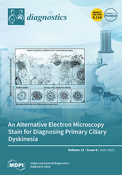
Cover Story (view full-size image): Primary ciliary dyskinesia is a disorder that affects motile cilia in the airway, and confirmation of a diagnosis commonly relies on the assessment of ciliary ultrastructure by transmission electron microscopy. Heavy metal staining is required to provide contrast when imaging cilia, and one of the most used stains is uranyl acetate. Uranyl acetate is radioactive and, due to growing safety concerns and restrictions, we show that UA zero provides a high contrast and is a suitable replacement for the diagnosis of primary ciliary dyskinesia. View this paper
  • Issues are regarded as officially published after their release is announced to the table of contents alert mailing list.
  • You may sign up for e-mail alerts to receive table of contents of newly released issues.
  • PDF is the official format for papers published in both, html and pdf forms. To view the papers in pdf format, click on the "PDF Full-text" link, and use the free Adobe Reader to open them.
Order results
Result details
Section
Select all
Export citation of selected articles as:
12 pages, 9093 KiB  
Article
High-Resolution Optical Coherence Tomography Angiography Characteristics of Limbal Stem Cell Deficiency
by Shobhit Varma, Swapna S. Shanbhag, Pragnya Rao Donthineni, Dilip Kumar Mishra, Vivek Singh and Sayan Basu
Diagnostics 2021, 11(6), 1130; https://doi.org/10.3390/diagnostics11061130 - 21 Jun 2021
Cited by 12 | Viewed by 2862
Abstract
This study aimed to identify the anterior segment high-resolution optical coherence tomography (HR-OCT) and HR-OCT angiography (HR-OCTA) features suggestive of limbal stem cell deficiency (LSCD) as confirmed by both impression cytology (IC) and in vivo confocal microscopy (IVCM). This was a single-centre prospective [...] Read more.
This study aimed to identify the anterior segment high-resolution optical coherence tomography (HR-OCT) and HR-OCT angiography (HR-OCTA) features suggestive of limbal stem cell deficiency (LSCD) as confirmed by both impression cytology (IC) and in vivo confocal microscopy (IVCM). This was a single-centre prospective cross-sectional study including 24 eyes of 22 patients with clinical suspicion of LSCD based on peripheral superficial corneal vascularisation and scarring. On IC and IVCM, performed and interpreted by blinded observers, 12 eyes each were diagnosed with and without LSCD. Additionally, 10 eyes of 5 healthy volunteers with no ocular pathology were also imaged. The 136 HR-OCT/A images of these 34 eyes were analysed with respect to 12 imaging parameters; the parameters most suggestive of LSCD were identified and the sensitivity and specificity were calculated. In the LSCD group, the most common aetiology was ocular chemical burns (83%), whereas in the non-LSCD group, the most common aetiology was viral keratitis (67%). Multiple logistic regression analysis revealed that mean epithelial reflectivity, mean stromal reflectivity, and mean superficial vascular density were the parameters that were diagnostic of LSCD on HR-OCT/A (p < 0.0001). A ratio of the mean epithelial reflectivity to stromal reflectivity of >1.29 corresponded with a high sensitivity (91.7%) and specificity (98.75%); while a mean superficial vascular density score of >0.38 corresponded with a sensitivity of 97.9% and specificity of 73.8%. In conclusion, HR-OCT/A as a non-invasive imaging modality could prove to be a useful tool for confirming the diagnosis of LSCD, with potential clinical and research applications. Full article
Show Figures

Figure 1

16 pages, 288 KiB  
Article
Sialoglyco-Conjugate Abnormalities, IL-6 Trans-Signaling and Anti-Ganglioside Immune Response—Potential Interferences in Lupus Nephritis Pathogenesis
by Corina-Daniela Ene, Mircea Nicolae Penescu and Ilinca Nicolae
Diagnostics 2021, 11(6), 1129; https://doi.org/10.3390/diagnostics11061129 - 21 Jun 2021
Cited by 2 | Viewed by 1626
Abstract
We have investigated glycoconjugates sialization profile, endogen synthesis rate of antiganglioside antibodies (AGA), IL-6 signaling pathways correlated with activity disease in systemic lupus erythematous (SLE) and lupus nephritis (LN). Material and methods. A case-control study was developed and included 109 patients with SLE [...] Read more.
We have investigated glycoconjugates sialization profile, endogen synthesis rate of antiganglioside antibodies (AGA), IL-6 signaling pathways correlated with activity disease in systemic lupus erythematous (SLE) and lupus nephritis (LN). Material and methods. A case-control study was developed and included 109 patients with SLE with or without renal impairment, 32 patients with IgA nephropathy and 60 healthy volunteers, clinically and paraclinically monitored. The following parameters were evaluated in volunteers serum: total sialic acid (TSA), orosomucoids, lipid bound sialic acid (LSA), interleukin-6 (IL-6), soluble factors IL-6R, gp130, anti –GM1, -GM2, -GM3, -GD1a, -GD1b, -GT1b, -GQ1b antigangliosides antibodies of IgG and IgM type. Results. Experimental data analysis showed: increase in synthesis rhythm of sialoglyco-conjugated in SLE (TSA increased in SLE and LN compared to control), accelerated catabolism of LSA in LN (LSA/TSA ratio was higher in SLE and LN than in control group), overexpression of IL-6 mediated trans-signaling (sIL-6R/sgp 130 ratio was subunit in SLE and IgA nephropathy and superunit in LN), large AGA profile synthesis of IgM isotype (over 45.1% in SLE and over 20.7% in LN). Conclusions. Hypersialization, accelerated glycosphingolipids degradation, IL-6 trans-signaling amplify and AGA pattern could represent essential mechanisms in LN pathogenesis. Full article
(This article belongs to the Section Pathology and Molecular Diagnostics)
10 pages, 1832 KiB  
Article
Arterial Digital Pulse Photoplethysmography in Patients with Suspected Thoracic Outlet Syndrome: A Study of the “Ca+Pra” Maneuver
by Jeanne Hersant, Pierre Ramondou, Francine Thouveny, Mickael Daligault, Mathieu Feuilloy, Patrick Saulnier, Pierre Abraham and Samir Henni
Diagnostics 2021, 11(6), 1128; https://doi.org/10.3390/diagnostics11061128 - 21 Jun 2021
Cited by 4 | Viewed by 2362
Abstract
The level of pulse amplitude (PA) change in arterial digital pulse plethysmography (A-PPG) that should be used to diagnose thoracic outlet syndrome (TOS) is debated. We hypothesized that a modification of the Roos test (by moving the arms forward, mimicking a prayer position [...] Read more.
The level of pulse amplitude (PA) change in arterial digital pulse plethysmography (A-PPG) that should be used to diagnose thoracic outlet syndrome (TOS) is debated. We hypothesized that a modification of the Roos test (by moving the arms forward, mimicking a prayer position (“Pra”)) releasing an eventual compression that occurs in the surrender/candlestick position (“Ca”) would facilitate interpretation of A-PPG results. In 52 subjects, we determined the optimal PA change from rest to predict compression at imaging (ultrasonography +/− angiography) with receiver operating characteristics (ROC). “Pra”-PA was set as 100%, and PA was expressed in normalized amplitude (NA) units. Imaging found arterial compression in 23 upper limbs. The area under ROC was 0.765 ± 0.065 (p < 0.0001), resulting in a 91.4% sensitivity and a 60.9% specificity for an increase of fewer than 3 NA from rest during “Ca”, while results were 17.4% and 98.8%, respectively, for the 75% PA decrease previously proposed in the literature. A-PPG during a “Ca+Pra” test provides demonstrable proof of inflow impairment and increases the sensitivity of A-PPG for the detection of arterial compression as determined by imaging. The absence of an increase in PA during the “Ca” phase of the “Ca+Pra” maneuver should be considered indicative of arterial inflow impairment. Full article
(This article belongs to the Section Pathology and Molecular Diagnostics)
Show Figures

Figure 1

10 pages, 2626 KiB  
Article
Development and Verification of a Deep Learning Algorithm to Evaluate Small-Bowel Preparation Quality
by Ji Hyung Nam, Dong Jun Oh, Sumin Lee, Hyun Joo Song and Yun Jeong Lim
Diagnostics 2021, 11(6), 1127; https://doi.org/10.3390/diagnostics11061127 - 20 Jun 2021
Cited by 11 | Viewed by 1873
Abstract
Capsule endoscopy (CE) quality control requires an objective scoring system to evaluate the preparation of the small bowel (SB). We propose a deep learning algorithm to calculate SB cleansing scores and verify the algorithm’s performance. A 5-point scoring system based on clarity of [...] Read more.
Capsule endoscopy (CE) quality control requires an objective scoring system to evaluate the preparation of the small bowel (SB). We propose a deep learning algorithm to calculate SB cleansing scores and verify the algorithm’s performance. A 5-point scoring system based on clarity of mucosal visualization was used to develop the deep learning algorithm (400,000 frames; 280,000 for training and 120,000 for testing). External validation was performed using additional CE cases (n = 50), and average cleansing scores (1.0 to 5.0) calculated using the algorithm were compared to clinical grades (A to C) assigned by clinicians. Test results obtained using 120,000 frames exhibited 93% accuracy. The separate CE case exhibited substantial agreement between the deep learning algorithm scores and clinicians’ assessments (Cohen’s kappa: 0.672). In the external validation, the cleansing score decreased with worsening clinical grade (scores of 3.9, 3.2, and 2.5 for grades A, B, and C, respectively, p < 0.001). Receiver operating characteristic curve analysis revealed that a cleansing score cut-off of 2.95 indicated clinically adequate preparation. This algorithm provides an objective and automated cleansing score for evaluating SB preparation for CE. The results of this study will serve as clinical evidence supporting the practical use of deep learning algorithms for evaluating SB preparation quality. Full article
(This article belongs to the Special Issue Capsule Endoscopy: Clinical Impacts and Innovation since 2001)
Show Figures

Figure 1

12 pages, 2104 KiB  
Article
Cytohistological Correlation in Pleural Effusions Based on the International System for Reporting Serous Fluid Cytopathology
by Daniel Pinto, Eduardo Cruz, Diamantina Branco, Cláudia Linares, Conceição Carvalho, Amélia Silva, Martinha Chorão and Fernando Schmitt
Diagnostics 2021, 11(6), 1126; https://doi.org/10.3390/diagnostics11061126 - 20 Jun 2021
Cited by 15 | Viewed by 2916
Abstract
The International System for Reporting Serous Fluid Cytology (TIS) was recently developed. Given its novelty, most studies looking into the risk of malignancy (ROM) of serous effusion diagnostic categories were published before the development of TIS. We searched the database of our department [...] Read more.
The International System for Reporting Serous Fluid Cytology (TIS) was recently developed. Given its novelty, most studies looking into the risk of malignancy (ROM) of serous effusion diagnostic categories were published before the development of TIS. We searched the database of our department for pleural effusions diagnosed in the last five years, excluding those without a corresponding pleural biopsy. Cases were reviewed and reclassified according to the TIS. A cytohistological correlation was performed. In total, 350 pleural effusion specimens with one or more corresponding pleural biopsies were included. After reclassification, 5 (1.43%) were nondiagnostic (ND), 253 (72.29%) were negative for malignancy (NFM), 7 (2.00%) had atypia of unknown significance (AUS), 14 (4.00%) were suspicious for malignancy (SFM), and 71 (20.57%) were malignant (MAL). Calculated ROM was 40% for ND, 20.16% for NFM, 42.86% for AUS, 78.57% for SFM, and 100% for MAL. Effusion cytology sensitivity and specificity were 60.29% and 98.56%, respectively. This is the first publication looking into the cytohistological correlation of a retrospective cohort of pleural effusions based on the TIS. We add to the body of data regarding the ROM for TIS categories, highlighting areas of potential future research. Full article
(This article belongs to the Special Issue Cyto-Histopathological Correlations in Pathology Diagnostics)
Show Figures

Figure 1

11 pages, 1015 KiB  
Article
Correlation of Native Liver Parenchyma T1 and T2 Relaxation Times and Liver Synthetic Function Tests: A Pilot Study
by Ute Lina Fahlenkamp, Jan Kunkel, Katharina Ziegeler, Konrad Neumann, Lisa Christine Adams, Günther Engel, Sarah Maria Böker and Marcus Richard Makowski
Diagnostics 2021, 11(6), 1125; https://doi.org/10.3390/diagnostics11061125 - 20 Jun 2021
Cited by 2 | Viewed by 2018
Abstract
MR relaxometry increasingly contributes to liver imaging. Studies on native relaxation times mainly describe relation to the presence of fibrosis. The hypothesis was that relaxation times are also influenced by other inherent factors, including changes in liver synthesis function. With the approval of [...] Read more.
MR relaxometry increasingly contributes to liver imaging. Studies on native relaxation times mainly describe relation to the presence of fibrosis. The hypothesis was that relaxation times are also influenced by other inherent factors, including changes in liver synthesis function. With the approval of the local ethics committee and written informed consent, data from 94 patients referred for liver MR imaging, of which 20 patients had cirrhosis, were included. Additionally to standard sequences, both native T1 and T2 parametric maps and T1 maps in the hepatobiliary phase of gadoxetate disodium were acquired. Associations with laboratory variables were assessed. Altogether, there was a negative correlation between albumin and all acquired relaxation times in cirrhotic patients. In non-cirrhotic patients, only T1 values exhibited a negative correlation with albumin. In all patients, bilirubin correlated significantly with post-contrast T1 relaxation times, whereas native relaxation times correlated only in cirrhotic patients. Evaluating patients with pathological INR values, post-contrast relaxation times were significantly higher, whereas native relaxation times did not correlate. In conclusion, apart from confirming the value of hepatobiliary phase T1 mapping, our results show a correlation of native T1 with serum albumin even in non-cirrhotic liver parenchyma, suggesting a direct influence of liver’s synthesis capacity on T1 relaxation times. Full article
(This article belongs to the Special Issue Advances and Novelties in Hepatobiliary and Pancreatic Imaging)
Show Figures

Figure 1

24 pages, 3313 KiB  
Review
International HIV Dementia Scale for HIV-Associated Neurocognitive Disorders: A Systematic Review and Meta-Analysis
by Elena Cecilia Rosca, Philippe Tadger, Amalia Cornea, Raluca Tudor, Cristian Oancea and Mihaela Simu
Diagnostics 2021, 11(6), 1124; https://doi.org/10.3390/diagnostics11061124 - 20 Jun 2021
Cited by 15 | Viewed by 3763
Abstract
The present study aims to systematically review the evidence on the accuracy of the International HIV Dementia Scale (IHDS) test for diagnosing human immunodeficiency virus (HIV)-associated neurocognitive disorders (HAND) and outline the quality and quantity of research evidence available on the accuracy of [...] Read more.
The present study aims to systematically review the evidence on the accuracy of the International HIV Dementia Scale (IHDS) test for diagnosing human immunodeficiency virus (HIV)-associated neurocognitive disorders (HAND) and outline the quality and quantity of research evidence available on the accuracy of IHDS in people living with HIV. We conducted a systematic literature review, searching five databases from inception until July 2020. We extracted dichotomized positive and negative test results at various thresholds and calculated the sensitivity and specificity of IHDS. Quality assessment was performed according to the Quality Assessment of Diagnostic Accuracy Studies 2 (QUADAS-2) criteria. Fifteen cross-sectional studies, published between 2011 and 2018, met the inclusion criteria for meta-analysis. Overall, 3760 patients were included, but most studies recruited small samples. We assessed most studies as being applicable to the review question, though we had concerns about the selection of participants in three studies. The accuracy of IHDS was investigated at thirteen cut-off points (scores 6–12). The threshold of 10 is the most useful for optimal HAND screening (including asymptomatic neurocognitive disorder, symptomatic HAND, and HIV-associated dementia) with fair diagnostic accuracy. Full article
(This article belongs to the Special Issue Diagnosis and Management of HIV Infection)
Show Figures

Figure 1

6 pages, 3672 KiB  
Communication
Usefulness of BATF3 Immunohistochemistry in Diagnosing Classical Hodgkin Lymphoma
by Julian Benckendorff, Johanna Kuchar, Frank Leithäuser, Malena Zahn and Peter Möller
Diagnostics 2021, 11(6), 1123; https://doi.org/10.3390/diagnostics11061123 - 20 Jun 2021
Cited by 3 | Viewed by 1741
Abstract
It is well recognized that the AP-1 transcription factor BATF3 is constitutively expressed in Hodgkin/Reed-Sternberg (HRS) cells, but its potential as a diagnostic marker for classical Hodgkin lymphoma (cHL) has not yet been addressed. In this study, we performed immunohistochemistry and analyzed the [...] Read more.
It is well recognized that the AP-1 transcription factor BATF3 is constitutively expressed in Hodgkin/Reed-Sternberg (HRS) cells, but its potential as a diagnostic marker for classical Hodgkin lymphoma (cHL) has not yet been addressed. In this study, we performed immunohistochemistry and analyzed the BATF3 expression in lymphoma cells on 218 lymphoma samples belonging to 14 different lymphoma entities. We observed varying degrees of BATF3 expression in nearly half of the cases (n = 100) with BATF3 expression being a constitutive feature of cHL (n = 53) and anaplastic large cell lymphoma (ALCL). By scoring BATF3 expression (BATF3-score) we observed constitutively high BATF3-scores in cHL and ALCL and low to moderate BATF3-scores in all other entities examined. Western blot analysis confirmed BATF3 protein expression in cell lysates from cHL cell lines (n = 7). Thus, BATF3 can be considered a useful IHC marker for the diagnosis of cHL as it is highly sensitive and sufficiently specific when analyzed by BATF3-scoring. Full article
(This article belongs to the Section Pathology and Molecular Diagnostics)
Show Figures

Figure 1

10 pages, 3591 KiB  
Article
Computed Tomography Imaging in Simulated Ongoing Cardiopulmonary Resuscitation: No Need to Switch Off the Chest Compression Device during Image Acquisition
by Jessica Graef, Bernd A. Leidel, Keno K. Bressem, Janis L. Vahldiek, Bernd Hamm and Stefan M. Niehues
Diagnostics 2021, 11(6), 1122; https://doi.org/10.3390/diagnostics11061122 - 19 Jun 2021
Cited by 1 | Viewed by 1991
Abstract
Computed tomography (CT) represents the current standard for imaging of patients with acute life-threatening diseases. As some patients present with circulatory arrest, they require cardiopulmonary resuscitation. Automated chest compression devices are used to continue resuscitation during CT examinations, but tend to cause motion [...] Read more.
Computed tomography (CT) represents the current standard for imaging of patients with acute life-threatening diseases. As some patients present with circulatory arrest, they require cardiopulmonary resuscitation. Automated chest compression devices are used to continue resuscitation during CT examinations, but tend to cause motion artifacts degrading diagnostic evaluation of the chest. The aim was to investigate and evaluate a CT protocol for motion-free imaging of thoracic structures during ongoing mechanical resuscitation. The standard CT trauma protocol and a CT protocol with ECG triggering using a simulated ECG were applied in an experimental setup to examine a compressible thorax phantom during resuscitation with two different compression devices. Twenty-eight phantom examinations were performed, 14 with AutoPulse® and 14 with corpuls cpr®. With each device, seven CT examinations were carried out with ECG triggering and seven without. Image quality improved significantly applying the ECG-triggered protocol (p < 0.001), which allowed almost artifact-free chest evaluation. With the investigated protocol, radiation exposure was 5.09% higher (15.51 mSv vs. 14.76 mSv), and average reconstruction time of CT scans increased from 45 to 76 s. Image acquisition using the proposed CT protocol prevents thoracic motion artifacts and facilitates diagnosis of acute life-threatening conditions during continuous automated chest compression. Full article
(This article belongs to the Section Medical Imaging and Theranostics)
Show Figures

Figure 1

14 pages, 5690 KiB  
Article
Cerebral CT Perfusion in Acute Stroke: The Effect of Lowering the Tube Load and Sampling Rate on the Reproducibility of Parametric Maps
by Georgios S. Ioannidis, Søren Christensen, Katerina Nikiforaki, Eleftherios Trivizakis, Kostas Perisinakis, Adam Hatzidakis, Apostolos Karantanas, Mauricio Reyes, Maarten Lansberg and Kostas Marias
Diagnostics 2021, 11(6), 1121; https://doi.org/10.3390/diagnostics11061121 - 19 Jun 2021
Cited by 8 | Viewed by 2362
Abstract
The aim of this study was to define lower dose parameters (tube load and temporal sampling) for CT perfusion that still preserve the diagnostic efficiency of the derived parametric maps. Ninety stroke CT examinations from four clinical sites with 1 s temporal sampling [...] Read more.
The aim of this study was to define lower dose parameters (tube load and temporal sampling) for CT perfusion that still preserve the diagnostic efficiency of the derived parametric maps. Ninety stroke CT examinations from four clinical sites with 1 s temporal sampling and a range of tube loads (mAs) (100–180) were studied. Realistic CT noise was retrospectively added to simulate a CT perfusion protocol, with a maximum reduction of 40% tube load (mAs) combined with increased sampling intervals (up to 3 s). Perfusion maps from the original and simulated protocols were compared by: (a) similarity using a voxel-wise Pearson’s correlation coefficient r with in-house software; (b) volumetric analysis of the infarcted and hypoperfused volumes using commercial software. Pearson’s r values varied for the different perfusion metrics from 0.1 to 0.85. The mean slope of increase and cerebral blood volume present the highest r values, remaining consistently above 0.7 for all protocol versions with 2 s sampling interval. Reduction of the sampling rate from 2 s to 1 s had only modest impacts on a TMAX volume of 0.4 mL (IQR −1–3) (p = 0.04) and core volume of −1.1 mL (IQR −4–0) (p < 0.001), indicating dose savings of 50%, with no practical loss of diagnostic accuracy. The lowest possible dose protocol was 2 s temporal sampling and a tube load of 100 mAs. Full article
(This article belongs to the Special Issue Radiation Dose and Image Quality in CT Imaging)
Show Figures

Figure 1

12 pages, 5448 KiB  
Article
Atypical Ductal Hyperplasia after Vacuum-Assisted Breast Biopsy: Can We Reduce the Upgrade to Breast Cancer to an Acceptable Rate?
by Luca Nicosia, Antuono Latronico, Francesca Addante, Rossella De Santis, Anna Carla Bozzini, Marta Montesano, Samuele Frassoni, Vincenzo Bagnardi, Giovanni Mazzarol, Oriana Pala, Matteo Lazzeroni, Germana Lissidini, Mauro Giuseppe Mastropasqua and Enrico Cassano
Diagnostics 2021, 11(6), 1120; https://doi.org/10.3390/diagnostics11061120 - 19 Jun 2021
Cited by 14 | Viewed by 4189
Abstract
(1) Background: to evaluate which factors can reduce the upgrade rate of atypical ductal hyperplasia (ADH) to in situ or invasive carcinoma in patients who underwent vacuum-assisted breast biopsy (VABB) and subsequent surgical excision. (2) Methods: 2955 VABBs were reviewed; 141 patients with [...] Read more.
(1) Background: to evaluate which factors can reduce the upgrade rate of atypical ductal hyperplasia (ADH) to in situ or invasive carcinoma in patients who underwent vacuum-assisted breast biopsy (VABB) and subsequent surgical excision. (2) Methods: 2955 VABBs were reviewed; 141 patients with a diagnosis of ADH were selected for subsequent surgical excision. The association between patients’ characteristics and the upgrade rate to breast cancer was evaluated in both univariate and multivariate analyses. (3) Results: the upgrade rates to ductal carcinoma in situ (DCIS) and invasive carcinoma (IC) were, respectively, 29.1% and 7.8%. The pooled upgrade rate to DCIS or IC was statistically lower at univariate analysis, considering the following parameters: complete removal of the lesion (p-value < 0.001); BIRADS ≤ 4a (p-value < 0.001); size of the lesion ≤15 mm (p-value: 0.002); age of the patients <50 years (p-value: 0.035). (4) Conclusions: the overall upgrade rate of ADH to DCIS or IC is high and, as already known, surgery should be recommended. However, ADH cases should always be discussed in multidisciplinary meetings: some parameters appear to be related to a lower upgrade rate. Patients presenting these parameters could be strictly followed up to avoid overtreatment. Full article
(This article belongs to the Special Issue Challenging Topics in Breast Cancer Diagnosis and Treatment)
Show Figures

Figure 1

17 pages, 1591 KiB  
Review
Detection of COVID-19 Virus on Surfaces Using Photonics: Challenges and Perspectives
by Bakr Ahmed Taha, Yousif Al Mashhadany, Nur Nadia Bachok, Ahmad Ashrif A Bakar, Mohd Hadri Hafiz Mokhtar, Mohd Saiful Dzulkefly Bin Zan and Norhana Arsad
Diagnostics 2021, 11(6), 1119; https://doi.org/10.3390/diagnostics11061119 - 19 Jun 2021
Cited by 34 | Viewed by 5785
Abstract
The propagation of viruses has become a global threat as proven through the coronavirus disease (COVID-19) pandemic. Therefore, the quick detection of viral diseases and infections could be necessary. This study aims to develop a framework for virus diagnoses based on integrating photonics [...] Read more.
The propagation of viruses has become a global threat as proven through the coronavirus disease (COVID-19) pandemic. Therefore, the quick detection of viral diseases and infections could be necessary. This study aims to develop a framework for virus diagnoses based on integrating photonics technology with artificial intelligence to enhance healthcare in public areas, marketplaces, hospitals, and airfields due to the distinct spectral signatures from lasers’ effectiveness in the classification and monitoring of viruses. However, providing insights into the technical aspect also helps researchers identify the possibilities and difficulties in this field. The contents of this study were collected from six authoritative databases: Web of Science, IEEE Xplore, Science Direct, Scopus, PubMed Central, and Google Scholar. This review includes an analysis and summary of laser techniques to diagnose COVID-19 such as fluorescence methods, surface-enhanced Raman scattering, surface plasmon resonance, and integration of Raman scattering with SPR techniques. Finally, we select the best strategies that could potentially be the most effective methods of reducing epidemic spreading and improving healthcare in the environment. Full article
Show Figures

Figure 1

27 pages, 1070 KiB  
Review
Cell-Secreted Vesicles: Novel Opportunities in Cancer Diagnosis, Monitoring and Treatment
by Cristina Catoni, Veronica Di Paolo, Elisabetta Rossi, Luigi Quintieri and Rita Zamarchi
Diagnostics 2021, 11(6), 1118; https://doi.org/10.3390/diagnostics11061118 - 19 Jun 2021
Cited by 5 | Viewed by 2834
Abstract
Extracellular vesicles (EVs) are important mediators of intercellular communication playing a pivotal role in the regulation of physiological and pathological processes, including cancer. In particular, there is significant evidence suggesting that tumor-derived EVs exert an immunosuppressive activity during cancer progression, as well as [...] Read more.
Extracellular vesicles (EVs) are important mediators of intercellular communication playing a pivotal role in the regulation of physiological and pathological processes, including cancer. In particular, there is significant evidence suggesting that tumor-derived EVs exert an immunosuppressive activity during cancer progression, as well as stimulate tumor cell migration, angiogenesis, invasion and metastasis. The use of EVs as a liquid biopsy is currently a fast-growing area of research in medicine, with the potential to provide a step-change in the diagnosis and treatment of cancer, allowing the prediction of both therapy response and prognosis. EVs could be useful not only as biomarkers but also as drug delivery systems, and may represent a target for anticancer therapy. In this review, we attempted to summarize the current knowledge about the techniques used for the isolation of EVs and their roles in cancer biology, as liquid biopsy biomarkers and as therapeutic tools and targets. Full article
Show Figures

Figure 1

7 pages, 9625 KiB  
Interesting Images
Magnetic Resonance Imaging during Proton Therapy Irradiation Allows for the Early Response Assessment of Pediatric Chordoma
by Sabina Vennarini, Dante Amelio, Stefano Lorentini, Giovanna Stefania Colafati, Antonella Cacchione, Rita De Vito, Andrea Carai, Benedetta Pettorini, Maurizio Amichetti and Angela Mastronuzzi
Diagnostics 2021, 11(6), 1117; https://doi.org/10.3390/diagnostics11061117 - 18 Jun 2021
Viewed by 2055
Abstract
Chordoma in pediatric patients is very rare. Proton therapy has become a gold standard in the treatment of these neoplasms, as high dose escalation can be achieved regarding the target while maximizing the sparing of the healthy tissues near the tumor. The aim [...] Read more.
Chordoma in pediatric patients is very rare. Proton therapy has become a gold standard in the treatment of these neoplasms, as high dose escalation can be achieved regarding the target while maximizing the sparing of the healthy tissues near the tumor. The aim of the work was to assess the evolution of morphological sequences during treatment using T1/T2-weighted magnetic resonance imaging (MRI) for the early response assessment of a classic chordoma of the skull base in a pediatric patient who had undergone surgical excision. Our results demonstrated a significant quantitative reduction in the residual nodule component adhered to the medullary bulb junction, with an almost complete recovery of normal anatomy at the end of the irradiation treatment. This was mainly shown in the T2-weighted MRI. On the other hand, the classic component of the lesion was predominantly present and located around the tooth of the axis. The occipital condyles were morphologically and dimensionally stable for the entire irradiation period. In conclusion, the application of this type of monitoring methodology, which is unusual during the administration of a proton treatment for chordoma, highlighted the unexpected early response of the disease. At the same time, it allowed the continuous assessment of the reliability of the treatment plan. Full article
(This article belongs to the Section Pathology and Molecular Diagnostics)
Show Figures

Figure 1

3 pages, 166 KiB  
Editorial
Assessment of Radiation Dose in Medical Imaging and Interventional Radiology Procedures for Patient and Staff Safety
by Kosuke Matsubara
Diagnostics 2021, 11(6), 1116; https://doi.org/10.3390/diagnostics11061116 - 18 Jun 2021
Cited by 6 | Viewed by 1964
Abstract
Medical imaging and interventional radiology procedures that use ionizing radiation play a significant role in patient healthcare [...] Full article
(This article belongs to the Special Issue Assessment of Radiation Dose in X-ray and CT Exams)
5 pages, 1564 KiB  
Interesting Images
Listeria Monocytogenes Brain Abscesses in a Patient with Disseminated Non-Small Cellular Lung Cancer: MRI Findings
by Anders Lykkemark Simonsen, Nitesh Shekhrajka, Frederik Boëtius Hertz, Jannik Helweg-Larsen, Åse Bengård Andersen and Anne-Mette Lebech
Diagnostics 2021, 11(6), 1115; https://doi.org/10.3390/diagnostics11061115 - 18 Jun 2021
Cited by 4 | Viewed by 2231
Abstract
Brain abscesses caused by Listeria monocytogenes (LM) are very rare and carry a high mortality risk. We present a patient with disseminated non-small cellular lung cancer (NSCLC) and multiple unusual LM brain abscesses. These abscesses have multiple elongated peripherally enhancing lesions in a [...] Read more.
Brain abscesses caused by Listeria monocytogenes (LM) are very rare and carry a high mortality risk. We present a patient with disseminated non-small cellular lung cancer (NSCLC) and multiple unusual LM brain abscesses. These abscesses have multiple elongated peripherally enhancing lesions in a characteristic formation that is “worm or tramtrack-like” following the white matter fiber tracts. Full article
(This article belongs to the Special Issue Imaging of Infections and Inflammatory Diseases)
Show Figures

Figure 1

16 pages, 1541 KiB  
Review
Imaging Cardiovascular Inflammation in the COVID-19 Era
by Andras Mester, Imre Benedek, Nora Rat, Cosmin Tolescu, Stefania Alexandra Polexa and Theodora Benedek
Diagnostics 2021, 11(6), 1114; https://doi.org/10.3390/diagnostics11061114 - 18 Jun 2021
Cited by 8 | Viewed by 2637
Abstract
Cardiac complications are among the most frequent extrapulmonary manifestations of COVID-19 and are associated with high mortality rates. Moreover, positive SARS-CoV-2 patients with underlying cardiovascular disease are more likely to require intensive care and are at higher risk of death. The underlying mechanism [...] Read more.
Cardiac complications are among the most frequent extrapulmonary manifestations of COVID-19 and are associated with high mortality rates. Moreover, positive SARS-CoV-2 patients with underlying cardiovascular disease are more likely to require intensive care and are at higher risk of death. The underlying mechanism for myocardial injury is multifaceted, in which the severe inflammatory response causes myocardial inflammation, coronary plaque destabilization, acute thrombotic events, and ischemia. Cardiac magnetic resonance (CMR) imaging is the non-invasive method of choice for identifying myocardial injury, and it is able to differentiate between underlying causes in various and often challenging clinical scenarios. Multimodal imaging protocols that incorporate CMR and computed tomography provide a complex evaluation for both respiratory and cardiovascular complications of SARS-CoV2 infection. This, in relation to biological evaluation of systemic inflammation, can guide appropriate therapeutic management in every stage of the disease. The use of artificial intelligence can further improve the diagnostic accuracy of these imaging techniques, thus enabling risk stratification and evaluation of prognosis. The present manuscript aims to review the current knowledge on the possible modalities for imaging COVID-related myocardial inflammation or post-COVID coronary inflammation and atherosclerosis. Full article
(This article belongs to the Special Issue Trends and Novelties in Cardiovascular Imaging)
Show Figures

Figure 1

11 pages, 594 KiB  
Review
Computer-Aided Detection False Positives in Colonoscopy
by Yu-Hsi Hsieh, Chia-Pei Tang, Chih-Wei Tseng, Tu-Liang Lin and Felix W. Leung
Diagnostics 2021, 11(6), 1113; https://doi.org/10.3390/diagnostics11061113 - 18 Jun 2021
Cited by 4 | Viewed by 2068
Abstract
Randomized control trials and meta-analyses comparing colonoscopies with and without computer-aided detection (CADe) assistance showed significant increases in adenoma detection rates (ADRs) with CADe. A major limitation of CADe is its false positives (FPs), ranked 3rd in importance among 59 research questions in [...] Read more.
Randomized control trials and meta-analyses comparing colonoscopies with and without computer-aided detection (CADe) assistance showed significant increases in adenoma detection rates (ADRs) with CADe. A major limitation of CADe is its false positives (FPs), ranked 3rd in importance among 59 research questions in a modified Delphi consensus review. The definition of FPs varies. One commonly used definition defines an FP as an activation of the CADe system, irrespective of the number of frames or duration of time, not due to any polypoid or nonpolypoid lesions. Although only 0.07 to 0.2 FPs were observed per colonoscopy, video analysis studies using FPs as the primary outcome showed much higher numbers of 26 to 27 per colonoscopy. Most FPs were of short duration (91% < 0.5 s). A higher number of FPs was also associated with suboptimal bowel preparation. The appearance of FPs can lead to user fatigue. The polypectomy of FPs results in increased procedure time and added use of resources. Re-training the CADe algorithms is one way to reduce FPs but is not practical in the clinical setting during colonoscopy. Water exchange (WE) is an emerging method that the colonoscopist can use to provide salvage cleaning during insertion. We discuss the potential of WE for reducing FPs as well as the augmentation of ADRs through CADe. Full article
Show Figures

Figure 1

14 pages, 4352 KiB  
Article
Mechanical Intermittent Compression Affects the Progression Rate of Malignant Melanoma Cells in a Cycle Period-Dependent Manner
by Takashi Morikura and Shogo Miyata
Diagnostics 2021, 11(6), 1112; https://doi.org/10.3390/diagnostics11061112 - 18 Jun 2021
Cited by 4 | Viewed by 2269
Abstract
Static mechanical compression is a biomechanical factor that affects the progression of melanoma cells. However, little is known about how dynamic mechanical compression affects the progression of melanoma cells. In the present study, we show that mechanical intermittent compression affects the progression rate [...] Read more.
Static mechanical compression is a biomechanical factor that affects the progression of melanoma cells. However, little is known about how dynamic mechanical compression affects the progression of melanoma cells. In the present study, we show that mechanical intermittent compression affects the progression rate of malignant melanoma cells in a cycle period-dependent manner. Our results suggest that intermittent compression with a cycle of 2 h on/2 h off could suppress the progression rate of melanoma cells by suppressing the elongation of F-actin filaments and mRNA expression levels related to collagen degradation. In contrast, intermittent compression with a cycle of 4 h on/4 h off could promote the progression rate of melanoma cells by promoting cell proliferation and mRNA expression levels related to collagen degradation. Mechanical intermittent compression could therefore affect the progression rate of malignant melanoma cells in a cycle period-dependent manner. Our results contribute to a deeper understanding of the physiological responses of melanoma cells to dynamic mechanical compression. Full article
Show Figures

Figure 1

11 pages, 2021 KiB  
Article
Aortic Origins of the Celiac Trunk and Superior Mesenteric Artery
by Mugurel Constantin Rusu, Adelina Maria Jianu, Bogdan Adrian Manta and Sorin Hostiuc
Diagnostics 2021, 11(6), 1111; https://doi.org/10.3390/diagnostics11061111 - 18 Jun 2021
Cited by 11 | Viewed by 2375
Abstract
(1) Background. The vertebral level of origin (VLO) of the celiac trunk (CT) and superior mesenteric artery (SMA) has been scarcely investigated. (2) Method. This study used 107 computed tomography angiograms and an eleven type grading system to classify the VLO of the [...] Read more.
(1) Background. The vertebral level of origin (VLO) of the celiac trunk (CT) and superior mesenteric artery (SMA) has been scarcely investigated. (2) Method. This study used 107 computed tomography angiograms and an eleven type grading system to classify the VLO of the CT and SMA. Each of the T12–L2 vertebra were divided in three horizontal levels. The intervertebral discs were considered distinct levels. (3) Results. The VLO of the CT ranged from the upper third of the T12 vertebra to the lower third of the L1 vertebra. The VLO of the SMA ranged from the lower third of the T12 vertebra to the upper third of the L2 vertebra. There was a highly significant association between the VLO of the CT and SMA (Chi2 = 201, p < 0.001), usually respecting a “plus two” rule. The mean CT–SMA distance was 1.82 +/− 0.66 cm in males and 1.55 +/− 0.411 cm in females, the difference being statistically significant. The mean CT–SMA distance tended to decrease with increasing CT–SMA types, the differences being statistically significant. (4) Conclusions. These characteristics of CT and SMA origins and their relations should be known by surgeons, as they could impact operative management and should be evaluated on a case-by-case basis. Full article
(This article belongs to the Special Issue Advances in Anatomy)
Show Figures

Figure 1

13 pages, 2633 KiB  
Article
Predictive Evaluation on Cytological Sample of Metastatic Melanoma: The Role of BRAF Immunocytochemistry in the Molecular Era
by Andrea Ronchi, Marco Montella, Federica Zito Marino, Michele Caraglia, Anna Grimaldi, Giuseppe Argenziano, Elvira Moscarella, Gabriella Brancaccio, Teresa Troiani, Stefania Napolitano, Renato Franco and Immacolata Cozzolino
Diagnostics 2021, 11(6), 1110; https://doi.org/10.3390/diagnostics11061110 - 18 Jun 2021
Cited by 4 | Viewed by 1947
Abstract
Background: Cutaneous malignant melanoma is an aggressive neoplasm. In advanced cases, the therapeutic choice depends on the mutational status of BRAF. Fine needle aspiration cytology (FNA) is often applied to the management of patients affected by melanoma, mainly for the diagnosis of metastases. [...] Read more.
Background: Cutaneous malignant melanoma is an aggressive neoplasm. In advanced cases, the therapeutic choice depends on the mutational status of BRAF. Fine needle aspiration cytology (FNA) is often applied to the management of patients affected by melanoma, mainly for the diagnosis of metastases. The evaluation of BRAF mutational status by sequencing technique on cytological samples may be inconvenient, as it is a time and biomaterial-consuming technique. Recently, BRAF immunocytochemistry (ICC) was applied for the evaluation of BRAF V600E mutational status. Although it may be useful mainly in cytological samples, data about BRAF ICC on cytological samples are missing. Methods: We performed BRAF ICC on a series of 50 FNA samples of metastatic melanoma. BRAF molecular analysis was performed on the same cytological samples or on the corresponding histological samples. Molecular analysis was considered the gold standard. Results: BRAF ICC results were adequate in 49 out of 50 (98%) cases, positive in 15 out of 50 (30%) cases and negative in 34 out of 50 (68%) of cases. Overall, BRAF ICC sensitivity, specificity, positive predictive value and negative predictive value results were 88.2%, 100%, 100% and 94.1%, respectively. The diagnostic performance of BRAF ICC results was perfect when molecular evaluation was performed on the same cytological samples. Hyperpigmentation represents the main limitation of the technique. Conclusions: BRAF ICC is a rapid, cost-effective method for detecting BRAF V600E mutation in melanoma metastases, applicable with high diagnostic performance to cytological samples. It could represent the first step to evaluate BRAF mutational status in cytological samples, mainly in poorly cellular cases. Full article
(This article belongs to the Special Issue Melanoma Pathology and Biomarkers)
Show Figures

Figure 1

13 pages, 8724 KiB  
Article
Cardiac Magnetic Resonance to Detect the Underlying Substrate in Patients with Frequent Idiopathic Ventricular Arrhythmias
by Chrysovalantou Nikolaidou, Christos P. Kotanidis, Rohan Wijesurendra, Joana Leal-Pelado, Konstantinos Kouskouras, Vassilios P. Vassilikos, Haralambos Karvounis, Ntobeko Ntusi, Charalambos Antoniades, Stefan Neubauer and Theodoros D. Karamitsos
Diagnostics 2021, 11(6), 1109; https://doi.org/10.3390/diagnostics11061109 - 18 Jun 2021
Cited by 4 | Viewed by 2264
Abstract
Background: A routine diagnostic work-up does not identify structural abnormalities in a substantial proportion of patients with idiopathic ventricular arrhythmias (VAs). We investigated the added value of cardiac magnetic resonance (CMR) imaging in this group of patients. Methods: A single-centre prospective study was [...] Read more.
Background: A routine diagnostic work-up does not identify structural abnormalities in a substantial proportion of patients with idiopathic ventricular arrhythmias (VAs). We investigated the added value of cardiac magnetic resonance (CMR) imaging in this group of patients. Methods: A single-centre prospective study was undertaken of 72 patients (mean age 46 ± 16 years; 53% females) with frequent premature ventricular contractions (PVCs ≥ 500/24 h) and/or non-sustained ventricular tachycardia (NSVT), an otherwise normal electrocardiogram, normal echocardiography and no coronary artery disease. Results: CMR provided an additional diagnostic yield in 54.2% of patients. The most prevalent diagnosis was previous myocarditis (23.6%) followed by possible PVC-related cardiomyopathy (20.8%), non-ischaemic cardiomyopathy (8.3%) and ischaemic heart disease (1.4%). The predictors of abnormal CMR findings were male gender, age and PVCs/NSVT non-outflow tract-related or with multiple morphologies. Patients with VAs had an impaired peak left ventricular (LV) global radial strain (GRS) compared with the controls (28.88% (IQR: 25.87% to 33.97%) vs. 36.65% (IQR: 33.19% to 40.2%), p < 0.001) and a global circumferential strain (GCS) (−17.66% (IQR: −19.62% to −16.23%) vs. −20.66% (IQR: −21.72% to −19.6%), p < 0.001). Conclusion: CMR reveals abnormalities in a significant proportion of patients with frequent idiopathic VAs. Male gender, age and non-outflow tract PVC origin can be clinical indicators for CMR referral. Full article
(This article belongs to the Special Issue Advances in Cardiovascular Imaging 2021)
Show Figures

Graphical abstract

12 pages, 285 KiB  
Review
Biomarkers in Prostate-Specific Membrane Antigen Theranostics
by Panagiotis J. Vlachostergios, Ioannis Zachos and Vassilios Tzortzis
Diagnostics 2021, 11(6), 1108; https://doi.org/10.3390/diagnostics11061108 - 18 Jun 2021
Cited by 5 | Viewed by 1986
Abstract
Theranostics of prostate cancer (PC) represents a growing area of development of imaging agents and targeted radionuclide therapeutics against a major target, prostate specific membrane antigen (PSMA). In view of the encouraging efficacy from the use of 177Lu and other radionuclides in [...] Read more.
Theranostics of prostate cancer (PC) represents a growing area of development of imaging agents and targeted radionuclide therapeutics against a major target, prostate specific membrane antigen (PSMA). In view of the encouraging efficacy from the use of 177Lu and other radionuclides in metastatic castration-resistant prostate cancer (mCRPC), it is becoming increasingly important to identify surrogate markers that can help predict which patients are more likely to respond and experience improved survival. This review discusses potential predictors of efficacy of PSMA-targeted radionuclide therapies (TRT) segregated in three major categories: imaging, clinical and molecular. Full article
(This article belongs to the Special Issue PSMA in Prostate Cancer)
13 pages, 7534 KiB  
Article
A Randomized Comparison of Delivered Energy in Cardioversion of Atrial Fibrillation: Biphasic Truncated Exponential Versus Pulsed Biphasic Waveforms
by Elina Trendafilova, Elena Dimitrova, Jean-Philippe Didon and Vessela Krasteva
Diagnostics 2021, 11(6), 1107; https://doi.org/10.3390/diagnostics11061107 - 17 Jun 2021
Viewed by 2175
Abstract
A few randomized trials have compared impedance-compensated biphasic defibrillators in clinical use. We aim to compare pulsed biphasic (PB) and biphasic truncated exponential (BTE) waveforms in a non-inferiority cardioversion (CVS) study. This was a prospective monocentric randomized clinical trial. Eligible patients admitted for [...] Read more.
A few randomized trials have compared impedance-compensated biphasic defibrillators in clinical use. We aim to compare pulsed biphasic (PB) and biphasic truncated exponential (BTE) waveforms in a non-inferiority cardioversion (CVS) study. This was a prospective monocentric randomized clinical trial. Eligible patients admitted for elective CVS of atrial fibrillation (AF) between February 2019 and March 2020 were alternately randomized to treatment with either a PB defibrillator (DEFIGARD TOUCH7, Schiller Médical, Wissembourg, France) or a BTE high-energy (BTE-HE) defibrillator (LIFEPAK15, Physio-Control Inc., Redmond, WA, USA). Fixed-energy protocol (200–200–200 J) was administered. CVS success was accepted if sinus rhythm was restored at 1 min post-shock. The study design considered non-inferiority testing of the primary outcome: cumulative delivered energy (CDE). Seventy-three out of 78 randomized patients received allocated intervention: 38 BTE-HE (52%), 35 PB (48%). Baseline characteristics were well-balanced between groups (p > 0.05). Both waveforms had similar CDE (mean ± standard deviation, 95% confidence interval): BTE-HE (253.9 ± 120.2 J, 214–293 J) vs. PB (226.0 ± 109.8 J, 188–264 J), p = 0.31. Indeed, effective PB shocks delivered significantly lower energies by mean of 25.6 J (95% CI 24–27.1 J, p < 0.001). Success rates were similar (BTE-HE vs. PB): 1 min first-shock (84.2% vs. 82.9%), 1 min CVS (97.4% vs. 94.3%), 2 h CVS (94.7% vs. 94.3%), 24 h CVS (92.1% vs. 94.3%), p > 0.05. Safety analysis did not find CVS hazards, reporting insignificant changes of myocardial-specific biomarkers, transient and rare ST-segment deviations, and no case of harmful tachyarrhythmias and apnea. Cardioversion of AF with fixed-energy protocol 200–200–200 J was highly efficient and safe for both PB and BTE-HE waveforms. These similar performances were achieved despite differences in the waveforms’ technical design, associated with significantly lower delivered energy for the effective PB shocks. Clinical Trial Registration: Registration number: NCT04032678, trial register: ClinicalTrials.gov. Full article
(This article belongs to the Special Issue Diagnostic Modalities in Critical Care)
Show Figures

Figure 1

9 pages, 35538 KiB  
Article
Development and Proof of Concept of a Low-Cost Ultrasound Training Model for Diagnosis of Giant Cell Arteritis Using 3D Printing
by Florian Recker, Lei Jin, Patrick Veith, Mark Lauterbach, Pantelis Karakostas and Valentin Sebastian Schäfer
Diagnostics 2021, 11(6), 1106; https://doi.org/10.3390/diagnostics11061106 - 17 Jun 2021
Cited by 4 | Viewed by 1921
Abstract
Objectives: Currently, ultrasound (US) is widely used for the diagnosis of giant cell arteritis (GCA). Our aim was to develop a low-cost US training model for diagnosis of GCA of the temporal and axillary artery using a modern 3D printing system. Methods: We [...] Read more.
Objectives: Currently, ultrasound (US) is widely used for the diagnosis of giant cell arteritis (GCA). Our aim was to develop a low-cost US training model for diagnosis of GCA of the temporal and axillary artery using a modern 3D printing system. Methods: We designed an US training model, which enables measurement of the intima-media thickness (IMT) of temporal and axillary arteries using Autodesk Fusion360. This model was printed using a modern 3D printer (Formlabs Form3) and embedded in ballistic gelatine. The ultrasound images including measurement of the IMT by ultrasound specialists in GCA were compared to ultrasound images in acute GCA and healthy subjects. Results: Our ultrasound training model of the axillary and temporal artery displayed a very similar ultrasound morphology compared to real US images and fulfilled the OMERACT ultrasound definitions of normal and pathological temporal and axillary arteries in GCA. The IMT measurements were in line with published cut-off values for normal and pathological IMT values in GCA and healthy individuals. When testing the models on blinded US specialists in GCA, they were identified correctly in all test rounds with an intra-class coefficient of 0.99. Conclusion: The production of low-cost ultrasound training models of normal and pathological temporal and axillary arteries in GCA, which fulfil the OMERACT ultrasound definitions and adhere to the published IMT cut-off values in GCA, is feasible. Ultrasound specialists identified each respective model correctly in every case. Full article
Show Figures

Figure 1

12 pages, 1976 KiB  
Article
Prognostic Factors Associated with Successful Salvage Surgery in Recurrent Oral Cancer
by Mateusz Szewczyk, Paweł Golusiński, Jakub Pazdrowski and Wojciech Golusiński
Diagnostics 2021, 11(6), 1105; https://doi.org/10.3390/diagnostics11061105 - 17 Jun 2021
Cited by 4 | Viewed by 1615
Abstract
Most patients with recurrent oral cancer are not eligible for salvage surgery. Among those who are candidates for surgical salvage, failure rates are high. Given the potential negative impact of salvage surgery on quality of life (QoL)—particularly in unsuccessful interventions—the decision to operate [...] Read more.
Most patients with recurrent oral cancer are not eligible for salvage surgery. Among those who are candidates for surgical salvage, failure rates are high. Given the potential negative impact of salvage surgery on quality of life (QoL)—particularly in unsuccessful interventions—the decision to operate must be weighed carefully. However, the variables associated with successful surgical salvage in oral cancer have not been clearly established. In the present retrospective study, we sought to determine the factors associated with disease recurrence and successful salvage surgery. We evaluated the following parameters in patients (n = 261) treated for primary oral cancer at our institution from 2010 to 2017: age; T/N status; perineurial invasion; lymphovascular invasion; extranodal extension; and margin status. In total, 36 patients (33%) were considered eligible for salvage surgery. Four variables were significantly associated with suitability for salvage surgery: early primary T stage, no primary neck disease (N0), no positive margins in the primary resection, and no adjuvant radiotherapy following primary resection. The only variable significantly associated with improved salvage outcomes was negative margin status after the primary tumor resection, underscoring the importance of margin status on treatment outcomes. Additional studies are needed to identify other factors associated with successful salvage surgery in order to better stratify patients according to the likelihood of success, thus potentially avoiding the negative impact on QoL in patients who undergo unsuccessful surgery. Full article
(This article belongs to the Special Issue Biomarkers of Oral Cancer 2.0)
Show Figures

Figure 1

16 pages, 1358 KiB  
Article
Group and Shuffle Convolutional Neural Networks with Pyramid Pooling Module for Automated Pterygium Segmentation
by Siti Raihanah Abdani, Mohd Asyraf Zulkifley and Nuraisyah Hani Zulkifley
Diagnostics 2021, 11(6), 1104; https://doi.org/10.3390/diagnostics11061104 - 17 Jun 2021
Cited by 13 | Viewed by 2137
Abstract
Pterygium is an eye condition that is prevalent among workers that are frequently exposed to sunlight radiation. However, most of them are not aware of this condition, which motivates many volunteers to set up health awareness booths to give them free health screening. [...] Read more.
Pterygium is an eye condition that is prevalent among workers that are frequently exposed to sunlight radiation. However, most of them are not aware of this condition, which motivates many volunteers to set up health awareness booths to give them free health screening. As a result, a screening tool that can be operated on various platforms is needed to support the automated pterygium assessment. One of the crucial functions of this assessment is to extract the infected regions, which directly correlates with the severity levels. Hence, Group-PPM-Net is proposed by integrating a spatial pyramid pooling module (PPM) and group convolution to the deep learning segmentation network. The system uses a standard mobile phone camera input, which is then fed to a modified encoder-decoder convolutional neural network, inspired by a Fully Convolutional Dense Network that consists of a total of 11 dense blocks. A PPM is integrated into the network because of its multi-scale capability, which is useful for multi-scale tissue extraction. The shape of the tissues remains relatively constant, but the size will differ according to the severity levels. Moreover, group and shuffle convolution modules are also integrated at the decoder side of Group-PPM-Net by placing them at the starting layer of each dense block. The addition of these modules allows better correlation among the filters in each group, while the shuffle process increases channel variation that the filters can learn from. The results show that the proposed method obtains mean accuracy, mean intersection over union, Hausdorff distance, and Jaccard index performances of 0.9330, 0.8640, 11.5474, and 0.7966, respectively. Full article
(This article belongs to the Special Issue Artificial Intelligence in Eye Disease)
Show Figures

Figure 1

14 pages, 1653 KiB  
Article
Microstructural MRI Correlates of Cognitive Impairment in Multiple Sclerosis: The Role of Deep Gray Matter
by Marco Pitteri, Ilaria Boscolo Galazzo, Lorenza Brusini, Federica Cruciani, Caterina Dapor, Damiano Marastoni, Gloria Menegaz and Massimiliano Calabrese
Diagnostics 2021, 11(6), 1103; https://doi.org/10.3390/diagnostics11061103 - 16 Jun 2021
Cited by 3 | Viewed by 2014
Abstract
Although cognitive impairment (CI) is frequently observed in people with multiple sclerosis (pwMS), its pathogenesis is still controversial. Conflicting results emerged concerning the role of microstructural gray matter (GM) damage especially when involving the deep GM structures. In this study, we aimed at [...] Read more.
Although cognitive impairment (CI) is frequently observed in people with multiple sclerosis (pwMS), its pathogenesis is still controversial. Conflicting results emerged concerning the role of microstructural gray matter (GM) damage especially when involving the deep GM structures. In this study, we aimed at evaluating whether differences in cortical and deep GM structures between apparently cognitively normal (ACN) and CI pwMS (36 subjects in total) are present, using an extensive set of diffusion MRI (dMRI) indices and conventional morphometry measures. The results revealed increased anisotropy and restriction over several deep GM structures in CI compared with ACN pwMS, while no changes in volume were present in the same areas. Conversely, reduced anisotropy/restriction values were detected in cortical regions, mostly the pericalcarine cortex and precuneus, combined with reduced thickness of the superior frontal gyrus and insula. Most of the dMRI metrics but none of the morphometric indices correlated with the Symbol Digit Modality Test. These results suggest that deep GM microstructural damage can be a strong anatomical substrate of CI in pwMS and might allow identifying pwMS at higher risk of developing CI. Full article
(This article belongs to the Section Pathology and Molecular Diagnostics)
Show Figures

Figure 1

14 pages, 2286 KiB  
Article
Comparison of Genetic Profiling between Primary Tumor and Circulating Tumor Cells Captured by Microfluidics in Epithelial Ovarian Cancer: Tumor Heterogeneity or Allele Dropout?
by Ting-Yu Chang, Sheng-Wen Chen, Wen-Hsiang Lin, Chung-Er Huang, Mark I. Evans, I-Fang Chung, Janne-Wha Wu, Gwo-Chin Ma and Ming Chen
Diagnostics 2021, 11(6), 1102; https://doi.org/10.3390/diagnostics11061102 - 16 Jun 2021
Cited by 3 | Viewed by 3275
Abstract
Epithelial ovarian cancer (EOC) is a leading cause of cancer mortality among women but unfortunately is usually not diagnosed until advanced stage. Early detection of EOC is of paramount importance to improve outcomes. Liquid biopsy of circulating tumor cells (CTCs) is emerging as [...] Read more.
Epithelial ovarian cancer (EOC) is a leading cause of cancer mortality among women but unfortunately is usually not diagnosed until advanced stage. Early detection of EOC is of paramount importance to improve outcomes. Liquid biopsy of circulating tumor cells (CTCs) is emerging as one of the promising biomarkers for early detection of solid tumors. However, discrepancies in terms of oncogenomics (i.e., different genetic defects detected) between the germline, primary tumor, and liquid biopsy are a serious concern and may adversely affect downstream cancer management. Here, we illustrate the potential and pitfalls of CTCs by presenting two patients of Stage I EOC. We successfully isolated and recovered CTCs by a silicon-based nanostructured microfluidics system, the automated Cell RevealTM. We examined the genomics of CTCs as well as the primary tumor and germline control (peripheral blood mononuclear cells) by whole exome sequencing. Different signatures were then investigated by comparisons of identified mutation loci distinguishing those that may only arise in the primary tumor or CTCs. A novel model is proposed to test if the highly variable allele frequencies, between primary tumor and CTCs results, are due to allele dropout in plural CTCs or tumor heterogeneity. This proof-of-principle study provides a strategy to elucidate the possible cause of genomic discrepancy between the germline, primary tumor, and CTCs, which is helpful for further large-scale use of such technology to be integrated into clinical management protocols. Full article
(This article belongs to the Section Pathology and Molecular Diagnostics)
Show Figures

Figure 1

11 pages, 1772 KiB  
Article
The Evaluation of Left Ventricle Ischemic Extent in Patients with Significantly Suspicious Cardiovascular Disease by 99mTc-Sestamibi Dynamic SPECT/CT and Myocardial Perfusion Imaging: A Head-to-Head Comparison
by Hung-Pin Chan, Chin-Chuan Chang, Chin Hu, Wen-Hwa Wang, Nan-Jing Peng, Yu-Chang Tyan and Ming-Hui Yang
Diagnostics 2021, 11(6), 1101; https://doi.org/10.3390/diagnostics11061101 - 16 Jun 2021
Cited by 3 | Viewed by 2082
Abstract
Heart disease is the second most common cause of mortality in Taiwan, mainly coronary artery disease (CAD).Quantitative coronary blood flow has been collected by dynamic single-photon emission computed tomography (Dynamic SPECT/CT) for CAD diagnosis in previous studies. However, few studies defined the extent [...] Read more.
Heart disease is the second most common cause of mortality in Taiwan, mainly coronary artery disease (CAD).Quantitative coronary blood flow has been collected by dynamic single-photon emission computed tomography (Dynamic SPECT/CT) for CAD diagnosis in previous studies. However, few studies defined the extent of left ventricle (LV) ischemia on Dynamic SPECT/CT for predicting significant coronary artery stenosis. This study evaluates the extent of LV ischemic blockage in patients suspected of CAD who were referred by cardiologists. A total of 181 patients with suspected CAD were enrolled. They underwent 99mTc-Sestamibi (MIBI) Dynamic SPECT/CT survey before cardiac intervention. Dynamic SPECT/CT has better sensitivity (88%), specificity (96%), and accuracy (94%) compared with those of semi-quantitative MIBI MPI (more than 10%). Results indicated that5% of the LV ischemic extent can yield positive PCI results (>70% stenosis in coronary arteries) compared with the moderate abnormal extent of at least 15% of LV. When the percentage of combined moderate abnormal extent and ischemia extent of LV reaches 27.3%, positive PCI results may be indicated. This study revealed Dynamic SPECT/CT has greater sensitivity, specificity, and accuracy as compared with MPI. Thus, the severity of abnormal perfusion extent of LV on Dynamic SPECT/CT might be beneficial to predict positive PCI results in patients with significant suspicion CAD. Full article
Show Figures

Figure 1

Previous Issue
Next Issue
Back to TopTop