Next Issue
Volume 11, May
Previous Issue
Volume 11, March


Diagnostics, Volume 11, Issue 4 (April 2021) – 157 articles

Cover Story (view full-size image): 
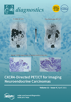
C-X-C motif chemokine receptor 4 (CXCR4) is overexpressed on a significant number of tumor entities. CXCR4-directed positron emission tomography (PET) can visualize receptor expression in vivo. We evaluated the potential of CXCR4-directed imaging in patients with poorly differentiated neuroendocrine carcinomas (NECs) and compared it to the established reference standard 18F-FDG PET/computed tomography (CT). Notably, the biopsy results of the tumor lesions showed weak-to-moderate CXCR4 expression; however, 18F-FDG PET/CT detected significantly more tumor lesions and was superior to CXCR-directed PET/CT imaging. View this paper
  • Issues are regarded as officially published after their release is announced to the table of contents alert mailing list.
  • You may sign up for e-mail alerts to receive table of contents of newly released issues.
  • PDF is the official format for papers published in both, html and pdf forms. To view the papers in pdf format, click on the "PDF Full-text" link, and use the free Adobe Reader to open them.
Order results
Result details
Section
Select all
Export citation of selected articles as:
16 pages, 1805 KiB  
Article
Predictive Markers of Early Cardiovascular Impairment and Insulin Resistance in Obese Pediatric Patients
by Laura Mihaela Trandafir, Elena Cojocaru, Mihaela Moscalu, Maria Magdalena Leon Constantin, Ingrith Miron, Alexandra Mastaleru, Oana Teslariu, Madalina Elena Datcu, Silvia Fotea and Otilia Frăsinariu
Diagnostics 2021, 11(4), 735; https://doi.org/10.3390/diagnostics11040735 - 20 Apr 2021
Cited by 3 | Viewed by 2016
Abstract
Background: The increased prevalence of obesity among children determined the rising number of its comorbidities in children and adults, too. This study aimed to evaluate certain markers of inflammation and insulin resistance in obese pediatric patients, identifying those who are more likely to [...] Read more.
Background: The increased prevalence of obesity among children determined the rising number of its comorbidities in children and adults, too. This study aimed to evaluate certain markers of inflammation and insulin resistance in obese pediatric patients, identifying those who are more likely to develop further complications. Methods: We included 115 obese pediatric patients: 85 overweight and obese patients in the study group and 30 normal-weight patients in the control group. We calculated the body mass index (BMI) and we evaluated markers (biological, inflammatory) and the hormones profile. Results: Low-threshold inflammation was assessed by measuring interleukin 6 IL-6 and Intercellular Adhesion Molecules (ICAM). The analysis showed that IL-6 is significantly correlated with glucose (p = 0.001) and BMI value (p = 0.031). ICAM correlates significantly with triglycerides (p = 0.001), glucose (p = 0.044) and BMI percentile (p = 0.037). For pediatric obese patients, endotoxemia has been significantly correlated only with BMI percentile (p = 0.001). Plasma cortisol did not show significant correlations with total cholesterol, triglycerides, glucose or BMI percentile. The results indicated a significant predictive power of BMI percentile on inflammatory markers: IL-6 (AUC = 0.803, p < 0.001), ICAM (AUC = 0.806, p < 0.001) and endotoxemia (AUC = 0.762, p = 0.019). Additionally, BMI percentile has a significant predictive power for metabolic markers of insulin resistance (insulin value: AUC = 0.72, p < 0.001 and HOMA index: AUC = 0.68, p = 0.003). Conclusions: The study highlighted the importance of early markers of cardiovascular risk in obese pediatric patients represented by IL-6, ICAM, endotoxemia and their correlation with metabolic markers of insulin resistance represented by insulinemia, HOMA index and plasma cortisol. It can clearly be considered that the BMI percentile has significant predictive power for metabolic markers of insulin resistance. Full article
(This article belongs to the Section Pathology and Molecular Diagnostics)
Show Figures

Figure 1

16 pages, 16514 KiB  
Review
Pediatric Abusive Head Trauma: A Systematic Review
by Aniello Maiese, Francesca Iannaccone, Andrea Scatena, Zoe Del Fante, Antonio Oliva, Paola Frati and Vittorio Fineschi
Diagnostics 2021, 11(4), 734; https://doi.org/10.3390/diagnostics11040734 - 20 Apr 2021
Cited by 15 | Viewed by 4917
Abstract
Abusive head trauma (AHT) represents a commonly misdiagnosed condition. In fact, there is no pathognomonic sign that allows the diagnosis in children. Therefore, it is such an important medico-legal challenge to evaluate reliable diagnostic tools. The aim of this review is to evaluate [...] Read more.
Abusive head trauma (AHT) represents a commonly misdiagnosed condition. In fact, there is no pathognomonic sign that allows the diagnosis in children. Therefore, it is such an important medico-legal challenge to evaluate reliable diagnostic tools. The aim of this review is to evaluate the current scientific evidence to assess what the best practice is in order to diagnose AHT. We have focused particularly on evaluating the importance of circumstantial evidence, clinical history, the use of postmortem radiological examinations (such as CT and MRI), and the performance of the autopsy. After autopsy, histological examination of the eye and brain play an important role, with attention paid to correlation with symptoms found in vivo. Full article
(This article belongs to the Special Issue Progress in the Forensic Diagnosis 2.0)
Show Figures

Figure 1

22 pages, 2689 KiB  
Article
Implementation of Precision Oncology for Patients with Metastatic Breast Cancer in an Interdisciplinary MTB Setting
by Elena Sultova, C. Benedikt Westphalen, Andreas Jung, Joerg Kumbrink, Thomas Kirchner, Doris Mayr, Martina Rudelius, Steffen Ormanns, Volker Heinemann, Klaus H. Metzeler, Philipp A. Greif, Anna Hester, Sven Mahner, Nadia Harbeck and Rachel Wuerstlein
Diagnostics 2021, 11(4), 733; https://doi.org/10.3390/diagnostics11040733 - 20 Apr 2021
Cited by 13 | Viewed by 2563
Abstract
The advent of molecular diagnostics and the rising number of targeted therapies have facilitated development of precision oncology for cancer patients. In order to demonstrate its impact for patients with metastatic breast cancer (mBC), we initiated a Molecular Tumor Board (MTB) to provide [...] Read more.
The advent of molecular diagnostics and the rising number of targeted therapies have facilitated development of precision oncology for cancer patients. In order to demonstrate its impact for patients with metastatic breast cancer (mBC), we initiated a Molecular Tumor Board (MTB) to provide treatment recommendations for mBC patients who had disease progression under standard treatment. NGS (next generation sequencing) was carried out using the Oncomine multi-gene panel testing system (Ion Torrent). The MTB reviewed molecular diagnostics’ results, relevant tumor characteristics, patient’s course of disease and made personalized treatment and/or diagnostic recommendations for each patient. From May 2017 to December 2019, 100 mBC patients were discussed by the local MTB. A total 72% of the mBC tumors had at least one molecular alteration (median 2 per case, range: 1 to 6). The most frequent genetic changes were found in the following genes: PIK3CA (19%) and TP53 (17%). The MTB rated 53% of these alterations as actionable and treatment recommendations were made accordingly for 49 (49%) patients. Sixteen patients (16%) underwent the suggested therapy. Nine out of sixteen patients (56%; 9% of all) experienced a clinical benefit with a progression-free survival ratio ≥ 1.3. Personalized targeted therapy recommendations resulting from MTB case discussions could provide substantial benefits for patients with mBC and should be implemented for all suitable patients. Full article
(This article belongs to the Special Issue Challenging Topics in Breast Cancer Diagnosis and Treatment)
Show Figures

Figure 1

12 pages, 3321 KiB  
Article
Efficiently Classifying Lung Sounds through Depthwise Separable CNN Models with Fused STFT and MFCC Features
by Shing-Yun Jung, Chia-Hung Liao, Yu-Sheng Wu, Shyan-Ming Yuan and Chuen-Tsai Sun
Diagnostics 2021, 11(4), 732; https://doi.org/10.3390/diagnostics11040732 - 20 Apr 2021
Cited by 42 | Viewed by 3861
Abstract
Lung sounds remain vital in clinical diagnosis as they reveal associations with pulmonary pathologies. With COVID-19 spreading across the world, it has become more pressing for medical professionals to better leverage artificial intelligence for faster and more accurate lung auscultation. This research aims [...] Read more.
Lung sounds remain vital in clinical diagnosis as they reveal associations with pulmonary pathologies. With COVID-19 spreading across the world, it has become more pressing for medical professionals to better leverage artificial intelligence for faster and more accurate lung auscultation. This research aims to propose a feature engineering process that extracts the dedicated features for the depthwise separable convolution neural network (DS-CNN) to classify lung sounds accurately and efficiently. We extracted a total of three features for the shrunk DS-CNN model: the short-time Fourier-transformed (STFT) feature, the Mel-frequency cepstrum coefficient (MFCC) feature, and the fused features of these two. We observed that while DS-CNN models trained on either the STFT or the MFCC feature achieved an accuracy of 82.27% and 73.02%, respectively, fusing both features led to a higher accuracy of 85.74%. In addition, our method achieved 16 times higher inference speed on an edge device and only 0.45% less accuracy than RespireNet. This finding indicates that the fusion of the STFT and MFCC features and DS-CNN would be a model design for lightweight edge devices to achieve accurate AI-aided detection of lung diseases. Full article
(This article belongs to the Special Issue Artificial Intelligence in Lung Diseases)
Show Figures

Figure 1

8 pages, 688 KiB  
Communication
Presence of SARS-CoV-2 Nucleoprotein in Cardiac Tissues of Donors with Negative COVID-19 Molecular Tests
by Gianluca Lorenzo Perrucci, Elena Sommariva, Veronica Ricci, Paola Songia, Yuri D’Alessandra, Paolo Poggio, Giulio Pompilio, Gianluca Polvani and Anna Guarino
Diagnostics 2021, 11(4), 731; https://doi.org/10.3390/diagnostics11040731 - 20 Apr 2021
Cited by 5 | Viewed by 2116
Abstract
The 2019 Coronavirus disease (COVID-19) outbreak had detrimental effects on essential medical services such as organ and tissue donation. Lombardy, one of the most active Italian regions in organ/tissue procurement, has been strongly affected by the COVID-19 pandemic. To date, data concerning the [...] Read more.
The 2019 Coronavirus disease (COVID-19) outbreak had detrimental effects on essential medical services such as organ and tissue donation. Lombardy, one of the most active Italian regions in organ/tissue procurement, has been strongly affected by the COVID-19 pandemic. To date, data concerning the risk of SARS-CoV-2 transmission after tissue transplantation are controversial. Here, we aimed to evaluate the presence/absence of SARS-CoV-2 in different cardiac tissues eligible for transplantation obtained from Lombard donors. We used cardiovascular tissues from eight donors potentially suitable for pulmonary valve transplantation. All donor subjects involved in the study returned negative results for the SARS-CoV-2 RNA molecular tests (quantitative real-time reverse-transcription PCR, qRT-PCR, and chip-based digital PCR) in nasopharyngeal swabs (NPS) or bronchoalveolar lavage (BAL). None of the eight donors included in this study revealed the presence of the SARS-CoV-2 viral genome. However, evaluation of the protein content of pulmonary vein wall (PVW) tissue revealed variable levels of SARS-CoV-2 nucleoprotein signal in all donors. Our study demonstrated for the first time, to the best of our knowledge, that viral nucleoprotein but not viral RNA was present in the examined tissue bank specimens, suggesting the need for caution and in-depth investigations on implantable tissue specimens collected during the COVID-19 pandemic period. Full article
Show Figures

Figure 1

10 pages, 23417 KiB  
Study Protocol
Development of the BWAT-CUA Scale to Assess Wounds in Patients with Calciphylaxis
by Lisa J. Gould, Thomas E. Serena and Smeeta Sinha
Diagnostics 2021, 11(4), 730; https://doi.org/10.3390/diagnostics11040730 - 20 Apr 2021
Cited by 5 | Viewed by 4500
Abstract
Calcific uremic arteriolopathy (CUA; calciphylaxis) is a severe form of vascular calcification with no approved therapies. A standardized wound assessment tool is needed to evaluate changes in calciphylaxis wounds over time. A prospective, single-arm study of 14 patients with calciphylaxis reported improvement for [...] Read more.
Calcific uremic arteriolopathy (CUA; calciphylaxis) is a severe form of vascular calcification with no approved therapies. A standardized wound assessment tool is needed to evaluate changes in calciphylaxis wounds over time. A prospective, single-arm study of 14 patients with calciphylaxis reported improvement for the primary endpoint of wound healing using the 13-item Bates-Jensen Wound Assessment Tool (BWAT), although that tool was developed for assessment of pressure ulcers. This report describes development of BWAT-CUA, an 8-item modification of BWAT focusing on prototypical features of calciphylaxis lesions. The BWAT-CUA has a range of 8 (best) to 40 (worst) and was used ad hoc to analyze BWAT data collected in the prospective study. Using BWAT-CUA, relative improvement in calciphylaxis wounds was 30% overall (from 21.2 to 14.9; p = 0.0016) and 34% in the subset of 12 patients with ulceration at baseline (from 23.3 to 15.3; p = 0.0002). BWAT-CUA is a primary endpoint in an ongoing randomized, placebo-controlled phase 3 study of SNF472 recruiting patients with end-stage kidney disease and at least one ulcerated calciphylaxis lesion. BWAT-CUA, a newly developed tool for assessment of calciphylaxis wound severity and improvements over time, may be used in clinical research and in clinical practice. Full article
(This article belongs to the Section Pathology and Molecular Diagnostics)
Show Figures

Figure 1

29 pages, 3900 KiB  
Article
miRNAs and Their Gene Targets—A Clue to Differentiate Pregnancies with Small for Gestational Age Newborns, Intrauterine Growth Restriction, and Preeclampsia
by Angelika V. Timofeeva, Ivan S. Fedorov, Alexander G. Brzhozovskiy, Anna E. Bugrova, Vitaliy V. Chagovets, Maria V. Volochaeva, Natalia L. Starodubtseva, Vladimir E. Frankevich, Evgeny N. Nikolaev, Roman G. Shmakov and Gennady T. Sukhikh
Diagnostics 2021, 11(4), 729; https://doi.org/10.3390/diagnostics11040729 - 20 Apr 2021
Cited by 5 | Viewed by 2320
Abstract
Despite the differences in the clinical manifestations of major obstetric syndromes, such as preeclampsia (PE) and intrauterine growth restriction (IUGR), their pathogenesis is based on the dysregulation of proliferation, differentiation, and invasion of cytotrophoblast cells that occur in the developing placenta, decidual endometrium, [...] Read more.
Despite the differences in the clinical manifestations of major obstetric syndromes, such as preeclampsia (PE) and intrauterine growth restriction (IUGR), their pathogenesis is based on the dysregulation of proliferation, differentiation, and invasion of cytotrophoblast cells that occur in the developing placenta, decidual endometrium, and myometrial parts of the spiral arteries. To understand the similarities and differences in the molecular mechanisms of PE and IUGR, samples of the placental bed and placental tissue were analyzed using protein mass spectrometry and the deep sequencing of small RNAs, followed by validation of the data obtained by quantitative RT-PCR in real time. A comparison of the transcriptome and proteomic profiles in the samples made it possible to conclude that the main changes in the molecular profile in IUGR occur in the placental bed, in contrast to PE, in which the majority of molecular changes occurs in the placenta. In placental bed samples, significant changes in the ratio of miRNA and its potential target gene expression levels were revealed, which were unique for IUGR (miR-30c-5p/VIM, miR-28-3p/VIM, miR-1-3p/ANXA2, miR-30c-5p/FBN1; miR-15b-5p/MYL6), unique for PE (miR-185-3p/FLNA), common for IUGR and PE (miR-30c-5p/YWHAZ and miR-654-3p/FGA), but all associated with abnormality in the hemostatic and vascular systems as well as with an inflammatory process at the fetal‒maternal interface. Full article
(This article belongs to the Section Pathology and Molecular Diagnostics)
Show Figures

Figure 1

10 pages, 549 KiB  
Review
Glucocorticoid Withdrawal—An Overview on When and How to Diagnose Adrenal Insufficiency in Clinical Practice
by Katarzyna Pelewicz and Piotr Miśkiewicz
Diagnostics 2021, 11(4), 728; https://doi.org/10.3390/diagnostics11040728 - 20 Apr 2021
Cited by 16 | Viewed by 4960
Abstract
Glucocorticoids (GCs) are widely used due to their anti-inflammatory and immunosuppressive effects. As many as 1–3% of the population are currently on GC treatment. Prolonged therapy with GCs is associated with an increased risk of GC-induced adrenal insufficiency (AI). AI is a rare [...] Read more.
Glucocorticoids (GCs) are widely used due to their anti-inflammatory and immunosuppressive effects. As many as 1–3% of the population are currently on GC treatment. Prolonged therapy with GCs is associated with an increased risk of GC-induced adrenal insufficiency (AI). AI is a rare and often underdiagnosed clinical condition characterized by deficient GC production by the adrenal cortex. AI can be life-threatening; therefore, it is essential to know how to diagnose and treat this disorder. Not only oral but also inhalation, topical, nasal, intra-articular and intravenous administration of GCs may lead to adrenal suppression. Moreover, recent studies have proven that short-term (<4 weeks), as well as low-dose (<5 mg prednisone equivalent per day) GC treatment can also suppress the hypothalamic–pituitary–adrenal axis. Chronic therapy with GCs is the most common cause of AI. GC-induced AI remains challenging for clinicians in everyday patient care. Properly conducted GC withdrawal is crucial in preventing GC-induced AI; however, adrenal suppression may occur despite following recommended GC tapering regimens. A suspicion of GC-induced AI requires careful diagnostic workup and prompt introduction of a GC replacement treatment. The present review provides a summary of current knowledge on the management of GC-induced AI, including diagnostic methods, treatment schedules, and GC withdrawal regimens in adults. Full article
(This article belongs to the Section Pathology and Molecular Diagnostics)
Show Figures

Figure 1

13 pages, 2152 KiB  
Communication
Imbalance of Mg Homeostasis as a Potential Biomarker in Colon Cancer
by Davide Schiroli, Chiara Marraccini, Eleonora Zanetti, Moira Ragazzi, Alessandra Gianoncelli, Eleonora Quartieri, Elisa Gasparini, Stefano Iotti, Roberto Baricchi and Lucia Merolle
Diagnostics 2021, 11(4), 727; https://doi.org/10.3390/diagnostics11040727 - 20 Apr 2021
Cited by 5 | Viewed by 1924
Abstract
Background: Increasing evidences support a correlation between magnesium (Mg) homeostasis and colorectal cancer (CRC). Nevertheless, the role of Mg and its transporters as diagnostic markers in CRC is still a matter of debate. In this study we combined X-ray Fluorescence Microscopy and databases [...] Read more.
Background: Increasing evidences support a correlation between magnesium (Mg) homeostasis and colorectal cancer (CRC). Nevertheless, the role of Mg and its transporters as diagnostic markers in CRC is still a matter of debate. In this study we combined X-ray Fluorescence Microscopy and databases information to investigate the possible correlation between Mg imbalance and CRC. Methods: CRC tissue samples and their non-tumoural counterpart from four patients were collected and analysed for total Mg level and distribution by X-Ray Fluorescence Microscopy. We also reviewed the scientific literature and the main tissue expression databases to collect data on Mg transporters expression in CRC. Results: We found a significantly higher content of total Mg in CRC samples when compared to non-tumoural tissues. Mg distribution was also impaired in CRC. Conversely, we evidenced an uncertain correlation between Mg transporters expression and colon malignancies. Discussion: Although further studies are necessary to determine the correlation between different cancer types and stages, this is the first report proposing the measurement of Mg tissue localisation as a marker in CRC. This study represents thus a proof-of-concept that paves the way for the design of a larger prospective investigation of Mg in CRC. Full article
Show Figures

Figure 1

19 pages, 8211 KiB  
Article
Analysis of LAGEs Family Gene Signature and Prognostic Relevance in Breast Cancer
by Hoang Dang Khoa Ta, Wan-Chun Tang, Nam Nhut Phan, Gangga Anuraga, Sz-Ying Hou, Chung-Chieh Chiao, Yen-Hsi Liu, Yung-Fu Wu, Kuen-Haur Lee and Chih-Yang Wang
Diagnostics 2021, 11(4), 726; https://doi.org/10.3390/diagnostics11040726 - 19 Apr 2021
Cited by 15 | Viewed by 3273
Abstract
Breast cancer (BRCA) is one of the most complex diseases and involves several biological processes. Members of the L-antigen (LAGE) family participate in the development of various cancers, but their expressions and prognostic values in breast cancer remain to be clarified. High-throughput methods [...] Read more.
Breast cancer (BRCA) is one of the most complex diseases and involves several biological processes. Members of the L-antigen (LAGE) family participate in the development of various cancers, but their expressions and prognostic values in breast cancer remain to be clarified. High-throughput methods for exploring disease progression mechanisms might play a pivotal role in the improvement of novel therapeutics. Therefore, gene expression profiles and clinical data of LAGE family members were acquired from the cBioportal database, followed by verification using the Oncomine and The Cancer Genome Atlas (TCGA) databases. In addition, the Kaplan-Meier method was applied to explore correlations between expressions of LAGE family members and prognoses of breast cancer patients. MetaCore, GlueGo, and GluePedia were used to comprehensively study the transcript expression signatures of LAGEs and their co-expressed genes together with LAGE-related signal transduction pathways in BRCA. The result indicated that higher LAGE3 messenger (m)RNA expressions were observed in BRCA tissues than in normal tissues, and they were also associated with the stage of BRCA patients. Kaplan-Meier plots showed that overexpression of LAGE1, LAGE2A, LAGE2B, and LAGE3 were highly correlated to poor survival in most types of breast cancer. Significant associations of LAGE family genes were correlated with the cell cycle, focal adhesion, and extracellular matrix (ECM) receptor interactions as indicated by functional enrichment analyses. Collectively, LAGE family members’ gene expression levels were related to adverse clinicopathological factors and prognoses of BRCA patients; therefore, LAGEs have the potential to serve as prognosticators of BRCA patients. Full article
(This article belongs to the Section Pathology and Molecular Diagnostics)
Show Figures

Figure 1

10 pages, 4697 KiB  
Case Report
Unilateral Orbital Metastasis as the Unique Symptom in the Onset of Breast Cancer in a Postmenopausal Woman: Case Report and Review of the Literature
by Cristina Marinela Oprean, Larisa Maria Badau, Nusa Alina Segarceanu, Andrei Dorin Ciocoiu, Ioana Alexandra Rivis, Vlad Norin Vornicu, Teodora Hoinoiu, Daciana Grujic, Cristina Bredicean and Alis Dema
Diagnostics 2021, 11(4), 725; https://doi.org/10.3390/diagnostics11040725 - 19 Apr 2021
Cited by 4 | Viewed by 2911
Abstract
The orbit represents an unusual metastases site for patients diagnosed with cancer, however, breast cancer is the main cause of metastases at this level. These orbital metastases were discovered in patients with a history of breast cancer as unique or synchronous lesions. We [...] Read more.
The orbit represents an unusual metastases site for patients diagnosed with cancer, however, breast cancer is the main cause of metastases at this level. These orbital metastases were discovered in patients with a history of breast cancer as unique or synchronous lesions. We present a rare case of a unique retroocular metastasis as the first initial symptom of a tubulo-lobular mammary carcinoma in a postmenopausal woman. A 57-year-old patient complains of diplopia, diminishing visual acuity, orbital tenderness, slight exophthalmia and ptosis of the left eyelid, with insidious onset. Clinical examination and subsequent investigations revealed a left breast cancer cT2 cN1 pM1 stage IV. Breast conserving surgery was performed on the left breast. Pathological examination with immunohistochemistry staining established the complete diagnostic: pT2pN3aM1 Stage IV breast cancer, luminal B subtype. After two years from the initial breast cancer diagnosis, the patient was diagnosed by the psychiatrist with a depressive disorder and was treated accordingly. Orbital metastases are usually discovered in known breast cancer patients and they are found in the context of a multi-system end-stage disease. Most reports cite that up to 25% of the total orbital metastases cases are discovered before the diagnosis of the primary tumor, as our case did. MRI is the gold standard for evaluating orbital tumors. The ILC histological subtype metastasizes in the orbitals more frequently than invasive ductal carcinoma. The prognosis of patients with orbital metastases is poor. The median survival after diagnosis of orbital metastases from a breast cancer primary is ranging from 22 to 31 months. Overall survival of our patient was 56 months, longer than the median survival reported in literature. Orbital metastases must be taken into account when patients accuse ophthalmologic symptoms even in the absence of a personal history of cancer. Objective examination of every patient that incriminates these types of symptoms is essential, and breast palpation must be made in every clinical setting. Orbital biopsy is necessary for the confirmation of the diagnosis and for an adequate treatment. Although recommendations for management of orbital metastases are controversial, it appears that multidisciplinary treatment of both metastases and primary cancer improves overall survival. Full article
(This article belongs to the Special Issue Challenging Topics in Breast Cancer Diagnosis and Treatment)
Show Figures

Figure 1

19 pages, 19122 KiB  
Article
Signature of Circulating Biomarkers in Recurrent Non-Infectious Anterior Uveitis. Immunomodulatory Effects of DHA-Triglyceride. A Pilot Study
by Maria D. Pinazo-Durán, Jose J. García-Medina, Silvia M. Sanz-González, Jose E. O’Connor, Ricardo P. Casaroli-Marano, Mar Valero-Velló, Maribel López-Gálvez, Cristina Peris-Martínez, Vicente Zanón-Moreno and Manuel Diaz-Llopis
Diagnostics 2021, 11(4), 724; https://doi.org/10.3390/diagnostics11040724 - 19 Apr 2021
Cited by 2 | Viewed by 2707
Abstract
The purpose of this study was to identify circulating biomarkers of recurrent non-infectious anterior uveitis (NIAU), and to address the anti-inflammatory effects of triglyceride containing docosahexaenoic acid (DHA-TG). A prospective multicenter study was conducted in 72 participants distributed into: patients diagnosed with recurrent [...] Read more.
The purpose of this study was to identify circulating biomarkers of recurrent non-infectious anterior uveitis (NIAU), and to address the anti-inflammatory effects of triglyceride containing docosahexaenoic acid (DHA-TG). A prospective multicenter study was conducted in 72 participants distributed into: patients diagnosed with recurrent NIAU in the quiescence stage (uveitis group (UG); n = 36) and healthy controls (control group (CG); n = 36). Each group was randomly assigned to the oral supplementation of one pill/day (+) containing DHA-TG (n = 18) or no-pill condition (−) (n = 17) for three consecutive months. Data from demographics, risk factors, comorbidities, eye complications and therapy were recorded. Blood was collected and processed to determine pro-inflammatory biomarkers by bead-base multiplex assay. Statistical processing with multivariate statistical analysis was performed. The mean age was 50, 12 (10, 31) years. The distribution by gender was 45% males and 55% females. The mean number of uveitis episodes was 5 (2). Higher plasma expression of interleukin (IL)-6 was detected in the UG versus the CG (p = 5 × 10⁻⁵). Likewise, significantly higher plasma levels were seen for IL-1β, IL-2, INFγ (p = 10−4), and TNFα (p = 2 × 10⁻⁴) in the UG versus the CG. Significantly lower values of the above molecules were found in the +DHA-TG than in the -DHA-TG subgroups, after 3 months of follow-up, TNFα (p = 10−7) and IL-6 (p = 3 × 10⁻⁶) being those that most significantly changed. Signatures of circulating inflammatory mediators were obtained in the quiescent stage of recurrent NIAU patients. This 3-month follow-up strongly reinforces that a regular oral administration of DHA-TG reduces the inflammatory load and may potentially supply a prophylaxis-adjunctive mediator for patients at risk of uveitis vision loss. Full article
(This article belongs to the Section Diagnostic Microbiology and Infectious Disease)
Show Figures

Figure 1

13 pages, 1095 KiB  
Article
FDG-PET/CT for Response Monitoring in Metastatic Breast Cancer: The Feasibility and Benefits of Applying PERCIST
by Marianne Vogsen, Jakob Lykke Bülow, Lasse Ljungstrøm, Hjalte Rasmus Oltmann, Tural Asgharzadeh Alamdari, Mohammad Naghavi-Behzad, Poul-Erik Braad, Oke Gerke and Malene Grubbe Hildebrandt
Diagnostics 2021, 11(4), 723; https://doi.org/10.3390/diagnostics11040723 - 19 Apr 2021
Cited by 12 | Viewed by 2113
Abstract
Background: We aimed to examine the feasibility and potential benefit of applying PET Response Criteria in Solid Tumors (PERCIST) for response monitoring in metastatic breast cancer (MBC). Further, we introduced the nadir scan as a reference. Methods: Response monitoring FDG-PET/CT scans in 37 [...] Read more.
Background: We aimed to examine the feasibility and potential benefit of applying PET Response Criteria in Solid Tumors (PERCIST) for response monitoring in metastatic breast cancer (MBC). Further, we introduced the nadir scan as a reference. Methods: Response monitoring FDG-PET/CT scans in 37 women with MBC were retrospectively screened for PERCIST standardization and measurability criteria. One-lesion PERCIST based on changes in SULpeak measurements of the hottest metastatic lesion was used for response categorization. The baseline (PERCISTbaseline) and the nadir scan (PERCISTnadir) were used as references for PERCIST analyses. Results: Metastatic lesions were measurable according to PERCIST in 35 of 37 (94.7%) patients. PERCIST was applied in 150 follow-up scans, with progression more frequently reported by PERCISTnadir (36%) than PERCISTbaseline (29.3%; p = 0.020). Reasons for progression were (a) more than 30% increase in SULpeak of the hottest lesion (n = 7, 15.9%), (b) detection of new metastatic lesions (n = 28, 63.6%), or both (a) and (b) (n = 9, 20.5%). Conclusions: PERCIST, with the introduction of PERCISTnadir, allows a graphical interpretation of disease fluctuation that may be beneficial in clinical decision-making regarding potential earlier termination of non-effective toxic treatment. PERCIST seems feasible for response monitoring in MBC but prospective studies are needed to come this closer. Full article
(This article belongs to the Special Issue Advances in Multimodality Molecular Imaging)
Show Figures

Figure 1

11 pages, 1128 KiB  
Case Report
Phenotypic Diversity of 15q11.2 BP1–BP2 Deletion in Three Korean Families with Development Delay and/or Intellectual Disability: A Case Series and Literature Review
by Ji Yoon Han and Joonhong Park
Diagnostics 2021, 11(4), 722; https://doi.org/10.3390/diagnostics11040722 - 19 Apr 2021
Viewed by 1832
Abstract
The 15q11.2 breakpoint (BP) 1–BP2 deletion syndrome is emerging as the most frequent pathogenic copy number variation in humans related to neurodevelopmental diseases, with changes in cognition, behavior, and brain morphology. Previous publications have reported that patients with 15q11.2 BP1–BP2 deletion showed intellectual [...] Read more.
The 15q11.2 breakpoint (BP) 1–BP2 deletion syndrome is emerging as the most frequent pathogenic copy number variation in humans related to neurodevelopmental diseases, with changes in cognition, behavior, and brain morphology. Previous publications have reported that patients with 15q11.2 BP1–BP2 deletion showed intellectual disability (ID), speech impairment, developmental delay (DD), and/or behavioral problems. We describe three new cases, aged 3 or 6 years old and belonging to three unrelated Korean families, with a 350-kb 15q11.2 BP1–BP2 deletion of four highly conserved genes, namely, the TUBGCP5, CYFIP1, NIPA2, and NIPA1 genes. All of our cases presented with global DD and/or ID, and the severity ranged from mild to severe, but common facial dysmorphism and congenital malformations in previous reports were not characteristic. The 15q11.2 BP1–BP2 deletion was inherited from an unaffected parent in all cases. Our three cases, together with previous findings from the literature review, confirm some of the features earlier reported to be associated with 15q11.2 BP1–BP2 deletion and help to further delineate the phenotype associated with 15q11.2 deletion. Identification of more cases with 15q11.2 BP1–BP2 deletion will allow us to obtain a better understanding of the clinical phenotypes. Further explanation of the functions of the genes within the 15q11.2 BP1–BP2 region is required to resolve the pathogenic effects on neurodevelopment. Full article
(This article belongs to the Section Pathology and Molecular Diagnostics)
Show Figures

Figure 1

12 pages, 750 KiB  
Article
Associations between Pre-Bariatric High-Sensitivity C-Reactive Protein and Post-Surgery Outcomes
by Tannaz Jamialahmadi, Mohsen Nematy, Simona Bo, Valentina Ponzo, Ali Jangjoo, Ladan Goshayeshi, Aida Tasbandi, Nikita G. Nikiforov and Amirhossein Sahebkar
Diagnostics 2021, 11(4), 721; https://doi.org/10.3390/diagnostics11040721 - 18 Apr 2021
Viewed by 1908
Abstract
Background: Obesity is a chronic inflammatory condition associated with increased circulating levels of C-reactive protein (CRP). Bariatric surgery has been reported to be effective in improving both inflammatory and liver status. Our aims were to elucidate the relationships between pre-surgery high sensitivity-CRP (hs-CRP) [...] Read more.
Background: Obesity is a chronic inflammatory condition associated with increased circulating levels of C-reactive protein (CRP). Bariatric surgery has been reported to be effective in improving both inflammatory and liver status. Our aims were to elucidate the relationships between pre-surgery high sensitivity-CRP (hs-CRP) values and post-surgery weight loss and liver steatosis and fibrosis in patients with severe obesity undergoing Roux-en-Y gastric bypass. Methods: We conducted an observational prospective study on 90 individuals with morbid obesity, who underwent gastric bypass. Anthropometric indices, laboratory assessment (lipid panel, glycemic status, liver enzymes, and hs-CRP), liver stiffness and steatosis were evaluated at baseline and 6-months after surgery. Results: There was a significant post-surgery reduction in all the anthropometric variables, with an average weight loss of 33.93 ± 11.79 kg; the mean percentage of total weight loss (TWL) was 27.96 ± 6.43%. Liver elasticity was significantly reduced (from 6.1 ± 1.25 to 5.42 ± 1.52 kPa; p = 0.002), as well as liver aminotransferases, nonalcoholic fatty liver disease fibrosis score (NFS) and the grade of steatosis. Serum hs-CRP levels significantly reduced (from 9.26 ± 8.45 to 3.29 ± 4.41 mg/L; p < 0.001). The correlations between hs-CRP levels and liver fibrosis (elastography), steatosis (ultrasonography), fibrosis-4 index, NFS, and surgery success rate were not significant. Regression analyses showed that serum hs-CRP levels were not predictive of liver status and success rate after surgery in both unadjusted and adjusted models. Conclusions: In patients with morbid obesity, bariatric surgery caused a significant decrease in hs-CRP levels, liver stiffness and steatosis. Baseline hs-CRP values did not predict the weight-loss success rate and post-surgery liver status. Full article
(This article belongs to the Special Issue Fatty Liver Disease: Diagnostic, Predictive and Prognostic Markers)
Show Figures

Figure 1

13 pages, 1286 KiB  
Article
Advantages of 18F-FDG PET/CT Imaging over Modified Duke Criteria and Clinical Presumption in Patients with Challenging Suspicion of Infective Endocarditis
by Valentin Pretet, Cyrille Blondet, Yvon Ruch, Matias Martinez, Soraya El Ghannudi, Olivier Morel, Yves Hansmann, Thomas H. Schindler and Alessio Imperiale
Diagnostics 2021, 11(4), 720; https://doi.org/10.3390/diagnostics11040720 - 18 Apr 2021
Cited by 6 | Viewed by 2631
Abstract
According to European Society of Cardiology guidelines (ESC2015) for infective endocarditis (IE) management, modified Duke criteria (mDC) are implemented with a degree of clinical suspicion degree, leading to grades such as “possible” or “rejected” IE despite a persisting high level of clinical suspicion. [...] Read more.
According to European Society of Cardiology guidelines (ESC2015) for infective endocarditis (IE) management, modified Duke criteria (mDC) are implemented with a degree of clinical suspicion degree, leading to grades such as “possible” or “rejected” IE despite a persisting high level of clinical suspicion. Herein, we evaluate the 18F-FDG PET/CT diagnostic and therapeutic impact in IE suspicion, with emphasis on possible/rejected IE with a high clinical suspicion. Excluding cases of definite IE diagnosis, 53 patients who underwent 18F-FDG PET/CT for IE suspicion were selected and afterwards classified according to both mDC (possible IE/Duke 1, rejected IE/Duke 0) and clinical suspicion degree (high and low IE suspicion). The final status regarding IE diagnosis (gold standard) was based on the multidisciplinary decision of the Endocarditis Team, including the ‘imaging specialist’. PET/CT images of the cardiac area were qualitatively interpreted and the intensity of each focus of extra-physiologic 18F-FDG uptake was evaluated by a maximum standardized uptake value (SUVmax) measurement. Extra-cardiac 18F-FDG PET/CT pathological findings were considered to be a possible embolic event, a possible source of IE, or even a concomitant infection. Based on the Endocarditis Team consensus, final diagnosis of IE was retained in 19 (36%) patients and excluded in 34 (64%). With a sensitivity, specificity, positive predictive value (PPV), negative predictive value (NPV), and global accuracy of 79%, 100%, 100%, 89%, and 92%, respectively, PET/CT performed significantly better than mDC (p = 0.003), clinical suspicion degree (p = 0.001), and a combination of both (p = 0.001) for IE diagnosis. In 41 patients with possible/rejected IE but high clinical suspicion, sensitivity, specificity, PPV, NPV, and global accuracies were 78%, 100%, 100%, 85%, and 90%, respectively. Moreover, PET/CT contributed to patient management in 24 out of 53 (45%) cases. 18F-FDG PET/CT represents a valuable diagnostic tool that could be proposed for challenging IE cases with significant differences between mDC and clinical suspicion degree. 18F-FDG PET/CT allows a binary diagnosis (definite or rejected IE) by removing uncertain diagnostic situations, thus improving patient therapeutic management. Full article
(This article belongs to the Special Issue Imaging of Infections and Inflammatory Diseases)
Show Figures

Figure 1

9 pages, 2186 KiB  
Article
Computer-Aided Assessment of Displacement and Reduction of Distal Radius Fractures
by Yuichi Yoshii, Yasukazu Totoki, Atsuo Shigi, Kunihiro Oka, Takeshi Ogawa, Tsuyoshi Murase and Tomoo Ishii
Diagnostics 2021, 11(4), 719; https://doi.org/10.3390/diagnostics11040719 - 18 Apr 2021
Cited by 3 | Viewed by 1726
Abstract
This study aims to investigate displacements and reductions of distal radius fractures using measurement indices based on the computer-aided three-dimensional (3D) radius shape model. Fifty-two distal radius fracture patients who underwent osteosynthesis were evaluated with pre- and post-operative distal radius 3D images. In [...] Read more.
This study aims to investigate displacements and reductions of distal radius fractures using measurement indices based on the computer-aided three-dimensional (3D) radius shape model. Fifty-two distal radius fracture patients who underwent osteosynthesis were evaluated with pre- and post-operative distal radius 3D images. In the 3D images, three reference points, i.e., the radial styloid process (1), sigmoid notch volar, and dorsal edge (2) (3) were marked. The three-dimensional coordinates of each reference point and the barycentric coordinates of the plane connecting the three reference points were evaluated. The distance and direction moved, due to the reductions for each reference point, were (1) 12.1 ± 8.1 mm in the ulnar-palmar-distal direction, (2) 7.5 ± 4.1 mm in the ulnar-palmar-proximal direction, and (3) 8.2 ± 4.7 mm in the ulnar-palmar-distal direction relative to the preoperative position. The barycentric coordinate moved 8.4 ± 5.3 mm in the ulnar-palmar-distal direction compared to the preoperative position. This analyzing method will be helpful to understand the three-dimensional direction and the extent of displacements in distal radius fractures. Full article
(This article belongs to the Section Machine Learning and Artificial Intelligence in Diagnostics)
Show Figures

Figure 1

10 pages, 857 KiB  
Article
Dual Effect of PER2 C111G Polymorphism on Cognitive Functions across Progression from Subjective Cognitive Decline to Mild Cognitive Impairment
by Salvatore Mazzeo, Valentina Bessi, Silvia Bagnoli, Giulia Giacomucci, Juri Balestrini, Sonia Padiglioni, Giulia Tomaiuolo, Assunta Ingannato, Camilla Ferrari, Laura Bracco, Sandro Sorbi and Benedetta Nacmias
Diagnostics 2021, 11(4), 718; https://doi.org/10.3390/diagnostics11040718 - 18 Apr 2021
Cited by 2 | Viewed by 1743
Abstract
Background: Periodic circadian protein homolog 2 (PER2) has a role in the intracellular signaling pathways of long-term potentiation and has implications for synaptic plasticity. We aimed to assess the association of PER2 C111G polymorphism with cognitive functions in subjective cognitive decline [...] Read more.
Background: Periodic circadian protein homolog 2 (PER2) has a role in the intracellular signaling pathways of long-term potentiation and has implications for synaptic plasticity. We aimed to assess the association of PER2 C111G polymorphism with cognitive functions in subjective cognitive decline (SCD). Methods: Forty-five SCD patients were included in this study. All participants underwent extensive neuropsychological investigation, analysis of apolipoprotein E (APOE) and PER2 genotypes, and neuropsychological follow-up every 12 or 24 months for a mean time of 9.87 ± 4.38 years. Results: Nine out of 45 patients (20%) were heterozygous carriers of the PER2 C111G polymorphism (G carriers), while 36 patients (80%) were not carriers of the G allele (G non-carriers). At baseline, G carriers had a higher language composite score compared to G non-carriers. During follow-up, 15 (34.88%) patients progressed to mild cognitive impairment (MCI). In this group, we found a significant interaction between PER2 G allele and follow-up time, as carriers of G allele showed greater worsening of executive function, visual-spatial ability, and language composite scores compared to G non-carriers. Conclusions: PER2 C111G polymorphism is associated with better language performance in SCD patients. Nevertheless, as patients progress to MCI, G allele carriers showed a greater worsening in cognitive performance compared to G non-carriers. The effect of PER2 C111G polymorphism depends on the global cognitive status of patients. Full article
(This article belongs to the Special Issue Diagnosis and Management of Alzheimer's Disease)
Show Figures

Figure 1

11 pages, 2149 KiB  
Case Report
Serous Carcinoma of the Endometrium with Mesonephric-Like Differentiation Initially Misdiagnosed as Uterine Mesonephric-Like Adenocarcinoma: A Case Report with Emphasis on the Immunostaining and the Identification of Splice Site TP53 Mutation
by Sangjoon Choi, Yoon Yang Jung and Hyun-Soo Kim
Diagnostics 2021, 11(4), 717; https://doi.org/10.3390/diagnostics11040717 - 18 Apr 2021
Cited by 10 | Viewed by 2635
Abstract
We present herein a rare case of uterine serous carcinoma with mesonephric-like differentiation (SC-MLD) initially misdiagnosed as mesonephric-like adenocarcinoma (MLA). A 51-year-old woman underwent total hysterectomy for a uterine tumor. Histologically, the tumor exhibited various architectures, including papillary, glandular, tubular, cribriform, and cystic. [...] Read more.
We present herein a rare case of uterine serous carcinoma with mesonephric-like differentiation (SC-MLD) initially misdiagnosed as mesonephric-like adenocarcinoma (MLA). A 51-year-old woman underwent total hysterectomy for a uterine tumor. Histologically, the tumor exhibited various architectures, including papillary, glandular, tubular, cribriform, and cystic. On the basis of this architectural diversity accompanied by intraluminal eosinophilic secretions and intermediate-grade nuclear atypia, the initial diagnosis was MLA. However, the tumor was diffusely and strongly positive for the expression of p16 and negative for the expression of GATA-binding protein 3 (GATA3). Furthermore, we identified a pathogenic tumor protein 53 (TP53) mutation affecting an acceptor splice site in intron 9, despite a wild-type p53 immunostaining pattern. The observations of diffuse and strong p16 expression, lack of GATA3 expression, pathogenic TP53 mutation, and wild-type Kirsten rat sarcoma viral oncogene homolog indicate that this tumor was not an MLA but an SC-MLD. Both uterine SC and MLA can exhibit various histological growth patterns. Our comprehensive clinicopathological and molecular analyses can serve to improve the understanding of this rare condition and help pathologists in making an accurate diagnosis. Full article
(This article belongs to the Section Pathology and Molecular Diagnostics)
Show Figures

Figure 1

15 pages, 9984 KiB  
Article
Using Dynamic Features for Automatic Cervical Precancer Detection
by Roser Viñals, Pierre Vassilakos, Mohammad Saeed Rad, Manuela Undurraga, Patrick Petignat and Jean-Philippe Thiran
Diagnostics 2021, 11(4), 716; https://doi.org/10.3390/diagnostics11040716 - 17 Apr 2021
Cited by 7 | Viewed by 7208
Abstract
Cervical cancer remains a major public health concern in developing countries due to financial and human resource constraints. Visual inspection with acetic acid (VIA) of the cervix was widely promoted and routinely used as a low-cost primary screening test in low- and middle-income [...] Read more.
Cervical cancer remains a major public health concern in developing countries due to financial and human resource constraints. Visual inspection with acetic acid (VIA) of the cervix was widely promoted and routinely used as a low-cost primary screening test in low- and middle-income countries. It can be performed by a variety of health workers and the result is immediate. VIA provides a transient whitening effect which appears and disappears differently in precancerous and cancerous lesions, as compared to benign conditions. Colposcopes are often used during VIA to magnify the view of the cervix and allow clinicians to visually assess it. However, this assessment is generally subjective and unreliable even for experienced clinicians. Computer-aided techniques may improve the accuracy of VIA diagnosis and be an important determinant in the promotion of cervical cancer screening. This work proposes a smartphone-based solution that automatically detects cervical precancer from the dynamic features extracted from videos taken during VIA. The proposed solution achieves a sensitivity and specificity of 0.9 and 0.87 respectively, and could be a solution for screening in countries that suffer from the lack of expensive tools such as colposcopes and well-trained clinicians. Full article
(This article belongs to the Section Machine Learning and Artificial Intelligence in Diagnostics)
Show Figures

Figure 1

15 pages, 3097 KiB  
Article
Magnetic Resonance-Guided High-Intensity Focused Ultrasound Ablation of Uterine Fibroids—Efficiency Assessment with the Use of Dynamic Contrast-Enhanced Magnetic Resonance Imaging and the Potential Role of the Administration of Uterotonic Drugs
by Tomasz Łoziński, Michał Ciebiera, Elżbieta Łuczyńska, Justyna Filipowska and Artur Czekierdowski
Diagnostics 2021, 11(4), 715; https://doi.org/10.3390/diagnostics11040715 - 16 Apr 2021
Cited by 4 | Viewed by 2869
Abstract
Objective: The assessment of the usefulness of dynamic contrast-enhanced magnetic resonance imaging (DCE-MRI) when qualifying patients with uterine fibroids (UFs) for magnetic resonance-guided high-intensity ultrasound (MR-HIFU). Material and methods: This retrospective, single center study included 283 women who underwent DCE-MRI and were treated [...] Read more.
Objective: The assessment of the usefulness of dynamic contrast-enhanced magnetic resonance imaging (DCE-MRI) when qualifying patients with uterine fibroids (UFs) for magnetic resonance-guided high-intensity ultrasound (MR-HIFU). Material and methods: This retrospective, single center study included 283 women who underwent DCE-MRI and were treated with MR-HIFU. The patients were divided according to non-perfused volume (NPV) as well as by the type of curve for patients with a washout curve in the DCE-MRI study and patients without a washout curve. The studied women were assessed in three groups according to the type of uterotonics administered. Group A (57 patients) received one dose of misoprostol/diclofenac transvaginally and group B (71 patients) received oxytocin intravenously prior to the MR-HIFU procedure. The remaining 155 women (group C) were treated with the traditional non-drug enhanced MR-HIFU procedure. Results: The average NPV value was higher in no washout group, and depended on the uterotonics used. Conclusions: We demonstrated a correlation between dynamic contrast enhancement curve types and the therapeutic efficacy of MR-HIFU. Our results suggest that DCE-MRI has the potential to assess treatment outcomes among patients with UFs, and patients with UFs that present with a washout curve may benefit from the use of uterotonic drugs. More studies are required to draw final conclusions. Full article
(This article belongs to the Special Issue Gynecological Tumor Imaging)
Show Figures

Figure 1

13 pages, 3888 KiB  
Article
Pretreatment [18F]FDG PET/CT Prognostic Factors in Patients with Squamous Cell Cervical Carcinoma FIGO IIIC1
by Ewa Burchardt, Wojciech Burchardt, Paulina Cegła, Anna Kubiak, Andrzej Roszak and Witold Cholewiński
Diagnostics 2021, 11(4), 714; https://doi.org/10.3390/diagnostics11040714 - 16 Apr 2021
Cited by 1 | Viewed by 2489
Abstract
Purpose: This study aims to determine whether semiquantitative parameters obtained from both the primary tumor and metastatic pelvic lymph nodes (PLN) diagnosed in fluoro-18-deoxy-glucose positron emission tomography (FDG-PET-CT) are associated with disease-free survival (DFS), local control (LC), distant metastasis-free survival (DMFS) and overall [...] Read more.
Purpose: This study aims to determine whether semiquantitative parameters obtained from both the primary tumor and metastatic pelvic lymph nodes (PLN) diagnosed in fluoro-18-deoxy-glucose positron emission tomography (FDG-PET-CT) are associated with disease-free survival (DFS), local control (LC), distant metastasis-free survival (DMFS) and overall survival (OS) in patients with locally advanced squamous cervical cancer (LACC) and metastatic pelvic lymph nodes. Materials: Retrospective analysis was performed on 93 female patients with FIGO IIIC1. The median age was 53 years (27–75). The PET parameters both in the primary tumor and metastatic pelvic lymph nodes, including SUVmax, SUVmean, TLG, MTV, heterogeneity, along with clinical variables, before radical cisplatin-based radiochemotherapy (RCT) were analyzed. The p-values < 0.05 were considered statistically significant. Results: Median follow-up was 38 months (4.5–92.6). Three years and five years OS were 75% and 70% respectively. Patients with SUVmax above 12.6, SUVmean above 7.6 and with TLG in tumors >245.7 lived longer (p < 0.05). The higher SUVmax or SUVmean reduced increased DMFS (HR 0.3 95%CI 0.56–0.96 and 0.59 95%CI 0.37–0.93). The clinical factors and other FDG PET CT parameters were not found to be statistically relevant in terms of OS, DFS, DM and LC. Conclusions: This study is the first report showing that in LACC patient population with PLN involvement treated with definitive RCT, high SUVmean, SUVmax and TLG of the primary tumor in FDG-PET-CT were linked with longer OS. Lower SUVmean and SUVmax were linked with shorter DMFS. None of the clinical factors and the nodal FDG-PET-CT parameters influenced the outcome. Full article
(This article belongs to the Special Issue Cervical Cancer Screening, Management, and Prevention)
Show Figures

Figure 1

11 pages, 5565 KiB  
Article
An Emerging Anti-p16 Antibody-BC42 Clone as an Alternative to the Current E6H4 for Use in the Female Genital Tract Pathological Diagnosis: Our Experience and a Review on p16ink4a Functional Significance, Role in Daily-Practice Diagnosis, Prognostic Potential, and Technical Pitfalls
by Giuseppe Angelico, Angela Santoro, Frediano Inzani, Patrizia Straccia, Saveria Spadola, Damiano Arciuolo, Michele Valente, Nicoletta D’Alessandris, Roberta Benvenuto, Antonio Travaglino, Antonio Raffone and Gian Franco Zannoni
Diagnostics 2021, 11(4), 713; https://doi.org/10.3390/diagnostics11040713 - 16 Apr 2021
Cited by 6 | Viewed by 3009
Abstract
Background: To date, useful diagnostic applications of p16 IHC have been documented in gynecological pathology both for HPV-related and non-HPV-related lesions. In the present article, we reported our experience with the novel anti-p16 INK4a antibody (clone BC42), whose expression was tested across all [...] Read more.
Background: To date, useful diagnostic applications of p16 IHC have been documented in gynecological pathology both for HPV-related and non-HPV-related lesions. In the present article, we reported our experience with the novel anti-p16 INK4a antibody (clone BC42), whose expression was tested across all different gynecologic neoplasms; we also compared it to the traditional E6H4 clone. Moreover, we discussed and explored all the diagnostic applications of p16 IHC in gynecologic pathology. Methods: Consultation cases covering a 5-year period (2016–2020) regarding gynecological neoplastic and non-neoplastic lesions in which immunohistochemistry for p16, clone E6H4 was originally performed, were retrospectively retrieved from the files of our institution. Immunohistochemical staining for p16ink4a (BC42) [Biocare Medical group-Paceco USA; Bioptica Milan] and p16ink4a (E6H4) [Ventana Medical Systems-Arizona USA; Roche] was performed by using the Ventana automated immunostainer (Ventana Medical Systems, Tucson, AZ, USA). The immunostaining pattern was defined as negative, focal/patchy, or diffuse. Results: A total of 196 cases, represented by 36 high-grade SIL/CIN3 of the uterine cervix, 30 cervical adenocarcinomas, 22 cervical squamous cell carcinoma, 70 endometrial carcinomas, 25 high grade serous ovarian carcinomas, 6 uterine adenomatoid tumors, and 10 uterine leiomyosarcomas were included in this study. Results showed concordant staining quality of both clones on all tested neoplastic tissues. Conclusions: The novel anti-p16 antibody (BC42 clone) appeared as an alternative to the current E6H4 for use in gynecological neoplasms, offering similar levels of positivity and equally reliable staining results. Full article
Show Figures

Figure 1

11 pages, 1756 KiB  
Article
Hypercoagulability as Measured by Thrombelastography May Be Associated with the Size of Acute Ischemic Infarct—A Pilot Study
by Adam Wiśniewski, Aleksandra Karczmarska-Wódzka, Joanna Sikora, Przemysław Sobczak, Adam Lemanowicz, Karolina Filipska and Robert Ślusarz
Diagnostics 2021, 11(4), 712; https://doi.org/10.3390/diagnostics11040712 - 15 Apr 2021
Cited by 6 | Viewed by 1781
Abstract
Background: Thromboelastography (TEG®) measures coagulation function in venous blood. Previous studies have reported that this device providing an integrated data on dynamics of clot formation may be useful for predicting clinical outcome in ischemic stroke. We investigated whether a hypercoagulability detected [...] Read more.
Background: Thromboelastography (TEG®) measures coagulation function in venous blood. Previous studies have reported that this device providing an integrated data on dynamics of clot formation may be useful for predicting clinical outcome in ischemic stroke. We investigated whether a hypercoagulability detected by thrombelastography may be associated with larger size of acute ischemic infarct. Methods: We included 40 ischemic stroke subjects with large artery atherosclerosis or small-vessel disease to a cross-sectional pilot study. Thrombelastography parameters related to time of clot formation (R- reaction time, K-clot kinetics), clot growth and strengthening (angle-alpha and MA-maximum amplitude) and lysis (Ly30) were performed within first 24 h after the onset of stroke. A volume of ischemic infarct was assessed on the basis of diffusion-weighted imaging (DWI) sequence of magnetic resonance imaging. Results: In the entire group, we reported that subjects with a large ischemic focus (>2 cm3) had a higher diameter of a clot (measured as MA) than subjects with a small ischemic focus (p = 0.0168). In the large artery atherosclerosis subgroup, we showed a significant correlation between MA and size of acute infarct (R = 0.64, p = 0.0138), between angle (alpha) and size of acute infarct (R = 0.55, p = 0.0428) and stroke subjects with hypercoagulability (MA > 69 mm) had significantly higher probability of a larger size of acute ischemic focus compared to normalcoagulable subjects (5.45 cm3 vs. 1.35 cm3; p = 0.0298). In multivariate logistic regression hypercoagulability was a predictor of a large size of ischemic infarct (Odds ratio OR = 59.5; 95% confidence interval (CI) 1.08–3558.8; p = 0.0488). Conclusions: We emphasized that thrombelastography, based on the parameters related to clot strength, may have clinical utility to identify the risk of the extensive ischemic infarct. Full article
(This article belongs to the Special Issue Diagnosis and Management of Ischemic and Hemorrhagic Stroke)
Show Figures

Figure 1

8 pages, 1604 KiB  
Communication
An aMMP-8 Point-of-Care and Questionnaire Based Real-Time Diagnostic Toolkit for Medical Practitioners
by Ismo T. Räisänen, Hanna Lähteenmäki, Shipra Gupta, Andreas Grigoriadis, Vaibhav Sahni, Juho Suojanen, Hanna Seppänen, Taina Tervahartiala, Dimitra Sakellari and Timo Sorsa
Diagnostics 2021, 11(4), 711; https://doi.org/10.3390/diagnostics11040711 - 15 Apr 2021
Cited by 15 | Viewed by 2290
Abstract
The aim of this cross-sectional study is to propose an efficient strategy based on biomarkers adjunct with an interview/questionnaire covering risk factors for periodontitis for the identification of undiagnosed periodontitis by medical professionals. Active matrix metalloproteinase (aMMP)-8 levels in mouthrinse were analyzed by [...] Read more.
The aim of this cross-sectional study is to propose an efficient strategy based on biomarkers adjunct with an interview/questionnaire covering risk factors for periodontitis for the identification of undiagnosed periodontitis by medical professionals. Active matrix metalloproteinase (aMMP)-8 levels in mouthrinse were analyzed by a point-of-care (PoC)/chairside lateral-flow immunotest, and salivary total MMP-8, total MMP-9 and calprotectin levels were analyzed by enzyme-linked immunosorbent assays (ELISAs) and active MMP-9 by gelatin zymography for 149 Greek patients. Patients underwent a full-mouth oral health examination for diagnosis according to the 2018 classification system of periodontal diseases. In addition, patient characteristics (risk factors: age, gender, education level, smoking and body mass index) were recorded. Receiver operating curve (ROC) analysis indicated better diagnostic precision to identify undiagnosed periodontitis for oral fluid biomarkers in adjunct with an interview/questionnaire compared with a plain questionnaire (i.e., risk factors): aMMP-8 AUC (95% confidence interval) = 0.834 (0.761−0.906), total MMP-8 = 0.800 (0.722–0.878), active MMP-9 = 0.787 (0.704–0.870), total MMP-9 = 0.773 (0.687−0.858) and calprotectin = 0.773 (0.687–0.858) vs. questionnaire = 0.764 (0.676–0.851). The findings of this study suggest that oral fluid biomarker analysis, such as a rapid aMMP-8 PoC immunotest, could be used as an adjunct to an interview/questionnaire to improve the precision of timely identification of asymptomatic, undiagnosed periodontitis patients by medical professionals. This strategy appears to be viable for referring patients to a dentist for diagnosis and treatment need assessment. Full article
(This article belongs to the Section Point-of-Care Diagnostics and Devices)
Show Figures

Figure 1

16 pages, 2632 KiB  
Review
Renal Diseases Associated with Hematologic Malignancies and Thymoma in the Absence of Renal Monoclonal Immunoglobulin Deposits
by Antoine Morel, Marie-Sophie Meuleman, Anissa Moktefi and Vincent Audard
Diagnostics 2021, 11(4), 710; https://doi.org/10.3390/diagnostics11040710 - 15 Apr 2021
Cited by 9 | Viewed by 6668
Abstract
In addition to kidney diseases characterized by the precipitation and deposition of overproduced monoclonal immunoglobulin and kidney damage due to chemotherapy agents, a broad spectrum of renal lesions may be found in patients with hematologic malignancies. Glomerular diseases, in the form of paraneoplastic [...] Read more.
In addition to kidney diseases characterized by the precipitation and deposition of overproduced monoclonal immunoglobulin and kidney damage due to chemotherapy agents, a broad spectrum of renal lesions may be found in patients with hematologic malignancies. Glomerular diseases, in the form of paraneoplastic glomerulopathies and acute kidney injury with various degrees of proteinuria due to specific lymphomatous interstitial and/or glomerular infiltration, are two major renal complications observed in the lymphoid disorder setting. However, other hematologic neoplasms, including chronic lymphocytic leukemia, thymoma, myeloproliferative disorders, Castleman disease and hemophagocytic syndrome, have also been associated with the development of kidney lesions. These renal disorders require prompt recognition by the clinician, due to the need to implement specific treatment, depending on the chemotherapy regimen, to decrease the risk of subsequent chronic kidney disease. In the context of renal disease related to hematologic malignancies, renal biopsy remains crucial for accurate pathological diagnosis, with the aim of optimizing medical care for these patients. In this review, we provide an update on the epidemiology, clinical presentation, pathophysiological processes and diagnostic strategy for kidney diseases associated with hematologic malignancies outside the spectrum of monoclonal gammopathy of renal significance. Full article
(This article belongs to the Special Issue Research Progress in NephroOncology)
Show Figures

Figure 1

9 pages, 16077 KiB  
Case Report
Melanocyte Colonization and Pigmentation of Breast Carcinoma: Description of Two Pathological Cases and Review of Literature
by Angela Santoro, Giuseppe Angelico, Vincenzo Fiorentino, Qianqian Zhang, Saveria Spadola, Angela Carlino, Alejandro Martin Sanchez, Gianluca Franceschini, Gian Franco Zannoni and Antonino Mulè
Diagnostics 2021, 11(4), 709; https://doi.org/10.3390/diagnostics11040709 - 15 Apr 2021
Cited by 2 | Viewed by 2225
Abstract
Colonization of breast carcinoma by non-neoplastic melanocytes of epidermal origin was first described by Azzopardi and Eusebi in 1977. We herein report two cases on the exceptional clinical and pathological features of this phenomenon in a 66-year-old and a 51-year-old patients. The pathogenesis [...] Read more.
Colonization of breast carcinoma by non-neoplastic melanocytes of epidermal origin was first described by Azzopardi and Eusebi in 1977. We herein report two cases on the exceptional clinical and pathological features of this phenomenon in a 66-year-old and a 51-year-old patients. The pathogenesis is not fully understood, but a disrupted basement membrane and the role of tumoral growth factors are considered essential. Full article
(This article belongs to the Special Issue Challenging Topics in Breast Cancer Diagnosis and Treatment)
Show Figures

Figure 1

11 pages, 1857 KiB  
Article
Copy Number Variant Detection with Low-Coverage Whole-Genome Sequencing Represents a Viable Alternative to the Conventional Array-CGH
by Marcel Kucharík, Jaroslav Budiš, Michaela Hýblová, Gabriel Minárik and Tomáš Szemes
Diagnostics 2021, 11(4), 708; https://doi.org/10.3390/diagnostics11040708 - 15 Apr 2021
Cited by 3 | Viewed by 3783
Abstract
Copy number variations (CNVs) represent a type of structural variant involving alterations in the number of copies of specific regions of DNA that can either be deleted or duplicated. CNVs contribute substantially to normal population variability, however, abnormal CNVs cause numerous genetic disorders. [...] Read more.
Copy number variations (CNVs) represent a type of structural variant involving alterations in the number of copies of specific regions of DNA that can either be deleted or duplicated. CNVs contribute substantially to normal population variability, however, abnormal CNVs cause numerous genetic disorders. At present, several methods for CNV detection are applied, ranging from the conventional cytogenetic analysis, through microarray-based methods (aCGH), to next-generation sequencing (NGS). In this paper, we present GenomeScreen, an NGS-based CNV detection method for low-coverage, whole-genome sequencing. We determined the theoretical limits of its accuracy and obtained confirmation in an extensive in silico study and in real patient samples with known genotypes. In theory, at least 6 M uniquely mapped reads are required to detect a CNV with the length of 100 kilobases (kb) or more with high confidence (Z-score > 7). In practice, the in silico analysis required at least 8 M to obtain >99% accuracy (for 100 kb deviations). We compared GenomeScreen with one of the currently used aCGH methods in diagnostic laboratories, which has mean resolution of 200 kb. GenomeScreen and aCGH both detected 59 deviations, while GenomeScreen furthermore detected 134 other (usually) smaller variations. When compared to aCGH, overall performance of the proposed GenemoScreen tool is comparable or superior in terms of accuracy, turn-around time, and cost-effectiveness, thus providing reasonable benefits, particularly in a prenatal diagnosis setting. Full article
(This article belongs to the Section Pathology and Molecular Diagnostics)
Show Figures

Figure 1

6 pages, 7161 KiB  
Interesting Images
Pneumomediastinum, Pneumoretroperitoneum, Pneumoperitoneum and Subcutaneous Emphysema Secondary to a Penetrating Anal Injury
by Yu-Ting Hsiao, Shyh-Wen Lin, Pei Wen Chuang and Ming-Jen Tsai
Diagnostics 2021, 11(4), 707; https://doi.org/10.3390/diagnostics11040707 - 15 Apr 2021
Viewed by 2794
Abstract
Simultaneous occurrence of pneumoperitoneum, pneumoretroperitoneum, pneumomediastinum and subcutaneous emphysema is rare. The most reported mechanisms are barotrauma, thermal injury and instrument puncture caused by colonoscopy. Ectopic air may travel into different body compartments through distinct anatomical fascial planes. Definite curative treatment involves surgical [...] Read more.
Simultaneous occurrence of pneumoperitoneum, pneumoretroperitoneum, pneumomediastinum and subcutaneous emphysema is rare. The most reported mechanisms are barotrauma, thermal injury and instrument puncture caused by colonoscopy. Ectopic air may travel into different body compartments through distinct anatomical fascial planes. Definite curative treatment involves surgical repair of the bowel wall defect. Conservative treatment is available in selected patients. Here, we present a case of traumatic penetrating rectal injury leading to developing air in the peritoneum, retroperitoneum, mediastinum, and subcutaneous space with good recovery under conservative treatment. Full article
(This article belongs to the Section Pathology and Molecular Diagnostics)
Show Figures

Figure 1

11 pages, 1389 KiB  
Review
Comparison of [18F]FDG PET/CT and MRI for Treatment Response Assessment in Multiple Myeloma: A Meta-Analysis
by Kota Yokoyama, Junichi Tsuchiya and Ukihide Tateishi
Diagnostics 2021, 11(4), 706; https://doi.org/10.3390/diagnostics11040706 - 15 Apr 2021
Cited by 8 | Viewed by 2348
Abstract
The present study was designed to assess the additional value of 2-deoxy-2[18F]fluoro-D-glucose ([18F]FDG) positron emission tomography/computed tomography (PET/CT) to magnetic resonance imaging (MRI) in the treatment response assessment of multiple myeloma (MM). We performed a meta-analysis of all available [...] Read more.
The present study was designed to assess the additional value of 2-deoxy-2[18F]fluoro-D-glucose ([18F]FDG) positron emission tomography/computed tomography (PET/CT) to magnetic resonance imaging (MRI) in the treatment response assessment of multiple myeloma (MM). We performed a meta-analysis of all available studies to compare the detectability of treatment response of [18F]FDG PET/CT and MRI in treated MM. We defined detecting a good therapeutic effect as positive, and residual disease as negative. We determined the sensitivities and specificities across studies, calculated the positive and negative likelihood ratios (LR), and made summary receiver operating characteristic curves (SROC) using hierarchical regression models. The pooled analysis included six studies that comprised 278 patients. The respective performance characteristics (95% confidence interval (CI)) of [18F]FDG PET/CT and MRI were as follows: sensitivity of 80% (56% to 94%) and 25% (19% to 31%); specificity of 58% (44% to 71%) and 83% (71% to 91%); diagnostic odds ratio (DOR) of 6.0 (3.0–12.0) and 1.7 (0.7–2.7); positive LR of 1.8 (1.3–2.4) and 1.4 (0.7–2.7); and negative LR of 0.33 (0.21–0.53) and 0.81 (0.62–1.1). In the respective SROC curves, the area under the curve was 0.77 (SE, 0.038) and 0.59 (SE, 0.079) and the Q* index was 0.71 and 0.57. Compared with MRI, [18F]FDG PET/CT had higher sensitivity and better DOR and SROC curves. Compared with MRI, [18F]FDG PET/CT had greater ability to detect the treatment assessment of MM. Full article
Show Figures

Figure 1

Previous Issue
Next Issue
Back to TopTop