Next Issue
Volume 10, March
Previous Issue
Volume 10, January


Diagnostics, Volume 10, Issue 2 (February 2020) – 70 articles

Cover Story (view full-size image): 
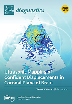
Quantification of displacement parameters was performed in the imaging of brain tissue endogenous motion using ultrasonic radiofrequency signals. Dedicated software and subject head–ultrasonic transducer stabilization allowed calculation of micrometer-range displacements. The method allows identifying brain areas where endogenous displacements waveforms show good intrasubject repeatability during a few heart-cycles. The method was tested in cases of Alzheimer’s disease (visible in cover picture) and healthy subjects and suggested to be useful for brain tissue characterization. View this paper.
  • Issues are regarded as officially published after their release is announced to the table of contents alert mailing list.
  • You may sign up for e-mail alerts to receive table of contents of newly released issues.
  • PDF is the official format for papers published in both, html and pdf forms. To view the papers in pdf format, click on the "PDF Full-text" link, and use the free Adobe Reader to open them.
Order results
Result details
Section
Select all
Export citation of selected articles as:
10 pages, 364 KiB  
Article
Tear Break-Up Time and Seasonal Variation in Intraocular Pressure in a Japanese Population
by Masahiko Ayaki, Kazuno Negishi, Kenya Yuki, Motoko Kawashima, Miki Uchino and Kazuo Tsubota
Diagnostics 2020, 10(2), 124; https://doi.org/10.3390/diagnostics10020124 - 24 Feb 2020
Cited by 5 | Viewed by 2673
Abstract
Purpose: To evaluate seasonal variation in intraocular pressure (IOP) with and without short tear break-up time (SBUT, BUT ≤5 s) since dry eye and IOP are known to have seasonal variation. Methods: This study enrolled 176 patients who visited one of six eye [...] Read more.
Purpose: To evaluate seasonal variation in intraocular pressure (IOP) with and without short tear break-up time (SBUT, BUT ≤5 s) since dry eye and IOP are known to have seasonal variation. Methods: This study enrolled 176 patients who visited one of six eye clinics, in Japan, four times for IOP measurement, in every season. The mean patient age was 67.9 years, including 79 males. Participants were divided into four groups based on the presence of glaucoma and/or SBUT and we compared the seasonal variation in IOP (winter and summer) among the four groups. Results: The IOP (mmHg) in winter and summer, respectively, was 12.8 ± 3.7 and 12.8 ± 3.1 for non-glaucoma patients without SBUT (n = 47, p = 0.964), 14.8 ± 3.4 and 13.3 ± 3.4 for non-glaucoma patients with SBUT (n = 57, p < 0.001), 14.3 ± 3.2 and 14.1 ± 3.4 for glaucoma patients without SBUT (n = 36, p = 0.489), and 13.3 ± 3.0 and 11.6 ± 2.9 for glaucoma with SBUT (n = 36, p < 0.001). Seasonal variation was largest across the seasons in the glaucoma with the SBUT group, and the magnitude of seasonal variation correlated with BUT (β = 0.228, p = 0.003). Conclusions: Seasonal variation tended to be larger in patients with SBUT than those without SBUT. Full article
(This article belongs to the Special Issue Dry Eye and Ocular Surface Disorders)
Show Figures

Figure 1

6 pages, 1320 KiB  
Article
Similarities and Differences between Clear Cell Tubulo-Papillary and Conventional Clear Cell Renal Cell Carcinoma: A Comparative Phenotypical and Mutational Analysis
by Francesca Giunchi, Tania Franceschini, Elisa Gruppioni, Annalisa Altimari, Elisa Capizzi, Francesco Massari, Riccardo Schiavina, Matteo Brunelli, Guido Martignoni and Michelangelo Fiorentino
Diagnostics 2020, 10(2), 123; https://doi.org/10.3390/diagnostics10020123 - 23 Feb 2020
Cited by 3 | Viewed by 2555
Abstract
Background: Clear cell tubulo-papillary renal cell carcinoma (cctpRCC) is characterized by clear cell morphology, but differs from conventional clear cell carcinoma (ccRCC) for its indolent clinical behavior and genetic background. The differential diagnosis between the two is based on histology and immunohistochemistry (IHC). [...] Read more.
Background: Clear cell tubulo-papillary renal cell carcinoma (cctpRCC) is characterized by clear cell morphology, but differs from conventional clear cell carcinoma (ccRCC) for its indolent clinical behavior and genetic background. The differential diagnosis between the two is based on histology and immunohistochemistry (IHC). Methods: We performed a comparative case-control histological, IHC, and genetic analysis by next generation sequencing (NGS), to point out the differences in 10 cases of cctpRCC, and six controls of ccRCC with low stage and grade. Results: All 16 cases showed the IHC profile with cytokeratin 7, racemase, and carbonic anhydrase IX expected for the histological features of each tumor type. By contrast, the NGS mutation analysis that covered 207 amplicons of 50 oncogenes or tumor suppressor genes provided conflicting results. Among the 10 cctpRCC cases, eight (80%) were wild type for all of the genes in the panel, while two (20%) harbored VHL mutations typical of ccRCC. Three of the six (50%) ccRCC control cases showed expected VHL mutations; two (33%) harbored pathogenic mutations in the p53 or the CKIT genes; and one (16%) was wild type. Conclusion: We can assume that histology and ICH are not sufficient for a definitive diagnosis of cctpRCC or ccRCC. Although with a panel covering 50 genes, we found that 80% of cctpRCC were genetically silent; thus, suggesting an indolent biology of these tumors. The differential diagnosis between ccptRCC and ccRCC for the choice of the best therapeutic strategy likely requires the comprehensive evaluation of histology, IHC, and at least VHL mutations. Full article
(This article belongs to the Section Pathology and Molecular Diagnostics)
Show Figures

Figure 1

13 pages, 1138 KiB  
Article
Decreased Cytoplasmic Expression of ADAMTS14 Is Correlated with Reduced Survival Rates in Oral Squamous Cell Carcinoma Patients
by Yueh-Min Lin, Chiao-Wen Lin, Jeng-Wei Lu, Kun-Tu Yeh, Shu-Hui Lin and Shun-Fa Yang
Diagnostics 2020, 10(2), 122; https://doi.org/10.3390/diagnostics10020122 - 23 Feb 2020
Cited by 10 | Viewed by 2908
Abstract
A disintegrin and metalloproteinase with thrombospondin motif 14 (ADAMTS14) is a member of the zinc-dependent protease family that is implicated in the occurrence and progression of tumors. Oral cancer (OC) is a common cancer worldwide, but it is particularly prevalent in Taiwan. However, [...] Read more.
A disintegrin and metalloproteinase with thrombospondin motif 14 (ADAMTS14) is a member of the zinc-dependent protease family that is implicated in the occurrence and progression of tumors. Oral cancer (OC) is a common cancer worldwide, but it is particularly prevalent in Taiwan. However, whether the expression of ADAMTS14 is correlated with the carcinogenesis and progression of oral squamous cell carcinoma (OSCC) has not yet been investigated. In this study, we used immunohistochemistry (IHC) to examine 250 OSCC specimens in order to identify correlations between the cytoplasmic expression of ADAMTS14 and (1) clinicopathological features of OSCC as well as (2) clinical outcomes of OSCC. Our results indicate that cytoplasmic expression of ADAMTS14 was lower in OSCC tissues than in normal tissues. In analyzing correlations between ADAMTS14 expression and clinicopathological features, we found that negative cytoplasmic expression of ADAMTS14 was significantly associated with higher frequencies of lymph node metastasis and more advanced AJCC stages (III/IV). Kaplan–Meier survival analysis revealed that negative cytoplasmic expression of ADAMTS14 was also associated with significantly worse OSCC survival. Univariate and multivariate analyses confirmed that cytoplasmic expression of ADAMTS14 was associated with lymph node metastasis, tumor stage, and tumor grade and also indicated that cytoplasmic ADAMTS14 expression may be an independent prognostic factor for OSCC. This is the first study to report that the cytoplasmic expression level of ADAMTS14 is associated with OSCC prognosis and tumor progression. Our data indicate that ADAMTS14 can serve as a prognostic marker and a potential therapeutic target for OSCC. Full article
(This article belongs to the Section Pathology and Molecular Diagnostics)
Show Figures

Figure 1

14 pages, 1707 KiB  
Article
Olaparib Combined with an ATR or Chk1 Inhibitor as a Treatment Strategy for Acquired Olaparib-Resistant BRCA1 Mutant Ovarian Cells
by Brian T. Burgess, Abigail M. Anderson, J. Robert McCorkle, Jianrong Wu, Frederick R. Ueland and Jill M. Kolesar
Diagnostics 2020, 10(2), 121; https://doi.org/10.3390/diagnostics10020121 - 22 Feb 2020
Cited by 32 | Viewed by 6107
Abstract
Objective: Despite the promise of PARP inhibitors (PARPi) for treating BRCA1/2 mutated ovarian cancer (OC), drug resistance invariably develops. We hypothesized rationale drug combinations, targeting key molecules in DNA repair pathways and the cell cycle may be synergistic and overcome acquired PARPi resistance. [...] Read more.
Objective: Despite the promise of PARP inhibitors (PARPi) for treating BRCA1/2 mutated ovarian cancer (OC), drug resistance invariably develops. We hypothesized rationale drug combinations, targeting key molecules in DNA repair pathways and the cell cycle may be synergistic and overcome acquired PARPi resistance. Methods: Drug sensitivity to PARPi alone and in combination with inhibitors of key DNA repair and cell cycle proteins, including ATR (VE-821), Chk1 (MK-8776), Wee1 (MK-1775), RAD51 (RI-1) was assessed in PARPi-sensitive (UWB1) and -resistant (UWB1-R) gBRCA1 mutant OC cell lines using a cell proliferation assay. The Bliss synergy model was used to estimate the two-drug combination effect and pharmacologic synergy (Bliss score ≥ 0) or antagonistic (Bliss score ≥ 0) response of the PARPi in combination with the inhibitors. Results: IC50 for olaparib alone was 1.6 ± 0.9 µM compared to 3.4 ± 0.6 µM (p = 0.05) for UWB1 and UWB1-R cells, respectively. UWB1-R demonstrated increased sensitivity to ATRi (p = 0.04) compared to UWB1. Olaparib (0.3–1.25 µM) and ATRi (0.8–2.5 µM) were synergistic with Bliss scores of 17.2 ± 0.2, 11.9 ± 0.6 for UWB1 and UWB1-R cells, respectively. Olaparib (0.3–1.25 µM) and Chk1i(0.05–1.25 µM) were synergistic with Bliss scores of 8.3 ± 1.6, 5.7 ± 2.9 for UWB1 and UWB1-R cells, respectively. Conclusions: Combining an ATRi or Chk1i with olaparib is synergistic in both PARPi-sensitive and -resistant BRCA1 mutated OC cell models, and are rationale combinations for further clinical development. Full article
(This article belongs to the Special Issue Ovarian Cancer: Characteristics, Screening, Diagnosis and Treatment)
Show Figures

Figure 1

15 pages, 315 KiB  
Review
Screening and Prevention for High-Grade Serous Carcinoma of the Ovary Based on Carcinogenesis—Fallopian Tube- and Ovarian-Derived Tumors and Incessant Retrograde Bleeding
by Isao Otsuka and Takuto Matsuura
Diagnostics 2020, 10(2), 120; https://doi.org/10.3390/diagnostics10020120 - 22 Feb 2020
Cited by 7 | Viewed by 3784
Abstract
High-grade serous carcinoma (HGSC) is the most common and lethal subtype of ovarian carcinoma. Many HGSCs are now believed to originate in the fallopian tube epithelium; ovarian surface epithelium is another possible origin. Thus, current screening methods, i.e., ultrasonography and serum CA-125 measurements, [...] Read more.
High-grade serous carcinoma (HGSC) is the most common and lethal subtype of ovarian carcinoma. Many HGSCs are now believed to originate in the fallopian tube epithelium; ovarian surface epithelium is another possible origin. Thus, current screening methods, i.e., ultrasonography and serum CA-125 measurements, have a limitation in their early detection. Recently, circulating biomarkers, such as tumor DNA, autoantibody, and microRNA, have been investigated to detect HGSCs. As cancer cells in the fallopian tube flow into the endometrial cavity, the detection of exfoliated cells, tumor DNA, and proteome from samples obtained from the endometrial cavity or the cervix may be useful. The risk of ovarian serous carcinoma is affected by the use of oral contraceptive and menopausal hormone therapy (MHT). MHT regimens causing endometrial bleeding increase serous carcinoma risk, hence, incessant retrograde bleeding from the endometrial cavity into the Douglas pouch appears to play an important role in high-grade serous carcinogenesis. In this review, we provide an overview of current and novel screening methods and prevention approaches for ovarian and fallopian tube HGSC. Full article
(This article belongs to the Special Issue Ovarian Cancer: Characteristics, Screening, Diagnosis and Treatment)
12 pages, 1397 KiB  
Article
BRCAness as an Important Prognostic Marker in Patients with Triple-Negative Breast Cancer Treated with Neoadjuvant Chemotherapy: A Multicenter Retrospective Study
by Yoshimasa Kosaka, Yutaka Yamamoto, Hirokazu Tanino, Hiroshi Nishimiya, Mutsuko Yamamoto-Ibusuki, Yuko Hirota, Hirotaka Iwase, Seigo Nakamura and Sadako Akashi-Tanaka
Diagnostics 2020, 10(2), 119; https://doi.org/10.3390/diagnostics10020119 - 21 Feb 2020
Cited by 6 | Viewed by 3441
Abstract
Triple-negative breast cancer (TNBC) has several subtypes. The identification of markers associated with recurrence and poor prognosis in patients with TNBC is urgently needed. BRCAness is a set of traits in which BRCA1 dysfunction, arising from gene mutation, methylation, or deletion, results in [...] Read more.
Triple-negative breast cancer (TNBC) has several subtypes. The identification of markers associated with recurrence and poor prognosis in patients with TNBC is urgently needed. BRCAness is a set of traits in which BRCA1 dysfunction, arising from gene mutation, methylation, or deletion, results in DNA repair deficiency. In the current study, we evaluated the clinical significance and prognosis of BRCAness in a multicenter retrospective study. Ninety-four patients with TNBC treated with neoadjuvant chemotherapy were enrolled from three university hospitals for this retrospective study. BRCAness was evaluated in 94 core needle biopsy (CNB) specimens prior to neoadjuvant chemotherapy and 49 surgical specimens without pathological complete response (pCR). The samples were assessed using multiplex ligation-dependent probe amplification, and the amplicons were scored. Of the 94 patients, 51 had BRCAness in CNB specimens. There were no significant differences in pCR rates or recurrence between the BRCAness and non-BRCAness groups. Among surgical specimens, the BRCAness group had a significantly shorter recurrence-free survival and overall survival compared with the non-BRCAness group. The BRCAness of surgical specimens was found to be an important marker to predict prognosis in patients with TNBC after neoadjuvant chemotherapy. A clinical trial to assess the clinical impact of carboplatin with BRCAness is planned. Full article
(This article belongs to the Collection Biomarkers in Medicine)
Show Figures

Figure 1

14 pages, 5001 KiB  
Review
Viscoelastic Hemostatic Assays: Moving from the Laboratory to the Site of Care—A Review of Established and Emerging Technologies
by Jan Hartmann, Matthew Murphy and Joao D. Dias
Diagnostics 2020, 10(2), 118; https://doi.org/10.3390/diagnostics10020118 - 21 Feb 2020
Cited by 52 | Viewed by 8175
Abstract
Viscoelastic-based techniques to evaluate whole blood hemostasis have advanced substantially since they were first developed over 70 years ago but are still based upon the techniques first described by Dr. Hellmut Hartert in 1948. Today, the use of thromboelastography, the method of testing [...] Read more.
Viscoelastic-based techniques to evaluate whole blood hemostasis have advanced substantially since they were first developed over 70 years ago but are still based upon the techniques first described by Dr. Hellmut Hartert in 1948. Today, the use of thromboelastography, the method of testing viscoelastic properties of blood coagulation, has moved out of the research laboratory and is now more widespread, used commonly during surgery, in emergency departments, intensive care units, and in labor wards. Thromboelastography is currently a rapidly growing field of technological advancement and is attracting significant investment. This review will first describe the history of the viscoelastic testing and the established first-generation devices, which were developed for use within the laboratory. This review will then describe the next-generation hemostasis monitoring devices, which were developed for use at the site of care for an expanding range of clinical applications. This review will then move on to experimental technologies, which promise to make viscoelastic testing more readily available in a wider range of clinical environments in the endeavor to improve patient care. Full article
(This article belongs to the Special Issue Point-of-Care Detection Devices for Healthcare)
Show Figures

Figure 1

8 pages, 1096 KiB  
Review
Diagnostic Problems in Diffuse Axonal Injury
by Sung Ho Jang
Diagnostics 2020, 10(2), 117; https://doi.org/10.3390/diagnostics10020117 - 21 Feb 2020
Cited by 14 | Viewed by 7449
Abstract
In this study, three problems associated with diagnosing diffuse axonal injury (DAI) in patients with traumatic brain injury are reviewed: the shortage of scientific evidence supporting the 6-hour loss of consciousness (LOC) diagnostic criterion to discriminate concussion and DAI, the low sensitivity of [...] Read more.
In this study, three problems associated with diagnosing diffuse axonal injury (DAI) in patients with traumatic brain injury are reviewed: the shortage of scientific evidence supporting the 6-hour loss of consciousness (LOC) diagnostic criterion to discriminate concussion and DAI, the low sensitivity of conventional brain MRI in the detection of DAI lesions, and the inappropriateness of the term diffuse in DAI. Pathological study by brain biopsy is required to confirm DAI; however, performing a brain biopsy for the diagnosis of DAI in a living patient is impossible. Therefore, the diagnosis of DAI in a living patient is clinically determined based on the duration of LOC, clinical manifestations, and the results of conventional brain MRI. There is a shortage of scientific evidence supporting the use of the 6-hour LOC criterion to distinguish DAI from concussion, and axonal injuries have been detected in many concussion cases with a less than 6-hour LOC. Moreover, due to the low sensitivity of conventional brain MRI, which can only detect DAI lesions in approximately half of DAI patients, diagnostic MRI criteria for DAI are not well established. In contrast, diffusion tensor imaging (DTI) has been shown to have high sensitivity for the detection of DAI lesions. As DTI is a relatively new method, further studies aimed at the establishment of diagnostic criteria for DAI detection using DTI are needed. On the other hand, because DAI distribution is not diffuse but multifocal, and because axonal injury lesions have been detected in concussion patients, steps to standardize the use of terms related to axonal injury in both concussion and DAI are necessary. Full article
(This article belongs to the Special Issue Brain Imaging/Neuroimaging)
Show Figures

Graphical abstract

11 pages, 1325 KiB  
Article
Modeling Morquio A Syndrome: An Anthropometric Study of Body Characteristics and Stature
by Agnieszka Różdżyńska-Świątkowska, Krzysztof Szklanny, Jolanta Marucha and Anna Tylki-Szymańska
Diagnostics 2020, 10(2), 116; https://doi.org/10.3390/diagnostics10020116 - 20 Feb 2020
Cited by 2 | Viewed by 6358
Abstract
Background: Morquio A syndrome or mucopolysaccharidosis (MPS) IVA is an autosomal recessive, life-limiting lysosomal storage disease caused by deficient activity of the enzyme galactosamine-6-sulfatase. Common early symptoms such as abnormalities of body stature can facilitate timely diagnosis. This study aimed to create a [...] Read more.
Background: Morquio A syndrome or mucopolysaccharidosis (MPS) IVA is an autosomal recessive, life-limiting lysosomal storage disease caused by deficient activity of the enzyme galactosamine-6-sulfatase. Common early symptoms such as abnormalities of body stature can facilitate timely diagnosis. This study aimed to create a pattern of face and body stature based on anthropometric measurements taken from a cohort of Polish patients with MPS IVA. Methods: Analysis of 11 somatometric and 14 craniofacial features was performed on 20 patients with MPS IVA, aged from 3 months to 26 years. The diagnosis of MPS IVA was confirmed by enzymatic and molecular analysis. Two-tailed t-tests were used to compare mean values for body length and weight at birth between the MPS IVA patients and the general population. To show the degree and direction of deviation z-scores were calculated and then used to construct a model of an average MPS IVA patient. Results: Mean values for body height and weight at birth were greater for boys than for the general population. The observed pattern of head and body shape indicated that dwarfism occurred with age as a result of the relatively short trunk and lower limbs. Skeletal abnormalities included a bell-shaped chest with the ratio of chest depth to chest width being significantly above the norm. The head and neck were relatively elongated, in comparison to body height, and tucked between narrow shoulders. The head had dolichocephalic shape, while the nose was short with wide nostrils. Conclusions: Multiple anthropometric measurements, including age ranges, allowed for the creation of a model that showed the most characteristic features of the MPS IVA phenotype. Full article
(This article belongs to the Special Issue Mucopolysaccharidoses: Diagnosis, Treatment, and Management)
Show Figures

Figure 1

14 pages, 1600 KiB  
Article
Prognostic Value of Circulating IGFBP2 and Related Autoantibodies in Children with Metastatic Rhabdomyosarcomas
by Elena Poli, Angelica Zin, Manuela Cattelan, Lucia Tombolan, Ilaria Zanetti, Angela Scagnellato, Paolo Bonvini and Gianni Bisogno
Diagnostics 2020, 10(2), 115; https://doi.org/10.3390/diagnostics10020115 - 20 Feb 2020
Cited by 8 | Viewed by 2443
Abstract
Insulin-like growth factor-binding protein 2 (IGFBP2) is a tumor-associated protein measurable in patients’ biopsies and blood samples. Increased IGFBP2 expression correlates with tumor severity in rhabdomyosarcoma (RMS). Thus, we examined the plasmatic IGFBP2 levels in 114 RMS patients and 15 healthy controls by [...] Read more.
Insulin-like growth factor-binding protein 2 (IGFBP2) is a tumor-associated protein measurable in patients’ biopsies and blood samples. Increased IGFBP2 expression correlates with tumor severity in rhabdomyosarcoma (RMS). Thus, we examined the plasmatic IGFBP2 levels in 114 RMS patients and 15 healthy controls by ELISA assay in order to evaluate its value as a plasma biomarker for RMS. Additionally, we looked for the presence of a humoral response against IGBFP2 protein measurable by the production of anti-IGFBP2 autoantibodies. We demonstrated that both circulating IGFBP2 protein and autoantibodies were significantly higher in RMS patients with respect to controls and their combination showed a better discriminative capacity. IGFBP2 protein identified metastatic patients with worse event-free survival, whereas both IGFBP2 and anti-IGFBP2 antibodies negatively correlated with overall survival. Our study suggests that IGFBP2 and anti-IGFBP2 antibodies are useful for diagnostic and prognostic purposes, mainly as independent negative prognostic markers in metastatic patients. This is the first study that reports a specific humoral response in RMS plasma samples and proves the value of blood-based biomarkers in improving risk assessment and outcome of metastatic RMS patients. Full article
(This article belongs to the Section Pathology and Molecular Diagnostics)
Show Figures

Figure 1

10 pages, 939 KiB  
Article
A CRISPR Test for Rapidly and Sensitively Detecting Circulating EGFR Mutations
by Jen-Hui Tsou, Qixin Leng and Feng Jiang
Diagnostics 2020, 10(2), 114; https://doi.org/10.3390/diagnostics10020114 - 19 Feb 2020
Cited by 21 | Viewed by 5065
Abstract
The detection of EGFR mutations in circulating cell-free DNA can enable personalized therapy for cancer. The current techniques for detecting circulating EGFR mutations are expensive and time-consuming with moderate sensitivity. Emerging CRISPR is revolutionizing medical diagnostics and showing a great promise for nucleic [...] Read more.
The detection of EGFR mutations in circulating cell-free DNA can enable personalized therapy for cancer. The current techniques for detecting circulating EGFR mutations are expensive and time-consuming with moderate sensitivity. Emerging CRISPR is revolutionizing medical diagnostics and showing a great promise for nucleic acid detection. This study aims to develop CRISPR-Cas12a as a simple test to sensitively detect circulating EGFR mutations in plasma. Serially diluted samples of DNA containing heterozygous EGFR mutations (L858R and T790M) in wild-type genomic DNA are concurrently tested for the mutations by a CRISPR-Cas12a system and droplet digital PCR (ddPCR). The CRISPR-Cas12a system can detect both L858R and T790M with a limit of detection of 0.005% in less than three hours. ddPCR detects the mutations with a limit of detection of 0.05% for more than five hours. Plasma samples of 28 lung cancer patients and 20 cancer-free individuals are tested for the EGFR mutations by CRISPR-Cas12a system and ddPCR. The CRISPR-Cas12a system could detect L858R in plasma of two lung cancer patients whose tissue biopsies are positive for L858R, and one plasma sample of three lung cancer patients whose tissue biopsies are positive for T790M. ddPCR detects L858R in the same two plasm samples, however, does not detect T790M in any of the plasma samples. This proof of principle study demonstrates that the CRISPR-Cas12a system could rapidly and sensitively detect circulating EGFR mutations, and thus, has potential prognostic or therapeutic implications. Full article
(This article belongs to the Section Pathology and Molecular Diagnostics)
Show Figures

Figure 1

9 pages, 1476 KiB  
Article
Urinary MicroRNA-21-5p as Potential Biomarker of Interstitial Fibrosis and Tubular Atrophy (IFTA) in Kidney Transplant Recipients
by Michal S. Gniewkiewicz, Izabela Paszkowska, Jolanta Gozdowska, Katarzyna Czerwinska, Anna Sadowska-Jakubowicz, Dominika Deborska-Materkowska, Agnieszka Perkowska-Ptasinska, Maciej Kosieradzki and Magdalena Durlik
Diagnostics 2020, 10(2), 113; https://doi.org/10.3390/diagnostics10020113 - 19 Feb 2020
Cited by 12 | Viewed by 3539
Abstract
Chronic renal allograft dysfunction (CAD) is a major limiting factor of long-term graft survival. The hallmarks of progressive CAD are interstitial fibrosis and tubular atrophy (IFTA). MicroRNAs are small, regulatory RNAs involved in many immunological processes. In particular, microRNA-21-5p (miR-21) is considered to [...] Read more.
Chronic renal allograft dysfunction (CAD) is a major limiting factor of long-term graft survival. The hallmarks of progressive CAD are interstitial fibrosis and tubular atrophy (IFTA). MicroRNAs are small, regulatory RNAs involved in many immunological processes. In particular, microRNA-21-5p (miR-21) is considered to be strongly associated with pathogenesis regarding tubulointerstitium. The aim of this study was to assess urinary miR-21 expression levels in the kidney transplant recipients and determine their application in the evaluation of IFTA and kidney allograft function. The expression levels of miR-21 were quantified in the urine of 31 kidney transplant recipients with biopsy-assessed IFTA (IFTA 0 + I: n = 17; IFTA II + III: n = 14) by real-time quantitative PCR. Urine samples were collected at the time of protocolar biopsies performed 1 or 2 years after kidney transplantation. MicroRNA-191-5p was used as reference gene. MiR-21 was significantly up-regulated in IFTA II + III group compared to IFTA 0 + I group (p = 0.003). MiR-21 correlated significantly with serum concentration of creatinine (r = 0.52, p = 0.003) and eGFR (r = −0.45; p = 0.01). ROC analysis determined the diagnostic value of miR-21 with an area under curve (AUC) of 0.80 (p = 0.0002), sensitivity of 0.86 and specificity of 0.71. miR-21 is associated with renal allograft dysfunction and IFTA. Therefore, it could be considered as a potential diagnostic, non-invasive biomarker for monitoring renal graft function. Full article
(This article belongs to the Section Pathology and Molecular Diagnostics)
Show Figures

Figure 1

13 pages, 1074 KiB  
Review
Pediatric Thyroid Cancer in Europe: An Overdiagnosed Condition? A Literature Review
by Andreea-Ioana Stefan, Andra Piciu, Alexandru Mester, Dragos Apostu, Marius Badan and Claudiu-Iulian Badulescu
Diagnostics 2020, 10(2), 112; https://doi.org/10.3390/diagnostics10020112 - 19 Feb 2020
Cited by 16 | Viewed by 3344
Abstract
Thyroid neoplastic pathology is the most common form of cancer associated with radiation exposure. The most common histopathological type of thyroid carcinoma is the differentiated thyroid cancer (these include papillary and follicular type), which represents over 90% of all cases, especially affecting girls [...] Read more.
Thyroid neoplastic pathology is the most common form of cancer associated with radiation exposure. The most common histopathological type of thyroid carcinoma is the differentiated thyroid cancer (these include papillary and follicular type), which represents over 90% of all cases, especially affecting girls rather than boys. Although patients are diagnosed in advanced stages as compared to adults, the prognosis of the disease is very good, with a 30-year survival rate of over 95% but post-therapeutic morbidity remains quite high. The treatment is based in particular on the therapeutic guidelines for adults, but as children have some histopathological and genetic characteristics of thyroid cancer, as well as different initial clinical presentations, we decided to review the literature on this pathology among the pediatric population, focusing on cases in Europe. The major interest is the impact of the Chernobyl accident. Full article
(This article belongs to the Special Issue New Insights in Thyroid Diagnostics)
Show Figures

Figure 1

18 pages, 16179 KiB  
Article
Viscoelastic Biomarkers of Ex Vivo Liver Samples via Torsional Wave Elastography
by Inas H. Faris, Juan Melchor, Antonio Callejas, Jorge Torres and Guillermo Rus
Diagnostics 2020, 10(2), 111; https://doi.org/10.3390/diagnostics10020111 - 19 Feb 2020
Cited by 9 | Viewed by 3276
Abstract
The clinical ultrasound community demands mechanisms to obtain the viscoelastic biomarkers of soft tissue in order to quantify the tissue condition and to be able to track its consistency. Torsional Wave Elastography (TWE) is an emerging technique proposed for interrogating soft tissue mechanical [...] Read more.
The clinical ultrasound community demands mechanisms to obtain the viscoelastic biomarkers of soft tissue in order to quantify the tissue condition and to be able to track its consistency. Torsional Wave Elastography (TWE) is an emerging technique proposed for interrogating soft tissue mechanical viscoelastic constants. Torsional waves are a particular configuration of shear waves, which propagate asymmetrically in-depth and are radially transmitted by a disc and received by a ring. This configuration is shown to be particularly efficient in minimizing spurious p-waves components and is sensitive to mechanical constants, especially in cylinder-shaped organs. The objective of this work was to validate (TWE) technique against Shear Wave Elasticity Imaging (SWEI) technique through the determination of shear wave velocity, shear moduli, and viscosity of ex vivo chicken liver samples and tissue mimicking hydrogel phantoms. The results of shear moduli for ex vivo liver tissue vary 1.69–4.0kPa using TWE technique and 1.32–4.48kPa using SWEI technique for a range of frequencies from 200 to 800Hz. Kelvin–Voigt viscoelastic parameters reported values of μ = 1.51kPa and η = 0.54Pa·s using TWE and μ = 1.02kPa and η = 0.63Pa·s using SWEI. Preliminary results show that the proposed technique successfully allows reconstructing shear wave velocity, shear moduli, and viscosity mechanical biomarkers from the propagated torsional wave, establishing a proof of principle and warranting further studies. Full article
(This article belongs to the Special Issue Elastography)
Show Figures

Figure 1

18 pages, 4186 KiB  
Article
SD-UNet: Stripping down U-Net for Segmentation of Biomedical Images on Platforms with Low Computational Budgets
by Pius Kwao Gadosey, Yujian Li, Enock Adjei Agyekum, Ting Zhang, Zhaoying Liu, Peter T. Yamak and Firdaous Essaf
Diagnostics 2020, 10(2), 110; https://doi.org/10.3390/diagnostics10020110 - 18 Feb 2020
Cited by 54 | Viewed by 10156
Abstract
During image segmentation tasks in computer vision, achieving high accuracy performance while requiring fewer computations and faster inference is a big challenge. This is especially important in medical imaging tasks but one metric is usually compromised for the other. To address this problem, [...] Read more.
During image segmentation tasks in computer vision, achieving high accuracy performance while requiring fewer computations and faster inference is a big challenge. This is especially important in medical imaging tasks but one metric is usually compromised for the other. To address this problem, this paper presents an extremely fast, small and computationally effective deep neural network called Stripped-Down UNet (SD-UNet), designed for the segmentation of biomedical data on devices with limited computational resources. By making use of depthwise separable convolutions in the entire network, we design a lightweight deep convolutional neural network architecture inspired by the widely adapted U-Net model. In order to recover the expected performance degradation in the process, we introduce a weight standardization algorithm with the group normalization method. We demonstrate that SD-UNet has three major advantages including: (i) smaller model size (23x smaller than U-Net); (ii) 8x fewer parameters; and (iii) faster inference time with a computational complexity lower than 8M floating point operations (FLOPs). Experiments on the benchmark dataset of the Internatioanl Symposium on Biomedical Imaging (ISBI) challenge for segmentation of neuronal structures in electron microscopic (EM) stacks and the Medical Segmentation Decathlon (MSD) challenge brain tumor segmentation (BRATs) dataset show that the proposed model achieves comparable and sometimes better results compared to the current state-of-the-art. Full article
(This article belongs to the Special Issue Artificial Intelligence in Diagnostics)
Show Figures

Figure 1

8 pages, 1824 KiB  
Article
Combination of Two Rapid Ophthalmic Test Kits for Improved Diagnosis in Cases of Severe Binocular Conjunctivitis
by Asako Kodama, Fumitaka Kobayashi, Hao Yung Yang, Kazumi Fukagawa, Hiroyuki Yazu and Hiroshi Fujishima
Diagnostics 2020, 10(2), 109; https://doi.org/10.3390/diagnostics10020109 - 17 Feb 2020
Cited by 4 | Viewed by 3230
Abstract
Introduction: Diagnosis of conjunctivitis can be sometimes difficult, especially in cases of severe conjunctivitis and those involving both eyes. In this study, we performed commercial tests for adenovirus (Capilia Adeno Eye®) and total tear IgE (Allerwatch®) in a single [...] Read more.
Introduction: Diagnosis of conjunctivitis can be sometimes difficult, especially in cases of severe conjunctivitis and those involving both eyes. In this study, we performed commercial tests for adenovirus (Capilia Adeno Eye®) and total tear IgE (Allerwatch®) in a single visit in patients with bilateral conjunctivitis to examine if, and by how much, the combination of these two tests would improve the diagnostic accuracy of conjunctivitis. Methods: The study included sixty patients with relatively severe conjunctivitis in both eyes within a week of consulting our clinic and who had no previous treatment. Capilia Adeno Eye® and Allerwatch® tests were performed. Results: A significantly higher number of cases (55/60) were diagnosed when both tests were evaluated than with either test (Capilia Adeno Eye® (12/60; p < 0.001) or Allerwatch® (44/60; p < 0.005)) alone. The positivity rate of Allerwatch® was significantly higher than that of Capilia Adeno Eye® (p < 0.001). The diagnosis rate of atopic keratoconjunctivitis was 100% in patients with allergic conjunctivitis, but there was no significant difference in positivity compared with other types of allergic conjunctivitis. Conclusions: Testing patients with both Capilia Adeno Eye® and Allerwatch® improves the diagnostic accuracy for conjunctivitis and can diagnose more than 90% of cases. Detection of adenovirus antigen and IgE in tears, using these simple and rapid methods, will be useful for early diagnosis and prevention of adenoviral conjunctivitis. Full article
(This article belongs to the Special Issue Dry Eye and Ocular Surface Disorders)
Show Figures

Figure 1

11 pages, 2348 KiB  
Case Report
Long-Term Outcomes of Early Enzyme Replacement Therapy for Mucopolysaccharidosis IV: Clinical Case Studies of Two Siblings
by Sharon Barak, Yair Anikster, Ifat Sarouk, Eve Stern, Etzyona Eisenstein, Tamar Yissar, Nir Sherr-Lurie, Annick Raas-Rothschild and Dafna Guttman
Diagnostics 2020, 10(2), 108; https://doi.org/10.3390/diagnostics10020108 - 17 Feb 2020
Cited by 4 | Viewed by 3755 | Correction
Abstract
Enzyme replacement therapy (ERT) is one of the available therapies for mucopolysaccharidosis (MPS). This study presents a follow-up of two siblings with MPS IVA (Morquio A disease) that received ERT. Both siblings received weekly intravenous infusions of elosulfase alfa for 4.5 years. One [...] Read more.
Enzyme replacement therapy (ERT) is one of the available therapies for mucopolysaccharidosis (MPS). This study presents a follow-up of two siblings with MPS IVA (Morquio A disease) that received ERT. Both siblings received weekly intravenous infusions of elosulfase alfa for 4.5 years. One sibling (patient 1, P1; male) started therapy at 54 months of age, and the other sibling (patient 2, P2; female) started at 11 months of age. ERT was well-tolerated. In comparison to P1, P2’s growth curves deviated less from the norm. The orthopedic deformities of P1 were more severe than those of P2 and required several surgical corrections. P1’s sleep test at 48 months revealed obstructive sleep apnea, while by the age of 102 months, parameters were normal. P2 never had sleep apnea. Only P1 demonstrated ear, nose, and throat clinical illnesses. In comparison to P1, P2’s physical function was better maintained. In conclusion, ERT was safe in both patients during a 4.5-year follow-up. Although the typical characteristics of this disease were similar in both patients, P1 had a complex clinical course in comparison to P2, which influenced function and quality of life. Therefore, in order to make the most of ERT, it may be more beneficial when initiated at a relatively young age. Full article
(This article belongs to the Special Issue Mucopolysaccharidoses: Diagnosis, Treatment, and Management)
Show Figures

Figure 1

17 pages, 2359 KiB  
Review
Role of pH Value in Clinically Relevant Diagnosis
by Shu-Hua Kuo, Ching-Ju Shen, Ching-Fen Shen and Chao-Min Cheng
Diagnostics 2020, 10(2), 107; https://doi.org/10.3390/diagnostics10020107 - 16 Feb 2020
Cited by 74 | Viewed by 9962
Abstract
As a highly influential physiological factor, pH may be leveraged as a tool to diagnose physiological state. It may be especially suitable for diagnosing and assessing skin structure and wound status. Multiple innovative and elegant smart wound dressings combined with either pH sensors [...] Read more.
As a highly influential physiological factor, pH may be leveraged as a tool to diagnose physiological state. It may be especially suitable for diagnosing and assessing skin structure and wound status. Multiple innovative and elegant smart wound dressings combined with either pH sensors or drug control-released carriers have been extensively studied. Increasing our understanding of the role of pH value in clinically relevant diagnostics should assist clinicians and improve personal health management in the home. In this review, we summarized a number of articles and discussed the role of pH on the skin surface as well as the factors that influence skin pH and pH-relevant skin diseases, but also the relationship of skin pH to the wound healing process, including its influence on the activity of proteases, bacterial enterotoxin, and some antibacterial agents. A great number of papers discussing physiological pH value have been published in recent decades, far too many to be included in this review. Here, we have focused on the impact of pH on wounds and skin with an emphasis on clinically relevant diagnosis toward effective treatment. We have also summarized the differences in skin structure and wound care between adults and infants, noting that infants have fragile skin and poor skin barriers, which makes them more vulnerable to skin damage and compels particular care, especially for wounds. Full article
(This article belongs to the Section Point-of-Care Diagnostics and Devices)
Show Figures

Figure 1

7 pages, 176 KiB  
Communication
Accuracy in Referrals to Gynecologic Oncologists Based on Clinical Presentation for Ovarian Mass
by Katherine Jane C. Chua, Ricky D. Patel, Radhika Trivedi, Patricia Greenberg, Kyle Beiter, Thomas Magliaro, Ushma Patel and Joyce Varughese
Diagnostics 2020, 10(2), 106; https://doi.org/10.3390/diagnostics10020106 - 16 Feb 2020
Cited by 5 | Viewed by 2669
Abstract
Ovarian cancer is one of the most lethal gynecological cancers in women due to late diagnosis. Despite technological advancements, experienced physicians have high sensitivities and specificities in subjective assessments when combining ultrasound findings and clinical history in analyzing adnexal masses. This study aims [...] Read more.
Ovarian cancer is one of the most lethal gynecological cancers in women due to late diagnosis. Despite technological advancements, experienced physicians have high sensitivities and specificities in subjective assessments when combining ultrasound findings and clinical history in analyzing adnexal masses. This study aims to demonstrate general obstetricians and gynecologists’ (OB/GYN) appropriateness in gynecologic oncologist referrals for malignant ovarian masses based on history and physical (H&P), imaging, and available tumor markers. Three board certified OB/GYNs were given 148 cases and determined whether or not they would refer them to a gynecologic oncologist. Results showed that OB/GYNs were 81–85% accurate in diagnosing patients with a benign or malignant disease. Among the malignant cases, reviewers had a high sensitivity ranging from 74–81% in appropriately referring a malignancy. In our study, OB/GYNs referred between 23–32% of ovarian masses to a gynecologic oncologist with only 9.5% of cases found to be malignant. Despite the high referral rates, generalists showed a high degree of sensitivity in accurately referring malignant diseases based solely on clinical experience and imaging studies, which could improve survival rates with early intervention by gynecologic oncologists. Full article
(This article belongs to the Special Issue Ovarian Cancer: Characteristics, Screening, Diagnosis and Treatment)
8 pages, 1742 KiB  
Case Report
Long-Term Follow-up Posthematopoietic Stem Cell Transplantation in a Japanese Patient with Type-VII Mucopolysaccharidosis
by Kenji Orii, Yasuyuki Suzuki, Shunji Tomatsu, Tadao Orii and Toshiyuki Fukao
Diagnostics 2020, 10(2), 105; https://doi.org/10.3390/diagnostics10020105 - 16 Feb 2020
Cited by 3 | Viewed by 2317
Abstract
The effectiveness of hematopoietic stem cell transplantation (HSCT) for type-VII mucopolysaccharidosis (MPS VII, Sly syndrome) remains controversial, although recent studies have shown that it has a clinical impact. In 1998, Yamada et al. reported the first patient with MPS VII, who underwent HSCT [...] Read more.
The effectiveness of hematopoietic stem cell transplantation (HSCT) for type-VII mucopolysaccharidosis (MPS VII, Sly syndrome) remains controversial, although recent studies have shown that it has a clinical impact. In 1998, Yamada et al. reported the first patient with MPS VII, who underwent HSCT at 12 years of age. Here, we report the results of a 22-year follow-up of that patient post-HSCT, who harbored the p.Ala619Val mutation associated with an attenuated phenotype. The purpose of this study was to evaluate changes in physical symptoms, the activity of daily living (ADL), and the intellectual status in the 34-year-old female MPS VII patient post-HSCT, and to prove the long-term effects of HSCT in MPS VII. Twenty-two years after HSCT, the β-glucuronidase activity in leukocytes remained at normal levels, and urinary glycosaminoglycan excretion was reduced and kept within normal levels. At present, she is capable of sustaining simple conversation, and her intellectual level is equivalent to that of a 6-year-old. She can walk alone and climb upstairs by holding onto a handrail, although she feels mild pain in the hip joint. The cervical vertebrae are fused with the occipital bone, causing dizziness and light-headedness when the neck is bent back. Overall, her clinical condition has been stabilized and kept well for long-term post-HSCT, indicating that HSCT is a therapeutic option for MPS VII. Full article
(This article belongs to the Special Issue Mucopolysaccharidoses: Diagnosis, Treatment, and Management)
Show Figures

Figure 1

11 pages, 2838 KiB  
Article
Microsatellite Instability and MMR Genes Abnormalities in Canine Mammary Gland Tumors
by Faiz Muhammad Khand, Da-Wei Yao, Pan Hao, Xin-Qi Wu, Asghar Ali Kamboh and De-Ji Yang
Diagnostics 2020, 10(2), 104; https://doi.org/10.3390/diagnostics10020104 - 14 Feb 2020
Cited by 4 | Viewed by 3068
Abstract
Early diagnosis of mammary gland tumors is a challenging task in animals, especially in unspayed dogs. Hence, this study investigated the role of microsatellite instability (MSI), MMR gene mRNA transcript levels and SNPs of MMR genes in canine mammary gland tumors (CMT). A [...] Read more.
Early diagnosis of mammary gland tumors is a challenging task in animals, especially in unspayed dogs. Hence, this study investigated the role of microsatellite instability (MSI), MMR gene mRNA transcript levels and SNPs of MMR genes in canine mammary gland tumors (CMT). A total of 77 microsatellite (MS) markers in 23 primary CMT were selected from four breeds of dogs. The results revealed that 11 out of 77 MS markers were unstable and showed MSI in all the tumors (at least at one locus), while the other markers were stable. Compared to the other markers, the ABC9TETRA, MEPIA, 9A5, SCNA11 and FJL25 markers showed higher frequencies of instability. All CMT demonstrated MSI, with eight tumors presenting MSI-H. The RT-qPCR results revealed significant upregulation of the mRNA levels of cMSH3, cMLH1, and cPMSI, but downregulation of cMSH2 compared to the levels in the control group. Moreover, single nucleotide polymorphisms (SNPs) were observed in the cMSH2 gene in four exons, i.e., 2, 6, 15, and 16. In conclusion, MSI, overexpression of MMR genes and SNPs in the MMR gene are associated with CMT and could be served as diagnostic biomarkers for CMT in the future. Full article
(This article belongs to the Special Issue Ovarian Cancer: Characteristics, Screening, Diagnosis and Treatment)
Show Figures

Figure 1

13 pages, 567 KiB  
Review
Breast Cancer Detection—A Synopsis of Conventional Modalities and the Potential Role of Microwave Imaging
by Brian M. Moloney, Declan O’Loughlin, Sami Abd Elwahab and Michael J. Kerin
Diagnostics 2020, 10(2), 103; https://doi.org/10.3390/diagnostics10020103 - 14 Feb 2020
Cited by 39 | Viewed by 6139
Abstract
Global statistics have demonstrated that breast cancer is the most frequently diagnosed invasive cancer and the leading cause of cancer death among female patients. Survival following a diagnosis of breast cancer is grossly determined by the stage of the disease at the time [...] Read more.
Global statistics have demonstrated that breast cancer is the most frequently diagnosed invasive cancer and the leading cause of cancer death among female patients. Survival following a diagnosis of breast cancer is grossly determined by the stage of the disease at the time of initial diagnosis, highlighting the importance of early detection. Improving early diagnosis will require a multi-faceted approach to optimizing the use of currently available imaging modalities and investigating new methods of detection. The application of microwave technologies in medical diagnostics is an emerging field of research, with breast cancer detection seeing the most significant progress in the last twenty years. In this review, the application of current conventional imaging modalities is discussed, and recurrent shortcomings highlighted. Microwave imaging is rapid and inexpensive. If the preliminary results of its diagnostic capacity are substantiated, microwave technology may offer a non-ionizing, non-invasive, and painless adjunct or stand-alone modality that could possibly be implemented in routine diagnostic breast care. Full article
(This article belongs to the Special Issue Multimodality Breast Imaging)
Show Figures

Figure 1

8 pages, 829 KiB  
Review
Serous Tubal Intraepithelial Carcinoma: A Concise Review for the Practicing Pathologist and Clinician
by S. Emily Bachert, Anthony McDowell, Jr., Dava Piecoro and Lauren Baldwin Branch
Diagnostics 2020, 10(2), 102; https://doi.org/10.3390/diagnostics10020102 - 13 Feb 2020
Cited by 15 | Viewed by 5094
Abstract
Ovarian cancer is the deadliest gynecologic malignancy, accounting for more than 14,000 deaths each year. With no established way to prevent or screen for it, the vast majority of cases are diagnosed as International Federation of Gynecology and Obstetrics (FIGO) stage III or [...] Read more.
Ovarian cancer is the deadliest gynecologic malignancy, accounting for more than 14,000 deaths each year. With no established way to prevent or screen for it, the vast majority of cases are diagnosed as International Federation of Gynecology and Obstetrics (FIGO) stage III or higher. Individuals with germline BRCA mutations are at particularly high risk for epithelial ovarian cancer and have been the subject of many risk-reducing strategies. In the past ten years, studies looking at risk-reducing salpingo-oophorectomy (RRSO) in this population have uncovered an interesting association: up to 8% of women with BRCA1 or BRCA2 mutations who underwent RRSO had an associated serous tubal intraepithelial carcinoma (STIC). The importance of this finding is highlighted by the fact that up to 60% of ovarian cancer patients will also have an associated STIC. These studies have led to a paradigm shift that a subset of epithelial ovarian cancer originates not in the ovarian epithelium, but rather in the distal fallopian tube. In response to this, many providers have changed their practice by expanding the role of routine salpingectomy, hysterectomy, and sterilization procedures. The American College of Obstetricians and Gynecologists (ACOG) has acknowledged opportunistic salpingectomy as a safe strategy to reduce the risk of epithelial ovarian cancer in Committee Opinion #774. It is thus important for pathologists and clinicians to understand the definition of STIC; how it is diagnosed; and, most importantly, its clinical significance. Full article
(This article belongs to the Special Issue Ovarian Cancer: Characteristics, Screening, Diagnosis and Treatment)
Show Figures

Figure 1

11 pages, 1366 KiB  
Review
Diagnostic Performance of 18F-FDG PET or PET/CT for Detection of Post-Transplant Lymphoproliferative Disorder: A Systematic Review and a Bivariate Meta-Analysis
by Veronika Ballova, Barbara Muoio, Domenico Albano, Francesco Bertagna, Luca Canziani, Michele Ghielmini, Luca Ceriani and Giorgio Treglia
Diagnostics 2020, 10(2), 101; https://doi.org/10.3390/diagnostics10020101 - 12 Feb 2020
Cited by 12 | Viewed by 2868
Abstract
Background: Some studies evaluated the diagnostic performance of fluorine-18-fluorodeoxyglucose (18F-FDG) positron emission tomography or positron emission tomography/computed tomography (PET or PET/CT) for the detection of post-transplant lymphoproliferative disorder (PTLD). As there is no clear consensus about the diagnostic accuracy of these [...] Read more.
Background: Some studies evaluated the diagnostic performance of fluorine-18-fluorodeoxyglucose (18F-FDG) positron emission tomography or positron emission tomography/computed tomography (PET or PET/CT) for the detection of post-transplant lymphoproliferative disorder (PTLD). As there is no clear consensus about the diagnostic accuracy of these imaging methods, we performed a meta-analysis on this topic. Methods: A comprehensive computer literature search of PubMed, Embase, and Cochrane library databases through December 2019 was performed. Pooled sensitivity, specificity, positive and negative likelihood ratios (LR+ and LR−), and diagnostic odds ratio (DOR) of 18F-FDG PET or PET/CT for detection of PTLD were calculated. Results: Five studies reporting data on the diagnostic performance of 18F-FDG PET or PET/CT in 336 transplant recipients were included in the systematic review and bivariate meta-analysis. Pooled sensitivity and specificity for detection of PTLD were 89.7% (95% confidence interval (95%CI): 84.6–93.2%) and 90.9% (95%CI: 85.9–94.3%), respectively. Pooled LR+, LR−, and DOR were 8.9 (95%CI: 5.7–14), 0.13 (95%CI: 0.08–0.2), and 70.4 (95%CI: 35.4–140), respectively. A significant heterogeneity among studies was not detected. Conclusions: Despite limited literature data, 18F-FDG PET or PET/CT demonstrated good diagnostic performance for the detection of PTLD, but large prospective studies are needed to strengthen these findings. Full article
(This article belongs to the Section Medical Imaging and Theranostics)
Show Figures

Figure 1

7 pages, 1587 KiB  
Review
Skeletal Muscle Metastasis in Papillary Thyroid Microcarcinoma Evaluated by F18-FDG PET/CT
by Liviu Hitu, Calin Cainap, Dragos Apostu, Katalin Gabora, Eduard-Alexandru Bonci, Marius Badan, Alexandru Mester and Andra Piciu
Diagnostics 2020, 10(2), 100; https://doi.org/10.3390/diagnostics10020100 - 12 Feb 2020
Cited by 9 | Viewed by 3449
Abstract
Papillary thyroid cancer (PTC) is the most common type of thyroid malignancy and is characterized by slow growth and an indolent biological behavior. Papillary thyroid microcarcinoma is the PTC with the maximum size of the tumor <1cm, considered the most indolent form of [...] Read more.
Papillary thyroid cancer (PTC) is the most common type of thyroid malignancy and is characterized by slow growth and an indolent biological behavior. Papillary thyroid microcarcinoma is the PTC with the maximum size of the tumor <1cm, considered the most indolent form of thyroid cancer. PTC is usually metastasizes in cervical lymph nodes, lungs and bones and, less commonly, in brain or liver. Skeletal muscle metastases from PTC are extremely rare, a retrospective review of the literature revealed only 13 case reports. Among them, six cases are solitary skeletal muscle metastases, and seven are multiple metastases, most of them being associated with lung lesions. It seems that PTC is prone to metastasizing to the erector spinae and thigh muscles groups with unique cases located in trapezoid, biceps, deltoid, gastrocnemius and rectus abdominis muscles. Although extremely rare, one must bear in mind the fact that muscle metastasis from PTC is possible, and that is the reason we would like to discuss the existing clinical cases and to add a unique case of solitary skeletal muscle metastasis from papillary microcarcinoma. Full article
(This article belongs to the Special Issue New Insights in Thyroid Diagnostics)
Show Figures

Figure 1

6 pages, 1015 KiB  
Case Report
A Rare Case of Sigmoid Colon Carcinoma in Incarcerated Inguinal Hernia
by Dario Baldi, Vincenzo Alfano, Bruna Punzo, Liberatore Tramontano, Simona Baselice, Gianluca Spidalieri, Osvaldo Micera and Carlo Cavaliere
Diagnostics 2020, 10(2), 99; https://doi.org/10.3390/diagnostics10020099 - 11 Feb 2020
Cited by 4 | Viewed by 3634
Abstract
Incarcerated inguinal hernia is a common diagnosis in patients presenting a painful and nonreducible groin mass. Although the diagnosis is usually made by physical examination, the content of the hernia sac and the extent of the surgical operation may vary and can require [...] Read more.
Incarcerated inguinal hernia is a common diagnosis in patients presenting a painful and nonreducible groin mass. Although the diagnosis is usually made by physical examination, the content of the hernia sac and the extent of the surgical operation may vary and can require multimodal imaging integration (e.g., ultrasonography, computed tomography); the usual finding is a segment of small bowel and, less commonly, large bowel. We present an extremely rare case of a sigmoid cancer incarcerated in a left inguinal hernia and infiltrating the spermatic cord. The patient underwent whole-body computed tomography (CT) with contrast agent injection for staging, followed by a left hemicolectomy paralleled by a unilateral orchiectomy. Full article
(This article belongs to the Section Medical Imaging and Theranostics)
Show Figures

Figure 1

14 pages, 928 KiB  
Article
Multidisciplinary Diagnostic Algorithm for Evaluation of Patients Presenting with a Prosthetic Problem in the Hip or Knee: A Prospective Study
by Vesal Khalid, Henrik Carl Schønheyder, Lone Heimann Larsen, Poul Torben Nielsen, Andreas Kappel, Trine Rolighed Thomsen, Ramune Aleksyniene, Jan Lorenzen, Iben Ørsted, Ole Simonsen, Peter Lüttge Jordal and Sten Rasmussen
Diagnostics 2020, 10(2), 98; https://doi.org/10.3390/diagnostics10020098 - 11 Feb 2020
Cited by 5 | Viewed by 2935
Abstract
The predominant indications for revision surgery after total hip (THA) or knee arthroplasty (TKA) are an aseptic failure (AF) and prosthetic joint infection (PJI). Accurate diagnosis is crucial. Therefore, we evaluated prospectively a multidisciplinary diagnostic algorithm including multi-modal radionucleid imaging (RNI) and extended [...] Read more.
The predominant indications for revision surgery after total hip (THA) or knee arthroplasty (TKA) are an aseptic failure (AF) and prosthetic joint infection (PJI). Accurate diagnosis is crucial. Therefore, we evaluated prospectively a multidisciplinary diagnostic algorithm including multi-modal radionucleid imaging (RNI) and extended microbiological diagnostics. If the surgeon suspected PJI or AF, revision surgery was performed with multiple samples obtained in parallel for special culture procedures and later molecular analyses. Alternatively, if the underlying cause was not evident, RNI was scheduled comprising 99Tc—HDP SPECT/CT, 111In-labeled white blood cells combined with 99Tc-nanocoll bone marrow SPECT/CT, and 18F-FDG PET/CT. A multidisciplinary clinical team made a recommendation on the indication for a diagnostic procedure guided by RNI images or revision surgery. A total of 156 patients with 163 arthroplasties were included. Fifty-five patients underwent RNI. In all, 118 revision surgeries were performed in 112 patients: 71 on the indication of AF and 41 revision of PJI. Thirty-four patients were concluded with chronic pain, and revision surgery refrained. The effective median follow-up period was 13 months. A structured approach offered by the algorithm was useful for the clinician in the evaluation of patients with a failing TKA or THA. Surgical revision was possibly obviated in approximately 20% of patients where an explanation or cause of failure was not found. The algorithm served as an effective tool. Full article
(This article belongs to the Section Diagnostic Microbiology and Infectious Disease)
Show Figures

Figure 1

11 pages, 545 KiB  
Article
Detection of Occult Metastases in Patients with T1 and T2 Stage Lower Lip Squamous Cell Carcinomas after Positive Lymphoscintigraphy
by Mergime Prekazi Loxha, David Stubljar, Tomislav Jukic and Sinan Rusinovci
Diagnostics 2020, 10(2), 97; https://doi.org/10.3390/diagnostics10020097 - 11 Feb 2020
Cited by 2 | Viewed by 2471
Abstract
The aim of this study was to detect lower lip squamous cell carcinomas (SCC) that had metastasized to the lymph nodes and to evaluate if neck dissection was necessary for patients with T1 or T2-stage lip cancer after a sentinel lymph node biopsy [...] Read more.
The aim of this study was to detect lower lip squamous cell carcinomas (SCC) that had metastasized to the lymph nodes and to evaluate if neck dissection was necessary for patients with T1 or T2-stage lip cancer after a sentinel lymph node biopsy (SLNB). The study was conducted as a prospective clinical study to detect occult neck metastases in patients with T1 or T2 stage SCC of the lower lip. Thirty-one patients were eligible and underwent echo-ultrasound, computer tomography, magnetic resonance and lymphoscintigraphy (LSG) as diagnostic procedures. LSG was performed on the same day as the surgical procedure, after intradermal injection of 37 Mbq Tc99m-Sn-colloid/mL at four peritumoral sites. In patients with positive LSG results, the sentinel lymph nodes were extracted surgically. The risk factors for cancer development were sun exposure and smoking. The highest accuracy for detecting lymph node enlargements was achieved with magnetic resonance imaging (MRI; 80.7%). LSG showed excellent sensitivity (100%) and negative predictive value (NPV; 100%). Overall, occult metastases were diagnosed with an SLNB in eight (25.8%) patients. According to the results, with great caution, we suggest that an SLNB is reasonable to initiate only for patients with positive sentinel nodes by positive LSG, to be used as a lower morbidity approach for selected patients with T1 and T2 stage cancers. Full article
(This article belongs to the Section Pathology and Molecular Diagnostics)
Show Figures

Figure 1

7 pages, 1900 KiB  
Case Report
Diagnostic Dilemma of Recurrent Pulmonary Embolism
by Alexandra Dadarlat-Pop, Irina Burian, Laura Cadis, Raluca Tomoaia and Alexandru Oprea
Diagnostics 2020, 10(2), 96; https://doi.org/10.3390/diagnostics10020096 - 11 Feb 2020
Viewed by 2196
Abstract
Popliteal venous aneurysms are rare vascular disorders associated with a high risk of pulmonary embolism. We present the case of a 56-year-old woman hospitalized for a third episode of unprovoked pulmonary embolism. Venous ultrasonography identified a popliteal aneurysm, repeatedly missed by two-point compression [...] Read more.
Popliteal venous aneurysms are rare vascular disorders associated with a high risk of pulmonary embolism. We present the case of a 56-year-old woman hospitalized for a third episode of unprovoked pulmonary embolism. Venous ultrasonography identified a popliteal aneurysm, repeatedly missed by two-point compression venous ultrasonography, which was eventually confirmed by a magnetic resonance examination. Because of its highly symptomatic nature despite optimal anticoagulant treatment, the decision was made to undergo surgery, consisting of aneurysmectomy followed by patch angioplasty. The goal of this paper is to report a rare case of popliteal venous aneurysm and its treatment strategies and postoperative evolution. Full article
(This article belongs to the Section Pathology and Molecular Diagnostics)
Show Figures

Figure 1

5 pages, 1478 KiB  
Interesting Images
Diagnosis of Complex Regional Pain Syndrome I Following Traumatic Axonal Injury of the Corticospinal Tract in a Patient with Mild Traumatic Brain Injury
by Sung Ho Jang and You Sung Seo
Diagnostics 2020, 10(2), 95; https://doi.org/10.3390/diagnostics10020095 - 10 Feb 2020
Cited by 6 | Viewed by 2931
Abstract
A 54-year-old male suffered from direct head trauma resulting from a fall while working. At approximately two months after the accident, he began to feel pain (burning sensation) and swelling of the dorsum of the right hand and wrist. He showed the following [...] Read more.
A 54-year-old male suffered from direct head trauma resulting from a fall while working. At approximately two months after the accident, he began to feel pain (burning sensation) and swelling of the dorsum of the right hand and wrist. He showed the following clinical features among the clinical signs and symptoms of revised diagnostic criteria for complex regional pain syndrome (CRPS): spontaneous pain, mechanical hyperalgesia, vasodilation, skin temperature asymmetries, skin color changes, swelling, motor weakness. No specific lesion was observed on brain MRI taken at ten weeks after onset. Plain X-ray, electromyography, and nerve conduction studies for the right upper extremity detected no abnormality. A three-phase bone scan showed hot uptake in the right wrist in the delayed image. On two-month diffusion tensor tractography, partial tearing of the corticospinal tract (CST) was observed at the subcortical white matter in both hemispheres (much more severe in the left CST). In addition, the fiber number of the right CST was significantly decreased than that of seven normal control subjects. CRPS I of the right hand in this patient appeared to be related to traumatic axonal injury of the left CST following mild traumatic brain injury. Full article
(This article belongs to the Special Issue Brain Imaging/Neuroimaging)
Previous Issue
Next Issue
Back to TopTop