Next Issue
Volume 11, January
Previous Issue
Volume 10, November


Diagnostics, Volume 10, Issue 12 (December 2020) – 122 articles

Cover Story (view full-size image): 
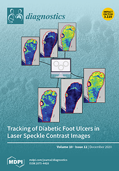
Foot ulcers are a severe complication of diabetes mellitus. Laser speckle contrast imaging (LSCI) is a promising approach for diagnosis and prognosis. However, manual assessment during analysis of LSCI limits clinical applicability. An algorithm for semi-automatic analysis of LSCI data was developed and validated. We observed good to excellent agreement between the algorithm and manual assessment of the LSCI data. The algorithm delivers assessment of diabetic foot ulcers with a 10-fold workload reduction. This enhances the clinical applicability of LSCI for assessment of diabetic foot ulcers. View this paper
  • Issues are regarded as officially published after their release is announced to the table of contents alert mailing list.
  • You may sign up for e-mail alerts to receive table of contents of newly released issues.
  • PDF is the official format for papers published in both, html and pdf forms. To view the papers in pdf format, click on the "PDF Full-text" link, and use the free Adobe Reader to open them.
Order results
Result details
Section
Select all
Export citation of selected articles as:
15 pages, 42128 KiB  
Article
Deep Learning Assisted Localization of Polycystic Kidney on Contrast-Enhanced CT Images
by Djeane Debora Onthoni, Ting-Wen Sheng, Prasan Kumar Sahoo, Li-Jen Wang and Pushpanjali Gupta
Diagnostics 2020, 10(12), 1113; https://doi.org/10.3390/diagnostics10121113 - 21 Dec 2020
Cited by 15 | Viewed by 4704
Abstract
Total Kidney Volume (TKV) is essential for analyzing the progressive loss of renal function in Autosomal Dominant Polycystic Kidney Disease (ADPKD). Conventionally, to measure TKV from medical images, a radiologist needs to localize and segment the kidneys by defining and delineating the kidney’s [...] Read more.
Total Kidney Volume (TKV) is essential for analyzing the progressive loss of renal function in Autosomal Dominant Polycystic Kidney Disease (ADPKD). Conventionally, to measure TKV from medical images, a radiologist needs to localize and segment the kidneys by defining and delineating the kidney’s boundary slice by slice. However, kidney localization is a time-consuming and challenging task considering the unstructured medical images from big data such as Contrast-enhanced Computed Tomography (CCT). This study aimed to design an automatic localization model of ADPKD using Artificial Intelligence. A robust detection model using CCT images, image preprocessing, and Single Shot Detector (SSD) Inception V2 Deep Learning (DL) model is designed here. The model is trained and evaluated with 110 CCT images that comprise 10,078 slices. The experimental results showed that our derived detection model outperformed other DL detectors in terms of Average Precision (AP) and mean Average Precision (mAP). We achieved mAP = 94% for image-wise testing and mAP = 82% for subject-wise testing, when threshold on Intersection over Union (IoU) = 0.5. This study proves that our derived automatic detection model can assist radiologist in locating and classifying the ADPKD kidneys precisely and rapidly in order to improve the segmentation task and TKV calculation. Full article
(This article belongs to the Special Issue Deep Learning for Computer-Aided Diagnosis in Biomedical Imaging)
Show Figures

Figure 1

6 pages, 1484 KiB  
Interesting Images
Usefulness of 18f-FDG PET-CT in Staging, Restaging, and Response Assessment in Pediatric Rhabdomyosarcoma
by Davide Donner, Paola Feraco, Linda Meneghello, Barbara Rombi, Lorena Picori and Franca Chierichetti
Diagnostics 2020, 10(12), 1112; https://doi.org/10.3390/diagnostics10121112 - 21 Dec 2020
Cited by 2 | Viewed by 2401
Abstract
Rhabdomyosarcoma is the most common soft-tissue sarcoma of childhood. Despite clinical advances, subsets of these patients continue to suffer high morbidity and mortality rates associated with their disease. Following the European guidelines for 18F-FDG PET and PET-CT imaging in pediatric oncology, the [...] Read more.
Rhabdomyosarcoma is the most common soft-tissue sarcoma of childhood. Despite clinical advances, subsets of these patients continue to suffer high morbidity and mortality rates associated with their disease. Following the European guidelines for 18F-FDG PET and PET-CT imaging in pediatric oncology, the routine use of 18F-FDG PET-CT may be useful for patients affected by rhabdomyosarcoma, in staging, in the evaluation of response to therapy, and for restaging/detection of relapse. The European Pediatric Protocols are very old, and for staging and restaging, they recommend only radionuclide bone scan. The 18F-FDG PET-CT exam is listed as an optional investigation prescribed according to local availability and local protocols in the investigations panel required at the end of the treatment. We present two cases highlighting the usefulness of 18F-FDG PET-CT in managing pediatric patients affected by rhabdomyosarcoma, providing some bibliographic references. Full article
(This article belongs to the Special Issue Positron Emission Tomography (PET) Imaging for Therapy Monitoring)
Show Figures

Figure 1

14 pages, 9000 KiB  
Article
Carotid Artery Plaque Identification and Display System (MRI-CAPIDS) Using Opensource Tools
by Felipe P. Vista IV, Minh Tri Ngo, Seung Bin Cho, Hyo Sung Kwak and Kil To Chong
Diagnostics 2020, 10(12), 1111; https://doi.org/10.3390/diagnostics10121111 - 21 Dec 2020
Cited by 1 | Viewed by 2248
Abstract
Magnetic resonance imaging (MRI) represents one modality in atherosclerosis risk assessment, by permitting the classification of carotid plaques into either high- or low-risk lesions. Although MRI is generally used for observing the impact of atherosclerosis on vessel lumens, it can also show both [...] Read more.
Magnetic resonance imaging (MRI) represents one modality in atherosclerosis risk assessment, by permitting the classification of carotid plaques into either high- or low-risk lesions. Although MRI is generally used for observing the impact of atherosclerosis on vessel lumens, it can also show both the size and composition of itself, as well as plaque information, thereby providing information beyond that of simple stenosis. Software systems are a valuable aid in carotid artery stenosis assessment wherein commercial software is readily available but is not accessible to all practitioners because of its often high cost. This study focuses on the development of a software system designed entirely for registration, marking, and 3D visualization of the wall and lumen, using freely available open-source tools and libraries. It was designed to be free from “feature bloat” and avoid “feature-creep.” The image loading and display module of the modified QDCM library was improved by a minimum of 10,000%. A Bezier function was used in order to smoothen the curve of the polygon (referring to the shape formed by the marked points) by interpolating additional points between the marked points. This smoother curve led to a smoother 3D view of the lumen and wall. Full article
(This article belongs to the Section Medical Imaging and Theranostics)
Show Figures

Figure 1

11 pages, 888 KiB  
Review
Right Heart Size and Right Ventricular Reserve in Pulmonary Hypertension: Impact on Management and Prognosis
by Ekkehard Grünig, Christina A. Eichstaedt, Rebekka Seeger and Nicola Benjamin
Diagnostics 2020, 10(12), 1110; https://doi.org/10.3390/diagnostics10121110 - 21 Dec 2020
Cited by 6 | Viewed by 5729
Abstract
Various parameters reflecting right heart size, right ventricular function and capacitance have been shown to be prognostically important in patients with pulmonary hypertension (PH). In the advanced disease, patients suffer from right heart failure, which is a main reason for an impaired prognosis. [...] Read more.
Various parameters reflecting right heart size, right ventricular function and capacitance have been shown to be prognostically important in patients with pulmonary hypertension (PH). In the advanced disease, patients suffer from right heart failure, which is a main reason for an impaired prognosis. Right heart size has shown to be associated with right ventricular function and reserve and is correlated with prognosis in patients with PH. Right ventricular reserve, defined as the ability of the ventricle to adjust to exercise or pharmacologic stress, is expressed by various parameters, which may be determined invasively by right heart catheterization or by stress-Doppler-echocardiography as a noninvasive approach. As the term “right ventricular contractile reserve” may be misleading, “right ventricular output reserve” seems desirable as a preferred term of increase in cardiac output during exercise. Both right heart size and right ventricular reserve have been shown to be of prognostic importance and may therefore be useful for risk assessment in patients with pulmonary hypertension. In this article we aim to display different aspects of right heart size and right ventricular reserve and their prognostic role in PH. Full article
(This article belongs to the Special Issue Pulmonary Hypertension: Diagnosis and Management)
Show Figures

Figure 1

5 pages, 1601 KiB  
Case Report
Dual TMPRSS2:ERG Fusion in a Patient with Lung and Prostate Cancers
by Francesca Giunchi, Francesco Massari, Annalisa Altimari, Elisa Gruppioni, Elisabetta Nobili, Michelangelo Fiorentino and Andrea Ardizzoni
Diagnostics 2020, 10(12), 1109; https://doi.org/10.3390/diagnostics10121109 - 20 Dec 2020
Cited by 2 | Viewed by 1865
Abstract
The TMPRSS2:ERG fusion is considered prostate specific and has been rarely described in other tumors. We describe the case of a patient who developed lung and prostate cancers, both harboring the TMPRSS2:ERG fusion. The patient developed a cancer of the prostate with lymph [...] Read more.
The TMPRSS2:ERG fusion is considered prostate specific and has been rarely described in other tumors. We describe the case of a patient who developed lung and prostate cancers, both harboring the TMPRSS2:ERG fusion. The patient developed a cancer of the prostate with lymph node metastases and after two years a nodule of the thoracic wall. The histology and immunohistochemical profile of the two tumors were typical of prostate and lung cancers. The presence of the TMPRSS2:ERG fusion was demonstrated by next-generation sequencing on both malignancies, leading to the assumption that the lung nodule was a metastasis from the prostate cancer. The patient failed to respond to antiandrogen therapy, while chemotherapy for lung cancer led to a significant objective response. To our knowledge, this is the first case of a lung cancer harboring the TMPRSS2:ERG fusion, widening the spectrum of lung cancer-associated molecular alterations. Full article
(This article belongs to the Section Pathology and Molecular Diagnostics)
Show Figures

Figure 1

15 pages, 2760 KiB  
Article
Prognostic Value of Admission Chest CT Findings for Invasive Ventilation Therapy in COVID-19 Pneumonia
by Eva Gresser, Johannes Rueckel, Daniel Puhr-Westerheide, Vincent Schwarze, Nicola Fink, Wolfgang G. Kunz, Dietmar Wassilowsky, Michael Irlbeck, Jens Ricke, Michael Ingrisch and Bastian O. Sabel
Diagnostics 2020, 10(12), 1108; https://doi.org/10.3390/diagnostics10121108 - 19 Dec 2020
Cited by 7 | Viewed by 2949
Abstract
(1) Background: To assess the value of chest CT imaging features of COVID-19 disease upon hospital admission for risk stratification of invasive ventilation (IV) versus no or non-invasive ventilation (non-IV) during hospital stay. (2) Methods: A retrospective single-center study was conducted including all [...] Read more.
(1) Background: To assess the value of chest CT imaging features of COVID-19 disease upon hospital admission for risk stratification of invasive ventilation (IV) versus no or non-invasive ventilation (non-IV) during hospital stay. (2) Methods: A retrospective single-center study was conducted including all patients admitted during the first three months of the pandemic at our hospital with PCR-confirmed COVID-19 disease and admission chest CT scans (n = 69). Using clinical information and CT imaging features, a 10-point ordinal risk score was developed and its diagnostic potential to differentiate a severe (IV-group) from a more moderate course (non-IV-group) of the disease was tested. (3) Results: Frequent imaging findings of COVID-19 pneumonia in both groups were ground glass opacities (91.3%), consolidations (53.6%) and crazy paving patterns (31.9%). Characteristics of later stages such as subpleural bands were observed significantly more often in the IV-group (52.2% versus 26.1%, p = 0.032). Using information directly accessible during a radiologist’s reporting, a simple risk score proved to reliably differentiate between IV- and non-IV-groups (AUC: 0.89 (95% CI 0.81–0.96), p < 0.001). (4) Conclusions: Information accessible from admission CT scans can effectively and reliably be used in a scoring model to support risk stratification of COVID-19 patients to improve resource and allocation management of hospitals. Full article
Show Figures

Figure 1

12 pages, 2848 KiB  
Article
Pericapillary Edema Assessment by Means of the Nailfold Capillaroscopy and Laser Scanning Microscopy
by Boris P. Yakimov, Yury I. Gurfinkel, Denis A. Davydov, Anastasia S. Allenova, Gleb S. Budylin, Vladimir Yu. Vasiliev, Vera Yu. Soldatova, Armais A. Kamalov, Simon T. Matskeplishvili, Alexander V. Priezzhev and Evgeny A. Shirshin
Diagnostics 2020, 10(12), 1107; https://doi.org/10.3390/diagnostics10121107 - 18 Dec 2020
Cited by 3 | Viewed by 3232
Abstract
Edema, i.e., fluid accumulation in the interstitial space, accompanies numerous pathological states of the human organism, including heart failure (HF), inflammatory response, and lymphedema. Nevertheless, techniques for quantitative assessment of the edema’s severity and dynamics are absent in clinical practice, and the analysis [...] Read more.
Edema, i.e., fluid accumulation in the interstitial space, accompanies numerous pathological states of the human organism, including heart failure (HF), inflammatory response, and lymphedema. Nevertheless, techniques for quantitative assessment of the edema’s severity and dynamics are absent in clinical practice, and the analysis is mainly limited to physical examination. This fact stimulates the development of novel methods for fast and reliable diagnostics of fluid retention in tissues. In this work, we focused on the possibilities of two microscopic techniques, nailfold video capillaroscopy (NVC) and confocal laser scanning microscopy (CLSM), in the assessment of the short-term and long-term cutaneous edema. We showed that for the patients with HF, morphological parameters obtained by NVC—namely, the apical diameter of capillaries and the size of the perivascular zone—indicate long-term edema. On the other hand, for healthy volunteers, the application of two models of short-term edema, venous occlusion, and histamine treatment of the skin, did not reveal notable changes in the capillary parameters. However, a significant reduction of the NVC image sharpness was observed in this case, which was suggested to be due to water accumulation in the epidermis. To verify these findings, we made use of CLSM, which provides the skin structure with cellular resolution. It was observed that for the histamine-treated skin, the areas of the dermal papillae become hyporefractive, leading to the loss of contrast and the lower visibility of capillaries. Similar effect was observed for patients undergoing infusion therapy. Collectively, our results reveal the parameters can be used for pericapillary edema assessment using the NVC and CLSM, and paves the way for their application in a clinical set-up. Full article
(This article belongs to the Section Optical Diagnostics)
Show Figures

Figure 1

12 pages, 1565 KiB  
Article
COPD Imaging on a 3rd Generation Dual-Source CT: Acquisition of Paired Inspiratory-Expiratory Chest Scans at an Overall Reduced Radiation Risk
by Joshua Gawlitza, Thomas Henzler, Frederik Trinkmann, Elke Nekolla, Holger Haubenreisser and Gunnar Brix
Diagnostics 2020, 10(12), 1106; https://doi.org/10.3390/diagnostics10121106 - 18 Dec 2020
Cited by 6 | Viewed by 2180
Abstract
As stated by the Fleischner Society, an additional computed tomography (CT) scan in expiration is beneficial in patients with chronic obstructive pulmonary disease (COPD). It was thus the aim of this study to evaluate the radiation risk of a state-of-the-art paired inspiratory-expiratory chest [...] Read more.
As stated by the Fleischner Society, an additional computed tomography (CT) scan in expiration is beneficial in patients with chronic obstructive pulmonary disease (COPD). It was thus the aim of this study to evaluate the radiation risk of a state-of-the-art paired inspiratory-expiratory chest scan compared to inspiration-only examinations. Radiation doses to 28 organs were determined for 824 COPD patients undergoing routine chest examinations at three different CT systems–a conventional multi-slice CT (MSCT), a 2nd generation (2nd-DSCT), and 3rd generation dual-source CT (3rd-DSCT). Patients examined at the 3rd-DSCT received a paired inspiratory-expiratory scan. Organ doses, effective doses, and lifetime attributable cancer risks (LAR) were calculated. All organ and effective doses were significantly lower for the paired inspiratory-expiratory protocol (effective doses: 4.3 ± 1.5 mSv (MSCT), 3.0 ± 1.2 mSv (2nd-DSCT), and 2.0 ± 0.8 mSv (3rd-DSCT)). Accordingly, LAR was lowest for the paired protocol with an estimate of 0.025 % and 0.013% for female and male patients (50 years) respectively. Image quality was not compromised. Paired inspiratory-expiratory scans can be acquired on 3rd-DSCT systems at substantially lower dose and risk levels when compared to inspiration-only scans at conventional CT systems, offering promising prospects for improved COPD diagnosis. Full article
(This article belongs to the Special Issue Assessment of Radiation Dose in X-ray and CT Exams)
Show Figures

Figure 1

10 pages, 682 KiB  
Article
Failure of Lactate Clearance Predicts the Outcome of Critically Ill Septic Patients
by Raphael Romano Bruno, Bernhard Wernly, Stephan Binneboessel, Philipp Baldia, Dragos Andrei Duse, Ralf Erkens, Malte Kelm, Behrooz Mamandipoor, Venet Osmani and Christian Jung
Diagnostics 2020, 10(12), 1105; https://doi.org/10.3390/diagnostics10121105 - 18 Dec 2020
Cited by 15 | Viewed by 2499
Abstract
Purpose: Early lactate clearance is an important parameter for prognosis assessment and therapy control in sepsis. Patients with a lactate clearance >0% might differ from patients with an inferior clearance in terms of intensive care management and outcomes. This study analyzes a large [...] Read more.
Purpose: Early lactate clearance is an important parameter for prognosis assessment and therapy control in sepsis. Patients with a lactate clearance >0% might differ from patients with an inferior clearance in terms of intensive care management and outcomes. This study analyzes a large collective with regards to baseline risk distribution and outcomes. Methods: In total, 3299 patients were included in this analysis, consisting of 1528 (46%) ≤0% and 1771 (54%) >0% patients. The primary endpoint was intensive care unit (ICU) mortality. Multilevel logistic regression analyses were used to compare both groups: A baseline model (model 1) with lactate clearance as a fixed effect and ICU as a random effect was installed. For model 2, patient characteristics (model 2) were included. For model 3, intensive care treatment (mechanical ventilation and vasopressors) was added to the model. Models 1 and 2 were used to evaluate the primary and secondary outcomes, respectively. Model 3 was only used to evaluate the primary outcomes. Adjusted odds ratios (aORs) with respective 95% confidence intervals (CI) were calculated. Results: The cohorts had no relevant differences regarding the gender, BMI, age, heart rate, body temperature, and baseline lactate. Neither the primary infection focuses nor the ethnic background differed between both groups. In both groups, the most common infection sites were of pulmonary origin, the urinary tract, and the gastrointestinal tract. Patients with lactate clearance >0% evidenced lower sepsis-related organ failure assessment (SOFA) scores (7 ± 6 versus 9 ± 6; p < 0.001) and creatinine (1.53 ± 1.49 versus 1.80 ± 1.67; p < 0.001). The ICU mortality differed significantly (14% versus 32%), and remained this way after multivariable adjustment for patient characteristics and intensive care treatment (aOR 0.43 95% CI 0.36–0.53; p < 0.001). In the additional sensitivity analysis, the lack of lactate clearance was associated with a worse prognosis in each subgroup. Conclusion: In this large collective of septic patients, the 6 h lactate clearance is an independent method for outcome prediction. Full article
Show Figures

Graphical abstract

15 pages, 1024 KiB  
Article
Changes in Short-Term and Ultra-Short Term Heart Rate, Respiratory Rate, and Time-Domain Heart Rate Variability Parameters during Sympathetic Nervous System Activity Stimulation in Elite Modern Pentathlonists—A Pilot Study
by Jakub S. Gąsior, Bartosz Hoffmann, Luiz Eduardo Virgilio Silva, Łukasz Małek, Andrew A. Flatt, Rafał Baranowski and Bożena Werner
Diagnostics 2020, 10(12), 1104; https://doi.org/10.3390/diagnostics10121104 - 17 Dec 2020
Cited by 6 | Viewed by 2766
Abstract
Monitoring of markers reflecting cardiac autonomic activity before and during stressful situations may be useful for identifying the physiological state of an athlete and may have medical or performance implications. The study aimed to determine group and individual changes in short-term (5 min) [...] Read more.
Monitoring of markers reflecting cardiac autonomic activity before and during stressful situations may be useful for identifying the physiological state of an athlete and may have medical or performance implications. The study aimed to determine group and individual changes in short-term (5 min) and ultra-short-term (1 min) heart rate (HR), respiratory rate (RespRate), and time-domain heart rate variability (HRV) parameters during sympathetic nervous system activity (SNSa) stimulation among professional endurance athletes. Electrocardiographic recordings were performed in stable measurement conditions (Baseline) and during SNSa stimulation via isometric handgrip in 12 elite modern pentathlonists. Significant increases in short-term HR and decreases in time-domain HRV parameters with no changes in RespRate were observed during SNSa stimulation. Significant differences were observed between Baseline (all minutes) and the last (i.e., 5th) minute of SNSa stimulation for ultra-short-term parameters. Analysis of intra-individual changes revealed some heterogeneity in responses. The study provides baseline responses of HR, RespRate, and time-domain HRV parameters to SNSa stimulation among elite pentathlonists, which may be useful for identifying abnormal responses among fatigued or injured (e.g., concussed) athletes. More attention to individual analysis seems to be necessary when assessing physiological responses to sympathetic stimuli in professional endurance athletes. Full article
(This article belongs to the Special Issue Diagnostic Challenges in Sports Cardiology)
Show Figures

Figure 1

12 pages, 4054 KiB  
Article
Multimodality Approach for Endovascular Left Atrial Appendage Closure: Head-To-Head Comparison among 2D and 3D Echocardiography, Angiography, and Computer Tomography
by Gianpiero Italiano, Anna Maltagliati, Valentina Mantegazza, Laura Fusini, Maria Elisabetta Mancini, Alessio Gasperetti, Denise Brusoni, Francesca Susini, Alberto Formenti, Gianluca Pontone, Gaetano Fassini, Claudio Tondo and Mauro Pepi
Diagnostics 2020, 10(12), 1103; https://doi.org/10.3390/diagnostics10121103 - 17 Dec 2020
Cited by 4 | Viewed by 2620
Abstract
Background: Percutaneous left atrial appendage closure (LAAC) requires accurate pre- and intraprocedural measurements, and multimodality imaging is an essential tool for guiding the procedure. Two-dimensional (2D TOE) and three-dimensional (3D TOE) transoesophageal echocardiography, cardiac computed tomography (CCT), and conventional cardiac angiography (CCA) are [...] Read more.
Background: Percutaneous left atrial appendage closure (LAAC) requires accurate pre- and intraprocedural measurements, and multimodality imaging is an essential tool for guiding the procedure. Two-dimensional (2D TOE) and three-dimensional (3D TOE) transoesophageal echocardiography, cardiac computed tomography (CCT), and conventional cardiac angiography (CCA) are commonly used to evaluate left atrial appendage (LAA) size. However, standardized approaches in measurement methods by different imaging modalities are lacking. The aims of the study were to evaluate the LAA dimension and morphology in patients undergoing LAAC and to compare data obtained by different imaging modalities: 2D and 3D TOE, CCT, and CCA. Methods: A total of 200 patients (mean age 70 ± 8 years, 128 males) were examined by different imaging techniques (161 2D TOE, 103 3D TOE, 98 CCT, and 200 CCA). Patients underwent preoperative CCT and intraoperative 2D and 3D TOE and CCA. Results: A significant correlation was found among all measurements obtained by different modalities. In particular, 3D TOE and CCT measurements were highly correlated with an excellent agreement for the landing zone (LZ) dimensions (LZ diameter: r = 0.87; LAA depth: r = 0.91, p < 0.001). Conclusions: Head-to-head comparison among imaging techniques (2D and 3D TOE, CCT, and CCA) showed a good correlation among LZ diameter measurements obtained by different imaging modalities, which is a parameter of paramount importance for the choice of the LAAC device size. LZ diameters and area by 3D TOE had the best correlation with CCT. Full article
(This article belongs to the Special Issue Trends and Novelties in Cardiovascular Imaging)
Show Figures

Figure 1

19 pages, 959 KiB  
Review
The Multifaceted Roles of MicroRNAs in Cystic Fibrosis
by Fatima Domenica Elisa De Palma, Valeria Raia, Guido Kroemer and Maria Chiara Maiuri
Diagnostics 2020, 10(12), 1102; https://doi.org/10.3390/diagnostics10121102 - 17 Dec 2020
Cited by 12 | Viewed by 3656
Abstract
Cystic fibrosis (CF) is a lifelong disorder affecting 1 in 3500 live births worldwide. It is a monogenetic autosomal recessive disease caused by loss-of-function mutations in the gene encoding the chloride channel cystic fibrosis transmembrane conductance regulator (CFTR), the impairment of which leads [...] Read more.
Cystic fibrosis (CF) is a lifelong disorder affecting 1 in 3500 live births worldwide. It is a monogenetic autosomal recessive disease caused by loss-of-function mutations in the gene encoding the chloride channel cystic fibrosis transmembrane conductance regulator (CFTR), the impairment of which leads to ionic disequilibria in exocrine organs. This translates into a chronic multisystemic disease characterized by airway obstruction, respiratory infections, and pancreatic insufficiency as well as hepatobiliary and gastrointestinal dysfunction. Molecular characterization of the mutational heterogeneity of CFTR (affected by more than 2000 variants) improved the understanding and management of CF. However, these CFTR variants are linked to different clinical manifestations and phenotypes, and they affect response to treatments. Expanding evidence suggests that multisystemic disease affects CF pathology via impairing either CFTR or proteins regulated by CFTR. Thus, altering the expression of miRNAs in vivo could constitute an appealing strategy for developing new CF therapies. In this review, we will first describe the pathophysiology and clinical management of CF. Then, we will summarize the current knowledge on altered miRNAs in CF patients, with a focus on the miRNAs involved in the deregulation of CFTR and in the modulation of inflammation. We will highlight recent findings on the potential utility of measuring circulating miRNAs in CF as diagnostic, prognostic, and predictive biomarkers. Finally, we will provide an overview on potential miRNA-based therapeutic approaches. Full article
(This article belongs to the Special Issue Diagnosis and Management of Cystic Fibrosis)
Show Figures

Figure 1

6 pages, 694 KiB  
Communication
Interrater Reliability of 99mTc-DMSA Scintigraphy Performed as Planar Scan vs. SPECT/Low Dose CT for Diagnosing Renal Scarring in Children
by Hrefna Sæunn Einarsdóttir, Ronan Martin Griffin Berg and Lise Borgwardt
Diagnostics 2020, 10(12), 1101; https://doi.org/10.3390/diagnostics10121101 - 17 Dec 2020
Cited by 6 | Viewed by 4041
Abstract
99mTc-dimercaptosuccinic acid (DMSA) scintigraphy is currently the method of choice for assessing renal scarring in children, but it is not established whether conducting the scan as a single photon emission tomography combined with low-dose CT (SPECT/ldCT) scan provides additional diagnostic benefits when [...] Read more.
99mTc-dimercaptosuccinic acid (DMSA) scintigraphy is currently the method of choice for assessing renal scarring in children, but it is not established whether conducting the scan as a single photon emission tomography combined with low-dose CT (SPECT/ldCT) scan provides additional diagnostic benefits when compared to conventional planar scintigraphy. In the present study, we evaluated the interrater reliability of DMSA SPECT/ldCT vs. planar DMSA scintigraphy for diagnosing renal scarring. Methods: Two nuclear medicine physicians blinded to patient data retrospectively analysed all paediatric 99mTc-DMSA scintigraphes that were conducted in our department for the assessment of post pyelonephritis renal scarring between 2011 and 2016. All scintigraphies included both a planar scan and SPECT/ldCT, and were performed on either a Phillips Precedence 16 slice CT or a Siemens Symbia 16 slice CT. The readers were blinded to each other’s readings and to patient data, and assessed all scans dichotomously for evidence of renal scarring. For each scan, the readers further noted if they were confident in their interpretation. Results: A total of 46 pairs of planar SPECT/ldCT DMSA scans were included. The readers were unconfident about their interpretation of 40% of the planar scans and 5% of the SPECT/ldCT scans. The interrater agreement rate was 72% for planar scans and 91% for SPECT/ldCT, and the corresponding Cohen’s kappa values were 0.38 and 0.79. Conclusion: DMSA SPECT/ldCT is associated with higher reader confidence and interrater reliability than conventional planar DMSA scintigraphy for the assessment of post pyelonephritis renal scarring in children. Full article
(This article belongs to the Collection Hybrid Imaging in Medicine)
Show Figures

Figure 1

24 pages, 2794 KiB  
Review
Characteristics of Fluorescent Intraoperative Dyes Helpful in Gross Total Resection of High-Grade Gliomas—A Systematic Review
by Marek Mazurek, Bartłomiej Kulesza, Filip Stoma, Jacek Osuchowski, Sławomir Mańdziuk and Radosław Rola
Diagnostics 2020, 10(12), 1100; https://doi.org/10.3390/diagnostics10121100 - 16 Dec 2020
Cited by 19 | Viewed by 2760
Abstract
Background: A very important aspect in the treatment of high-grade glioma is gross total resection to reduce the risk of tumor recurrence. One of the methods to facilitate this task is intraoperative fluorescence navigation. The aim of the study was to compare [...] Read more.
Background: A very important aspect in the treatment of high-grade glioma is gross total resection to reduce the risk of tumor recurrence. One of the methods to facilitate this task is intraoperative fluorescence navigation. The aim of the study was to compare the dyes used in this technique fluorescent intraoperative navigation in terms of the mechanism of action and influence on the treatment of patients. Methods: The review was carried out on the basis of articles found in PubMed, Google Scholar, and BMC search engines, as well as those identified by searched bibliographies and suggested by experts during the preparation of the article. The database analysis was performed for the following phrases: “glioma”, “glioblastoma”, “ALA”, “5ALA”, “5-ALA”, “aminolevulinic acid”, ”levulinic acid”, “fluorescein”, “ICG”, “indocyanine green”, and “fluorescence navigation”. Results: After analyzing 913 citations identified on the basis of the search criteria, we included 36 studies in the review. On the basis of the analyzed articles, we found that 5-aminolevulinic acid and fluorescein are highly effective in improving the percentage of gross total resection achieved in high-grade glioma surgery. At the same time, the limitations resulting from the use of these methods are marked—higher costs of the procedure and the need to have neurosurgical microscope in combination with a special light filter in the case of 5-aminolevulinic acid (5-ALA), and low specificity for neoplastic cells and the dependence on the degree of damage to the blood–brain barrier in the intensity of fluorescence in the case of fluorescein. The use of indocyanine green in the visualization of glioma cells is relatively unknown, but some researchers have suggested its utility and the benefits of using it simultaneously with other dyes. Conclusion: The use of intraoperative fluorescence navigation with the use of 5-aminolevulinic acid and fluorescein allows the range of high-grade glioma resection to be increased. Full article
(This article belongs to the Special Issue Brain Imaging/Neuroimaging)
Show Figures

Figure 1

11 pages, 1856 KiB  
Review
Exploring the Spectrum of Kidney Ciliopathies
by Matteo Santoni, Francesco Piva, Alessia Cimadamore, Matteo Giulietti, Nicola Battelli, Rodolfo Montironi, Laura Cosmai and Camillo Porta
Diagnostics 2020, 10(12), 1099; https://doi.org/10.3390/diagnostics10121099 - 16 Dec 2020
Cited by 8 | Viewed by 8362
Abstract
Ciliopathies are a group of multi-organ diseases caused by the disruption of the primary cilium. This event leads to a variety of kidney disorders, including nephronophthisis, renal cystic dysplasia, and renal cell carcinoma (RCC). Primary cilium contributes to the regulation of the cell [...] Read more.
Ciliopathies are a group of multi-organ diseases caused by the disruption of the primary cilium. This event leads to a variety of kidney disorders, including nephronophthisis, renal cystic dysplasia, and renal cell carcinoma (RCC). Primary cilium contributes to the regulation of the cell cycle and protein homeostasis, that is, the balance between protein synthesis and degradation by acting on the ubiquitin-proteasome system, autophagy, and mTOR signaling. Many proteins are involved in renal ciliopathies. In particular, fibrocystin (PKHD1) is involved in autosomal recessive polycystic kidney disease (ARPKD), while polycystin-1 (PKD1) and polycystin-2 (PKD2) are implicated in autosomal dominant polycystic kidney disease (ADPKD). Moreover, primary cilia are associated with essential signaling pathways, such as Hedgehog, Wnt, and Platelet-Derived Growth Factor (PDGF). In this review, we focused on the ciliopathies associated with kidney diseases, exploring genes and signaling pathways associated with primary cilium and the potential role of cilia as therapeutic targets in renal disorders. Full article
(This article belongs to the Special Issue Novel Diagnostic and Predictive Strategies in Renal Cell Tumors)
Show Figures

Graphical abstract

13 pages, 1756 KiB  
Review
Immune Checkpoint Inhibitors with or without Radiotherapy in Non-Small Cell Lung Cancer Patients with Brain Metastases: A Systematic Review and Meta-Analysis
by Dong Yeong Kim, Pyeong Hwa Kim, Chong Hyun Suh, Kyung Won Kim and Ho Sung Kim
Diagnostics 2020, 10(12), 1098; https://doi.org/10.3390/diagnostics10121098 - 16 Dec 2020
Cited by 8 | Viewed by 2777
Abstract
This study aimed to evaluate the radiologic response and adverse event rates of immune checkpoint inhibitor (ICI) therapy with or without radiotherapy for the treatment of non-small cell lung cancer (NSCLC) brain metastases. A systematic literature search was performed up to January 3, [...] Read more.
This study aimed to evaluate the radiologic response and adverse event rates of immune checkpoint inhibitor (ICI) therapy with or without radiotherapy for the treatment of non-small cell lung cancer (NSCLC) brain metastases. A systematic literature search was performed up to January 3, 2020. Studies evaluating the intracranial objective response rates (ORR) and/or disease control rates (DCR) of ICI with or without radiotherapy for treating NSCLC brain metastases were included. Consequently, twelve studies satisfied inclusion criteria. ICI combined with radiotherapy (pooled ORR, 95%; DCR, 97%) showed better local efficacy compared to ICI monotherapy (pooled ORR, 24%; DCR, 44%; p < 0.01 for both ORR and DCR). Grade 3 or 4 central nervous system (CNS)-related adverse event rates were not different (5% vs. 4%; p = 0.93). In conclusion, ICI combined with radiotherapy showed better intracranial efficacy than ICI monotherapy for treating NSCLC brain metastases. CNS-related grade 3 or 4 adverse event rate was not statistically different between the two groups. Several prospective trials are needed to compare the efficacy of ICI combined with radiotherapy and ICI monotherapy. Full article
Show Figures

Figure 1

11 pages, 1479 KiB  
Article
Nuclear Expression Loss of SSBP2 Is Associated with Poor Prognostic Factors in Colorectal Adenocarcinoma
by Yumin Chung, Hyunsung Kim, Seongsik Bang, Kiseok Jang, Seung Sam Paik and Su-Jin Shin
Diagnostics 2020, 10(12), 1097; https://doi.org/10.3390/diagnostics10121097 - 16 Dec 2020
Cited by 6 | Viewed by 1860
Abstract
Single-stranded DNA binding protein 2 (SSBP2) is involved in DNA damage response and may induce growth arrest in cancer cells, having a potent tumor suppressor role. SSBP2 is ubiquitously expressed and the loss of its expression has been reported in various tumor types. [...] Read more.
Single-stranded DNA binding protein 2 (SSBP2) is involved in DNA damage response and may induce growth arrest in cancer cells, having a potent tumor suppressor role. SSBP2 is ubiquitously expressed and the loss of its expression has been reported in various tumor types. However, the correlation between SSBP2 expression and colorectal cancer (CRC) prognosis remains unclear. SSBP2 nuclear expression was evaluated immunohistochemically in 48 normal colonic mucosae, 47 adenomas, 391 primary adenocarcinomas, and 131 metastatic carcinoma tissue samples. The clinicopathological factors, overall survival (OS), and recurrence-free survival were evaluated, and associations with the clinicopathological parameters were analyzed in 391 colorectal adenocarcinoma patients. A diffuse nuclear SSBP2 expression was detected in all normal colonic mucosa and adenoma samples. SSBP2 expression loss was observed in 131 (34.3%) primary adenocarcinoma and 100 (76.3%) metastatic carcinoma samples. SSBP2 expression was significantly associated with poor prognostic factors, such as vascular invasion (p = 0.005), high pT category (p = 0.045), and shorter OS (p = 0.038), using univariate survival analysis. Nuclear SSBP2 expression loss was significantly observed in colorectal carcinoma and metastatic carcinoma tissues, being associated with poor prognostic factors. SSBP2 acts as a tumor suppressor and may be used as a CRC prognostic biomarker. Full article
(This article belongs to the Collection Biomarkers in Medicine)
Show Figures

Figure 1

16 pages, 4586 KiB  
Article
Heart Rate Influence on the QT Variability Risk Factors
by Irena Andršová, Katerina Hnatkova, Martina Šišáková, Ondřej Toman, Peter Smetana, Katharina M. Huster, Petra Barthel, Tomáš Novotný, Georg Schmidt and Marek Malik
Diagnostics 2020, 10(12), 1096; https://doi.org/10.3390/diagnostics10121096 - 16 Dec 2020
Cited by 4 | Viewed by 1934
Abstract
QT interval variability, mostly expressed by QT variability index (QTVi), has repeatedly been used in risk diagnostics. Physiologic correlates of QT variability expressions have been little researched especially when measured in short 10-second electrocardiograms (ECGs). This study investigated different QT variability indices, including [...] Read more.
QT interval variability, mostly expressed by QT variability index (QTVi), has repeatedly been used in risk diagnostics. Physiologic correlates of QT variability expressions have been little researched especially when measured in short 10-second electrocardiograms (ECGs). This study investigated different QT variability indices, including QTVi and the standard deviation of QT interval durations (SDQT) in 657,287 10-second ECGs recorded in 523 healthy subjects (259 females). The indices were related to the underlying heart rate and to the 10-second standard deviation of RR intervals (SDRR). The analyses showed that both QTVi and SDQT (as well as other QT variability indices) were highly statistically significantly (p < 0.00001) influenced by heart rate and that QTVi showed poor intra-subject reproducibility (coefficient of variance approaching 200%). Furthermore, sequential analysis of regression variance showed that SDQT was more strongly related to the underlying heart rate than to SDRR, and that QTVi was influenced by the underlying heart rate and SDRR more strongly than by SDQT (p < 0.00001 for these comparisons of regression dependency). The study concludes that instead of QTVi, simpler expressions of QT interval variability, such as SDQT, appear preferable for future applications especially if multivariable combination with the underlying heart rate is used. Full article
(This article belongs to the Section Point-of-Care Diagnostics and Devices)
Show Figures

Figure 1

18 pages, 2239 KiB  
Article
Salivary Metabolic Profile of Patients with Lung Cancer, Chronic Obstructive Pulmonary Disease of Varying Severity and Their Comorbidity: A Preliminary Study
by Lyudmila V. Bel’skaya, Elena A. Sarf, Denis V. Solomatin and Victor K. Kosenok
Diagnostics 2020, 10(12), 1095; https://doi.org/10.3390/diagnostics10121095 - 15 Dec 2020
Cited by 2 | Viewed by 1929
Abstract
The aim of the work was to study the features of the salivary biochemical composition in the combined pathology of lung cancer and chronic obstructive pulmonary disease (COPD) of varying severity (COPD I, COPD II). The study group included patients with lung cancer [...] Read more.
The aim of the work was to study the features of the salivary biochemical composition in the combined pathology of lung cancer and chronic obstructive pulmonary disease (COPD) of varying severity (COPD I, COPD II). The study group included patients with lung cancer (n = 392), non-malignant lung pathologies (n = 168) and healthy volunteers (n = 500). Before treatment, the salivary biochemical composition was determined according to 34 indicators. Survival analysis performed by the Kaplan-Meier method. Biochemical parameters (catalase, imidazole compounds ICs, sialic acids, lactate dehydrogenase (LDH)) that can be used to monitor patients at risk (COPD I) for timely diagnosis of lung cancer are determined. A complex of salivary biochemical indicators with prognostic value in lung cancer was revealed. For patients with lung cancer without COPD, a group of patients with a favorable prognosis can be distinguished with a combination of ICs < 0.478 mmol/L and LDH >1248 U/L (HR = 1.56, 95% CI 0.40–6.07, p = 0.03891). For COPD I, a level of ICs < 0.182 mmol/L are prognostically favorable (HR = 1.74, 95% CI 0.71–4.21, p = 0.07270). For COPD II, combinations of pH < 6.74 and LDH >1006 U/L are prognostically favorable. In general, for patients with lung cancer in combination with COPD I, the prognosis is more favorable than without COPD. Full article
(This article belongs to the Special Issue Diagnosis, Treatment, and Management of COPD and Asthma)
Show Figures

Figure 1

11 pages, 5595 KiB  
Article
Simple Stepwise Approach to Differentiate Cyst-Like Soft-Tissue Masses by Using Time-Resolved Magnetic Resonance Angiography
by Ying-Chieh Lai, Yu-Hsiang Juan, Shu-Hang Ng, Tzu-Chin Lo, Wen-Yu Chuang, Chun-Chieh Chen, Chi-Ting Liau, Gigin Lin, Yu-Jr Lin and Yu-Ching Lin
Diagnostics 2020, 10(12), 1094; https://doi.org/10.3390/diagnostics10121094 - 15 Dec 2020
Viewed by 1692
Abstract
This retrospective study aimed to differentiate cyst-like musculoskeletal soft-tissue masses by using time-resolved magnetic resonance angiography (MRA). During May 2015 to November 2019, patients with cyst-like soft-tissue masses examined through contrast-enhanced MRI followed by histologic diagnosis were included. The masses were classified into [...] Read more.
This retrospective study aimed to differentiate cyst-like musculoskeletal soft-tissue masses by using time-resolved magnetic resonance angiography (MRA). During May 2015 to November 2019, patients with cyst-like soft-tissue masses examined through contrast-enhanced MRI followed by histologic diagnosis were included. The masses were classified into vascular lesions, solid lesions, and true cysts. Size, T1 hyperintensity, T2 composition, perilesional edema, time-resolved MRA, and static internal enhancement were assessed. The time-resolved MRA manifestations were classified into vascular pooling, solid stain, and occult lesion. Imaging predictors for each type of mass were identified through logistic regression and were used to develop a diagnostic flowchart. A total of 80 patients (47 men; median age, 42 years) were included, with 22 vascular lesions, 38 solid lesions, and 20 true cysts. The T2 composition, time-resolved MRA, and static internal enhancement were significantly different among the masses. Vascular pooling on time-resolved MRA was the sole predictor of vascular lesions (odds ratio = 722.0, p < 0.001). Solid stain on time-resolved MRA was the sole predictor of solid lesions (odds ratio = 73.6, p < 0.001). Occult lesion on time-resolved MRA (odds ratio = 7.4, p = 0.001) and absence of static internal enhancement (odds ratio = 80.0, p < 0.001) both predicted true cysts, while the latter was the sole predictor of true cysts after multivariate analysis. A diagnostic flowchart based on time-resolved MRA correctly classified 89% of the masses. In conclusion, time-resolved MRA accurately differentiates cyst-like soft-tissue masses and provides guidance for management. Full article
(This article belongs to the Section Medical Imaging and Theranostics)
Show Figures

Figure 1

11 pages, 2457 KiB  
Article
Introduction of a Simplified Psoriatic Arthritis Magnetic Resonance Imaging Score (sPsAMRIS): A Potential Tool for Treatment Monitoring in Peripheral Psoriatic Arthritis
by Daniel B. Abrar, Christoph Schleich, Ralph Brinks, Christine Goertz, Miriam Frenken, Matthias Schneider, Sven Nebelung and Philipp Sewerin
Diagnostics 2020, 10(12), 1093; https://doi.org/10.3390/diagnostics10121093 - 15 Dec 2020
Cited by 1 | Viewed by 3393
Abstract
Background: To evaluate whether a simplified (s) version of the psoriatic arthritis magnetic resonance imaging score (PsAMRIS), sPsAMRIS, is a potential tool for therapy monitoring in psoriatic arthritis (PsA). Methods: Seventeen patients with active psoriatic arthritis (PsA) underwent magnetic resonance imaging (MRI) at [...] Read more.
Background: To evaluate whether a simplified (s) version of the psoriatic arthritis magnetic resonance imaging score (PsAMRIS), sPsAMRIS, is a potential tool for therapy monitoring in psoriatic arthritis (PsA). Methods: Seventeen patients with active psoriatic arthritis (PsA) underwent magnetic resonance imaging (MRI) at 3 T of the clinically dominant hand at baseline and after 6 months. Scoring was performed by two musculoskeletal radiologists in terms of the PsAMRIS and sPsAMRIS, which is a simplified version with reduced item numbers based on prior evaluation of responsiveness to change by standardized response means (SRMs). Both scores were compared by calculation of overall and each sub-score’s SRMs and relative efficacy (RE) after bootstrapping. Results: PsAMRIS sub-scores of MCP joints 3 and 4, and proximal interphalangeal (PIP) joint 4 had the highest SRM (−0.07 each), indicating highest responsiveness to change, and were, therefore, included in sPsAMRIS. Compared to PsAMRIS, sPsAMRIS was characterized by higher SRMs (sPsAMRIS: −0.13 vs. PsAMRIS: −0.02) and higher RE (29.46). sPsAMRIS and PsAMRIS were highly correlated at baseline (r = 0.75, p < 0.01 (Pearson’s correlation)) and at 6-month follow-up (r = 0.64, p = 0.01). Mean time burden for completion of scoring per MRI study was significantly reduced when using PsAMRIS (469 ± 87.03 s) as compared to sPsAMRIS (140.1 ± 21.25 s) (p < 0.001). Conclusion: Due to its similar responsiveness to change compared to standard PsAMRIS, and time efficiency, sPsAMRIS might be a potential diagnostic tool to quantitatively assess and monitor therapy in PsA. Full article
Show Figures

Figure 1

12 pages, 822 KiB  
Review
Next Generation Sequencing in Non-Small Cell Lung Cancer: Pitfalls and Opportunities
by Chiara Lazzari, Alessandra Bulotta, Maria Giulia Cangi, Gabriele Bucci, Lorenza Pecciarini, Silvia Bonfiglio, Vincenza Lorusso, Stefania Ippati, Gianluigi Arrigoni, Greta Grassini, Claudio Doglioni and Vanesa Gregorc
Diagnostics 2020, 10(12), 1092; https://doi.org/10.3390/diagnostics10121092 - 15 Dec 2020
Cited by 13 | Viewed by 2662
Abstract
Lung cancer remains the first cause of cancer-related deaths worldwide. Thanks to the improvement in the knowledge of the biology of non-small cell lung cancer (NSCLC), patients’ survival has significantly improved. A growing number of targetable molecular alterations have been identified. Next-generation sequencing [...] Read more.
Lung cancer remains the first cause of cancer-related deaths worldwide. Thanks to the improvement in the knowledge of the biology of non-small cell lung cancer (NSCLC), patients’ survival has significantly improved. A growing number of targetable molecular alterations have been identified. Next-generation sequencing (NGS) has become one of the methodologies entered in clinical practice and was recently recommended by the European society for medical oncology (ESMO) to perform a comprehensive molecular characterization in patients with cancer. The current review provides an overview of the clinical trials that have explored the impact of NGS in patients with cancer, its limits, and advantages. Full article
(This article belongs to the Special Issue Lung Cancer Molecular Pathology)
Show Figures

Figure 1

11 pages, 260 KiB  
Article
Neurocognitive Functioning and Suicidal Behavior in Violent Offenders with Schizophrenia Spectrum Disorders
by Miriam Sánchez-Sansegundo, Irene Portilla-Tamarit, María Rubio-Aparicio, Natalia Albaladejo-Blazquez, Nicolás Ruiz-Robledillo, Rosario Ferrer-Cascales and Ana Zaragoza-Martí
Diagnostics 2020, 10(12), 1091; https://doi.org/10.3390/diagnostics10121091 - 15 Dec 2020
Cited by 9 | Viewed by 2247
Abstract
Suicide is one of the main premature causes of death in patients with schizophrenia. However, little is known about the relationship between neurocognitive functioning and suicidality in violent offenders with schizophrenia who have been sentenced to psychiatric treatment after committing violent crimes. We [...] Read more.
Suicide is one of the main premature causes of death in patients with schizophrenia. However, little is known about the relationship between neurocognitive functioning and suicidality in violent offenders with schizophrenia who have been sentenced to psychiatric treatment after committing violent crimes. We examined the neurocognitive functioning of a sample of 61 violent offenders, most of them murderers with schizophrenia who were classified as suicide attempters (n = 26) and non-attempters (n = 35). We compared the neurocognitive functioning of both groups using a neuropsychological battery. Suicide attempters showed similar performance to non-attempters in a neuropsychological test across all domains of cognitive functioning, memory, attention, verbal fluency, and executive functioning. However, after controlling for demographic and clinical variables, suicide attempters performed better than non-attempters in two planning-related tasks: the Tower of London (p < 0.01) and the Zoo Map (p < 0.01). Suicide attempters were also characterized as having more family histories of suicidality and as displaying more depressive symptoms and negative symptoms of psychopathology on the Positive and Negative Syndrome Scale (PANSS) scale. These results suggest that suicide attempters have a greater ability to formulate plans and initiate goals directed at making a suicide attempt. Full article
(This article belongs to the Section Pathology and Molecular Diagnostics)
11 pages, 922 KiB  
Article
Can MRI Biomarkers Predict Triple-Negative Breast Cancer?
by Giuliana Moffa, Francesca Galati, Emmanuel Collalunga, Veronica Rizzo, Endi Kripa, Giulia D’Amati and Federica Pediconi
Diagnostics 2020, 10(12), 1090; https://doi.org/10.3390/diagnostics10121090 - 15 Dec 2020
Cited by 23 | Viewed by 3312
Abstract
The purpose of this study was to investigate MRI features of triple-negative breast cancer (TNBC) compared with non-TNBC, to predict histopathological results. In the study, 26 patients with TNBC and 24 with non-TNBC who underwent multiparametric MRI of the breast on a 3 [...] Read more.
The purpose of this study was to investigate MRI features of triple-negative breast cancer (TNBC) compared with non-TNBC, to predict histopathological results. In the study, 26 patients with TNBC and 24 with non-TNBC who underwent multiparametric MRI of the breast on a 3 T magnet over a 10-months period were retrospectively recruited. MR imaging sets were evaluated by two experienced breast radiologists in consensus and classified according to the 2013 American College of Radiology (ACR) BI-RADS lexicon. The comparison between the two groups was performed using the Chi-square test and followed by logistic regression analyses. We found that 92% of tumors presented as mass enhancements (p = 0.192). 41.7% of TNBC and 86.4% of non-TNBC had irregular shape (p = 0.005); 58.3% of TNBC showed circumscribed margins, compared to 9.1% of non-TNBC masses (p = 0.001); 75% of TNBC and 9.1% of non-TNBC showed rim enhancement (p < 0.001). Intralesional necrosis was significantly associated with TNBC (p = 0.016). Rim enhancement and intralesional necrosis risulted to be positive predictors at univariate analysis (OR = 29.86, and 8.10, respectively) and the multivariate analysis confirmed that rim enhancement is independently associated with TNBC (OR = 33.08). The mean ADC values were significantly higher for TNBC (p = 0.011). In conclusion, TNBC is associated with specific MRI features that can be possible predictors of pathological results, with a consequent prognostic value. Full article
(This article belongs to the Special Issue Advances in Breast MRI)
Show Figures

Figure 1

15 pages, 2179 KiB  
Article
Prognostic Implications of Epilepsy Onset Age According to Relapse Pattern in Patients with Four-Year Remission
by Soochul Park and Myeongjee Lee
Diagnostics 2020, 10(12), 1089; https://doi.org/10.3390/diagnostics10121089 - 14 Dec 2020
Cited by 2 | Viewed by 1676
Abstract
A total of 472 epilepsy patients with a 4-year remission period were divided into 10-year age groups according to age of onset. The relapse patterns during at least 3 years of follow-up were classified as early relapse (ER), late relapse (LR), and seizure-free [...] Read more.
A total of 472 epilepsy patients with a 4-year remission period were divided into 10-year age groups according to age of onset. The relapse patterns during at least 3 years of follow-up were classified as early relapse (ER), late relapse (LR), and seizure-free (SF). The remission probability and multiplicity of prognostic factors were evaluated using univariate and multivariate multinomial logistic analyses. The weighted risk score based on odd ratios (ORs) was used for comparisons of the relative risk of relapse between groups. The group with onset in their 20s had the lowest remission probability among the groups. The risks of relapse in the LR patients and the relative weighted risk score of ER patients in the group with onset in their 20s were 3.11 and 19.44, respectively, which was the highest risk among the age groups. Patients without remission within 1 year had the highest relapse risk, with an OR of 7.18 in ER patients. The OR of relapse in patients with >10 generalized tonic–clonic (GTC) seizures was the second most important prognostic factor in LR patients. The distinct risk and corresponding prognostic factors in LR and ER patients reflected inherent differences between these relapse patterns. Full article
(This article belongs to the Section Pathology and Molecular Diagnostics)
Show Figures

Figure 1

17 pages, 27049 KiB  
Review
Cardiac Masses: The Role of Cardiovascular Imaging in the Differential Diagnosis
by Constantina Aggeli, Yannis Dimitroglou, Leonidas Raftopoulos, Georgia Sarri, Sophie Mavrogeni, Joyce Wong, Eleftherios Tsiamis and Costas Tsioufis
Diagnostics 2020, 10(12), 1088; https://doi.org/10.3390/diagnostics10121088 - 14 Dec 2020
Cited by 28 | Viewed by 4080
Abstract
Cardiac masses are space occupying lesions within the cardiac cavities or adjacent to the pericardium. They include frequently diagnosed clinical entities such as clots and vegetations, common benign tumors such as myxomas and papillary fibroelastomas and uncommon benign or malignant primary or metastatic [...] Read more.
Cardiac masses are space occupying lesions within the cardiac cavities or adjacent to the pericardium. They include frequently diagnosed clinical entities such as clots and vegetations, common benign tumors such as myxomas and papillary fibroelastomas and uncommon benign or malignant primary or metastatic tumors. Given their diversity, there are no guidelines or consensus statements regarding the best diagnostic or therapeutic approach. In the past, diagnosis used to be made by the histological specimens after surgery or during the post-mortem examination. Nevertheless, evolution and increased availability of cardiovascular imaging modalities has enabled better characterization of the masses and the surrounding tissue. Transthoracic echocardiography using contrast agents can evaluate the location, the morphology and the perfusion of the mass as well as its hemodynamic effect. Transesophageal echocardiography has increased spatial and temporal resolution; hence it is superior in depicting small highly mobile masses. Cardiac magnetic resonance and cardiac computed tomography are complementary providing tissue characterization. The scope of this review is to present the role of cardiovascular imaging in the differential diagnosis of cardiac masses and to propose a step-wise diagnostic algorithm, taking into account the epidemiology and clinical presentation of the cardiac masses, as well as the availability and the incremental value of each imaging modality. Full article
(This article belongs to the Special Issue Advances in Cardiovascular Imaging 2021)
Show Figures

Figure 1

24 pages, 9704 KiB  
Article
Simultaneous fMRI-EEG-Based Characterisation of NREM Parasomnia Disease: Methods and Limitations
by Marek Piorecky, Vlastimil Koudelka, Eva Miletinova, Jitka Buskova, Jan Strobl, Jiri Horacek, Martin Brunovsky, Stanislav Jiricek, Jaroslav Hlinka, David Tomecek and Vaclava Piorecka
Diagnostics 2020, 10(12), 1087; https://doi.org/10.3390/diagnostics10121087 - 14 Dec 2020
Cited by 6 | Viewed by 3329
Abstract
Functional magnetic resonance imaging (fMRI) techniques and electroencephalography (EEG) were used to investigate sleep with a focus on impaired arousal mechanisms in disorders of arousal (DOAs). With a prevalence of 2–4% in adults, DOAs are significant disorders that are currently gaining attention among [...] Read more.
Functional magnetic resonance imaging (fMRI) techniques and electroencephalography (EEG) were used to investigate sleep with a focus on impaired arousal mechanisms in disorders of arousal (DOAs). With a prevalence of 2–4% in adults, DOAs are significant disorders that are currently gaining attention among physicians. The paper describes a simultaneous EEG and fMRI experiment conducted in adult individuals with DOAs (n=10). Both EEG and fMRI data were validated by reproducing well established EEG and fMRI associations. A method for identification of both brain functional areas and EEG rhythms associated with DOAs in shallow sleep was designed. Significant differences between patients and controls were found in delta, theta, and alpha bands during awakening epochs. General linear models of the blood-oxygen-level-dependent signal have shown the secondary visual cortex and dorsal posterior cingulate cortex to be associated with alpha spectral power fluctuations, and the precuneus with delta spectral power fluctuations, specifically in patients and not in controls. Future EEG–fMRI sleep studies should also consider subject comfort as an important aspect in the experimental design. Full article
(This article belongs to the Special Issue Advanced Techniques in Body Magnetic Resonance Imaging)
Show Figures

Figure 1

9 pages, 1116 KiB  
Article
Endoscopic Ultrasonography Diagnosis of Early Pancreatic Cancer
by Keisuke Kurihara, Keiji Hanada and Akinori Shimizu
Diagnostics 2020, 10(12), 1086; https://doi.org/10.3390/diagnostics10121086 - 14 Dec 2020
Cited by 16 | Viewed by 2758
Abstract
Early diagnosis of pancreatic cancer (PC) can improve patients’ prognosis. We aimed to investigate the utility of endoscopic ultrasonography (EUS) for the early diagnosis of PC. This study included 64 patients with PC at an early stage treated at Onomichi General Hospital between [...] Read more.
Early diagnosis of pancreatic cancer (PC) can improve patients’ prognosis. We aimed to investigate the utility of endoscopic ultrasonography (EUS) for the early diagnosis of PC. This study included 64 patients with PC at an early stage treated at Onomichi General Hospital between January 2007 and January 2020. Diagnostic procedures included contrast computed tomography (CT), magnetic resonance cholangiopancreatography, EUS fine-needle aspiration, and endoscopic retrograde cholangiopancreatography (ERCP) for pancreatic juice cytology. The mean age was 71.3 years. In all, 32 patients were stage 0, and 32 were stage I. As for image findings, the main pancreatic duct (MPD) stenosis was detected in several cases, although CT and MRCP seldom detected tumors. EUS had a high detection rate for stage 0 tumor lesions. The median observation period was 3.9 years. In cases with stage 0, the 1 year and 5 year survival rates were 100% and 78.9%, respectively. In cases with stage I, the 1 year and 5 year survival rates were 96.4% and 66.7%, respectively. EUS has the highest sensitivity among all imaging modalities for detecting small pancreatic tumors. Cases with MPD dilation or stenosis, especially with tumors that cannot be identified on CT and MRI, should have EUS performed. In some cases, EUS was not able to detect any tumor lesions, and ERCP-based pancreatic juice cytology should be useful for pathological diagnosis. Full article
(This article belongs to the Special Issue The Advanced Role of Diagnostic Endoscopic Ultrasonography)
Show Figures

Figure 1

11 pages, 4025 KiB  
Article
Comparing Absorbable and Nonabsorbable Suture Materials for Repair of Achilles Tendon Rupture: A Magnetic Resonance Imaging-Based Study
by Jaeho Cho, Hyun-Joo Kim, Jeong Seok Lee, Jahyung Kim, Sung Hun Won, Young Yi and Dong-Il Chun
Diagnostics 2020, 10(12), 1085; https://doi.org/10.3390/diagnostics10121085 - 13 Dec 2020
Cited by 6 | Viewed by 4154
Abstract
We aimed to compare magnetic resonance imaging (MRI) findings and corresponding clinical outcomes of repaired Achilles tendons using absorbable and nonabsorbable sutures. Patients who underwent Achilles tendon repair were divided into 2 groups, with 11 in the absorbable group (group A) and 11 [...] Read more.
We aimed to compare magnetic resonance imaging (MRI) findings and corresponding clinical outcomes of repaired Achilles tendons using absorbable and nonabsorbable sutures. Patients who underwent Achilles tendon repair were divided into 2 groups, with 11 in the absorbable group (group A) and 11 in the nonabsorbable group (group B). For all patients, MRI findings taken 6 months postoperatively were evaluated for morphological changes in the tendon. Concurrently, the American Orthopedic Foot and Ankle Society (AOFAS) ankle-hind foot score and incidence of postoperative complications were evaluated. Regarding MRI findings, the extent to which the cross-sectional area of the repaired tendon was thicker than that of the preoperative tendon was significantly greater in group B than in group A (p = 0.0012). Notably, more stitches remained within the tendon in group B than in group A (p = 0.0063). No other MRI findings showed a significant difference between the two groups. No significant difference was observed in the AOFAS score, and there was one re-rupture each in both groups. Because nonabsorbable suture material in the treatment of Achilles tendon rupture yielded a thicker postoperative MRI cross-sectional area, enhanced rehabilitation is recommended in order to prevent scar formation. Full article
(This article belongs to the Section Medical Imaging and Theranostics)
Show Figures

Figure 1

11 pages, 5313 KiB  
Article
Shortened Tracer Uptake Time in GA-68-DOTATOC-PET of Meningiomas Does Not Impair Diagnostic Accuracy and PET Volume Definition
by Josefine Graef, Carolin Senger, Christoph Wetz, Alexander D. J. Baur, Anne K. Kluge, Mathias Lukas, Julian M. M. Rogasch, Thula C. Walter-Rittel, David Kohnert, Marcus Makowski, Güliz Acker, Kai Huang, Volker Budach, Holger Amthauer, Imke Schatka and Christian Furth
Diagnostics 2020, 10(12), 1084; https://doi.org/10.3390/diagnostics10121084 - 13 Dec 2020
Cited by 3 | Viewed by 2509
Abstract
Ga-68-DOTATOC-PET/MRI can affect the planning target volume (PTV) definition of meningiomas before radiosurgery. A shorter tracer uptake time before image acquisition could allow the examination of more patients. The aim of this study was to investigate if shortening uptake time is possible without [...] Read more.
Ga-68-DOTATOC-PET/MRI can affect the planning target volume (PTV) definition of meningiomas before radiosurgery. A shorter tracer uptake time before image acquisition could allow the examination of more patients. The aim of this study was to investigate if shortening uptake time is possible without compromising diagnostic accuracy and PET volume. Fifteen patients (f = 12; mean age 52 years (34–80 years)) with meningiomas were prospectively examined with dynamic [68Ga]Ga-68-labeled [DOTA0-Phe1-Tyr3] octreotide (Ga-68-DOTATOC)-PET/MRI over 70 min before radiosurgery planning. Meningiomas were delineated manually in the PET dataset. PET volumes at each time point were compared to the reference standard 60 min post tracer injection (p.i.) using the Friedman test followed by a Wilcoxon signed-rank test and Bonferroni correction. In all patients, the earliest time point with 100% lesion detection compared to 60 min p.i. was identified. PET volumes did not change significantly from 15 min p.i. (p = 1.0) compared to 60 min p.i. The earliest time point with 100% lesion detection in all patients was 10 min p.i. In patients with meningiomas undergoing Ga-68-DOTATOC-PET, the tracer uptake time can safely be reduced to 15 min p.i. with comparable PET volume and 100% lesion detection compared to 60 min p.i. Full article
(This article belongs to the Special Issue Novel Theranostic Agents)
Show Figures

Figure 1

Previous Issue
Next Issue
Back to TopTop