
Analytical Determination of Heavy Metals in Human Seminal Plasma—A Systematic Review
 , ,
, ,  and
and
Abstract
:1. Introduction
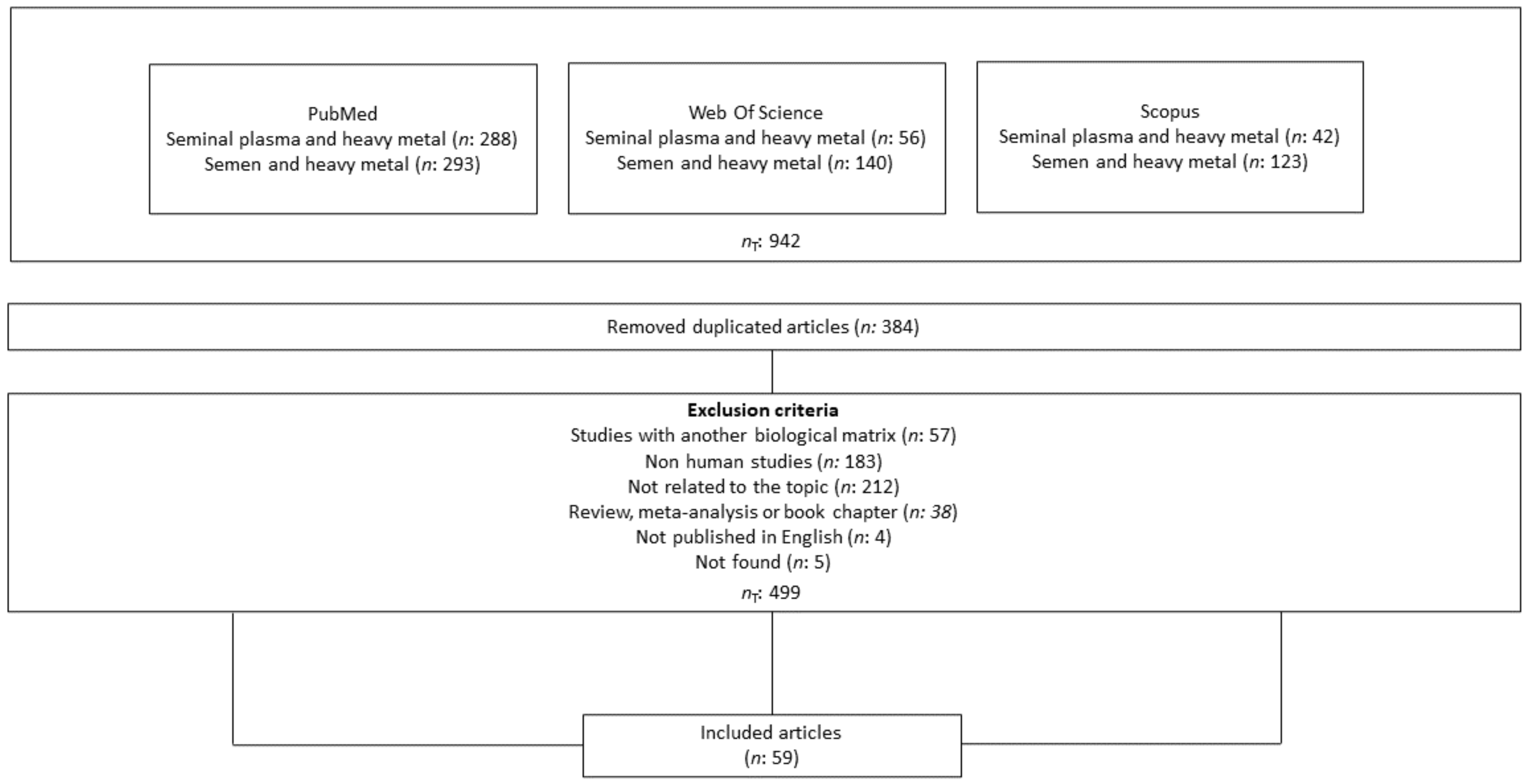
2. Materials and Methods
2.1. Search Strategy
Heavy Metals in Seminal Plasma
2.2. Selection of Relevant Studies and Data Analysis
Heavy Metals in Seminal Plasma
3. Results and Discussion
3.1. Compilation of Relevant Bibliographical Sources
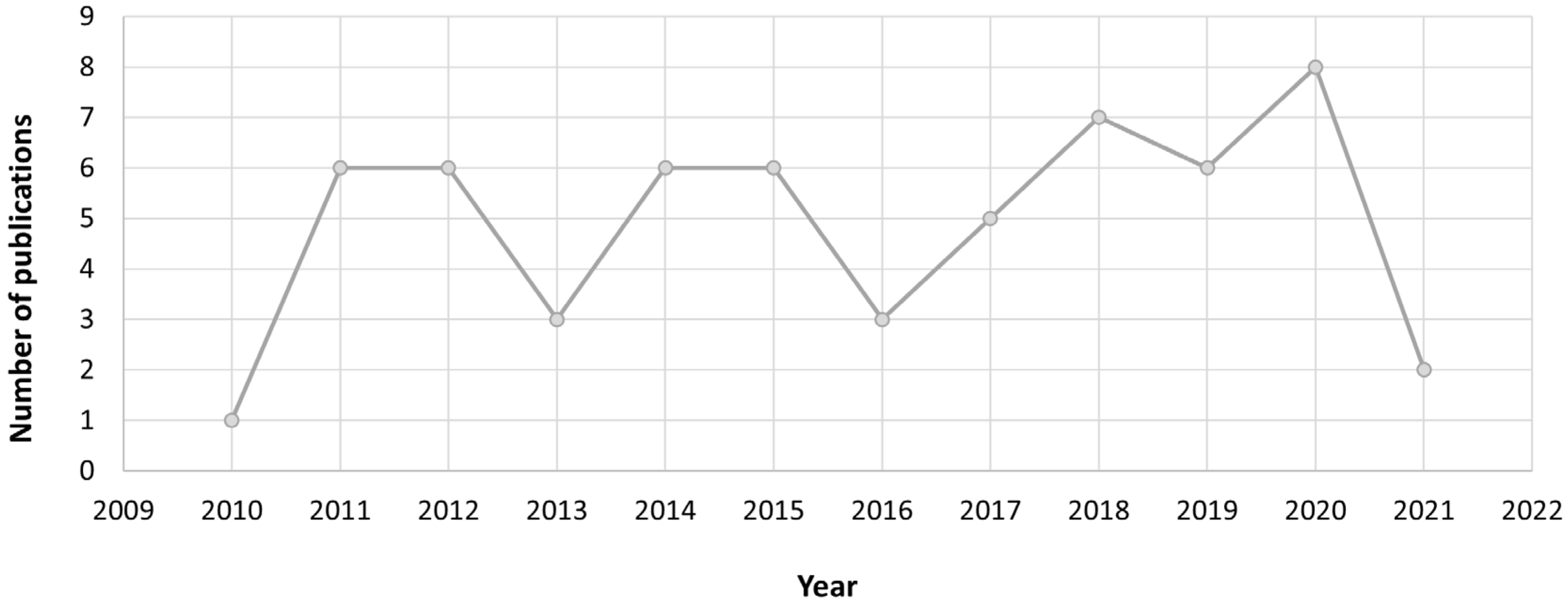
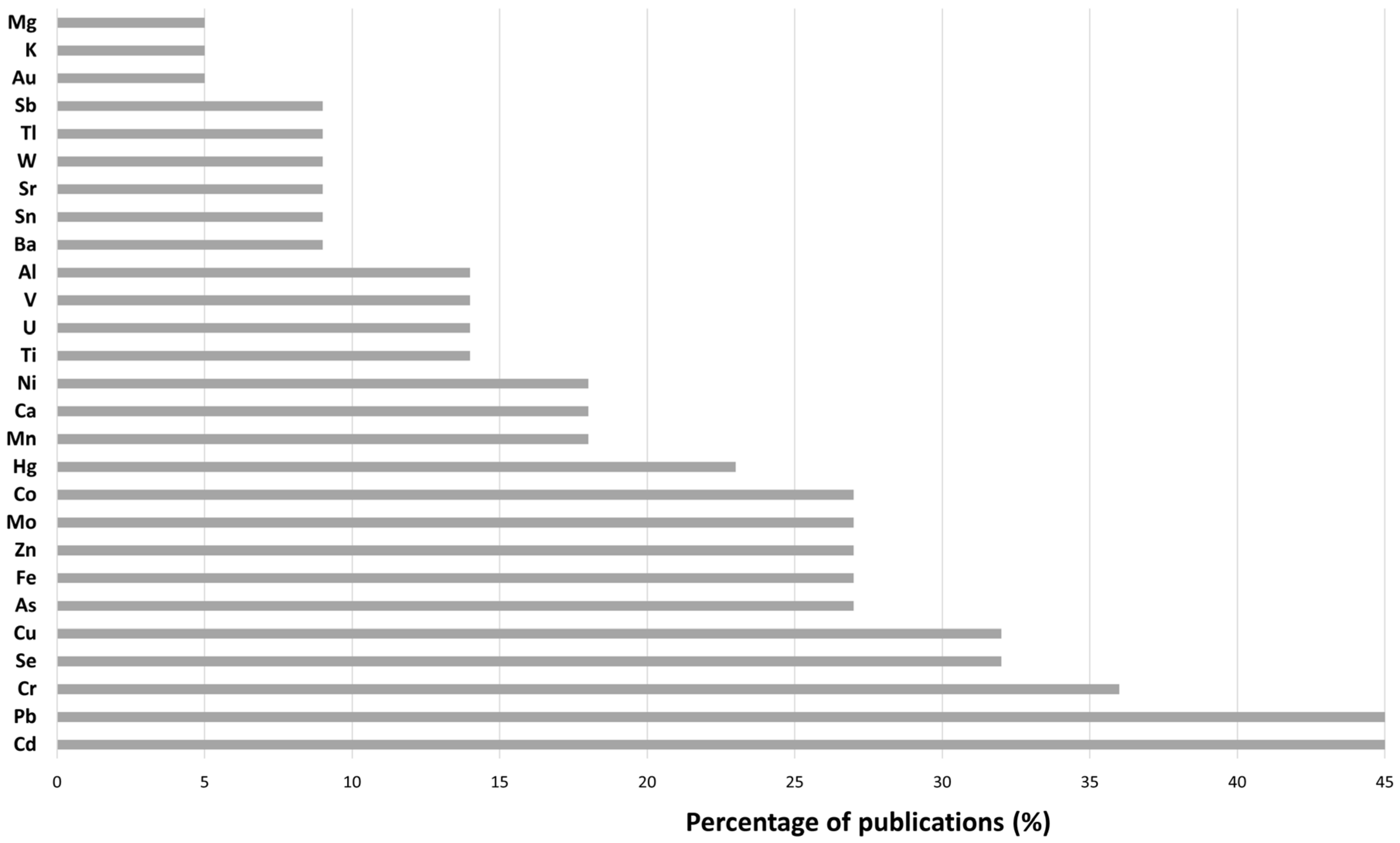
3.2. Bibliometric Analysis
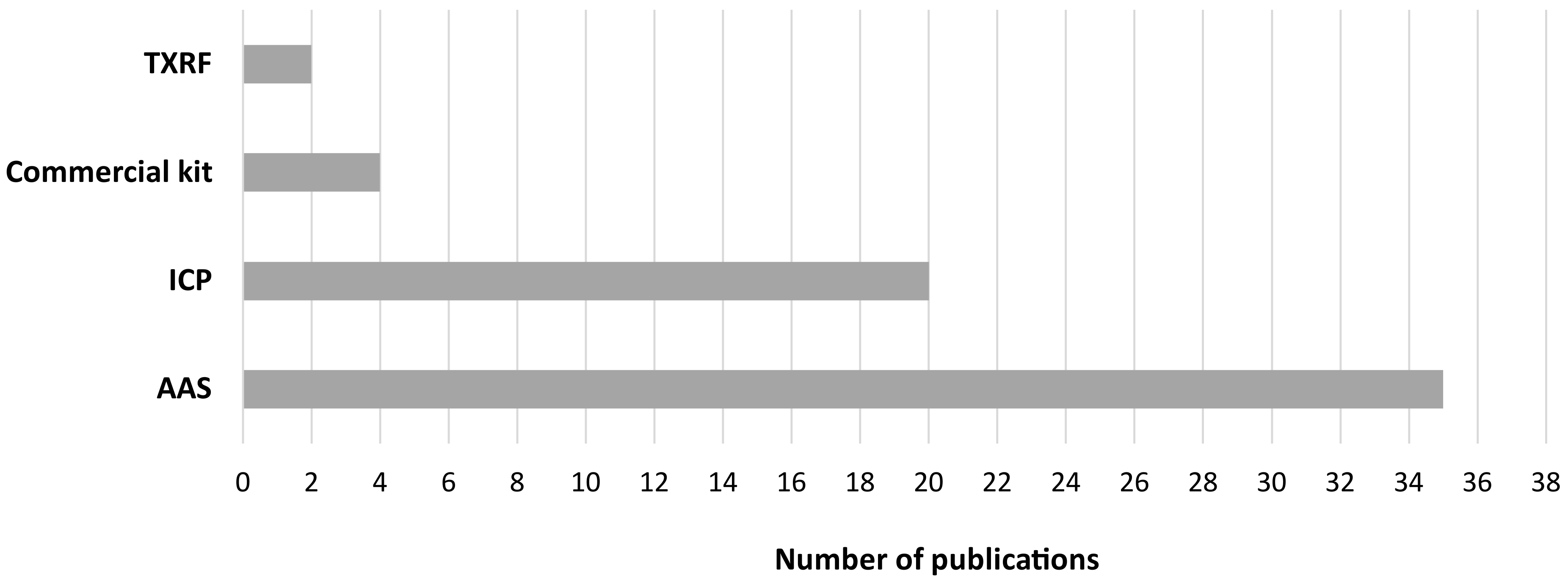
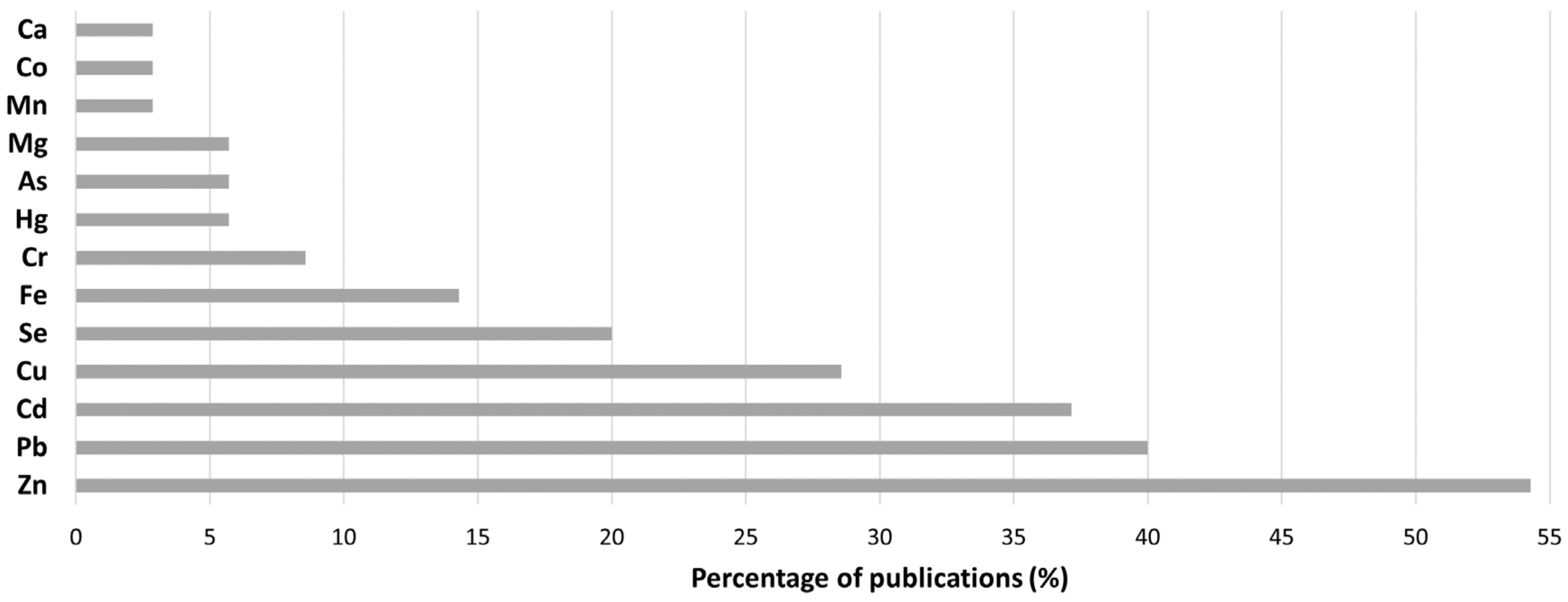
3.3. Bibliographical Analysis
3.3.1. Heavy Metal Detection and Determination in Human Seminal Plasma
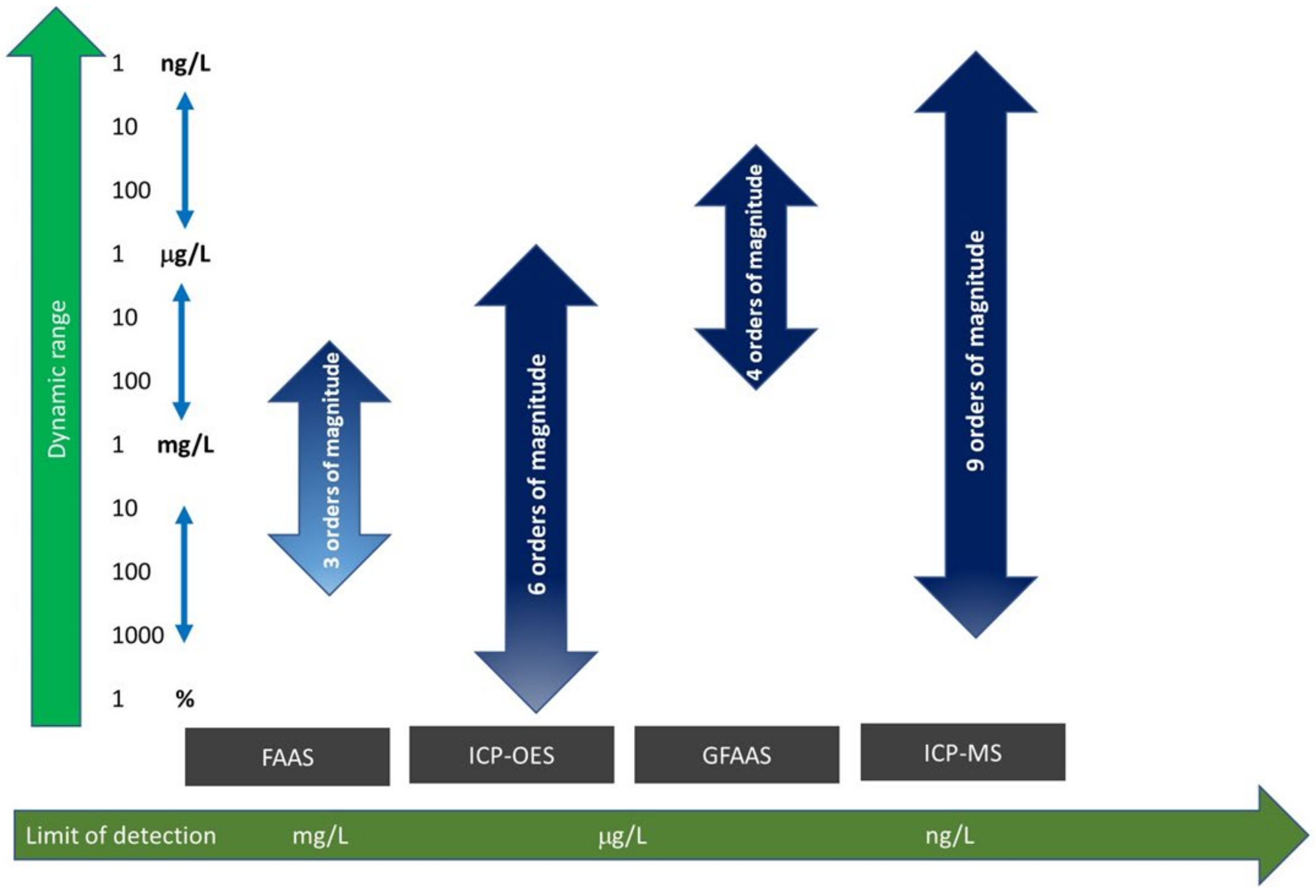
3.3.2. Atomic Absorption Spectroscopy
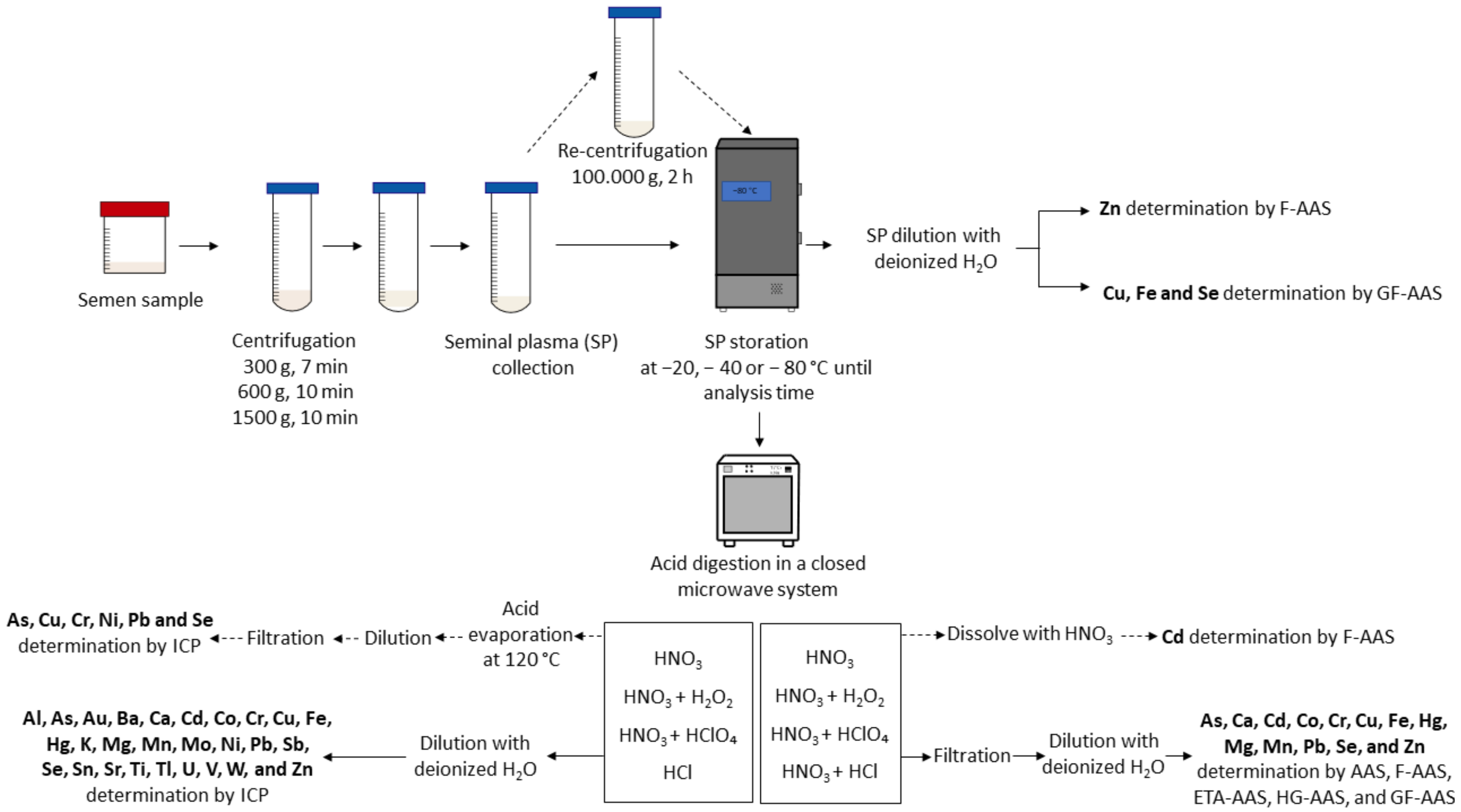
Sample Preparation for AAS Determination
Instrumental Settings for Heavy Metal Detection and Quantification
Heavy Metal Detection and Quantification
Limitations of Atomic Absorption Techniques
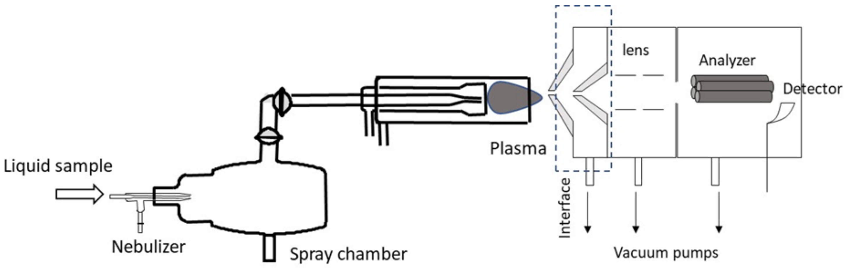
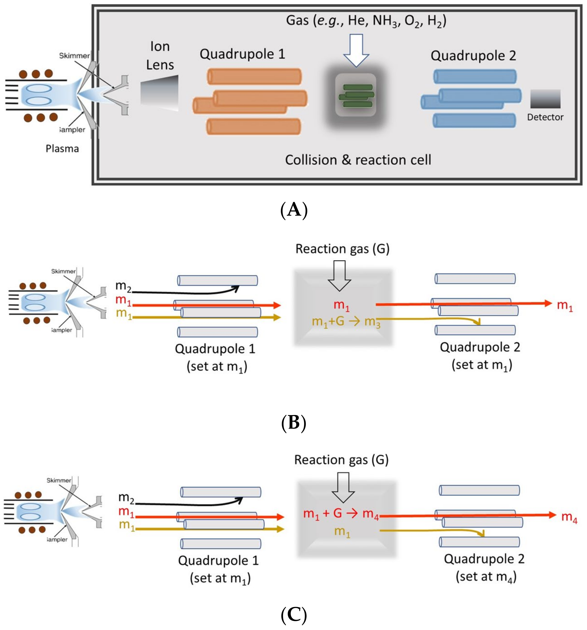
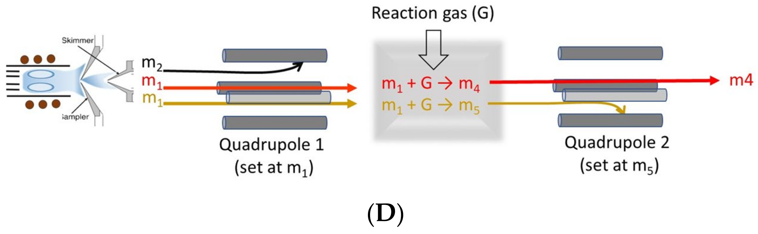
3.3.3. Inductively Coupled Plasma (ICP)
Sample Preparation for Determination
Instrumental Settings for Heavy Metal Detection and Quantification
Heavy Metal Detection and Quantification
3.3.4. Total Reflection X-ray Fluorescence and Commercial Kits
Instrumental Settings for Heavy Metal Detection and Quantification
Heavy Metal Detection and Quantification
Sample Preparation
4. Conclusions
Supplementary Materials
Author Contributions
Funding
Institutional Review Board Statement
Conflicts of Interest
References
- Bracke, A.; Peeters, K.; Punjabi, U.; Hoogewijs, D.; Dewilde, S. A search for molecular mechanisms underlying male idiopathic infertility. Reprod. Biomed. Online 2018, 36, 327–339. [Google Scholar] [CrossRef] [PubMed]
- Agarwal, A.; Mulgund, A.; Hamada, A.; Chyatte, M.R. A unique view on male infertility around the globe. Reprod. Biol. Endocrinol. 2015, 13, 37. [Google Scholar] [CrossRef] [PubMed]
- Sengupta, P.; Dutta, S.; Krajewska-Kułak, E. The Disappearing Sperms: Analysis of Reports Published Between 1980 and 2015. Am. J. Men’s Health 2016, 11, 1279–1304. [Google Scholar] [CrossRef] [PubMed]
- Magalhães, J.A.; Ribeiro, L.S.; Rego, J.P.A.; de Andrade, C.R. Current markers for infertility in men. JBRA Assist. Reprod. 2021, 25, 592–600. [Google Scholar] [CrossRef] [PubMed]
- Ma, Y.; He, X.; Qi, K.; Wang, T.; Qi, Y.; Cui, L.; Wang, F.; Song, M. Effects of environmental contaminants on fertility and reproductive health. J. Environ. Sci. 2018, 77, 210–217. [Google Scholar] [CrossRef]
- Mann, U.; Shiff, B.; Patel, P. Reasons for worldwide decline in male fertility. Curr. Opin. Urol. 2020, 30, 296–301. [Google Scholar] [CrossRef]
- Figa-Talamanca, I.; Traina, M.E.; Urbani, E. Occupational exposures to metals, solvents and pesticides: Recent evidence on male reproductive effects and biological markers. Occup. Med. 2001, 51, 174–188. [Google Scholar] [CrossRef]
- Ali, H.; Khan, E. What are heavy metals? Long-standing controversy over the scientific use of the term ‘heavy metals’—Proposal of a comprehensive definition. Toxicol. Environ. Chem. 2018, 100, 6–19. [Google Scholar] [CrossRef]
- Pourret, O.; Hursthouse, A. It’s Time to Replace the Term “Heavy Metals” with “Potentially Toxic Elements” When Reporting Environmental Research. Int. J. Environ. Res. Public Health 2019, 16, 4446. [Google Scholar] [CrossRef]
- López-Botella, A.; Velasco, I.; Acién, M.; Sáez-Espinosa, P.; Todolí-Torró, J.-L.; Sánchez-Romero, R.; Gómez-Torres, M.J. Impact of Heavy Metals on Human Male Fertility—An Overview. Antioxidants 2021, 10, 1473. [Google Scholar] [CrossRef]
- Balabanič, D.; Rupnik, M.S.; Klemenčič, A.K. Negative impact of endocrine-disrupting compounds on human reproductive health. Reprod. Fertil. Dev. 2011, 23, 403–416. [Google Scholar] [CrossRef] [PubMed]
- Kabir, E.R.; Rahman, M.S.; Rahman, I. A review on endocrine disruptors and their possible impacts on human health. Environ. Toxicol. Pharmacol. 2015, 40, 241–258. [Google Scholar] [CrossRef] [PubMed]
- Mirnamniha, M.; Faroughi, F.; Tahmasbpour, E.; Ebrahimi, P.; Harchegani, A.B. An overview on role of some trace elements in human reproductive health, sperm function and fertilization process. Rev. Environ. Health 2019, 34, 339–348. [Google Scholar] [CrossRef]
- Louis, G.M.B.; Smarr, M.M.; Sun, L.; Chen, Z.; Honda, M.; Wang, W.; Karthikraj, R.; Weck, J.; Kannan, K. Endocrine disrupting chemicals in seminal plasma and couple fecundity. Environ. Res. 2018, 163, 64–70. [Google Scholar] [CrossRef]
- Wang, C.; Swerdloff, R.S. Limitations of semen analysis as a test of male fertility and anticipated needs from newer tests. Fertil. Steril. 2014, 102, 1502–1507. [Google Scholar] [CrossRef]
- Khatun, A.; Rahman, S.; Pang, M.-G. Clinical assessment of the male fertility. Obstet. Gynecol. Sci. 2018, 61, 179–191. [Google Scholar] [CrossRef]
- Ong, C.-N.; Shen, H.-M.; Chia, S.-E. Biomarkers for Male Reproductive health hazards: Are they available? Toxicol. Lett. 2002, 134, 17–30. [Google Scholar] [CrossRef]
- Muratori, M.; Marchiani, S.; Maggi, M.; Forti, G.; Baldi, E. Origin and biological significance of DNA fragmentation in human spermatozoa. Front. Biosci. 2006, 11, 1491–1499. [Google Scholar] [CrossRef] [PubMed]
- Mitra, S.; Varghese, A.C.; Mandal, S.; Bhattacharyya, S.; Nandi, P.; Rahman, S.M.; Kar, K.K.; Saha, R.; Roychoudhury, S.; Murmu, N. Lead and cadmium exposure induces male reproductive dysfunction by modulating the expression profiles of apoptotic and survival signal proteins in tea-garden workers. Reprod. Toxicol. 2020, 98, 134–148. [Google Scholar] [CrossRef] [PubMed]
- Cescon, M.; Chianese, R.; Tavares, R. Environmental Impact on Male (In)Fertility via Epigenetic Route. J. Clin. Med. 2020, 9, 2520. [Google Scholar] [CrossRef]
- Quintanilla-Vega, B.; Hoover, D.J.; Bal, W.; Silbergeld, E.K.; Waalkes, M.P.; Anderson, L.D. Lead Interaction with Human Protamine (HP2) as a Mechanism of Male Reproductive Toxicity. Chem. Res. Toxicol. 2000, 13, 594–600. [Google Scholar] [CrossRef]
- Gao, F.; Zhang, P.; Zhang, H.; Zhang, Y.; Zhang, Y.; Hao, Q.; Zhang, X. Dysregulation of long noncoding RNAs in mouse testes and spermatozoa after exposure to cadmium. Biochem. Biophys. Res. Commun. 2017, 484, 8–14. [Google Scholar] [CrossRef] [PubMed]
- Lv, Y.; Zhang, P.; Guo, J.; Zhu, Z.; Li, X.; Xu, D.; Zeng, W. Melatonin protects mouse spermatogonial stem cells against hexavalent chromium-induced apoptosis and epigenetic histone modification. Toxicol. Appl. Pharmacol. 2017, 340, 30–38. [Google Scholar] [CrossRef] [PubMed]
- Galimov, S.N.; Gromenko, Y.Y.; Galimova, E.F.; Bodrova, E.S.; Bulygin, K.V.; Litvitsky, P.F. Molecular Mechanisms of Male Infertility: Main Directions of Scientific Research. Urologiia 2022, 4, 114–117. [Google Scholar] [CrossRef]
- Hernández-Falcó, M.; Sáez-Espinosa, P.; López-Botella, A.; Aizpurua, J.; Gómez-Torres, M.J. The Role of Sperm Proteins IZUMO1 and TMEM95 in Mammalian Fertilization: A Systematic Review. Int. J. Mol. Sci. 2022, 23, 3929. [Google Scholar] [CrossRef]
- Page, M.J.; McKenzie, J.E.; Bossuyt, P.M.; Boutron, I.; Hoffmann, T.C.; Mulrow, C.D.; Shamseer, L.; Tetzlaff, J.M.; Akl, E.A.; Brennan, S.E.; et al. The PRISMA 2020 Statement: An Updated Guideline for Reporting Systematic Reviews. BMJ 2021, 372, n71. [Google Scholar] [CrossRef]
- Sengupta, M.; Deb, I.; Sharma, G.D.; Kar, K.K. Human sperm and other seminal constituents in male infertile patients from arsenic and cadmium rich areas of Southern Assam. Syst. Biol. Reprod. Med. 2013, 59, 199–209. [Google Scholar] [CrossRef]
- Skandhan, K.; Valsa, J.; Sumangala, B.; Jaya, V. Level of Copper in Human Split Ejaculate. Urol. J. 2016, 84, 51–54. [Google Scholar] [CrossRef]
- Ranganathan, P.; Rao, K.A.; Sudan, J.J.; Balasundaram, S. Cadmium effects on sperm morphology and semenogelin with relates to increased ROS in infertile smokers: An in vitro and in silico approach. Reprod. Biol. 2018, 18, 189–197. [Google Scholar] [CrossRef]
- Pant, N.; Kumar, G.; Upadhyay, A.D.; Gupta, Y.K.; Chaturvedi, P.K. Correlation between lead and cadmium concentration and semen quality. Andrologia 2014, 47, 887–891. [Google Scholar] [CrossRef]
- Pant, N.; Kumar, G.; Upadhyay, A.D.; Patel, D.K.; Gupta, Y.K.; Chaturvedi, P.K. Reproductive toxicity of lead, cadmium, and phthalate exposure in men. Environ. Sci. Pollut. Res. 2014, 21, 11066–11074. [Google Scholar] [CrossRef] [PubMed]
- Skandhan, K.; Mazumdar, B.; Sumangala, B. Study into the Iron Content of Seminal Plasma in Normal and Infertile Subjects. Urol. J. 2012, 79, 54–57. [Google Scholar] [CrossRef] [PubMed]
- Nishihara, T.; Matsumoto, K.; Hosoi, Y.; Morimoto, Y. Evaluation of antioxidant status and oxidative stress markers in follicular fluid for human in vitro fertilization outcome. Reprod. Med. Biol. 2018, 17, 481–486. [Google Scholar] [CrossRef] [PubMed]
- Khan, P.S.; Skandhan, K.P.; Ajesh, K.; Siraj, M.V.P. Gold in Human Semen Around and Away From a Gold Deposit Area. Biol. Trace Element Res. 2010, 142, 302–308. [Google Scholar] [CrossRef]
- Li, R.; Zhao, L.; Li, L.; Hou, Z.; Zhang, D.; Wan, L.; Wei, L.; Yang, Y.; Lv, J.; Ma, M.; et al. A Preliminary Study about the Potential Effects of Heavy Metals on the Human Male Reproductive Parameters in HIV-Infected Population in China. Biol. Trace Element Res. 2017, 180, 39–47. [Google Scholar] [CrossRef]
- Lu, J.-C.; Jing, J.; Chen, L.; Ge, Y.-F.; Feng, R.-X.; Liang, Y.-J.; Yao, B. Analysis of human sperm DNA fragmentation index (DFI) related factors: A report of 1010 subfertile men in China. Reprod. Biol. Endocrinol. 2018, 16, 23. [Google Scholar] [CrossRef]
- Wu, S.; Wang, M.; Deng, Y.; Qiu, J.; Zhang, X.; Tan, J. Associations of toxic and essential trace elements in serum, follicular fluid, and seminal plasma with In vitro fertilization outcomes. Ecotoxicol. Environ. Saf. 2020, 204, 110965. [Google Scholar] [CrossRef]
- Wang, Y.-X.; Chen, H.-G.; Li, X.-D.; Chen, Y.-J.; Liu, C.; Feng, W.; Zeng, Q.; Wang, P.; Pan, A.; Lu, W.-Q. Concentrations of vanadium in urine and seminal plasma in relation to semen quality parameters, spermatozoa DNA damage and serum hormone levels. Sci. Total Environ. 2018, 645, 441–448. [Google Scholar] [CrossRef]
- Liu, R.-Z.; Gao, J.-C.; Zhang, H.-G.; Wang, R.-X.; Zhang, Z.-H.; Liu, X.-Y. Seminal Plasma Zinc Level May be Associated with the Effect of Cigarette Smoking on Sperm Parameters. J. Int. Med. Res. 2010, 38, 923–928. [Google Scholar] [CrossRef]
- Liu, P.; Yuan, G.; Zhou, Q.; Liu, Y.; He, X.; Zhang, H.; Guo, Y.; Wen, Y.; Huang, S.; Ke, Y.; et al. The association between metal exposure and semen quality in Chinese males: The mediating effect of androgens. Environ. Pollut. 2020, 264, 113975. [Google Scholar] [CrossRef]
- Kasperczyk, A.; Dobrakowski, M.; Czuba, Z.P.; Horak, S.; Kasperczyk, S. Environmental exposure to lead induces oxidative stress and modulates the function of the antioxidant defense system and the immune system in the semen of males with normal semen profile. Toxicol. Appl. Pharmacol. 2015, 284, 339–344. [Google Scholar] [CrossRef] [PubMed]
- Kasperczyk, A.; Dobrakowski, M.; Czuba, Z.P.; Kapka-Skrzypczak, L.; Kasperczyk, S. Environmental exposure to zinc and copper influences sperm quality in fertile males. Ann. Agric. Environ. Med. 2015, 23, 138–143. [Google Scholar] [CrossRef] [PubMed]
- Wdowiak, A.; Bakalczuk, G.; Bakalczuk, S. Evaluation of effect of selected trace elements on dynamics of sperm DNA fragmentation. Postepy Hig. Med. Dosw. 2015, 69, 1405–1410. [Google Scholar] [PubMed]
- Marzec-Wróblewska, U.; Kamiński, P.; Łakota, P.; Szymański, M.; Wasilow, K.; Ludwikowski, G.; Kuligowska-Prusińska, M.; Odrowąż-Sypniewska, G.; Stuczyński, T.; Michałkiewicz, J. Zinc and Iron Concentration and SOD Activity in Human Semen and Seminal Plasma. Biol. Trace Element Res. 2010, 143, 167–177. [Google Scholar] [CrossRef] [PubMed]
- Marzec-Wróblewska, U.; Kamiński, P.; Łakota, P.; Szymański, M.; Wasilow, K.; Ludwikowski, G.; Jerzak, L.; Stuczyński, T.; Woźniak, A.; Buciński, A. Human Sperm Characteristics with Regard to Cobalt, Chromium, and Lead in Semen and Activity of Catalase in Seminal Plasma. Biol. Trace Element Res. 2018, 188, 251–260. [Google Scholar] [CrossRef]
- Habib, S.; Toson, E.A.; El-Baz, R.A.; Elafify, M.E. The Impact of Heavy Metals in Impairment of Spermatogenesis and Sperm Density in the Human. J. Biochem. Technol. 2018, 9, 1–8. [Google Scholar]
- Taha, E.A.; Ez-Aldin, A.M.; Sayed, S.K.; Ghandour, N.M.; Mostafa, T. Effect of Smoking on Sperm Vitality, DNA Integrity, Seminal Oxidative Stress, Zinc in Fertile Men. Urology 2012, 80, 822–825. [Google Scholar] [CrossRef]
- Taha, E.A.; Ezz-Aldin, A.M.; Sayed, S.K.; Ghandour, N.M.; Mostafa, T. Smoking influence on sperm vitality, DNA fragmentation, reactive oxygen species and zinc in oligoasthenoteratozoospermic men with varicocele. Andrologia 2013, 46, 687–691. [Google Scholar] [CrossRef] [PubMed]
- Awadalla, N.J.; El-Helaly, M.; Gouida, M.; Mandour, R.; Mansour, M. Sperm Chromatin Structure, Semen Quali-ty and Lead in Blood and Seminal Fluid of Infertile Men. Int. J. Occup. Environ. Med. 2011, 2, 27–36. [Google Scholar]
- Elsamanoudy, A.Z.; Shaalan, D.; Gaballah, M.; El-Atta, H.M.A.; Helaly, A.M. Possible Effects of Metallosis on Spermatozoal Apoptotic Genes Expression in Individuals with Intramedullary Nailing Prosthesis. Biol. Trace Element Res. 2014, 158, 334–341. [Google Scholar] [CrossRef]
- Owen, D.H. A Review of the Physical and Chemical Properties of Human Semen and the Formulation of a Semen Simulant. J. Androl. 2005, 26, 459–469. [Google Scholar] [CrossRef] [PubMed]
- Souza, L.R.R.; Zanatta, M.B.T.; da Silva, I.A.; da Veiga, M.A.M.S. Mercury determination in soil and sludge samples by HR CS GFAAS: Comparison of sample preparation procedures and chemical modifiers. J. Anal. At. Spectrom. 2018, 33, 1477–1485. [Google Scholar] [CrossRef]
- Tsai, S.-J.J.; Shiue, C.-C.; Chang, S.-I. Electrothermal atomic absorption spectrometric determination of copper in nickel-base alloys with various chemical modifiers. Spectrochim. Acta Part B At. Spectrosc. 1997, 52, 1497–1508. [Google Scholar] [CrossRef]
- Pohl, P.; Stecka, H.; Jamroz, P. Interference-free determination of trace copper in freshly ripened honeys by flame atomic absorption spectrometry following a preconcentration by solid-phase extraction and a two-step elution process. Arch. Environ. Contam. Toxicol. 2013, 66, 287–294. [Google Scholar] [CrossRef] [PubMed]
- Schlemmer, G.; Balcaen, L.; Todolí, J.L. Elemental Analysis. An Introduction to Modern Spectrometric Techniques; Walter de Gruyter GmbH: Berlin, Germany, 2019. [Google Scholar]
- Hashemi, M.M.; Behnampour, N.; Nejabat, M.; Tabandeh, A.; Ghazi-Moghaddam, B.; Joshaghani, H.R. Impact of Seminal Plasma Trace Elements on Human Sperm Motility Parameters. Romanian J. Intern. Med. 2018, 56, 15–20. [Google Scholar] [CrossRef]
- Wijesekara, G.; Fernando, D.; Wijerathna, S.; Bandara, N. Environmental and occupational exposures as a cause of male infertility: A caveat. Ceylon Med, J. 2015, 60, 52–56. [Google Scholar] [CrossRef]
- Mendiola, J.; Moreno, J.M.; Roca, M.; Vergara-Juárez, N.; Martínez-García, M.J.; García-Sánchez, A.; Elvira-Rendueles, B.; Moreno-Grau, S.; López-Espín, J.J.; Ten, J.; et al. Relationships between heavy metal concentrations in three different body fluids and male reproductive parameters: A pilot study. Environ. Health 2011, 10, 6. [Google Scholar] [CrossRef]
- Nsonwu-Anyanwu, A.C.; Ekong, E.R.; Offor, S.J.; Awusha, O.F.; Orji, O.C.; Umoh, E.I.; Owhorji, J.A.; Emetonjor, F.R.; Usoro, C.A.O. Heavy metals, biomarkers of oxidative stress and changes in sperm function: A case-control study. Int. J. Reprod. Biomed. 2019, 17, 163–174. [Google Scholar] [CrossRef]
- Atig, F.; Raffa, M.; Ben Ali, H.; Abdelhamid, K.; Saad, A.; Ajina, M. Altered Antioxidant Status and Increased Lipid Per-Oxidation in Seminal Plasma of Tunisian Infertile Men. Int. J. Biol. Sci. 2012, 8, 139–149. [Google Scholar] [CrossRef]
- Nenkova, G.; Petrov, L.; Alexandrova, A. Role of Trace Elements for Oxidative Status and Quality of Human Sperm. Balk. Med. J. 2017, 34, 343–348. [Google Scholar] [CrossRef]
- Wu, H.-M.; Lin-Tan, D.-T.; Wang, M.-L.; Huang, H.-Y.; Lee, C.-L.; Wang, H.-S.; Soong, Y.-K.; Lin, J.-L. Lead level in seminal plasma may affect semen quality for men without occupational exposure to lead. Reprod. Biol. Endocrinol. 2012, 10, 91. [Google Scholar] [CrossRef] [PubMed]
- Wijesekara, G.; Fernando, D.; Wijeratne, S. The effects of Pb on sperm parameters and sperm DNA fragmentation of men investigated for infertility. J. Basic Clin. Physiol. Pharmacol. 2020, 31, 20190239. [Google Scholar] [CrossRef] [PubMed]
- Calogero, A.E.; Fiore, M.; Giacone, F.; Altomare, M.; Asero, P.; Ledda, C.; Romeo, G.; Mongioì, L.M.; Copat, C.; Giuffrida, M.; et al. Exposure to multiple metals/metalloids and human semen quality: A cross-sectional study. Ecotoxicol. Environ. Saf. 2021, 215, 112165. [Google Scholar] [CrossRef] [PubMed]
- Akinloye, O.; Abbiyesuku, F.M.; Oguntibeju, O.O.; Arowojolu, A.O.; Truter, E.J. The impact of blood and seminal plasma zinc and copper concentrations on spermogram and hormonal changes in infertile Nigerian men. Reprod. Biol. 2011, 11, 83–97. [Google Scholar] [CrossRef] [PubMed]
- Riaz, M.; Mahmood, Z.; Shahid, M.; Saeed, M.U.Q.; Tahir, I.M.; Shah, S.A.; Munir, N.; El-Ghorab, A. Impact of reactive oxygen species on antioxidant capacity of male reproductive system. Int. J. Immunopathol. Pharmacol. 2015, 29, 421–425. [Google Scholar] [CrossRef]
- Kahraman, S.; Hassa, H.; Karataş, A.; Ilgin, H. The Effect of Blood and Seminal Plasma Heavy Metal and Trace Element Levels on Sperm Quality. Turk. Klin. J. Med. Sci. 2012, 32, 1560–1568. [Google Scholar] [CrossRef]
- Habib, S.A.E.-H.; Toson, E.A.M.; Al-Mutairi, F.M.; Al-Alawy, A.I.; Elfaki, I.; El-Baz, R.A.; Elafify, M.E. Effects of Trace Metals Levels and Hyaluronic Acid Degrading Enzymes Activities on Human Sperm Function. Pak. J. Biol. Sci. 2019, 22, 444–451. [Google Scholar] [CrossRef]
- Alexandrino, A.P.; Rodrigues, M.A.F.; Matsuo, T.; Gregório, E.P.; Santilli, J.C. Evaluation of seminal zinc levels by atomic absorption in men with spinal cord injury. Spinal Cord 2010, 49, 435–438. [Google Scholar] [CrossRef]
- Atig, F.; Raffa, M.; Habib, B.-A.; Kerkeni, A.; Saad, A.; Ajina, M. Impact of seminal trace element and glutathione levels on semen quality of Tunisian infertile men. BMC Urol. 2012, 12, 6. [Google Scholar] [CrossRef]
- Milostić-Srb, A.; Včev, A.; Tandara, M.; Marić, S.; Kuić-Vadlja, V.; Srb, N.; Holik, D. Importance of Zinc Concentration in Seminal Fluid of Men Diagnosed with Infertility. Acta Clin. Croat. 2020, 59, 154–159. [Google Scholar] [CrossRef]
- Chen, S.-Y.; Chang, C.-H.; Hu, C.-C.; Chen, C.-C.; Chang, Y.-H.; Hsieh, P.-H. Metal ion concentrations and semen quality in patients undergoing hip arthroplasty: A prospective comparison between metal-on-metal and metal-on-polyethylene implants. J. Orthop. Res. 2015, 34, 544–551. [Google Scholar] [CrossRef] [PubMed]
- Jeng, H.A.; Huang, Y.-L.; Pan, C.-H.; Diawara, N. Role of low exposure to metals as male reproductive toxicants. Int. J. Environ. Health Res. 2014, 25, 405–417. [Google Scholar] [CrossRef] [PubMed]
- Abbas, A.; Khan, J.; Hassan, M.A.; Qayyum, A.; Shafiq, H. Bioaccumulation of Heavy Metals, Even in Low Quantities, is the Main Causative Agent of Male Human Infertility in D. I. Khan Division. Pak. J. Zool. 2021, 53, 2001–2521. [Google Scholar] [CrossRef]
- Hasan, S.H.; Hasan, H.R. Oxidative stress status in sera and seminal plasma and their correlation with lead (Pb) and cadmium (Cd) in Iraqi infertile male. Int. J. Pharm. Res. 2020, 12, 887–896. [Google Scholar] [CrossRef]
- Kumar, S.; Mishra, V.; Thaker, R.; Gor, M.; Perumal, S.; Joshi, P.; Sheth, H.; Shaikh, I.; Gautam, A.K.; Verma, Y. Role of environmental factors & oxidative stress with respect to in vitro fertilization outcome. Indian J. Med. Res. 2018, 148, S125–S133. [Google Scholar] [CrossRef]
- Bassey, I.; Essien, O.; Isong, I.; Udoh, A.; Agbara, G. Seminal Plasma Chromium, Cadmium and Lead Levels in Infertile Men. J. Med. Sci. 2013, 13, 497–500. [Google Scholar] [CrossRef]
- Infante, H.G.; Warren, J.; Chalmers, J.; Dent, G.; Todoli, J.L.; Collingwood, J.; Telling, N.; Resano, M.; Limbeck, A.; Schoenberger, T.; et al. Glossary of methods and terms used in analytical spectroscopy (IUPAC Recommendations 2019). Pure Appl. Chem. 2021, 93, 647–776. [Google Scholar] [CrossRef]
- Todoli, J.-L.; Mermet, J.-M. Liquid Sample Introduction in ICP Spectrometry. A Practical Guide; Elsevier: Amsterdam, The Netherlands, 2008. [Google Scholar]
- Todolí, J.L. Atomic Mass Spectrometry. Inductively Coupled Plasma Mass Spectrometry. In Encyclopedia of Analytical Science; Elsevier: Amsterdam, The Netherlands, 2019; Volume 1, pp. 209–217. [Google Scholar]
- Sukhn, C.; Awwad, J.; Ghantous, A.; Zaatari, G. Associations of semen quality with non-essential heavy metals in blood and seminal fluid: Data from the Environment and Male Infertility (EMI) study in Lebanon. J. Assist. Reprod. Genet. 2018, 35, 1691–1701. [Google Scholar] [CrossRef]
- Zafar, A.; Eqani, S.A.M.A.S.; Bostan, N.; Cincinelli, A.; Tahir, F.; Shah, S.T.A.; Hussain, A.; Alamdar, A.; Huang, Q.; Peng, S.; et al. Toxic metals signature in the human seminal plasma of Pakistani population and their potential role in male infertility. Environ. Geochem. Health 2014, 37, 515–527. [Google Scholar] [CrossRef]
- Ali, S.; Chaspoul, F.; Anderson, L.; Bergé-Lefranc, D.; Achard, V.; Perrin, J.; Gallice, P.; Guichaoua, M. Mapping Fifteen Trace Elements in Human Seminal Plasma and Sperm DNA. Biol. Trace Element Res. 2016, 175, 244–253. [Google Scholar] [CrossRef]
- Wang, Y.-X.; Wang, P.; Feng, W.; Liu, C.; Yang, P.; Chen, Y.-J.; Sun, L.; Sun, Y.; Yue, J.; Gu, L.-J.; et al. Relationships between seminal plasma metals/metalloids and semen quality, sperm apoptosis and DNA integrity. Environ. Pollut. 2017, 224, 224–234. [Google Scholar] [CrossRef] [PubMed]
- Ai, C.-E.; Li, C.-J.; Tsou, M.-C.; Chen, J.-L.; Hsi, H.-C.; Chien, L.-C. Blood and seminal plasma mercury levels and predatory fish intake in relation to low semen quality. Environ. Sci. Pollut. Res. 2019, 26, 19425–19433. [Google Scholar] [CrossRef] [PubMed]
- Nikolaou, V.S.; Petit, A.; Zukor, D.J.; Papanastasiou, C.; Huk, O.L.; Antoniou, J. Presence of Cobalt and Chromium Ions in the Seminal Fluid of Young Patients with Metal-on-Metal Total Hip Arthroplasty. J. Arthroplast. 2013, 28, 161–167. [Google Scholar] [CrossRef] [PubMed]
- McDiarmid, M.A.; Gucer, P.; Centeno, J.A.; Todorov, T.; Squibb, K.S. Semen Uranium Concentrations in Depleted Uranium Exposed Gulf War Veterans: Correlations with Other Body Fluid Matrices. Biol. Trace Element Res. 2018, 190, 45–51. [Google Scholar] [CrossRef] [PubMed]
- Kim, K.; Bloom, M.S.; Kruger, P.C.; Parsons, P.J.; Arnason, J.G.; Byun, Y.; Goins, S.; Fujimoto, V.Y. Toxic metals in seminal plasma and in vitro fertilization (IVF) outcomes. Environ. Res. 2014, 133, 334–337. [Google Scholar] [CrossRef]
- Lettieri, G.; Marra, F.; Moriello, C.; Prisco, M.; Notari, T.; Trifuoggi, M.; Giarra, A.; Bosco, L.; Montano, L.; Piscopo, M. Molecular Alterations in Spermatozoa of a Family Case Living in the Land of Fires. A First Look at Possible Transgenerational Effects of Pollutants. Int. J. Mol. Sci. 2020, 21, 6710. [Google Scholar] [CrossRef]
- Türk, S.; Mändar, R.; Mahlapuu, R.; Viitak, A.; Punab, M.; Kullisaar, T. Male infertility: Decreased levels of selenium, zinc and antioxidants. J. Trace Elements Med. Biol. 2014, 28, 179–185. [Google Scholar] [CrossRef]
- Marguí, E.; Dumić, J.; Queralt, I.; Baković, L.; Jablan, J. Simple and reliable determination of Zn and some additional elements in seminal plasma samples by using total reflection X-ray fluorescence spectroscopy. Anal. Methods 2020, 12, 4899–4905. [Google Scholar] [CrossRef]
- Martínez, S.; Sánchez, R.; Lefevre, J.; Todolí, J.-L. Multi-elemental analysis of oil renewable fuel feedstock. Spectrochim. Acta Part B At. Spectrosc. 2022, 189, 106356. [Google Scholar] [CrossRef]
- Nascimento, M.S.; Druzian, G.T.; Pereira, L.S.F.; Mesko, M.F.; Picoloto, R.S.; Mello, P.A.; Flores, E.M.M. Microwave-assisted extraction for further Cl, Br, and I determination in medicinal plants by ICP-MS: A study of carbon interferences. J. Anal. At. Spectrom. 2022, 37, 535–543. [Google Scholar] [CrossRef]
- Leclercq, A.; Nonell, A.; Torró, J.L.T.; Bresson, C.; Vio, L.; Vercouter, T.; Chartier, F. Introduction of organic/hydro-organic matrices in inductively coupled plasma optical emission spectrometry and mass spectrometry: A tutorial review. Part I. Theoretical considerations. Anal. Chim. Acta 2015, 885, 33–56. [Google Scholar] [CrossRef] [PubMed]
- Todolí, J.L.; Gras, L.; Hernandis, V.; Mora, J. Elemental matrix effects in ICP-AES. J. Anal. At. Spectrom. 2002, 17, 142–169. [Google Scholar] [CrossRef]
- Chan, G.C.-Y.; Hieftje, G.M. In-situ determination of cross-over point for overcoming plasma-related matrix effects in inductively coupled plasma-atomic emission spectrometry. Spectrochim. Acta Part B At. Spectrosc. 2008, 63, 355–366. [Google Scholar] [CrossRef]
- Grotti, M.; Todolí, J.-L. Nitric acid effect in inductively coupled plasma mass spectrometry: New insights on possible causes and correction. J. Anal. At. Spectrom. 2020, 35, 1959–1968. [Google Scholar] [CrossRef]
- Wilschefski, S.; Baxter, M. Inductively Coupled Plasma Mass Spectrometry: Introduction to Analytical Aspects. Clin. Biochem. Rev. 2019, 40, 115–133. [Google Scholar] [CrossRef]
- Lum, T.-S.; Leung, K.S.-Y. Strategies to overcome spectral interference in ICP-MS detection. J. Anal. At. Spectrom. 2016, 31, 1078–1088. [Google Scholar] [CrossRef]
- Balcaen, L.; Bolea-Fernandez, E.; Resano, M.; Vanhaecke, F. Inductively coupled plasma—Tandem mass spectrometry (ICP-MS/MS): A powerful and universal tool for the interference-free determination of (ultra)trace elements—A tutorial review. Anal. Chim. Acta 2015, 894, 7–19. [Google Scholar] [CrossRef]
- Nguyen, T.T.; Trieu, T.S.; Tran, T.O.; Luong, T.L.A. Evaluation of sperm DNA fragmentation index, Zinc concentration and seminal parameters from infertile men with varicocele. Andrologia 2018, 51, e13184. [Google Scholar] [CrossRef]
- Ammar, O.; Houas, Z.; Mehdi, M. The association between iron, calcium, and oxidative stress in seminal plasma and sperm quality. Environ. Sci. Pollut. Res. 2019, 26, 14097–14105. [Google Scholar] [CrossRef]
- Camejo, M.I.; Abdala, L.; Vivas-Acevedo, G.; Lozano-Hernández, R.; Angeli-Greaves, M.; Greaves, E.D. Selenium, Copper and Zinc in Seminal Plasma of Men with Varicocele, Relationship with Seminal Parameters. Biol. Trace Element Res. 2011, 143, 1247–1254. [Google Scholar] [CrossRef]
- Michaelis, M.; Gralla, O.; Behrends, T.; Scharpf, M.; Endermann, T.; Rijntjes, E.; Pietschmann, N.; Hollenbach, B.; Schomburg, L. Selenoprotein P in seminal fluid is a novel biomarker of sperm quality. Biochem. Biophys. Res. Commun. 2014, 443, 905–910. [Google Scholar] [CrossRef] [PubMed]


| Country | Number of Publications | Ref. |
|---|---|---|
| India | 9 | [19,27,28,29,30,31,32,33,34] |
| China | 6 | [35,36,37,38,39,40] |
| Poland | 5 | [41,42,43,44,45] |
| Egypt | 5 | [46,47,48,49,50] |
| Symbol | Chemical Element | Symbol | Chemical Element |
|---|---|---|---|
| Au | Gold | Mo | Molybdenum |
| Al | Aluminum | Na | Sodium |
| As | Arsenic | Ni | Nickel |
| Ba | Barium | Pb | Lead |
| Ca | Calcium | Sb | Antimony |
| Cd | Cadmium | Se | Selenium |
| Co | Cobalt | Sn | Tin |
| Cr | Chromium | Sr | Strontium |
| Cu | Copper | Ti | Titanium |
| Fe | Iron | Tl | Thallium |
| Hg | Mercury | U | Uranium |
| K | Potassium | V | Vanadium |
| Mg | Magnesium | W | Wolframium |
| Mn | Manganese | Zn | Zinc |
| Ref. | Detected Analyte | LOD | Analytical Tool |
|---|---|---|---|
| [19] | Cd and Pb | n.a | FAAS |
| [27] | Cd and As | As: 0.05 μg/L and Cd: 0.002 μg/L | AAS |
| [28] | Cu | n.a | AAS |
| [29] | Cd and Zn | Zn: 6.6 mg /L | FAAS |
| [32] | Fe | n.a | AAS |
| [35] | Cd, Zn, and Pb | n.a | AAS, GFAAS, and FAAS |
| [41] | Pb | n.a | GFAAS |
| [42] | Zn and Cu | n.a | AAS |
| [43] | Cd, Zn, Se, Pb, and Cu | n.a | ETA-AAS |
| [46] | Fe and Cd | n.a | AAS |
| [47] | Zn | n.a | FAAS |
| [48] | Zn | n.a | AAS |
| [49] | Pb | n.a | GFAAS |
| [56] | Fe, Zn, Cu, and Mg | n.a | AAS |
| [57] | Cd and Pb | Pb: 0.32 and Cd: 0.50 μg/L | GFAAS |
| [58] | Hg | 0.1 μg/L | AAS |
| [59] | Cd, Zn, Se, and Pb | n.a | AAS |
| [60] | Zn | 0.47 μg /L | GFAAS |
| [61] | Fe, Zn, Se, and Cu | n.a | FAAS and GFAAS |
| [62] | Pb | 0.1 μg/L | AAS |
| [63] | Pb | 0.32 μg/L | GFAAS |
| [64] | Hg | 0.098 μg/L | AAS |
| [65] | Zn | 5 μg/L | FAAS |
| [66] | Cd, Se, and Pb | n.a | AAS |
| [67] | Fe, Zn, Se, Cu, Ca, and Mg | n.a | FAAS |
| [68] | Zn, Mn, and Cu | n.a | AAS |
| [69] | Zn | n.a | AAS |
| [70] | Zn and Se | Zn: 0.47μg/L and Se: 0.78μg/L | AAS and FAAS |
| [71] | Zn | n.a | AAS |
| [72] | Cr and Co | 0.02 mg/L | ETA-AAS |
| [73] | As, Cd, Cu, Pb, Se, and Zn | As: 2.59, Cd: 0.03, Cu:1.03, Pb: 0.98, Se: 0.02, and Zn: 0.065 μg/L | AAS, ETA-AAS, and HGAAS |
| [74] | Cd, Zn, Pb, Cr, and Cu | n.a | AAS |
| [75] | Cd and Pb | n.a | FAAS |
| [76] | Zn | n.a | AAS |
| [77] | Cd, Pb, and Cr | n.a | AAS |
| Ref. | Study Group | Cd | Pb | Zn |
|---|---|---|---|---|
| [19] | Semen samples were collected from infertile male tea garden workers (n: 200) visiting fertility clinics in southern Assam, India. Age-matched donors as the control group (n: 200) who were not working in the tea gardens. All participants were residents of areas surrounding the tea gardens | Infertile: 0.0145 ppm | Infertile: 0.08 ppm | N.d |
| [27] | Proven fertile healthy volunteers (N, n: 32) and infertile individuals (n: 68): Oligo (n: 35) and Azo (n: 33) subjects, according to the WHO | N: 0.28, O: 0.41, Azo: 0.59 ppm | N.d | N.d |
| [28] | donors (n: 28) submitted semen samples for this study. They belonged to the province of Gujarat and were in the age group of 20–27 years. They were physically and mentally healthy | N.d | N.d | N.d |
| [29] | G1: fertile smokers (n: 68), G2: infertile non-smokers (history of genital examination (testis and scrotum), family inheritance) (n: 72), G3: infertile smokers (n: 76), and G4: non-smoker fertile subjects (n: 74), as the control group | G1: 6.8 ± 0.65, G2: 6.9 ± 1.77, G3: 12 ± 1.52, G4: 4.02 ± 0.62 μg/mL | N.d | G1: 0.71 ± 0.08, G2: 0.12 ± 0.03, G3: 0.07 ± 0.02, G4: 1.99 ± 0.09 mg/mL |
| [32] | N (n: 19), O (n: 11), Azo (n: 12), OA (n: 18), A (n: 15) according to the WHO | N.d | N.d | N.d |
| [35] | Subjects infected with HIV were recruited from Chongqing AIDS Hospice, Chongqing, China (n: 50) | 1.69 ± 0.33 μg/L | 8.57 ± 0.86 μg/L | 30.66 ± 1.60 mg/L |
| [41] | Based on the medians of the levels of Zn in seminal plasma, the study subjects were divided into different groups: men with low environmental exposure to Zn (Zn-L) (n: 33) and men with high environmental exposure to Cu (Zn-H) (n: 32) | N.d | N.d | Zn-L: 85 ± 42.5, Zn-H: 55.0 ± 58.2 mg/dL |
| [42] | Based on the median of the values of Pb concentration in seminal plasma (PbS = 1.00 μg/dL), the subjects were divided into two groups: low environmental exposure to Pb (LE)–Pb concentration in seminal plasma between 0.40 and 1.00 μg/dL and a group with high environmental exposure to Pb (HE)–Pb concentration in seminal plasma between 1.01 and 2.70 μg/dL. NT: 65 study participants | N.d | LE: 0.72 ± 0.19, HE: 1.54 ± 0.47 μg/dL | N.d |
| [43] | Men were divided into two groups of proven fertile men (n: 85) and infertile patients (n: 131), who had been treated due to infertility for a period longer than 1 year. NT: 216, men | Proven fertility: 0.76, infertile patients: 0.92 μg/dL | Proven fertility: 0.76, infetile patients: 0.92 μg/dL | Proven fertility: 0.47, infetile patients: 0.42 μg/dL |
| [46] | Subjects (NT: 216), G1 (n: 70) N samples as the control, G2 (n: 48) A samples, G3 (n: 18) OA samples, G4 (n: 41) OAT samples, G5 (n: 39) Azo samples, according to the WHO | N: 0.015 ± 0.005, A: 0.065 ± 0.017, OA: 0.114 ± 0.018, OAT: 0.072 ± 0.013, Azo: 0.083 ± 0.028 μg/mg | N.d | N.d |
| [47] | Men (n: 160) recruited from the Andrology Unit, University Hospital. They were divided into healthy fertile non-smokers, G1 (n: 80), and fertile smokers, G2 (n: 80) | N.d | N.d | G1: 139.51 ± 7.78, G2: 101.21 ± 11.03 μg/mL |
| [48] | OAT non-smokers (n: 72), OAT smokers (n: 84), OAT non-smokers ith Vx (n: 23), and OAT smokers with Vx (n: 67) | N.d | N.d | OAT non-smoker: 97.2 ± 7.3, OAT smoker: 79.16 ± 5.6, OAT non-smoker with Vx: 68.2 ± 4.6, OAT smoker with Vx: 52.73 ± 9.96 μg /mL |
| [49] | Men with primary infertility (n: 29) attending the outpatient clinic of infertility in Mansoura University Hospital, Egypt | N.d | 11.40 ± 7.53 μg/dL | N.d |
| [56] | Men referred to clinical laboratories in Gorgan (northern Iran) for routine semen analysis as the primary approach for male fertility testing. The control group included N subjects (n: 96) and the case group included A subjects (n: 96). NT: 192 | N.d | N.d | N: 188.42 ± 99.61, A: 187.15 ± 87.38 mg/mL |
| [57] | Male partners (n: 300) of couples investigated for infertility. Males with known causes of infertility were excluded. Environmentally and occupationally exposed (n: 164) and non-exposed (n: 136) men | EE: 0.7, N-EE: 1.3; OE:1.4, N-OE: 1; E and/or OE: 1.2, N-EE and/or OE 1.1 μg/dL | EE: 19.7, N-EE: 14.3; OE: 15.9, N-OE: 15.7; E and/or OE: 17.7, N-EE and/or OE 13.5 μg/dL | x |
| [58] | Case subjects (n: 30) composed of men with OAT and control subjects (n: 31) composed of N patients, according to the WHO | N.d | N.d | N.d |
| [59] | Azo (n: 30), Oligo (n: 50), and N (n: 50) according to the WHO | N: 0.41 ± 0.15, O: 0.47 ± 0.16, Azo: 0.37 ± 0.13 μg/L | N: 0.47 ± 0.17, O: 0.53 ± 0.17, Azo: 0.43 ± 0.15 μg/L | N: 100.53 ± 16.85, O: 104.56 ± 15.94, Azo: 100.310 ± 3.14 μg/L |
| [60] | Male partners (n: 120) from couples in infertility treatment. G1 (n: 40) consisted of males with normal ejaculate (N), G2 (n: 45) consisted of A patients, and G3 which consisted of OAT | N.d | N.d | Control: 97.6 ± 34.9, A: 43.4 ± 24.9, OAT: 32.4 ± 7.9 mg/L |
| [61] | Semen samples were obtained from men aged between 24 and 38 years attending a fertility clinic. N (n: 6), T (n: 6), AT (n: 6), OT (n: 6), according to WHO guidelines | N.d | N.d | N: 150.67 ± 4.75, T: 127.00 ± 12.37, AT: 127.50 ± 5.30, OT: 120.40 ± 13.98 mg/L |
| [62] | Male partners of infertile couples attending the reproductive center of Lin-Kou Medical Center (n: 341), Chang Gung Memorial Hospital and undergoing infertility evaluation were recruited to the study | N.d | 2.19 ± 1.45 μg/L | N.d |
| [63] | Male partners (n: 300) of couples investigated for infertility at Vindana Reproductive Health Center, Colombo, were recruited after informed consent was obtained. Normozoospermics (n: 77) and pathozoospermics (n: 38) following the WHO guidelines | N.d | Total: 15.7 μg/dL; P: 17.2 ± 3.02, N: 15.0 ± 1.70 μg/dL | N.d |
| [64] | Men (n: 179), men with normal sperm parameter (n: 48), and men with abnormal sperm parameter (n: 131) | N.d | N.d | N.d |
| [65] | Three male groups (age: 20–55 years) based on sperm count: O (n: 40); Azo (n: 20); and healthy fertile control males (n: 40) | N.d | N.d | Control: 29.85 ± 1.67, O: 32.77 ± 2.41, Azo: 38.20 ± 3.68 μmol/L |
| [66] | Infertile men (test group, n: 20) attending various fertility centers in Faisalabad, Pakistan were recruited in the study and fertile male volunteers (control group n: 20), according to the WHO | Fertile: 1.43 ± 0.85, infertile: 9.11± 2.34 μg/L | Fertile: 3.2 ± 1.43, infertile: 10.20 ± 4.54 μg/L | N.d |
| [67] | Two groups: a low level of metals group (Me-L, n: 44) and a high level of metals group (Me-H, n: 44) | N.d | N.d | ME-L: 119.29 ± 39.66, ME-H: 139.14 ± 70.45 mg/dL |
| [68] | Semen samples (n: 216) were divided into 5 groups based on WHO criteria. G1 (n: 70) N as the control and 4 groups of infertile males: G2 (n: 48) A samples, G3 (n: 18) OA, G4 (n: 41) OAT samples, and G5 (n: 39) Azo samples | N.d | N.d | G1: 3.93 ± 0.76, G2: 1.91 ± 0.56, G3: 1.1 ± 0.29, G4: 2.17 ± 0.56, G5: 2.13 ± 0.61 μg/mg |
| [69] | Patients with SCI (n: 24) were selected from the multidisciplinary clinic and controls (n: 24) were selected from the family planning clinic of the university hospital matched for age | N.d | N.d | SCI patients: 85.20 ± 65.52 and controls: 147.16 ± 72.17 mg/L |
| [70] | N (n: 60), A (n: 74), O (n: 56), T (n: 60) subjects according to the WHO | N.d | N.d | N: 144 ± 42.13, A: 122 ± 34.69, O: 120.51 ± 25.33, T: 126 ± 24.82 mg/L |
| [71] | Men (n: 276) diagnosed with O (n: 100) and N (n: 176) according to the 2010 WHO guidelines. Subjects diagnosed with low sperm count (n: 100, n: 26 were Azo) | N.d | N.d | Azo: 1.1, O: 1.75 mmol/L |
| [72] | Patients (n: 50) were enrolled for analysis. Patients in each group were based on the implant type: metal-on-metal (n: 25) and metal-on-polyethylene (n: 25) implants | N.d | N.d | N.d |
| [73] | Male human subjects from Taiwan (n: 196) | 0.5 ± 0.3 μg/L | 0.6 ± 1.1 μg/L | 169.3 ± 97.9 μg/L |
| [74] | Semen samples were collected from clinically diagnosed O males (n: 120). Groups per age: 18–23 (n: 20), 24–29 (n: 20), 30–35 (n: 20), 36–41 (n: 20), 42–47 (n: 20), >47 (n: 20) and a control group of N men (n: 30) | 18–23: 0.039, 24–29: 0.037, 30–35: 0.046, 36–41: 0.047, 42–47: 0.050, >47: 0.091, and control: 0.008 μg/mL | 18–23: 0.028, 24–29: 0.117, 30–35: 0.229, 36–41: 0.110, 42–47: 0.231, >47: 0.222, and control: 0.010 μg/mL | 18–23: 2.913, 24–29: 1.628, 30–35: 1.299, 36–41: 0.143, 42–47: 2.469, >47: 1.830, and control: 0.115 μg/mL |
| [75] | Males (n: 101) at Kamal-Alsamerae Hospital, including infertile patients (n: 70) with ages ranging (22–50) years compared to fertile males as the control (n: 40). N individuals (control group) (n: 37), infertile individuals (OA) (n: 32), and infertile individuals (Azo) (n: 32) | N: 0.141 ± 0.028, OA: 0.134 ± 0.020, Azo: 0.089 ± 0.032 μg/mL | N: 0.830 ± 0.213, OA: 0.693 ± 0.131, Azo: 0.045 ± 0.020 mg/mL | N.d |
| [76] | Couples (n: 253) were enrolled from an IVF center, Department of Obstetrics and Gynaecology, Institute of Kidney Diseases and Research Centre, Ahmedabad, India. ET conducted (n: 176) and ET not conducted (n: 77) | N.d | N.d | ET conducted: 78.63 ± 4.79, ET not conducted: 65.25 ± 5.22 mg/L |
| [77] | Males attending a fertility clinic (n: 78) at the University of Calabar Teaching Hospital with confirmed infertility were used as subjects. They were classified as O (n: 18), OA (n: 40) and Azo (n: 20). Fertile men with a history of fathering at least one child were used as the controls (n: 62) | Control: 3.66 ± 0.22, OA: 3.65± 0.23, O: 3.32 ± 0.4, Azo: 3.49 ± 0.53 μg/dL | Control: 17.13 ± 0.69, OA: 26.81 ± 7.55, O: 15.41 ± 1.48, Azo: 12.2 ± 0.82 μg/dL | N.d |
| Ref. | Detected Analytes | LOD/LOQ | Analytical Tool |
|---|---|---|---|
| [30] | Cd and Pb | LOD were Pb:1.9 and Cd:0.28 μg/L | ICP-AES |
| [31] | Cd and Pb | LOD were Pb:1.9 and Cd:0.28 μg/L | ICP-AES |
| [34] | Au | n.a | ICP-AES |
| [37] | Cd, Se, Pb, As, Cr, and Ni | n.a | ICP-MS |
| [38] | Cd, Zn, Se, As, V, Cu, and Sn | LOQ for V was 0.0046 μg/L | ICP-MS |
| [40] | Cd, Zn, Se, As, Mn, Co, Cu, Mo, and Tl | n.a | ICP-MS |
| [44] | Fe and Zn | n.a | ICP-MS |
| [45] | Pb, Cr, and Co | n.a | ICP-MS |
| [50] | Cr, Co, and Mo | n.a | ICP-MS |
| [81] | Cd, Pb, Hg, As, Ba, and U | LOD Hg: 0.5 μg/L and 1 μg/L for all other metals | ICP-MS |
| [82] | Cd, Zn, Pb, Hg, Ba, Mg, Ca, Al, Ti, V, Cr, Mn, Co, Ni, Cu, Sr, Sn, Sb, and Mo | n.a | ICP MS |
| [83] | Cd, Se, Pb, Hg, As, Al, Ti, V, Cr, Mn, Ni, Cu, Sr, Mo, and W | LOD Zn and Al: 2.5 μg/L, and 0.5 μg/L for all other metals | ICP-MS |
| [84] | Fe, Cd, Se, Pb, As, U, Al, Cr, Mn, Co, Ni, Cu, Sb, Mo, W, and Tl | LOQ were Al: 0.25, Cr: 0.032, Mn: 0.060, Fe: 0.41, Co: 0.0040, Ni: 0.036, Cu: 0.13, Zn: 0.49, As: 0.014, Se: 0.047, Mo: 0.033, Cd: 0.0037, Sn: 0.020, Sb: 0.0030, W: 0.055, Tl: 0.0026, Pb: 0.0063, and U: 0.0020 μg/L | ICP-MS |
| [85] | Se, Pb, and Hg | LOD were Hg: 0.11, Pb: 0.74, and Se: 0.49 μg/L. | ICP-MS |
| [86] | Cr, Co, and Mo | LOD: 0.01 to 0.03 μg/L | ICP-MS |
| [87] | U | n.a | ICP-MS |
| [88] | Zn, Pb, and Hg | LOD were Hg: 0.1 μg/L, Cd: 0.1 μg/L, and Pb: 0.23 μg/L | ICP-MS |
| [89] | Cr and Cu | n.a | ICP-MS |
| [90] | Zn and Se | n.a | ICP-AES |
| [91] | Fe, Zn, and Ca | n.a | ICP-OES |
| Isotope | Abundance (%) | Interference from the Matrix/from the Argon Plasma |
|---|---|---|
| 24Mg+ | 79.0 | 12C2+ |
| 25Mg+ | 10.0 | 12C21H+, 13C12C+ |
| 26Mg+ | 11.0 | 12C14N+, 12C21H2+, 12C13C1H+ |
| 27Al+ | 100 | 12C15N+, 13C14N+, 1H12C14N+ |
| 47Ti+ | 7.4 | 12C35Cl+ |
| 48Ti+ | 73.7 | 12C4+, 36Ar12C+ |
| 49Ti+ | 5.4 | 36Ar13C+, 36Ar12C1H+, 12C37Cl+ |
| 52Cr+ | 83.8 | 40Ar12C+, 35Cl17O+ |
| 53Cr+ | 9.5 | 40Ar13C+ |
| 56Fe+ | 91.7 | 40Ca16O+,40Ar16O+ |
| 60Ni+ | 26.2 | 12C16O3+ |
| 63Cu+ | 69.2 | 36Ar12C14N1H+, 14N12C37Cl+, 16O12C35Cl+ |
| 65Cu+ | 30.9 | 12C16O37Cl+, 12C18O35Cl+ |
| 75As+ | 100 | 23Na12C40Ar, 12C31P16O2+, 40Ar35Cl+, 36Ar39K+ |
| 77Se+ | 7.6 | 12C19F14N16O2+ |
| 80Se+ | 49.8 | 40Ar40Ca+, 40Ar40K+, 40Ar2 |
| 112Cd+ | 24.1 | 40Ca216O2+ |
| Ref. | Study Groups | Cd | Pb | Cr |
|---|---|---|---|---|
| [30] | Fertile group (n: 46) (fertile men with proven fertility) and infertile men (n: 73) | Fertile: 4.03 ± 0.32 and infertile: 5.91 ± 0.25 μg/dL | Fertile: 5.30 ± 0.3, infertile: 7.24 ± 0.27 μg/dL | N.d |
| [31] | Male partners attending the Andrology Laboratory of the Reproductive Biology Department (n: 60) from the India Institute of Medical Sciences (AIIMS) in New Delhi, India | 4.91 ± 2.12 μg/dL | 6.18 ± 2.16 μg/dL | N.d |
| [34] | Subjects from a Au deposit area (n: 11) and from the control area (n: 13) | N.d | N.d | N.d |
| [37] | Men from couples (n: 103) undergoing their first IVF treatment in a reproductive center (Shenyang, China) were recruited | 1.23 ± 0.85 μg/L | 7.44 ± 44.61 μg/L | 13.65 ± 17.53 μg/L |
| [38] | Eligible men (n: 746) with no self-reported diseases | 0.37 μg/L | N.d | N.d |
| [40] | Subjects recruited (n: 1136). 464 men were tested for seminal androgen levels (testosterone, dihydrotestosterone (DHT), dehydroepiandrosterone (DHEA), and androstenedione (ADD)). | Total: 0.57, subjects with androgens: 0.53, without androgens: 0.60 μg/L | N.d | N.d |
| [44] | Samples from two groups: N men (n: 38) and men with abnormal seminal samples (n: 124) | N.d | N.d | N.d |
| [45] | Samples collected from G1 (N, n: 38) and G2 (pathological spermiogram, n: 124) | N.d | Normo: 0.2569 ± 0.4295, no normo: 0.1898 ± 0.27 (mg/kg of dry weight) | Normo: 0.0001, no normo: 0.0001 mg/kg |
| [50] | Subjects with IMN (n: 60) ranging in ages from 29 to 58 years and 30 age-matched healthy controls. G1, with no metal implant in their bodies. Two subgroups: individuals with IMN of less than 5 years (group 2A) (n: 30) and those with IMN of more than 5 years (group 2B) (n: 30) | N.d | N.d | G1: 42.55 ± 2.73, 2A: 47.46 ± 0.35, 2B: 85.42 ± 6.13 μg/L |
| [81] | Low (n: 61) and normal (control group, n: 55) quality semen groups, according to the WHO | Low-quality semen group: 6.22 and control: 3.67 μg/L | Low-quality semen group: 5.88 and control: 4.70 μg/L | N.d |
| [82] | N (n: 25), O (n: 25), and Azo (n: 25) according to the WHO | N: 1.6, O: 1.08, Azo: 4.99 μg/L. | N: 17.8, O: 11, Azo: 44.87 μg/L | N: 8.41, O: 11.44, Azo: 21.71 μg/L |
| [83] | Normal (n: 64) and abnormal (n: 30) patients were recruited for routine semen analysis in the Reproduction Biology Laboratory of the University Hospital of Marseille (France) | N: 0.2 ± 0.4, Abnormal: 1.01 ± 0.7 μg/L | N: 0.5 ± 1.1, Abnormal: 1.75 ± 0.9 μg/L | N: 26.45 ± 9.5, Abnormal: 28.14 ± 10.1 μg/L |
| [84] | Subjects (n: 764) from subfertile couples from Wuhan reproductive medicine center for examination with no prior knowledge of male factor infertility and a control group (N, n: 482) | Abnormal: 0.37, control: 0.37 μg/L | Abnormal: 0.28, normal: 0.24 μg/L | Abnormal: 0.42, normal:0.42 μg/L |
| [85] | Male participants (n: 84) were enrolled from the Center for Reproductive Medicine and Science, Taipei Medical University (Taiwan). Groups were the low-quality semen group (n: 27) and high-quality semen group (n: 57) | N.d | Total: 13.5 ± 10.1, low-quality sperm: 14.7 ± 14.2, high-quality sperm: 12.8 ± 6.99 μg/L | N.d |
| [86] | Patients with MM THA (mean time from implantation, 4.1 years, n: 11) and control patients of comparable ages with no metal implant in their body (n: 5) | N.d | N.d | Non-detailed |
| [87] | Samples were collected from a cohort of the First Gulf War veterans (n: 34) | N.d | N.d | N.d |
| [88] | Male partners (n: 36) recruited to the Study of Metals and Assisted Reproductive Technologies | 0.25 μg/L | 0.66 μg/L | N.d |
| [89] | Semen samples of the father (50 years old) and son (18 years old) came from the “Land of Fires” (Italy) obtained by the Medicina Futura Center (Acerra Province of Naples). Two age-matched men as the controls | N.d | N.d | Control: 10, son: 225, and father: 350 μg/L |
| [90] | Men with severe inflammation in semen (n: 29), men with severe inflammation in their EPS and/or post-M (n: 31), men with mild inflammation (n: 24), men with oligozoospermia with no inflammation (n: 32), and the control group (n: 27) (asymptomatic inflammation-free fertile men–partners of pregnant women) | N.d | N.d | N.d |
| [91] | SP samples of three different diagnostic groups (N, A, and OA) were analyzed (n: 21) | N.d | N.d | N.d |
| Ref. | Equipment Specification | Detected Analytes |
|---|---|---|
| [36] | Zn commercial kit from Nanjing Xindi Biological Pharmaceutical Engineering Co., Ltd. (Nanjing, China) | Zn |
| [101] | Zn concentration was determined according to the colour intensity of samples after reaction with Zn and 5-Br-3′-phosphoadenosine-5′-phosphosulfate (5-Br-PAPS) in an alkalic buffer | Zn |
| [102] | Commercial reagent Cobas kit (IRON2Iron Gen.2) and Cobas kit (CA2 Calcium Gen.2) | Fe, Ca |
| [39] | Seminal plasma Zn quantitative assay kit (Shenzhen HuaKang Co. Ltd., China) | Zn |
| [103] | Spectrometer S2 PICOFOX (Bruker AXS, Madison, WI, USA) | Zn and Cu |
| [104] | Spectrometer S2 PICOFOX (Bruker-nano, Berlin, Germany) | Zn and Se |
| Ref. | Study Groups | Sample Preparation | Zn Values (Means ± SD) |
|---|---|---|---|
| [36] | Subfertile men whose partners had not conceived within 12 months after stopping use of contraception attending an infertility clinic (Nanjing Jinling Hospital), (n: 1010) | After liquefaction, semen samples were centrifuged at 12,000× g for 5 min. The upper layer of SP was collected | 9592.62 ± 1.16 mmol/L |
| [101] | Male subjects (n: 179) with a more than 1-year history of infertility and clinical Vx: Grade 1 (n: 9), Grade 2 (n: 36), and Grade 3 (n: 134). The control group included healthy subjects (n: 179) | To analyze Zn concentration in SP, sperm samples were centrifuged during 10 min at 1500× g to acquire the supernatant. Zn concentration was determined according to the colour intensity of samples after reaction with Zn and 5-Br-PAPS in an alkalic buffer | Vx Grade 1: 0.294 ± 0.045, Vx Grade 2: 0.240 ± 0.067, Vx Grade 3: 0.194 ± 0.085, and control subjects: 0.297 ± 0.045 μmol/mL |
| [102] | Three patient groups based on the ejaculate parameters: group T (n: 32), group AT (n: 27), and TL (n: 27). Healthy donors (with normal semen profiles and proven fertility) were considered the control group (n: 29) | After semen analysis, each aliquot of the fresh semen was centrifugated at 3500 rpm for 10 min to obtain the SP. It was frozen at −20 °C until trace element analysis | N.d |
| [39] | Male patients attending infertility investigation. Non-smoker (n: 79) and smoker (n: 68) patients | The SP was diluted in 1 mol/l ammonia/ammonium chloride buffer (1:150), pH 10.0, and estimated using a spectrophotometer | Non-smokers: 2.38 ± 1.04, smokers: 1.84 ± 0.85 mmol/L |
| [103] | Patients (n: 67) who had Vx Grade 2 (n: 42) and Grade 3 (n: 25). N men without varicocele were the control group (n: 44) | SP was mixed 1:1 with gallium as the internal standard. Mixed fluid (10 μL) was deposited on pure quartz-polished sample carriers and air dried in a laminar flow cabinet. Then, they were analyzed by TXRF | N (n: 44): 160.0 ± 99.4, Vx (Grade 2 and 3): 136.9 ± 76.4 mg/L |
| [104] | SP samples were obtained from men visiting the Department of Urology (University Hospital Charité) for infertility diagnosis (vasectomized men, n: 4) and healthy volunteers (n: 18) | Samples were diluted 1:2 using HPLC-H2O. Gallium was added as the internal standard | 143.6 ± 95.6 mg/L |
Disclaimer/Publisher’s Note: The statements, opinions and data contained in all publications are solely those of the individual author(s) and contributor(s) and not of MDPI and/or the editor(s). MDPI and/or the editor(s) disclaim responsibility for any injury to people or property resulting from any ideas, methods, instructions or products referred to in the content. |
© 2023 by the authors. Licensee MDPI, Basel, Switzerland. This article is an open access article distributed under the terms and conditions of the Creative Commons Attribution (CC BY) license (https://creativecommons.org/licenses/by/4.0/).
Share and Cite
López-Botella, A.; Sánchez, R.; Paul, R.; Aizpurua, J.; Gómez-Torres, M.J.; Todolí-Torró, J.-L. Analytical Determination of Heavy Metals in Human Seminal Plasma—A Systematic Review. Life 2023, 13, 925. https://doi.org/10.3390/life13040925
López-Botella A, Sánchez R, Paul R, Aizpurua J, Gómez-Torres MJ, Todolí-Torró J-L. Analytical Determination of Heavy Metals in Human Seminal Plasma—A Systematic Review. Life. 2023; 13(4):925. https://doi.org/10.3390/life13040925
Chicago/Turabian StyleLópez-Botella, Andrea, Raquel Sánchez, Raiza Paul, Jon Aizpurua, María José Gómez-Torres, and José-Luis Todolí-Torró. 2023. "Analytical Determination of Heavy Metals in Human Seminal Plasma—A Systematic Review" Life 13, no. 4: 925. https://doi.org/10.3390/life13040925