Article

Antimicrobial and Antioxidant Properties of Chemically Analyzed Essential Oil of Artemisia annua L. (Asteraceae) Native to Mediterranean Area

by
Khalid Chebbac
1,
Zineb Benziane Ouaritini
2,
Abdelfattah El Moussaoui
3,
Mohammed Chalkha
4,
Soufyane Lafraxo
3,
Yousef A. Bin Jardan
5,
Hiba-Allah Nafidi
6,
Mohammed Bourhia
7,* and
Raja Guemmouh
1
1
Laboratory of Biotechnology Conservation and Valorisation of Natural Resources, Faculty of Sciences Dhar El Mahraz, Sidi Mohamed Ben Abdellah University, Fez 30000, Morocco
2
Laboratory of Natural Substances, Pharmacology, Environment, Modeling, Health and Quality of Life, Faculty of Sciences, Sidi Mohamed Ben Abdellah University, Fez 30000, Morocco
3
Laboratory of Biotechnology, Environment, Agri-Food and Health, Faculty of Sciences Dhar El Mahraz, Sidi Mohamed Ben Abdellah University, Fez 30000, Morocco
4
Engineering Laboratory of Organometallic, Molecular, Materials and Environment, Faculty of Sciences Dhar EL Mahraz, Sidi Mohamed Ben Abdellah University, Fez 30000, Morocco
5
Department of Pharmaceutics, College of Pharmacy, King Saud University, Riyadh 11451, Saudi Arabia
6
Department of Food Science, Faculty of Agricultural and Food Sciences, Laval University, Quebec City, QC G1V 0A6, Canada
7
Laboratory of Chemistry and Biochemistry, Faculty of Medicine and Pharmacy, Laayoune 70000, Morocco
*
Author to whom correspondence should be addressed.
Life 2023, 13(3), 807; https://doi.org/10.3390/life13030807
Submission received: 1 February 2023 / Revised: 1 March 2023 / Accepted: 6 March 2023 / Published: 16 March 2023

Abstract

:
Artemisia annua (AA) is an aromatic plant belonging to the Asteraceae family, which has long been known for its several medicinal virtues. In addition, essential oils (EOs) extracted from AA have a wide range of therapeutic properties. Therefore, this study aimed to investigate the phytochemical composition, anti-microbial, and anti-oxidant properties of Artemisia annua essential oil (EOAA). EO was extracted, and its chemical constituents were ascertained by the use of GC-MS analysis. EOAA shows remarkable antioxidant capacities of DPPH free radical scavenging with an IC50 value of 29 ± 5.3 μg/mL and ferric reducing antioxidant power with an EC50 value of 9.21 ± 0.3 µg/mL, and it also has a good total antioxidant capacity of 911.59 ± 115.71 milligrams of ascorbic acid equivalence per gram of EO (mg AAE/g EO). Moreover, the in vitro antimicrobial screening results indicate that EOAA has shown promising antibacterial activity, especially against the Escherichia coli strain, and it also shows significant antifungal activity against Fusarium oxysporum and Candida albicans yeasts. Taken together, our findings highlight the importance of EOAA as a source of strong antioxidant and antimicrobial agents, which could be used as an alternative form to control free radicals and combat drug-resistant microbes.

1. Introduction

The emergence and spread of infectious diseases caused by bacteria and fungi have become a serious threat and an important cause of mortality of living beings [1,2]. The fight against pathogens is mainly performed through the use of antibiotics. However, the development of multidrug-resistant microorganisms has serious consequences on the healthcare system and makes the treatment of these infections a great challenge [3]. Furthermore, the imbalance between the generation and detoxification of reactive oxygen species leads to oxidative stress that poses serious risks to human health [4,5]. This phenomenon leads to oxidative damage of cell membranes, the degradation of several biological molecules, and causes many severe pathologies [6,7]. Consequently, it is crucial to find new powerful substances that can address the issues caused by free radicals and drug-resistant microorganisms.
To overcome these problems, scientists have paid more attention to natural products for the development of new drugs with high efficiency and low toxicity [8,9]. Medicinal and aromatic plants (MAP) are a gift of nature to mankind to help them lead a healthy and disease-free life [10]. The practice of harnessing natural products to cure and relieve pain and disease in both humans and domestic animals is as old as the history of human development [11]. Recently, there has been a growing interest in the utilization of natural medications as a means to reduce the risk of adverse effects and the expense of synthetic drugs [12].
The species of the family Asteraceae are among the natural plants that have been widely used for a long time in popular cooking all over the world owing to their excellent dietary value. It is especially consumed as an herbal tea by diabetics (Moroccan tea) and in salads, used as appetizers, spices, and condiments, as well as coloring agents in Moroccan and Chinese dishes [13]. They contain chemical constituents that serve as an important sources for the design and development of drugs competitive with synthetic products, and some of them are approved for clinical use [14].
The genus Artemisia comprises woody shrubs and herbs that grow mainly in semi-arid (steppe climate) regions. This genus includes more than 500 species as recorded in the literature [15]. It is distinguished by a great morphological and phytochemical variety and variability, which are related to the specimen’s various geographic origins [16,17]. Besides, many studies on the genus Artemisia are conducted to discover its evolutionary and taxonomic relationships [18,19]. Furthermore, the scientific importance of the Artemisia plant comes from its biologically active substances, such as sesquiterpenes and terpenoids (mainly monoterpenes in EOs) [20]. These phytochemical classes are recognized for a wide variety of interesting biological activities [21].
Artemisia annua (AA) is a herbal plant from the family Asteraceae (subfamily Asteroideae), native to the Old World (Eurasia, North Africa). A. annua has been widely used in Chinese folk medicine for many centuries [22]. This plant has been recognized for its many extraordinary medicinal properties. The extracts of A. annua demonstrate many interesting biological properties, such as having anti-malarial, anti-cancer, anti-inflammatory, and antiulcerogenic activities [23,24]. The medicinal efficacy of A. annua is attributable to the variety of active substances it contains, among which is the artemisinin, which is used to treat the chloroquine-resistant Plasmodium falciparum [25]. In addition, essential oils (EOs) extracted from AA have been extensively reported to have a wide range of therapeutic properties [26].
Relying on the aforementioned statement and ensuring the continuity of our ongoing research focused on the assessment of the biological and pharmacological properties of EOs of the genus Artemisia plant [9,27], we report in the current study the analysis of the chemical composition and the evaluation of the antimicrobial and antioxidant properties of the EO extracted from Artemisia annua, a native of Morocco.

2. Materials and Methods

2.1. Reagents and Chemicals

All reagents and chemicals used, such as ammonium molybdate ((NH4)6Mo7O24), sodium phosphate, 2,2-diphenylpicrylhydrazyl radical (DPPH), quercetin, ascorbic acid, gallic acid, potassium ferricyanide (K3[Fe (CN)6]), ferric chloride (FeCl3), resazurin, agar, fluconazole, and trichloroacetic acid (TCA), were purchased from Sigma-Aldrich (St. Louis, MO, USA) and other commercial suppliers.

2.2. Plant Material Collection, Preparation, and EO Extraction

Artemisia annua was collected in October 2021 at the southern slopes of the Middle Atlas Mountains–Morocco (latitude: 33.76965638; longitude: 3.49256285; altitude: 741 m). The identification was conducted by a botanist Bari Amina under the voucher number AAN001JB241021 before being deposited in the university herbarium. After that, the plant’s aerial parts were sliced into medium-sized pieces and dried at room temperature in a chamber for two weeks. Next, the dried plant material was ground into a fine powder and subjected to hydro-distillation with a Clevenger-type apparatus for 2 h and 15 min until the total extraction of the oil. The resulting EO was desiccated over anhydrous sodium sulfate (Na2SO4) and preserved in amber worm bottles and stored at 4 °C until tested [28].

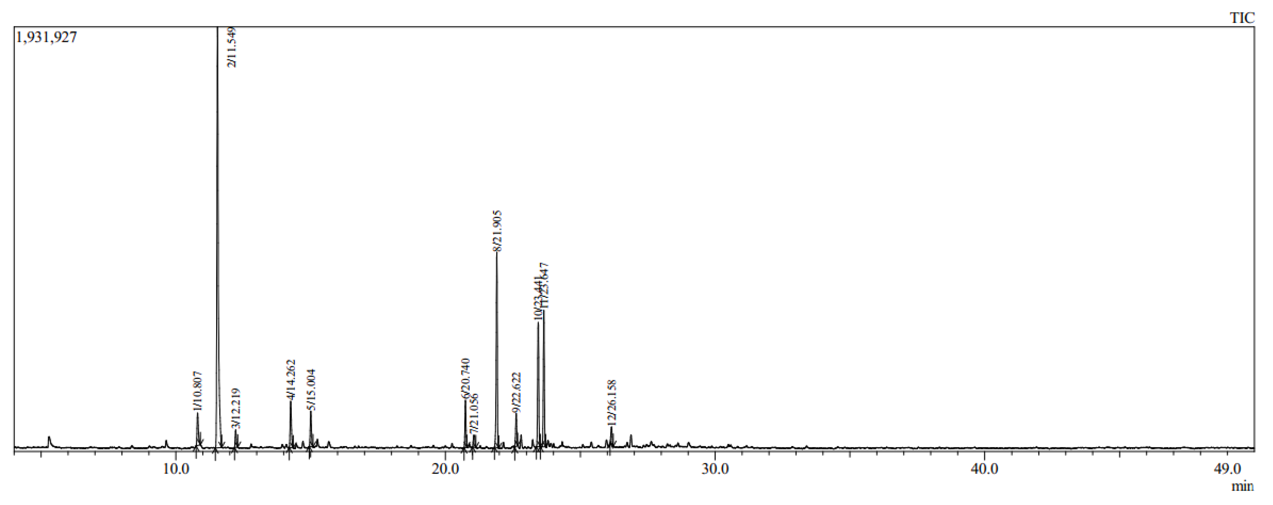
2.3. Analysis of the Chemical Composition by Use of GCMS

GC-MS analysis was used to identify the chemical composition of EOAA. Briefly, after diluting the EOAA in hexane (10:100 dilution), 1 µL was used for the identification and quantification of constituents by the use of gas chromatography–mass spectrometry (GC-MS-TQ8040 NX, brand Shimadzu, Tokyo, Japan), equipped with an apolar capillary column (RTxi-5 Sil MS-30m × 0.25 mm ID × 0.25 µm). The initial oven temperature program was maintained at 50 °C for two minutes and ramped at 5 °C/min until 160 °C for 2 min, then ramped at 5 °C/min up to 280 °C for 2 min. Notably, the analysis time was 50 min, and high-purity helium was employed as the carrier gas at a flow rate of 1 mL/min, with a split ratio of 1:20. The injection temperature was held at 250 °C, while that of the detector was held at 280 °C [29]. The injected volume of the EO sample was 1 μL, and the ion source temperature operated at 200 °C with an electron ionization energy of 70 eV utilizing a spectral range of m/z 40–650. Chemical compounds found in the EOAA were identified through the comparison of their mass spectra with those found in the MS library (using the NIST-MS Search Version 2.0 program). Additionally, their Kovats index was compared with the reference Adams database [30].

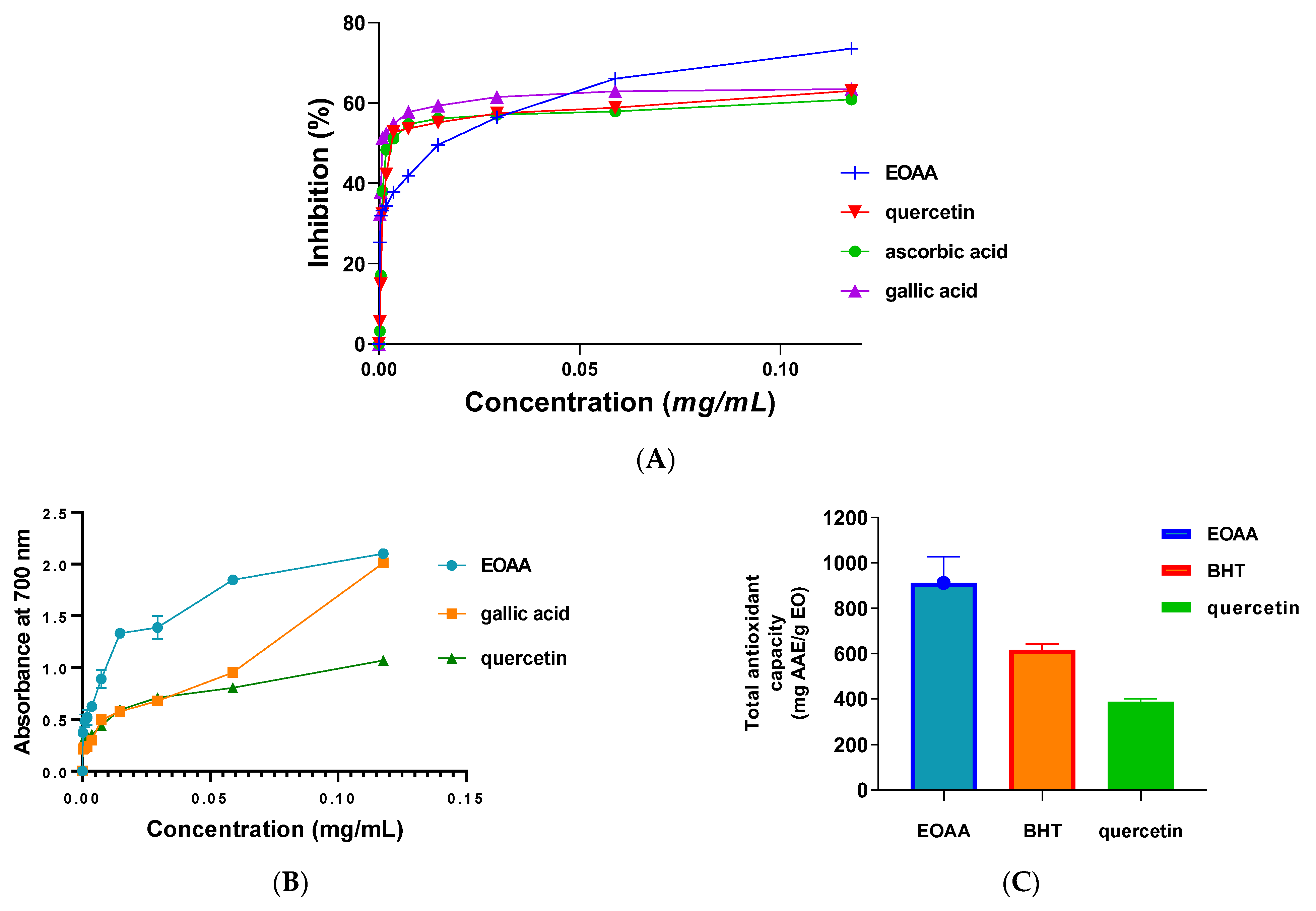
2.4. Antioxidant Activity

2.4.1. DPPH Test

The DPPH assay was performed according to the method of Shams Moattar and co-authors, with slight modifications. [31] was used for the DPPH test. Briefly, from a methanolic solution of EOAA, a dilution series was prepared, and 100 µL of each of its concentrations was taken and placed in an Eppendorf tube. After that, 1000 μL of a methanolic solution of DPPH (0.005%) was added. Then, the mixture was stirred and incubated in the darkness for 40 min. Finally, the optical density was well measured at 517 nm using a Shimadzu 160-UV spectrophotometer virus a blank (negative control). Ascorbic acid, gallic acid, and quercetin were used as standards. DPPH free radical inhibition was determined as follows:
P I   % = A 0 A A 0 × 100
PI: Percentage of inhibition,
A0: Absorbance of the control,
A: Absorbance of EOAA

2.4.2. Ferric Reducing Antioxidant Power (FRAP) Test

The reducing power of EOAA was ascertained following our previously described procedure with slight modifications [27]. Briefly, 0.05 mL of EOAA were added to 0.2 mL of phosphate buffer (200mM, pH = 6.60) and 0.2 mL of (NH4)6Mo7O24 (1% each). After shaking, the solution was left in a water bath at 50 °C for 25 min. Next, 0.6 mL of water stirred with 0.12 mL of 0.1% FeCl3 and 0.2 mL of trichloroacetic acid (10% TCA). The absorbance of the mixture was then determined with a UV-visible spectrophotometer at 700 nm. Efficacy concentration (EC-50) was used to report results.

2.4.3. Total Antioxidant Capacity (TAC) Test

TAC of EOAA was conducted as follows; one milliliter of reagent prepared by use of sodium phosphate, sulfuric acid, and (NH4)6Mo7O24 was mixed with 0.025 mL of EOAA [32]. Next, the tubes were stirred, screwed, and incubated at 96 °C for 1 h and 25 min, and then they were allowed to cool at room temperature. The optical density of the mixture and positive controls was measured at 695 nm by means of a UV-visible spectrophotometer and compared to a blank consisting of 25 µL of methanol instead of EOAA. Butylhydroxytoluene (BHT) and quercetin were used as standards, and the TAC was expressed as milligrams of ascorbic acid equivalence per gram of EO (mg AAE/g EO).

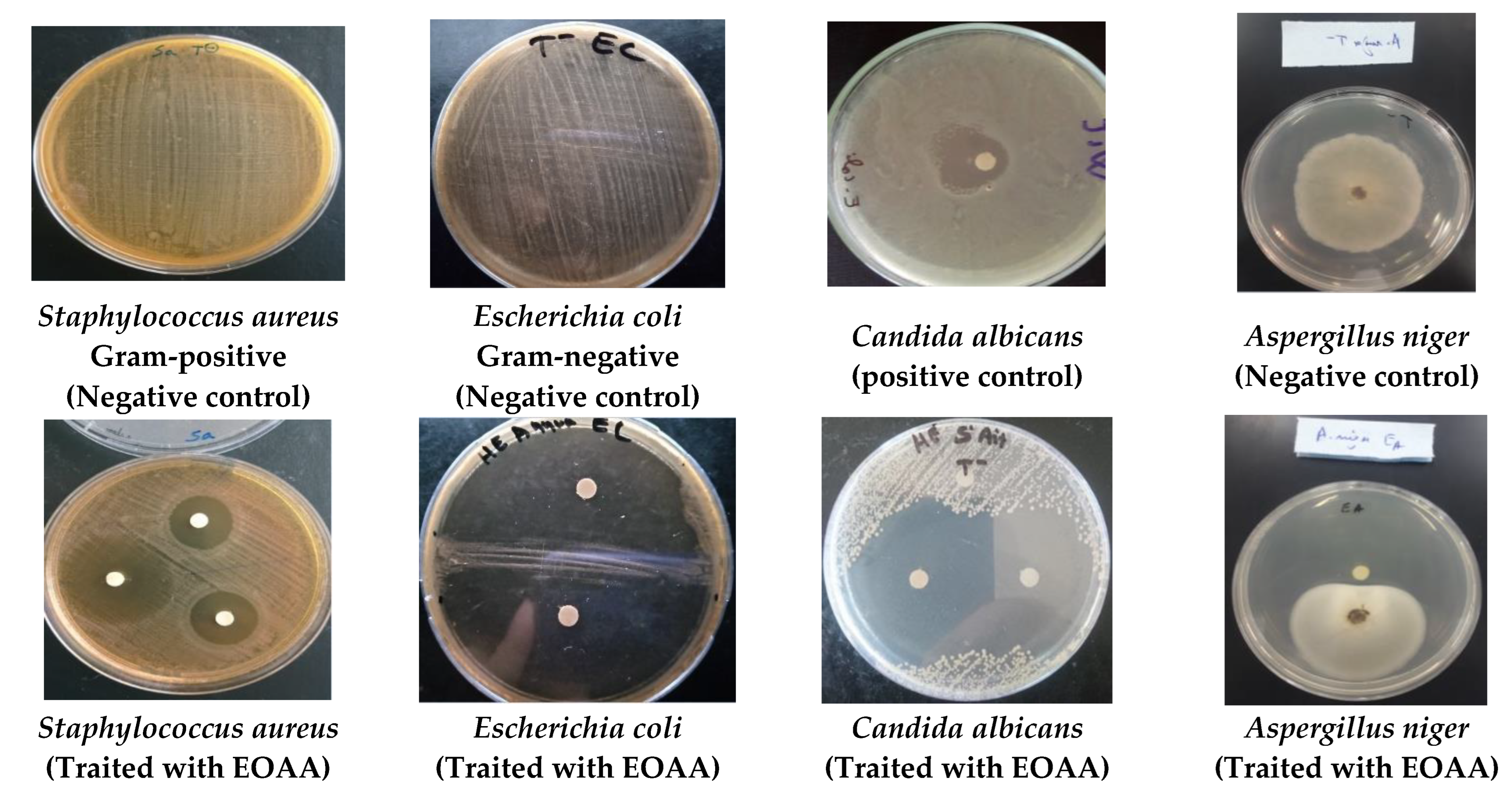
2.5. Antimicrobial Activity

2.5.1. Microbial Strains Tested

The antimicrobial activity of EOAA was performed against three fungal strains, which are Candida albicans (ATCC 10231), Aspergillus niger (MTCC 282), and Fusarium oxysporum (LBEAH/FS/17), as well as three Gram-negative bacterial strains, which are Escherichia coli (K12), Klebsiella pneumoniae, Salmonella sp., and three Gram-positive bacterial strains, which are Staphylococcus aureus (ATCC 6633), Bacillus subtilis (DSM 6333), and Bacillus cereus.

2.5.2. Agar Diffusion Method

The Agar diffusion method was used to assess the antimicrobial activity of EOAA as reported previously [33]. Briefly, bacterial and fungi suspensions were prepared to be 0.5 McFarland approximately 1–2 × 10⁵ CFU/mL. Next, bacteria and fungi were seeded in Petri dishes inoculated with Mueller-Hinton Agar (MHB) and yeast-peptone-glucose (YPG) extract media, respectively. After that, 6 mm Wattman N°4 paper discs were placed on the agar surface of Petri dishes and soaked with 10 µL of EOAA and kanamycin (233398) used as a positive control (30 µg). Then, the plates were placed in the dark and incubated for 24 h at 37 °C for bacterial strains and 48 h at 30 °C for fungal strains, especially, and Fusarium oxysporum and Aspergillus niger were incubated for seven days [34]. The zones of inhibition were measured and expressed in millimeters (mm).

2.5.3. Minimal Inhibitory Concentration (MIC)

To determine the MIC of EOAA against the aforementioned microbial strains, the microdilution method (96-well microplate) was used as described by Sadiki [35]. The emulsifier used (0.15% (w/v) bacterial agar) was mixed with Mueller-Hinton broth (MHB). EOAA was serially diluted in agar-supplemented broth at concentrations ranging from 870 to 1.4 µg/mL. Then, 50 μL of bacteria (106 CFU/mL) was applied. Finally, bacterial growth was visualized by turning resazurin from purple to pink.
Similarly for Candida albicans, the MIC values were determined according to the protocol described by Clinical and Laboratory Standards Institute (CLSI) [36]. The EOAA dilution was prepared in YPG broth containing 0.15% (w/v) agar. Then, the fungal inoculum (50 µL containing 103 CFU/mL) was added to each well of the plate, which was incubated at 30 °C for 48 h.
For the Fusarium oxysporum and Aspergillus niger strains, a similar protocol was followed, using 0.15% sauton agar as the culture medium and incubating for 72 h. The culture medium was 0.15% sauton agar. The experiments were repeated multiple times, and results are expressed as means [27].

2.6. Statistical Analysis

The data are displayed as the mean value and the standard deviation value (mean ± SD). In order to carry out the analysis, GraphPad Prism 9 was utilized. In order to make comparisons, we used analysis of variance (ANOVA), and then we used Tukey’s highly significant difference test. A statistically significant difference was thought to exist when the p-value was less than 0.05.

3. Results

3.1. Yield and Chemical Composition

The yield of Eos recovered from the aerial parts of A. annua was about 0.51%. This rate is comparable to that found in the genus Artemisia e.g., A. mesatlantica, and A. annua, which grow in the Mediterranean area, and they have an essential oil yield of about 0.5% [37,38]. Similarly, A. aragonensis, A. negrei, and A. campestris have around 1.2% of the EOs [9,27,39]. In addition, twelve compounds were identified by GC-MS analysis with a predominance of artemisia ketone (43.19%) followed by a Caryophyllene (15.75%) and β-Selinene (10.32%). The gas chromatogram of EOAA is provided in Figure 1, and the detailed qualitative and quantitative chemical composition of EOAA is presented in Table 1.

3.2. Antioxidant Activity of EOAA

The effectiveness of the free radical scavenging capability of EOAA was tested with the DPPH assay. The results showed that EOAA possessed potent antioxidant power with an IC50 value of 29 ± 5.3 μg/mL, lower than of the positive controls, quercetin (IC50 = 43 ± 0.5 μg/mL), and ascorbic acid (IC50 = 42 ± 0.9 μg/mL). However, this value is higher than that of the antioxidant gallic acid (IC50 = 2.2 ± 0.6 μg/mL) (Figure 2A). Notably, a smaller value of IC50 indicates potent antioxidant activity of the tested agent [27]. Furthermore, according to the ferric reducing power assay (FRAP), EOAA exhibited an EC50 value in the range of 9.218 ± 0.3 µg/mL, while gallic acid and quercetin recorded EC50 values of 17.28 ± 0.4 µg/mL and 20.87 ± 1.1 µg/mL, respectively (Figure 2B). Besides, the total antioxidant capacity of EOAA was 911.59 ± 115.71 mg AAE/g EO, whereas quercetin and BHT used as antioxidant standards recorded 388.744 ± 12.289 and 616.244 ± 25 mg AAE/g EO, respectively (Figure 2C).

3.3. Antimicrobial Activity of EOAA

The results obtained from the antimicrobial screening of EOAA and standard drugs against Gram-negative bacteria, Gram-positive bacteria, and the fungi Fusarium oxysporum, Aspergillus niger, and Candida albicans are provided in Table 2, Table 3 and Table 4 and Figure 3. The tested microbial strains are characterized by their resistance and high pathogenicity [27,40]. The outcomes revealed strong antimicrobial capacity of EOAA against Gram-negative bacteria, especially Escherichia coli with an inhibition zone of 75.67 ± 2.05 mm and MIC of 5.34 ± 0.50 µg/mL (Table 2 and Table 3). Likewise, EOAA showed a MIC value of 1.09 ± 00 µg/mL and an inhibition zone of 16 ± 1 mm against the Salmonella sp. strain. However, Bacillus cereus as Gram-positive bacteria was less sensitive to EOAA, which had an inhibition zone of 11 ± 1 mm and a MIC value of 5.44 µg/mL (Table 2 and Table 3 and Figure 3).
The EOAA showed also significant activity against the fungal strains as compared to the antifungal drug. According to the findings, EOAA was effectively inhibited by the fungi Fusarium oxysporum, Aspergillus niger, and Candida albicans, with inhibition zones of 57.67 ± 2.08, 45.71 ± 4.06, and 45.67 ± 2.05 mm, respectively (Table 4). Similarly, the EOAA showed MICs values of 14.38 ± 2.37 µg/mL and 21.75 ± 0.34 µg/mL against Fusarium oxysporum and Aspergillus niger, respectively, while the MIC value obtained against Candida albicans was 3.12 ± 00 µg/mL (Table 4 and Figure 3). All bacterial strains tested showed resistance to the antibiotics used as positive controls. However, Fusarium oxysporum, Aspergillus niger, and Candida albicans showed sensitivity to fluconazole, with an inhibition zone of 23.33 ± 1.25, 36.12 ± 1.70 and 34.67 ± 2.62 mm and a MICs values of 2.78 ± 0.12, 2.01 ± 0.01 and 3.47 ± 0.05 µg/mL, respectively (Table 4 and Figure 3).

4. Discussion

The demand for essential oils derived from natural plants has increased over the years owing to their use as medicines against many severe diseases and their low toxicity [41]. For this purpose, we examined the antimicrobial and antioxidant properties of EOs from Artemisia annua L. indigenous to the Mediterranean area. The extraction of the EO that we carried out allowed us to obtain a yield of approximately 0.51%. We have noticed that the yield changes as a function of the use of the leaves or flowers, the site of culture, the origin of the seeds, and the moment of the harvest. Importantly, the variation of chemical compounds is generally influenced by the drying conditions, harvest season, geographical location, fertilization, soil pH, as well as the chemotype, the part of the plant used, genotype, or extraction method [42]. The analysis of EOAA showed a high content of oxygenated monoterpenes (55.35%) and hydrocarbon sesquiterpenes (13.43%). However, oxygenated monoterpenes (1.62%) were detected in lower quantity while recording the absence of oxygenated monoterpenes. Our results agree with a previous study by Verdian, which showed that oxygenated monoterpenes (83.7%) and sesquiterpenes (12.5%) constitute the major compounds in Iranian Artemisia annua EOs [43]. Additionally, we have observed that EOAA are rich in active substances, such as artemisia ketone (43.19%), caryophyllene (15.75%), β-selinene (10.32%), and germacrene D (9.56%) according to GC-MS analysis. Most of these components are present in the EOs extracted from the genus Artemisia collected in the Mediterranean area (A. arborescens, A. caerulescens subsp. and A. annua) [44,45], as well as in other countries, such as India, France, Turkey, and North America [46,47,48,49]. In addition, compounds in essential oils from A. mesatlantica endemic to Morocco are slightly similar to those revealed in EOAA [38,50]. Importantly, many compounds in EOAA are also detected in nearby plant species, including Artemisia herba-alba L., Artemisia pontica L., and Artemisia absinthium L, which is in agreement with our study. Notably, 1,8 cineol, camphor, and borneol are among the main compounds of EOAA, and they have been widely found in the EO of the genus Artemisia [51]. There are few reports on the analysis of EOs from Mediterranean plants in the literature [44]. Interestingly, due to the presence of different chemotypes within species, variations in essential oil concentration between subspecies have been reported. Notably, GC-MS showed some special compounds in Moroccan A. annua, which may confirm its indigenous to this country [52]. Thus, it seems that EO possesses a chemical polymorphism, with different chemotypes related to intrinsic and extrinsic factors of the species [53].
The presented result here agrees with those reported by Chirane et al., who showed that artemisia ketone detected in EOAA had a good antioxidant power when compared to BHT. Because of this fitting, the antioxidant potency of EOAA may be due to its richness in artemisia ketone [54]. Artemisia ketone showed higher activity than 1,8-cineole and camphor in the DPPH test, as well as in the recorded literature [45,55]. Previous studies showed that the antioxidant effect of EOs is frequently related to their major compounds [56,57]. The synergistic effect of minor chemicals in EOs can determine their antioxidant powder of EO [27,58]. In general, OEs rich in oxygenated monoterpenes have a more pronounced antiradical activity than those with hydrocarbon terpenes [52]. Importantly, the antioxidant results are in agreement with those found in previous works, where it was reported that the genus Artemisia has promising antioxidant activity by all bioassays used, such as DPPH, β-carotene bleaching, and total antioxidant capacity [9].
Concerning the antibacterial power, our results are comparable with those reported elsewhere [59,60], which showed that EOs from the genus Artemisia possess antimicrobial potential. EOAA showed better antibacterial activity when compared to Iranian EOAA [61]. The antimicrobial action demonstrated by EOAA is mostly related to its high content of oxygenated monoterpene and hydrocarbon sesquiterpenes (55.33 and 43.03% respectively), which are known for their antibacterial action [61,62]. The current results agreed with those reported by Habibi et al. [63], who showed that EOAA had significant antibacterial capacity against bacterial strains, particularly versus Escherichia coli, Staphylococcus aureus, and Bacillus subtilis. In addition, our findings are in accordance with the findings of a number of previous research efforts, such as the one conducted by Radulovic et al. [55], who reported that the compound artemisia ketone possessed antibacterial power vs. pathogenic microorganisms. Some studies indicated that Gram-positive bacteria are more sensitive to the effect of Eos, unlike Gram-positive bacteria [29]. The findings of the current research, on the other hand, suggested that the difference in the cell walls of Gram-positive and Gram-negative bacteria was not a significant factor in determining the antibacterial activity of EOAA, since Escherichia coli was the most sensitive to essential oil with a zone of inhibition of 75.67 mm and minimum inhibitory concentration of 5.34 μg/mL. These results may be explained by the broad-spectrum activity of EOAA. According to the previous findings, the essential oils of the genus Artemisia is effective against Gram-negative (Escherichia coli 57, Escherichia coli 97, Klebsiella pneumonia, and Pseudomonas aeruginosa) and Gram-positive (Staphylococcus aureus) bacteria, with maximum inhibition zones ranging from 18–37 mm and MIC values ranging from 3.25 to 12.5 mg/mL, respectively [9].
The antibacterial properties of EOs can be attributed to their lipophilic nature, which allows them to effectively infiltrate into bacteria cells. In this regard, it was found that hydrocarbons in EOs (43.03% of EOAA) are predominantly accumulated in cytoplasmic membranes, which leads to impaired membrane permeability and, ultimately, the rapid death of microorganisms [64]. The phytochemicals in EOAA (artemisia ketone, caryophyllene, β-selinene and germacrene D) may act synergistically, rather than individually, as previous research showed that the antibacterial activity of EOs is more potent than their compounds examined individually [65,66].
The antifungal activity of EOAA can be explained by the synergistic effect between the different chemical substances of essential oil, notably artemisia ketone, caryophyllene, and β-selinene, which are often responsible for the antifungal activity, as well as the minority compounds that can also contribute significantly to this activity [67]. Some studies reported that the antimicrobial activity of EOs may be more important than that of their major compounds when tested separately [58]. Importantly, the activity of the essences is frequently resultant from synergic effect of minor components in EOs. Moreover, a proportional correlation between the presence of oxygenated terpenes and the antimicrobial potency is reported in the work of Agour [68], which is in agreement with our results regarding oxygenated monoterpenes in EOAA, found to be 55.35%.

5. Conclusions

The EO extracted from Artemisia annua grown in the Middle Atlas region of Morocco showed potent antioxidant and antimicrobial activities against antibiotic resistant microbes. The remarkable bioactivities observed can be attributed to the chemicals found in the EO, especially the main constituents, such as artemisia ketones (43.19%), caryophyllene (15.75%), β-selinene (10.32%), and germacrene D (9.56%). Altogether, the explored EOAA can be considered as a possible alternative to combat drug-resistant microbes and oxidative stress. Additionally, it will be crucial to conduct in vitro and in vivo studies on other biological targets, as well as to further assess any possible adverse effects on non-target organisms.

Author Contributions

Conceptualization, K.C., Z.B.O. and S.L.; methodology, A.E.M.; software, M.B. and R.G.; validation, S.L.; formal analysis H.-A.N., Z.B.O. and M.B; investigation, Y.A.B.J. and A.E.M.; resources, M.B.; data curation, A.E.M. and K.C.; writing—original draft preparation, M.B.; review and editing the revised version, improve the manuscript, M.C.; writing—review and editing, H.-A.N.; visualization, R.G.; supervision, R.G.; project administration, S.L. and Y.A.B.J.; funding acquisition, R.G. All authors have read and agreed to the published version of the manuscript.

Funding

This work is financially supported by the Researchers Supporting Project (RSP2023R457). King Saud University, Riyadh, Saudi Arabia.

Institutional Review Board Statement

Not applicable.

Informed Consent Statement

Not applicable.

Data Availability Statement

Not applicable.

Conflicts of Interest

The authors declare that there is no conflict of interest.

References

  1. Bloom, D.E.; Cadarette, D. Infectious disease threats in the twenty-first century: Strengthening the global response. Front. Immunol. 2019, 10, 549. [Google Scholar] [CrossRef] [Green Version]
  2. Vouga, M.; Greub, G. Emerging bacterial pathogens: The past and beyond. Clin. Microbiol. Infect. 2016, 22, 12–21. [Google Scholar] [CrossRef] [PubMed] [Green Version]
  3. Catalano, A.; Iacopetta, D.; Ceramella, J.; Scumaci, D.; Giuzio, F.; Saturnino, C.; Aquaro, S.; Rosano, C.; Sinicropi, M.S. Multidrug resistance (MDR): A widespread phenomenon in pharmacological therapies. Molecules 2022, 27, 616. [Google Scholar] [CrossRef] [PubMed]
  4. Pisoschi, A.M.; Pop, A.; Iordache, F.; Stanca, L.; Predoi, G.; Serban, A.I. Oxidative stress mitigation by antioxidants-an overview on their chemistry and influences on health status. Eur. J. Med. Chem. 2021, 209, 112891. [Google Scholar] [CrossRef] [PubMed]
  5. Gilgun-Sherki, Y.; Melamed, E.; Offen, D. Oxidative stress induced-neurodegenerative diseases: The need for antioxidants that penetrate the blood brain barrier. Neuropharmacology 2001, 40, 959–975. [Google Scholar] [CrossRef]
  6. Aruoma, O.I. Free radicals, oxidative stress, and antioxidants in human health and disease. J. Am. Oil Chem. Soc. 1998, 75, 199–212. [Google Scholar] [CrossRef] [PubMed]
  7. AlSalhi, M.S.; Elumalai, K.; Devanesan, S.; Govindarajan, M.; Krishnappa, K.; Maggi, F. The aromatic ginger Kaempferia galanga L.(Zingiberaceae) essential oil and its main compounds are effective larvicidal agents against Aedes vittatus and Anopheles maculatus without toxicity on the non-target aquatic fauna. Ind. Crops Prod. 2020, 158, 113012. [Google Scholar] [CrossRef]
  8. El Moussaoui, A.; Bourhia, M.; Jawhari, F.Z.; Salamatullah, A.M.; Ullah, R.; Bari, A.; Majid Mahmood, H.; Sohaib, M.; Serhii, B.; Rozhenko, A.; et al. Chemical Profiling, Antioxidant, and Antimicrobial Activity against Drug-Resistant Microbes of Essential Oil from Withania frutescens L. Appl. Sci. 2021, 11, 5168. [Google Scholar] [CrossRef]
  9. Chebbac, K.; Moussaoui, A.E.; Bourhia, M.; Salamatullah, A.M.; Alzahrani, A.; Guemmouh, R. Chemical analysis and antioxidant and antimicrobial activity of essential oils from Artemisia negrei L. Against drug-resistant microbes. Evid. -Based Complement. Altern. Med. 2021, 2021, 5902851. [Google Scholar] [CrossRef]
  10. Chaachouay, N.; Douira, A.; Hassikou, R.; Brhadda, N.; Dahmani, J.; Belahbib, N.; Ziri, R.; Zidane, L. Mr Chaachouay Noureddine Sous le thème" Etude floristique et ethnomédicinale des plantes aromatiques et médicinales dans le Rif (Nord du Maroc). Ph.D. Thesis, Département de Biologie-Université Ibn Tofail-Kénitra, Kenitra, Morocco, 2020. [Google Scholar]
  11. Tahri, N.; El Basti, A.; Zidane, L.; Rochdi, A.; Douira, A. Etude ethnobotanique des plantes medicinales dans la province de Settat (Maroc). Kast. Univ. J. For. Fac. 2012, 12, 192–208. [Google Scholar]
  12. Yuan, H.; Ma, Q.; Ye, L.; Piao, G. The traditional medicine and modern medicine from natural products. Molecules 2016, 21, 559. [Google Scholar] [CrossRef] [PubMed] [Green Version]
  13. Gruessner, B.M.; Cornet-Vernet, L.; Desrosiers, M.R.; Lutgen, P.; Towler, M.J.; Weathers, P.J. It is not just artemisinin: Artemisia sp. for treating diseases including malaria and schistosomiasis. Phytochem. Rev. 2019, 18, 1509–1527. [Google Scholar] [CrossRef] [PubMed]
  14. Nigam, M.; Atanassova, M.; Mishra, A.P.; Pezzani, R.; Devkota, H.P.; Plygun, S.; Salehi, B.; Setzer, W.N.; Sharifi-Rad, J. Bioactive compounds and health benefits of Artemisia species. Nat. Prod. Commun. 2019, 14, 1934578X19850354. [Google Scholar]
  15. Abad, M.J.; Bedoya, L.M.; Apaza, L.; Bermejo, P. The Artemisia L. genus: A review of bioactive essential oils. Molecules 2012, 17, 2542–2566. [Google Scholar] [CrossRef] [Green Version]
  16. Jaradat, N.; Qneibi, M.; Hawash, M.; Al-Maharik, N.; Qadi, M.; Abualhasan, M.N.; Ayesh, O.; Bsharat, J.; Khadir, M.; Morshed, R.; et al. Assessing Artemisia arborescens essential oil compositions, antimicrobial, cytotoxic, anti-inflammatory, and neuroprotective effects gathered from two geographic locations in Palestine. Ind. Crops Prod. 2022, 176, 114360. [Google Scholar] [CrossRef]
  17. Hinane, D.; Oubaha, S.; Satrani, B.; Ghanmi, M.; Bourkhiss, B. Domestication of the endemic plant of Morocco and high added value: Artemisia herba alba. J. Mater. Environ. Sci. 2020, 11, 283–292. [Google Scholar]
  18. Şenkal, B.C.; Kiralan, M.; Yaman, C. The Effect of Different Harvest Stages on Chemical Composition and Antioxidant Capacity of Essential Oil from Artemisia annua L. Artemisia annua L.’dan Elde Edilen Uçucu Yağın Kimyasal Kompozisyonu ve Antioksidan Kapasitesi Üzerine Farklı Hasat Dönemlerin. J. Agric. Sci. 2015, 21, 71–77. [Google Scholar]
  19. Howyzeh, M.S.; Noori, S.A.S.; Shariati, V. Essential oil profiling of Ajowan (Trachyspermum ammi) industrial medicinal plant. Ind. Crops Prod. 2018, 119, 255–259. [Google Scholar] [CrossRef]
  20. Trendafilova, A.; Moujir, L.M.; Sousa, P.M.C.; Seca, A.M.L. Research advances on health effects of edible artemisia species and some sesquiterpene lactones constituents. Foods 2021, 10, 65. [Google Scholar] [CrossRef]
  21. Ur Rashid, M.; Alamzeb, M.; Ali, S.; Ullah, Z.; Shah, Z.A.; Naz, I.; Khan, M.R. The chemistry and pharmacology of alkaloids and allied nitrogen compounds from Artemisia species: A review. Phytother. Res. 2019, 33, 2661–2684. [Google Scholar] [CrossRef]
  22. Verma, R.K.; Chauhan, A.; Verma, R.S.; Gupta, A.K. Influence of planting date on growth, artemisinin yield, seed and oil yield of Artemisia annua L. under temperate climatic conditions. Ind. Crops Prod. 2011, 34, 860–864. [Google Scholar] [CrossRef]
  23. He, G.; Sun, H.; Liao, R.; Wei, Y.; Zhang, T.; Chen, Y.; Lin, S. Effects of herbal extracts (Foeniculum vulgare and Artemisia annua) on growth, liver antioxidant capacity, intestinal morphology and microorganism of juvenile largemouth bass, Micropterus salmoides. Aquac. Rep. 2022, 23, 101081. [Google Scholar] [CrossRef]
  24. Ćavar, S.; Maksimović, M.; Vidic, D.; Parić, A. Chemical composition and antioxidant and antimicrobial activity of essential oil of Artemisia annua L. from Bosnia. Ind. Crops Prod. 2012, 37, 479–485. [Google Scholar] [CrossRef]
  25. Talman, A.M.; Clain, J.; Duval, R.; Ménard, R.; Ariey, F. Artemisinin bioactivity and resistance in malaria parasites. Trends Parasitol. 2019, 35, 953–963. [Google Scholar] [CrossRef] [PubMed]
  26. Malhotra, A.; Rawat, A.; Prakash, O.; Kumar, R.; Srivastava, R.M.; Kumar, S. Chemical composition and pesticide activity of essential oils from Artemisia annua L. harvested in the rainy and winter seasons. Biochem. Syst. Ecol. 2023, 107, 104601. [Google Scholar] [CrossRef]
  27. Chebbac, K.; Ghneim, H.K.; El Moussaoui, A.; Bourhia, M.; El Barnossi, A.; Benziane Ouaritini, Z.; Salamatullah, A.M.; Alzahrani, A.; Aboul-Soud, M.A.; Giesy, J.P.; et al. Antioxidant and Antimicrobial Activities of Chemically-Characterized Essential Oil from Artemisia aragonensis Lam. against Drug-Resistant Microbes. Molecules 2022, 27, 1136. [Google Scholar] [CrossRef]
  28. Boutabia, L.; Telailia, S.; Guenadil, F.; Chefrour, A. Chemical composition and antibacterial activity of essential oils from Mentha pulegium L. and Mentha suaveolens Ehrh. growing in North-East of Algeria. An. Univ. Din Oradea Fasc. Biol. 2020, 2, 143–148. [Google Scholar]
  29. Lafraxo, S.; El Barnossi, A.; El Moussaoui, A.; Bourhia, M.; Salamatullah, A.M.; Alzahrani, A.; Akka, A.A.; Choubbane, A.; Akhazzane, M.; Aboul-Soud, M.A.M.; et al. Essential Oils from Leaves of Juniperus thurifera L., Exhibiting Antioxidant, Antifungal and Antibacterial Activities against Antibiotic-Resistant Microbes. Horticulturae 2022, 8, 321. [Google Scholar] [CrossRef]
  30. Adams, D.R.P. Identification of Essential Oil Components by Gas Chromatography/Mass Spectrometry, 4th ed.; Texensis Publishing: Devon, UK, 2017; p. 811. [Google Scholar]
  31. Moattar, F.S.; Sariri, R.; Yaghmaee, P.; Giahi, M. Enzymatic and Non-Enzymatic Antioxidants of Calamintha Officinalis Moench Extracts. J. Appl. Biotechnol. Rep. 2016, 3, 489–494. [Google Scholar]
  32. Lafraxo, S.; El Moussaoui, A.A.; Bin Jardan, Y.; El Barnossi, A.; Chebaibi, M.; Baammi, S.; Akka, A.A.; Chebbac, K.; Akhazzane, M.; Chelouati, T.; et al. GC-MS Profiling, In Vitro Antioxidant, Antimicrobial, and In Silico NADPH Oxidase Inhibition Studies of Essential Oil of Juniperus thurifera Bark. Evid. Based Complement. Altern. Med. 2022, 2022, e6305672. [Google Scholar] [CrossRef]
  33. Suurbaar, J.; Mosobil, R.; Donkor, A.-M. Antibacterial and antifungal activities and phytochemical profile of leaf extract from different extractants of Ricinus communis against selected pathogens. BMC Res. Notes 2017, 10, 660. [Google Scholar] [CrossRef] [PubMed] [Green Version]
  34. Sarker, S.D.; Nahar, L.; Kumarasamy, Y. Microtitre plate-based antibacterial assay incorporating resazurin as an indicator of cell growth, and its application in the in vitro antibacterial screening of phytochemicals. Methods 2007, 42, 321–324. [Google Scholar] [CrossRef] [PubMed]
  35. Sadiki, M.; Elabed, A.; Elaabedy, A.; Elabed, A.; Farah, A.; Iraqui, M.; Koraichi, S.I. Characterization and antibacterial activity of the essential oil from Thymus vulgaris cultivated in morocco (Taounate) against ten bacteria. World J. Pharm. Res. 2015, 4, 314–325. [Google Scholar]
  36. Wayne, P. Clinical and Laboratory Standards Institute: Reference method for broth dilution antifungal susceptibility testing of yeasts; approved standard. CLSI Doc. M27-A3 Suppl. S 2008, 3, 6–12. [Google Scholar]
  37. Juteau, F.; Masotti, V.; Bessière, J.M.; Dherbomez, M.; Viano, J. Antibacterial and antioxidant activities of Artemisia annua essential oil. Fitoterapia 2002, 73, 532–535. [Google Scholar] [CrossRef] [PubMed]
  38. Bencheqroun, H.K.; Ghanmi, M.; Satrani, B.; Aafi, A. Activité antimicrobienne des huiles essentielles d’Artemisia mesatlantica, plante endémique du Maroc. Bull. Société R. Des Sci. Liège 2012, 81, 18. [Google Scholar]
  39. Akrout, A.; El Jani, H.; Amouri, S.; Neffati, M. Screening of antiradical and antibacterial activities of essential oils of Artemisia campestris L., Artemisia herba alba asso, & thymus capitatus hoff. Et link. Growing wild in the southern of Tunisia. Recent Res. Sci. Technol. 2009, 2, 29–39. [Google Scholar]
  40. Thomas, V.M.; Brown, R.M.; Ashcraft, D.S.; Pankey, G.A. Synergistic effect between nisin and polymyxin B against pandrug-resistant and extensively drug-resistant Acinetobacter baumannii. Int. J. Antimicrob. Agents 2019, 53, 663–668. [Google Scholar] [CrossRef]
  41. Chabosseau, S.; Derbré, S. Cancer du sein: Recommandations sur l’usage de la phytothérapie. Actual. Pharm. 2016, 55, 45–49. [Google Scholar] [CrossRef] [Green Version]
  42. Akrout, A.A.; Chemli, R.; Simmonds, M.; Kite, G.; Hammami, M.; Chreif, I. Seasonal variation of the essential oil of Artemisia campestris L. J. Essent. Oil Res. 2003, 15, 333–336. [Google Scholar] [CrossRef]
  43. Verdian, R.M.; Sadat, E.E.; Haji, A.A.; Fazeli, M.R.; Pirali, H.M. Chemical composition and antimicrobial activity of Artemisia annua L. essential oil from Iran. J. Med. Plant 2008, 7, 58–62. [Google Scholar]
  44. Ornano, L.; Venditti, A.; Ballero, M.; Sanna, C.; Quassinti, L.; Bramucci, M.; Lupidi, G.; Papa, F.; Vittori, S.; Maggi, F.; et al. Chemopreventive and antioxidant activity of the chamazulene-rich essential oil obtained from Artemisia arborescens L. growing on the Isle of La Maddalena, Sardinia, Italy. Chem. Biodivers. 2013, 10, 1464–1474. [Google Scholar] [CrossRef]
  45. Zhigzhitzhapova, S.V.; Dylenova, E.P.; Gulyaev, S.M.; Randalova, T.E.; Taraskin, V.V.; Tykheev, Z.A.; Radnaeva, L.D. Composition and antioxidant activity of the essential oil of Artemisia annua L. Nat. Prod. Res. 2020, 34, 2668–2671. [Google Scholar] [CrossRef] [PubMed]
  46. Soylu, E.M.; Yiğitbaş, H.; Tok, F.M.; Soylu, S.; Kurt, Ş.; Baysal, Ö.; Kaya, A.D. Chemical composition and antifungal activity of the essential oil of Artemisia annua L. against foliar and soil-borne fungal pathogens/Die chemische Zusammensetzung und antimikrobielle Aktivität das ätherischen Öls von Artemisia annua L. gegen blatt-und bodenbürtige pilzliche Krankheitserreger. Z. Für Pflanzenkrankh. Und Pflanzenschutz/J. Plant Dis. Prot. 2005, 112, 229–239. [Google Scholar]
  47. Chalchat, J.-C.; Garry, R.-P.; Lamy, J. Influence of harvest time on yield and composition of Artemisia annua oil produced in France. J. Essent. Oil Res. 1994, 6, 261–268. [Google Scholar] [CrossRef]
  48. Charles, D.J.; Cebert, E.; Simon, J.E. Characterization of the essential oil of Artemisia annua L. J. Essent. Oil Res. 1991, 3, 33–39. [Google Scholar] [CrossRef]
  49. Ahmad, A.; Misra, L.N. Terpenoids from Artemisia annua and constituents of its essential oil. Phytochemistry 1994, 37, 183–186. [Google Scholar] [CrossRef]
  50. Burt, S. Essential oils: Their antibacterial properties and potential applications in foods—A review. Int. J. Food Microbiol. 2004, 94, 223–253. [Google Scholar] [CrossRef]
  51. Balouiri, M.; Sadiki, M.; Ibnsouda, S.K. Methods for in vitro evaluating antimicrobial activity: A review. J. Pharm. Anal. 2016, 6, 71–79. [Google Scholar] [CrossRef] [Green Version]
  52. Woerdenbag, H.J.; Bos, R.; Salomons, M.C.; Hendriks, H.; Pras, N.; Malingré, T.M. Volatile constituents of Artemisia annua L.(Asteraceae). Flavour Fragr. J. 1993, 8, 131–137. [Google Scholar] [CrossRef]
  53. Maggio, A.; Rosselli, S.; Bruno, M.; Spadaro, V.; Raimondo, F.M.; Senatore, F. Chemical composition of essential oil from Italian populations of Artemisia alba Turra (Asteraceae). Molecules 2012, 17, 10232–10241. [Google Scholar] [CrossRef] [PubMed]
  54. Chirane, M.S.; Benchabane, O.; Bousbia, N.; Zenia, S. Antioxydant and antimicrobial activities of essential oil and ethanol extract of Santolina chamaecyparissus L. Rev. Agrobiol. 2019, 9, 1660–1668. [Google Scholar]
  55. Radulović, N.S.; Randjelović, P.J.; Stojanović, N.M.; Blagojević, P.D.; Stojanović-Radić, Z.Z.; Ilić, I.R.; Djordjević, V.B. Toxic essential oils. Part II: Chemical, toxicological, pharmacological and microbiological profiles of Artemisia annua L. volatiles. Food Chem. Toxicol. 2013, 58, 37–49. [Google Scholar] [CrossRef] [PubMed]
  56. Tepe, B.; Sokmen, M.; Akpulat, H.A.; Daferera, D.; Polissiou, M.; Sokmen, A. Antioxidative activity of the essential oils of Thymus sipyleus subsp. sipyleus var. sipyleus and Thymus sipyleus subsp. sipyleus var. rosulans. J. Food Eng. 2005, 66, 447–454. [Google Scholar]
  57. Baser, K.H.C.; Buchbauer, G. Essential Oils Science, Technology, and Applications; CRC Press: Boca Raton, FL, USA, 2010; Volume 10, p. 9781420063165–c3. [Google Scholar]
  58. Bakkali, F.; Averbeck, S.; Averbeck, D.; Idaomar, M. Biological effects of essential oils–A review. Food Chem. Toxicol. 2008, 46, 446–475. [Google Scholar] [CrossRef]
  59. Rani, S.J.; Supraja, P.; Sujitha, A.; Kiranmayee, P.; Usha, R. Evaluation of antibacterial and antifungal activity of Artemisia annua during pre and post flowering stages. Int. J. Curr. Res. 2015, 7, 21581–21587. [Google Scholar]
  60. Kumar, A.R.; Rathinam, K.M.S. Antibacterial activity of extracts of Artemesia annua. Int. J. Res. Pharm. Chem. 2013, 3, 376–377. [Google Scholar]
  61. Gorris, L.G.; Smid, E.J. Bactericidal activity of carvacrol towards the food-borne pathogen Bacillus cereus. J. Appl. Microbiol. 1998, 85, 211–218. [Google Scholar]
  62. Ultee, A.; Slump, R.A.; Steging, G.; Smid, E.J. Antimicrobial activity of carvacrol toward Bacillus cereus on rice. J. Food Prot. 2000, 63, 620–624. [Google Scholar] [CrossRef]
  63. Habibi, Z.; Ghanian, S.; Ghasemi, S.; Yousefi, M. Chemical composition and antibacterial activity of the volatile oil from seeds of Artemisia annua L. from Iran. Nat. Prod. Res. 2013, 27, 198–200. [Google Scholar] [CrossRef]
  64. Amrati, F.E.-Z.; Bourhia, M.; Saghrouchni, H.; Slighoua, M.; Grafov, A.; Ullah, R.; Ezzeldin, E.; Mostafa, G.A.; Bari, A.; Ibenmoussa, S.; et al. Caralluma europaea (Guss.) NE Br.: Anti-inflammatory, antifungal, and antibacterial activities against nosocomial antibiotic-resistant microbes of chemically characterized fractions. Molecules 2021, 26, 636. [Google Scholar] [CrossRef] [PubMed]
  65. Yu, Z.; Tang, J.; Khare, T.; Kumar, V. The alarming antimicrobial resistance in ESKAPEE pathogens: Can essential oils come to the rescue? Fitoterapia 2020, 140, 104433. [Google Scholar] [CrossRef]
  66. Bilia, A.R.; Santomauro, F.; Sacco, C.; Bergonzi, M.C.; Donato, R. Essential Oil of Artemisia annua L.: An Extraordinary Component with Numerous Antimicrobial Properties. Evid. Based Complement. Altern. Med. 2014, 2014, 159819. [Google Scholar] [CrossRef] [Green Version]
  67. Mssillou, I.; Agour, A.; Slighoua, M.; Tourabi, M.; Nouioura, G.; Lyoussi, B.; Derwich, E. Phytochemical characterization, antioxidant activity, and in vitro investigation of antimicrobial potential of Dittrichia viscosa L. leaf extracts against nosocomial infections. Acta Ecol. Sin. 2021, 42, 661–669. [Google Scholar] [CrossRef]
  68. Agour, A.; Mssillou, I.; Saghrouchni, H.; Bari, A.; Lyoussi, B.; Derwich, E. Chemical composition, antioxidant potential and antimicrobial Properties of the essential Oils of Haplophyllum tuberculatum (Forsskal) A. Juss from Morocco. Trop. J. Nat. Prod. Res. 2021, 4, 1108–1115. [Google Scholar]
Figure 1. Chromatographic profile of EOAA profiled by GC-MS analysis.
Life 13 00807 g001
Figure 2. Antioxidant capacity using DPPH (A), FRAP (B), and TAC assays (C).
Life 13 00807 g002
Figure 3. Photo displaying the effects of EOAA on the tested bacteria and fungi (the number of discs in the Petri dishes signifies the number of repetitions).
Life 13 00807 g003
Table 1. Chemical constituents identified in EOAA by GC/MS analysis.
PR.T.NameArea%R. I.M.W.
(g/mol)
Chemical StructureChemical Classes
LitObs
110.8071,8-cineole3.2110311059154Life 13 00807 i001M.O
211.549artemisia ketone43.1910481042152Life 13 00807 i002M.O
312.219artemisia alcohol1.4810711068154Life 13 00807 i003M.O
414.262camphor4.4111271121152Life 13 00807 i004M.O
515.004borneol3.0711521138154.25Life 13 00807 i005M.O
620.740copaene3.5213631221204Life 13 00807 i006S.H
721.056γ-cadinene1.2514901435204Life 13 00807 i007S.H
821.905caryophyllene15.7514401494204Life 13 00807 i008S.H
922.622β-farnesene2.6214421440204Life 13 00807 i009S.H
1023.441germacrene D9.5614911515204Life 13 00807 i010S.H
1123.647β-selinene10.3214731469204Life 13 00807 i011S.H
1226.158caryophyllene oxide1.621549150720Life 13 00807 i012S.O
Chemical classes
Oxygenated Monoterpenes (M.O) 55.35%
Oxygenated Sesquiterpenes (S.O) 1.62%
Sesquiterpene Hydrocarbons (S.H) 43.03%
Total Identification 100%
P: Peak; R.T: Retention time; Obs: Observed; Lit: Literature; R.I: Retention index. M.W.: Molecular weight.
Table 2. Antibacterial activity of EOAA and Kanamycine by use of inhibition zone diameter (mm).
CompoundGram-Positive BacteriaGram-Negative Bacteria
BacillussubtilisBacillus cereusStaphylococcus aureusEscherichia coliKlebsiella pneumoniaeSalmonella sp.
EOAA37.0 ± 0.0 a11.0 ± 1.0 d23.33 ± 5.86 c75.67 ± 2.05 e22.33 ± 1.15 c16.0 ± 1.0 d
KanRst bRst bRst bRst bRst bRst b
Rst: Resistant (inhibition zone diameter ≤ 15 mm); Kan: Kanamycine; EOAA: A. annua essential oil. Values that have the same sign (a, b, c, …) do not represent a significant difference.
Table 3. Antibacterial activity of EOAA and Kanamycin by use of minimum inhibitory concentration (µg/mL).
CompoundGram-Positive BacteriaGram-Negative Bacteria
B. subtilisB. cereusStaphylococcus aureusEscherichia coliK. pneumoniaSalmonella sp.
EOAA 5.64 ± 0.97 a5.44 ± 0.0 a4.98 ± 1.82 b5.34 ± 0.5 a5.44 ± 0.0 a1.09 ± 0.0 d
Kan 2.65 ± 0.45 c2.75 ± 0.01 c2.44 ± 0.64 c1.78 ± 0.35 d2.33 ± 0.27 c1.04 ± 0.0 d
Kan: Kanamycine; EOAA: A. annua essential oil. Values that have the same sign (a, b, c, …) do not represent a significant difference.
Table 4. Antifungal activity of EOAA and fluconazole by use of minimum inhibitory concentration (MIC) and inhibition zone diameter.
MIC (μg/mL)Inhibition Diameter (mm)
Fungal StrainsEOAAFluEOAAFlu
Candida albicans3.12 ± 0.00 a3.47 ± 0.05 a45.67 ± 2.05 c34.67 ± 2.62 e
Fusarium oxysporum14.38 ± 2.37 b2.78 ± 0.12 a57.67 ± 2.08 d23.33 ± 1.25 f
Aspergillus niger21.75 ± 0.34 f2.01 ± 0.01 a45.71 ± 4.06 c36.12 ± 1.70 e
Flu: Fluconazole; EOAA: A. annua essential oil. Values with the same sign (a, b, c, …) do not represent a significant difference.
Disclaimer/Publisher’s Note: The statements, opinions and data contained in all publications are solely those of the individual author(s) and contributor(s) and not of MDPI and/or the editor(s). MDPI and/or the editor(s) disclaim responsibility for any injury to people or property resulting from any ideas, methods, instructions or products referred to in the content.

