
Emerging Role of Plant-Based Dietary Components in Post-Translational Modifications Associated with Colorectal Cancer


1
Department of Health Sciences, Faculty of Experimental Sciences, University of Jaen, Campus las Lagunillas s/n, 23071 Jaen, Spain
2
University Institute of Research in Olive Groves and Olive Oils, University of Jaen, Campus las Lagunillas s/n, 23071 Jaen, Spain
3
Department of Experimental Biology, Faculty of Experimental Sciences, University of Jaen, Campus las Lagunillas s/n, 23071 Jaen, Spain
*
Authors to whom correspondence should be addressed.
Life 2023, 13(2), 264; https://doi.org/10.3390/life13020264
Submission received: 30 December 2022
/
Revised: 12 January 2023
/
Accepted: 16 January 2023
/
Published: 18 January 2023
(This article belongs to the Collection The Role of Bioactive Natural Compounds in the Treatment of Diseases)
Abstract
:Colorectal cancer (CRC) is one of the most common cancers worldwide. Its main modifiable risk factors are diet, alcohol consumption, and smoking. Thus, the right approach through lifestyle changes may lead to its prevention. In fact, some natural dietary components have exhibited chemopreventive activity through modulation of cellular processes involved in CRC development. Although cancer is a multi-factorial process, the study of post-translational modifications (PTMs) of proteins associated with CRC has recently gained interest, as inappropriate modification is closely related to the activation of cell signalling pathways involved in carcinogenesis. Therefore, this review aimed to collect the main PTMs associated with CRC, analyse the relationship between different proteins that are susceptible to inappropriate PTMs, and review the available scientific literature on the role of plant-based dietary compounds in modulating CRC-associated PTMs. In summary, this review suggested that some plant-based dietary components such as phenols, flavonoids, lignans, terpenoids, and alkaloids may be able to correct the inappropriate PTMs associated with CRC and promote apoptosis in tumour cells.
Keywords:
cancer risk factor; flavonoids; lignans; phenols; phosphorylation; prognosis; natural products1. Introduction
Colorectal cancer (CRC) is currently the second type of cancer with the highest mortality rate in the population according to Global Cancer Statistics 2020 [1]. Metastatic CRC has a poor prognosis, with less than a 15% of five-year survival rate [2]. Its carcinogenesis is a process of many years of development and some early life risk factors are important contributors [3]. Among them, cigarette smoking, obesity, and a sedentary lifestyle are closely related to CRC incidence [4,5]. However, its quickly increasing incidence is mainly due to lifestyle westernization associated with changes in dietary behaviour such as heavy alcohol consumption and diets rich in sugars, saturated fats, and red and processed meat [6]. Thus, some protective lifestyle factors against CRC include a diet rich in minerals and vitamins, dairy, dietary fibre, fish, vegetables, and fruits. An alternative strategy for CRC prevention is the use of a chemopreventive supplement providing greater individual exposure to some nutrients than can be obtained from the diet (such as phytochemicals) [7].
The pathogenesis of CRC is a complex multi-stage process which includes gut microbiota imbalances, cell DNA disruption, and carcinogenic signalling pathways activation [8]. The aetiology underlying the mechanism of action of specific nutrients in CRC has been mainly attributed to their anti-inflammatory and antioxidant properties, and their modulation of gut microbiota populations, maintaining gut homeostasis and regulating the host immune response [9,10]. However, their effects on epigenetic modulation associated with CRC pathogenesis remains unknown. There is increasing evidence that the disruption of epigenetic control over gene expression has an important role in carcinogenesis [11,12,13,14]. Together with non-coding RNAs and DNA methylation, histone and protein post-translational modifications (PTMs) have an important role in carcinogenesis and gene regulation [15,16]. PTMs occur once the mRNA has been translated into the protein sequence in the ribosomes and produce marginal chemical modifications to lipoproteins and native proteins. Among these modifications, PTMs may mark proteins for degradation, inhibit or promote interactions with other proteins, redirect cellular protein localization, and modify enzyme activity [17,18]. Most PTMs are reversible, so normal cells use them as a switch to control proliferating or quiescent cells [19]. The role of PTMs in the onset and progression of diseases such as cancer has been investigated. Their involvement in the process of carcinogenesis could be due to their function in processes such as the cell cycle, cell survival, and cell proliferation [20]. Therefore, PTM-focused analysis of enzyme phosphorylation and the involvement of protein kinases in cancer formation and progression have led to the use of PTM-based therapeutic approaches (i.e., tyrosine kinase inhibitors) [21,22]. Furthermore, in the case of CRC, PTMs develop key role-playing as a tight junction protein and regulate the epithelial barrier function [23,24]. Thus, PTMs may be essential to work with the external impact and could provide an excellent opportunity for intervention through feeding and promoting clinical strategies for CRC patients regarding predictive, preventive, and personalized medicine. Overall, the aims of this article were (1) to summarize the main PTMs involved in CRC development; (2) to identify the connection between the main PTMs involved in CRC via the Search Tool for the Retrieval of Interacting Proteins 11 (STRING 11) [25]; and (3) to review the plant-based dietary components that can modulate these modifications.
2. Post-Translational Modifications in Colorectal Cancer
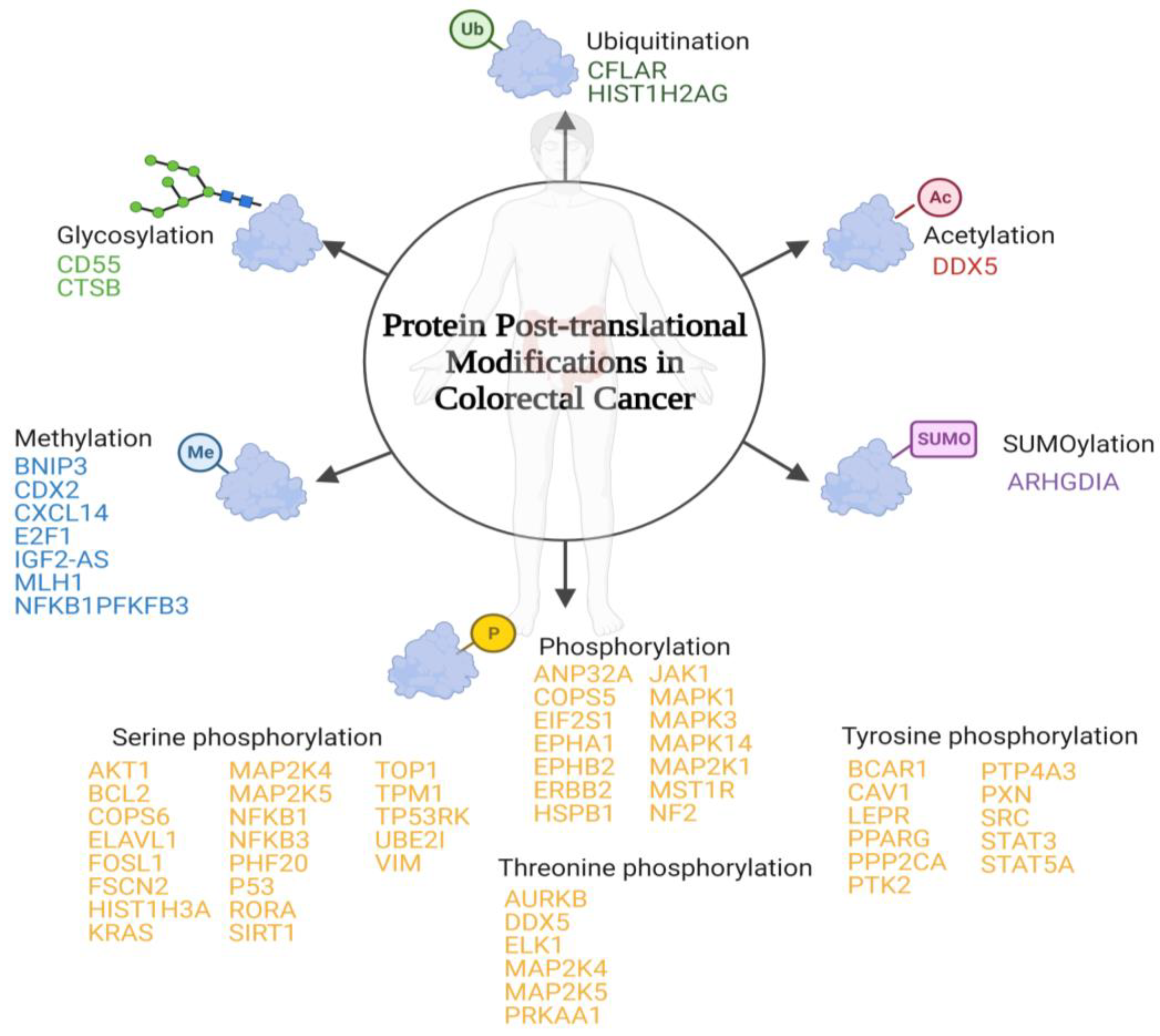
PTMs are protein-specific modifications that control many physiological processes to ensure the dynamic and quick response of cells to intracellular and extracellular stimuli [26]. Any proteome protein may be modified post-translationally or during translation. These reversible modifications may alter not only the protein’s stability, conformation, and charge state, but also its function modulating its intracellular conformation, its interactions, and the life span of the target protein [27]. In some cases, PTMs are inadequate and modulate positively some signal transduction pathways that are involved in tumourigenesis regulation and cancer development [28]. To date, more than 450 unique protein modifications have been described, including ubiquitination, acylation, SUMOylation, methylation, and phosphorylation [29]. In the case of CRC, the most important modifications involved have been summarized below (Figure 1).
2.1. SUMOylation
2.2. Glycosylation
A carbohydrate is attached to specific proteins. In mammals, there are two types: (1) O-glycosylation, where glycosyl groups are connected to tyrosine, hydroxylysine, serine, or threonine side chains with glycosidic linkages by glycosyltransferases, and (2) N-glycosylation, where glycosyl groups are connected to Asn side chains with amide linkages by oligosaccharyltransferase [32,33]. The upregulation of this PTM in complement decay-accelerating factor and cathepsin B has been identified in tumour tissue samples of CRC patients (Table 1) [34,35].
2.3. O-GlcNAcylation
There is a covalent attachment of N-acetylglucosamine residue O-linked to the hydroxyl group of threonine and serine residues of multiple cytosolic and nuclear proteins [36,37]. The upregulation of O-GlcNAcylation in ATP-dependent RNA helicase DDX5 has been associated with CRC in cell lines and murine models (Table 1) [38].
2.4. Ubiquitination
There is an attachment of ubiquitin molecules to the lysine residue of the substrate proteins. This process is based on an enzymatic cascade of ubiquitin-activating, ubiquitin-conjugating, and ubiquitin-ligase enzymes [39,40]. There have been two ubiquitination-susceptible proteins identified that are related to CRC (Table 1): (1) caspase homolog that is an apoptosis regulator [41] and (2) histone H2A type 1 that is involved in chromosomal stability, DNA replication, and DNA repair [42].

Table 1.
Post-translational glycosylation, O-GlcNAcylation, SUMOylation, and ubiquitination associated with CRC.
Table 1.
Post-translational glycosylation, O-GlcNAcylation, SUMOylation, and ubiquitination associated with CRC.
| Protein Name | Gene Name | PTM | PTM Site | Type | Ref |
|---|---|---|---|---|---|
| Rho GDP-dissociation inhibitor 1 | ARHGDIA | SUMOylation | K138 | Downregulated | [31] |
| Complement decay-accelerating factor | CD55 | O-linked glycosylation | NA | Upregulated | [34] |
| Cathepsin B | CTSB | Glycosylation | NA | Upregulated | [35] |
| Probable ATP-dependent RNA helicase DDX5 | DDX5 | O-GlcNAcylation | NA | Upregulated | [38] |
| Caspase homolog | CFLAR | Ubiquitination | K195 | Upregulated | [41] |
| Histone H2A type 1 | HIST1H2AG | Ubiquitination | NA | Upregulated | [42] |
NA: data not available; K: lysine.
2.5. Methylation
Methylation occurs mainly in arginine or lysine residues. One of the most biologically important roles of methylation is in histone modification [43]. Among the different proteins that suffer dysregulated post-translational methylation associated with CRC (Table 2), the one that is involved in cell growth suppression has downregulated methylation (putative insulin-like growth factor 2 antisense gene protein) [44,45]. The other proteins identified have an upregulated methylation, among them are (1) BCL2/adenovirus E1B 19 kDa protein-interacting protein 3 that is involved in apoptosis [46]; (2) homeobox protein CDX-2 that is involved in the transcriptional regulation of different genes expressed in the intestine [47]; (3) C-X-C motif chemokine 14 that is involved in immunoregulatory and inflammatory processes [48]; (4) transcription factor E2F1 that participates in the cell cycle [49]; (5) DNA mismatch repair protein Mlh1 that participates in DNA repair [50]; (6) nuclear factor NF-kappa-B p105 subunit that is a pleiotropic transcription factor involved in several signal transduction events which are initiated by stimuli such as oxidative stress or inflammation [51]; and (7) 6-phosphofructo-2-kinase/fructose-2,6-bisphosphatase 3 that is an essential protein for cell cycle progression and apoptosis prevention [52].
Table 2.
Post-translational methylation associated with CRC.
| Protein Name | Gene Name | PTM Site | Type | Ref |
|---|---|---|---|---|
| BCL2/adenovirus E1B 19 kDa protein-interacting protein 3 | BNIP3 | NA | Upregulated | [53] |
| Homeobox protein CDX-2 | CDX2 | NA | Upregulated | [54] |
| C-X-C motif chemokine 14 | CXCL14 | T72 | Upregulated | [55] |
| Transcription factor E2F1 | E2F1 | K109/111/113 | Upregulation | [56] |
| Putative insulin-like growth factor 2 antisense gene protein | IGF2-AS | NA | Downregulated | [45] |
| DNA mismatch repair protein Mlh1 | MLH1 | NA | Upregulated | [57] |
| Nuclear factor NF-kappa-B p105 subunit | NFKB1 | K218/221 | Upregulated | [58] |
| 6-phosphofructo-2-kinase/fructose-2,6-bisphosphatase 3 | PFKFB3 | R131/134 | Upregulated | [59] |
NA: data not available; K: lysine; R: arginine.
2.6. Phosphorylation
Phosphorylation is the most prevalent and widely studied type of PTM. It is inversely regulated by phosphatases and protein kinases in the amino acids’ hydroxyl tyrosine, threonine, or serine [32,60]. In the case of CRC, inadequate PTMs have been identified in the following proteins (Table 3): (1) acidic leucine-rich nuclear phosphoprotein 32 family member A that is involved in cell growth [61]; (2) COP9 signalosome complex subunit 5 that develops an important role in the degradation of cyclin-dependent kinase inhibitor [62]; (3) eukaryotic translation initiation factor 2 subunit 1 that is a translation initiation factor [63]; (4) ephrin type-A receptor 1 and ephrin type-B receptor 2 that are members of the ephrin receptor subfamily of the protein tyrosine kinase family [64]; (5) receptor tyrosine-protein kinase erbB-2 that is a member of the epidermal growth factor receptor family [65]; (6) heat shock protein beta-1 which plays an important role in cancer cells proliferation [66]; (7) tyrosine-protein kinase JAK1 that is a tyrosine kinase of the non-receptor type [67]; (8) mitogen-activated protein kinase 1, 3, and 14 that are serine/threonine kinases that are essential components of the MAP kinase signal transduction pathway [68,69,70,71]; (9) dual specificity mitogen-activated protein kinase kinase 1 which acts as an essential component of the MAP kinase signal transduction pathway [72]; (10) macrophage-stimulating protein receptor that is a tyrosine kinase receptor [73]; and (11) merlin that plays a pivotal role in tumour suppression through apoptosis promotion [74].
2.7. Serine Phosphorylation
Serine phosphorylation includes proto-oncogene c-Ak and Fos-related antigen 1 that regulates many processes including proliferation cell survival, growth, and angiogenesis [75,76]; apoptosis regulator Bcl-2 that is a regulator of apoptosis [77]; COP9 signalosome complex subunit 6 which is a component of the COP9 signalosome complex [78]; ELAV-like protein 1 that stabilizes mRNAs and regulates gene expression [79]; fascin-2 that acts as an actin bundling protein [80]; histone H3.1 which plays a central role in transcription regulation and DNA repair [81]; Kirsten rat sarcoma virus which is involved in the propagation of growth factors [82]; MAP kinase kinase 4 and 5 that are dual specificity protein kinase which act as an essential component of the MAP kinase signal transduction pathway [83,84]; NFKB1 and NFKB3 which are pleiotropic transcription factors involved in several signal transduction [85,86]; PHD finger protein 20 that contributes to p53 stabilization after DNA damage [87]; cellular tumour antigen p53 that acts as a tumour suppressor [88]; nuclear receptor ROR-alpha which is a key regulator of glucose metabolism [89]; sirtuin 1 that is an intracellular regulatory protein [90]; DNA topoisomerase 1 that releases the supercoiling tension of DNA introduced during the DNA replication [91]; tropomyosin-1 which is a member of the tropomyosin family of highly conserved proteins [92]; TP53-regulating kinase which is a protein kinase that phosphorylates ‘Ser-15’ of p53/TP53 protein [93]; SUMO-protein ligase that is essential for nuclear architecture and chromosome segregation [94]; and vimentin which is responsible for maintaining cell shape and stabilizing cytoskeletal interactions [95].
Table 4.
Post-translational serine, threonine, and tyrosine phosphorylation associated with CRC.
| Protein | Gene Name | PTM Site | Type | Ref |
|---|---|---|---|---|
| Serine Phosphorylation | ||||
| Proto-oncogene c-Akt | AKT1 | S473 | Upregulated | [75] |
| Apoptosis regulator Bcl-2 | BCL2 | S87 | Upregulated | [77] |
| COP9 signalosome complex subunit 6 | COPS6 | S148 | Upregulated | [78] |
| ELAV-like protein 1 | ELAVL1 | S318 | Upregulated | [79] |
| Fos-related antigen 1 | FOSL1 | S252; S265 | Upregulated | [76] |
| Fascin-2 | FSCN2 | S39 | Upregulated | [80] |
| Histone H3.1 | HIST1H3A | S28 | Upregulated | [81] |
| Kirsten rat sarcoma virus | KRAS | S181 | Upregulated | [82] |
| MAP kinase kinase 4 | MAP2K4 | S257 | Upregulated | [83] |
| MAP kinase kinase 5 | MAP2K5 | S311 | Upregulated | [84] |
| Nuclear factor NF-kappa-B p105 subunit | NFKB1 | S536 | Upregulated | [85] |
| Nuclear factor NF-kappa-B p65 subunit | NFKB3 | S276 | Upregulated | [86] |
| PHD finger protein 20 | PHF20 | S291 | Upregulated | [87] |
| Cellular tumour antigen p53 | P53 | S15 | Upregulated | [88] |
| Nuclear receptor ROR-alpha | RORA | S35 | Downregulated | [89] |
| Sirtuin 1 | SIRT1 | S27 | Upregulated | [90] |
| DNA topoisomerase 1 | TOP1 | S506 | Upregulated | [91] |
| Tropomyosin-1 | TPM1 | S283 | Upregulated | [92] |
| TP53-regulating kinase | TP53RK | S250 | Upregulated | [93] |
| SUMO-protein ligase | UBE2I | S71 | Upregulated | [94] |
| Vimentin | VIM | S72 | Upregulated | [95] |
| Threonine Phosphorylation | ||||
| Aurora kinase B | AURKB | T232 | Upregulated | [96] |
| Probable ATP-dependent RNA helicase DDX5 | DDX5 | T564/446 | Upregulated | [97] |
| ETS domain-containing protein Elk-1 | ELK1 | T417 | Upregulated | [98] |
| Dual specificity mitogen-activated protein kinase kinase 4 | MAP2K4 | T261 | Upregulated | [83] |
| MAP kinase kinase 5 | MAP2K5 | T315 | Upregulated | [84] |
| 5’-AMP-activated protein kinase catalytic subunit alpha-1 | PRKAA1 | T183 | Downregulated | [99] |
| Tyrosine Phosphorylation | ||||
| Breast cancer anti-estrogen resistance protein 1 | BCAR1 | Y12; Y128 | Upregulated | [100,101] |
| Caveolin-1 | CAV1 | Y14 | Upregulated | [102] |
| Leptin receptor | LEPR | Y1141 | Upregulated | [103] |
| Peroxisome proliferator-activated receptor gamma | PPARG | Y102 | Upregulated | [104] |
| Serine/threonine-protein phosphatase 2A catalytic subunit alpha isoform | PPP2CA | Y307 | Upregulated | [105] |
| Focal adhesion kinase 1 | PTK2 | Y397; Y407; Y925 | Downregulated | [106,107] |
| Protein tyrosine phosphatase type IVA 3 | PTP4A3 | Y53 | Upregulated | [108] |
| Paxillin | PXN | Y88 | Upregulated | [109] |
| Proto-oncogene tyrosine-protein kinase Src | SRC | Y419 | Upregulated | [110] |
| Signal transducer and activator of transcription 3 | STAT3 | Y705 | Upregulated | [111,112] |
| Signal transducer and activator of transcription 5A | STAT5A | Y694 | Downregulated | [113] |
S: serine; T: threonine; Y: tyrosine.
2.8. Threonine Phosphorylation
Threonine phosphorylation includes Aurora kinase B which is a serine/threonine-protein kinase component of the chromosomal passenger complex [96]; probable ATP-dependent RNA helicase DDX5 which is involved in the alternative regulation of pre-mRNA splicing [97]; ETS domain-containing protein Elk-1 which is a transcription factor that binds to purine-rich DNA sequences [98]; dual specificity mitogen-activated protein kinase kinase 4 which is an essential component of the MAP kinase signal transduction pathway [83]; MAP kinase kinase 5 that acts as a scaffold for the formation of a ternary MAP3K2/MAP3K3-MAP3K5-MAPK7 signalling complex [84]; and 5′-AMP-activated protein kinase catalytic subunit alpha-1 which is the catalytic subunit of AMP-activated protein kinase that plays a key role in regulating cellular energy metabolism [99].
2.9. Tyrosine Phosphorylation
Tyrosine phosphorylation includes breast cancer anti-estrogen resistance protein 1 which plays a central role in cell adhesion [100,101]; caveolin-1 that act as a scaffolding protein within caveolar membranes [102]; leptin receptor that mediates leptin central and peripheral effects [103]; peroxisome proliferator-activated receptor gamma that is a nuclear receptor [104]; serine/threonine-protein phosphatase 2A catalytic subunit alpha isoform which is the major phosphatase for microtubule-associated proteins [105]; focal adhesion kinase 1 which is a non-receptor protein-tyrosine kinase that plays an essential role in regulating cell migration and apoptosis [106,107]; protein tyrosine phosphatase type IVA 3 that stimulates progression from G1 into S phase during mitosis [108]; paxillin which is a cytoskeletal protein involved in actin-membrane attachment at sites of cell adhesion to the extracellular matrix [109]; proto-oncogene tyrosine-protein kinase Src that is a non-receptor protein tyrosine kinase [110]; signal transducer and activator of transcription 3 which mediates cellular responses to interleukins and other growth factors [111,112]; and signal transducer and activator of transcription 5A that is involved in signal transduction and activation of transcription [113].
3. Relationship between Post-Translational Modifications Associated with Colorectal Cancer
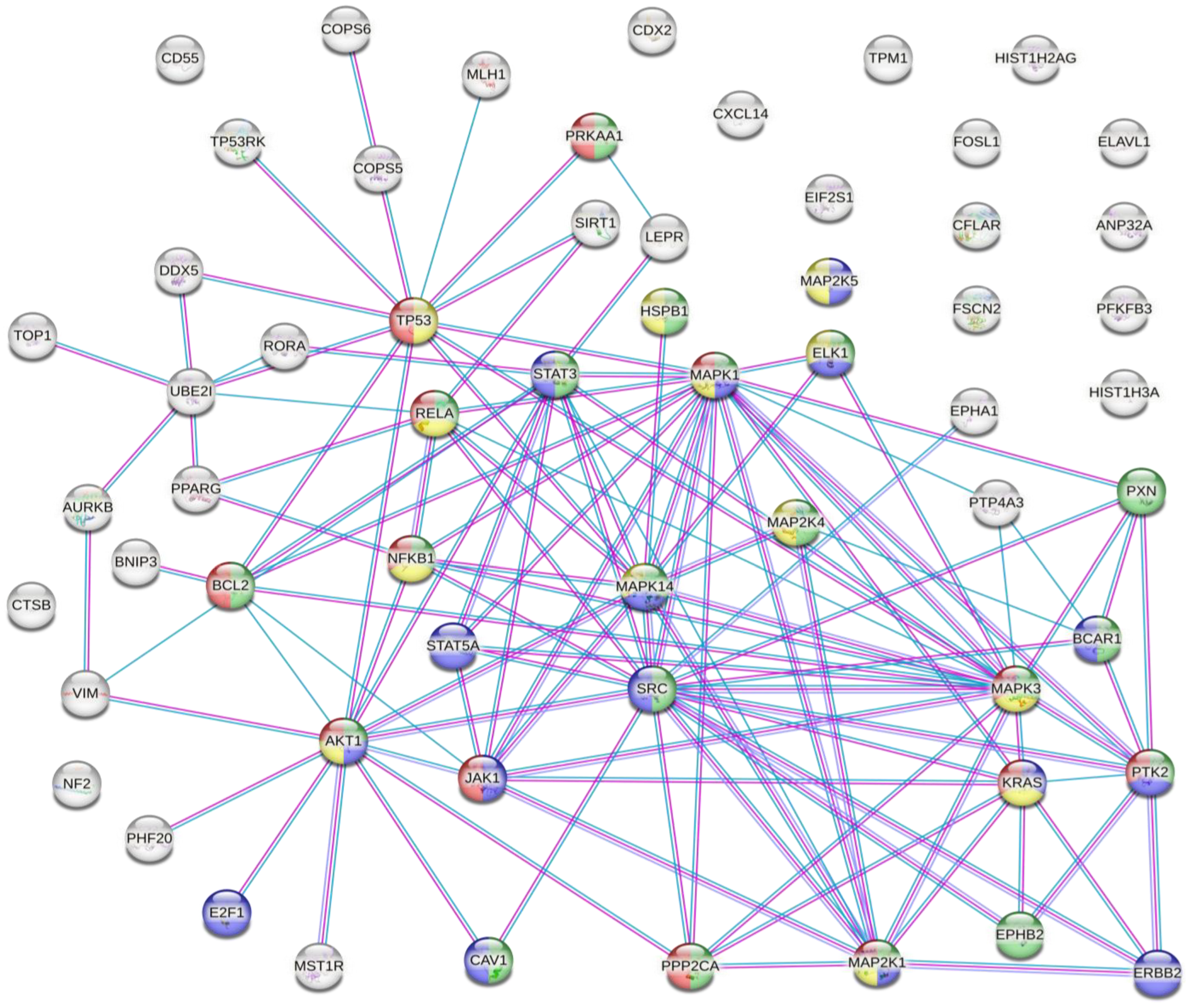
The results of the analysis showed that there were several interactions between some of the proteins susceptible to inappropriate PTMs associated with CRC (Figure 2).
This analysis showed that there were strong interactions between TP53, AKT1, STAT3, STAT5A, JAK1, MAPK1, MAPK14, MAP2K1, and SRC. In fact, this network had significantly more interactions than expected, which means that proteins have more interactions among themselves than what would be expected from a random set of proteins, demonstrating that the proteins may be partially biologically connected as a group. This group of proteins is mainly involved in the PI3K-Akt, EGF-EFGR, MAPK, and VEGFA-VEGFR2 signalling pathways. On the one hand, PI3K-Akt is the classical signalling pathway involved in glucose metabolism that promotes cancer metabolic reprogramming by elevation of aerobic glycolysis (known as the “Warburg effect”) [114,115]. Both EGF-EGFR and MAPK signalling pathways are involved in proliferation, differentiation, and apoptosis. Its regulation in cancer cells allows the maintenance of proliferative signalling, promoting cancer cell survival [60,116]. On the other hand, VEGF and its receptors (such as VEGFR2) develop an important role in tumour-associated angiogenesis. This process is essential for tumour progression because it favours oxygen and nutrient uptake by cancer cells [117,118]. Therefore, the main PTMs identified in CRC are involved in cancer progression and cancer cell survival. The next step was to identify how the inadequate PTMs in these proteins associated with CRC may be modulated by plant-based dietary components.
4. Nutrigenomic Effects of Plant-Based Dietary Components on Protein Post-Translational Modifications Associated with Colorectal Cancer
The available scientific literature showed several plant-based dietary components that may modulate CRC-associated PTMs (Table 5). These components can be grouped according to bioactive compounds as follows: phenols, flavonoids, lignans, terpenoids/alkaloids, vitamins, phytochemicals, and plant extracts.
Among the different articles shown in Table 5, 39% of the studies evaluated PTMs in STAT3, 18% of the studies measured PTMs in AKT, 18% of the studies analysed PTMs in p53, 9% of the studies measured PTMs in ERK, and the rest of the studies analyzed PTMs in other proteins such as AMPK, BCL2, CDX2, EGFR, JAK2, EphA1, EphB2, PI3K, SCR, MLH1, and Nf-Kβ. All the studies referred to post-translational phosphorylation, and four of them also mentioned post-translational ubiquitination, acetylation, and methylation.
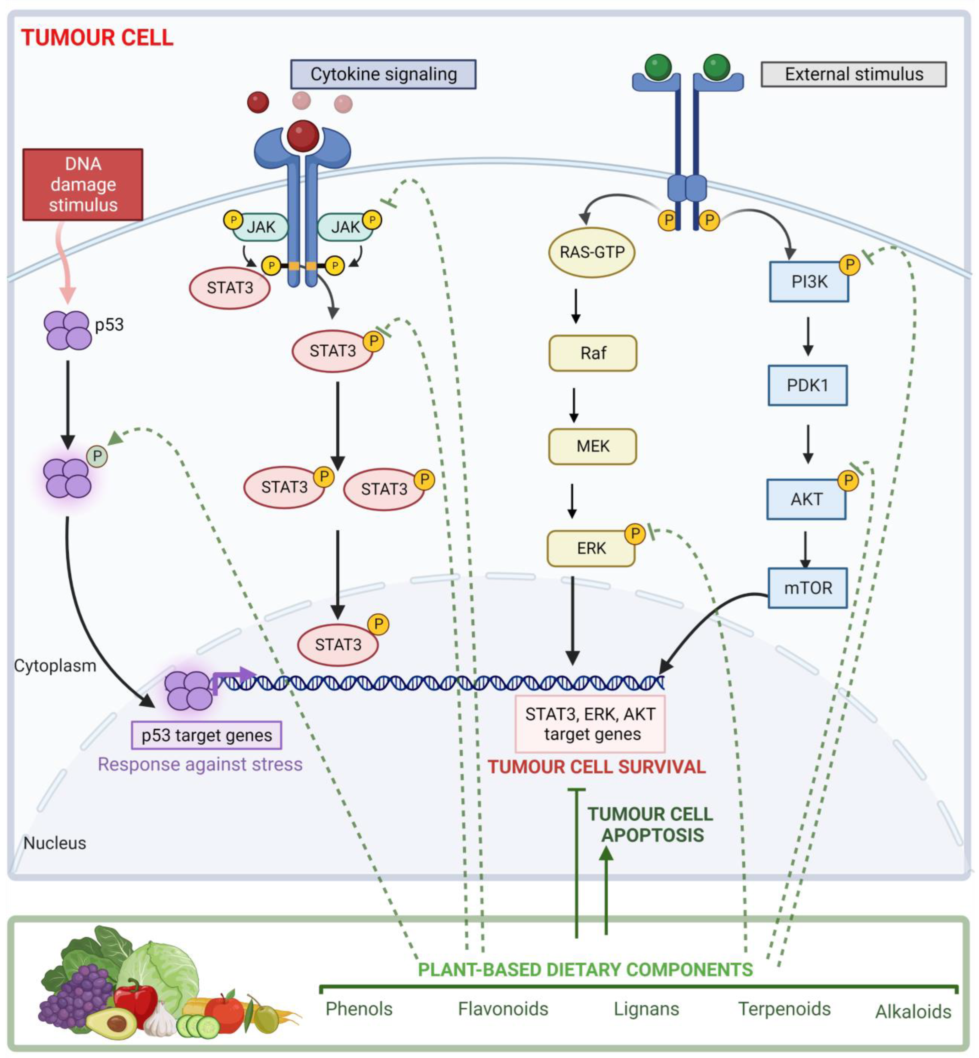
The PTMs induced by plant-based dietary components mainly consist of modulating those modifications observed in CRC (Figure 3). Concerning STAT3, tyrosine phosphorylation (Tyr 705) was upregulated in CRC. Several articles showed that some phenols, flavonoids, terpenoids, alkaloids, phytochemicals, and plant extracts were able to downregulate not only this phosphorylation but also STAT3 serine phosphorylation (Ser 727) in some cases [120,121,124,130]. Some of them are also involved in reduced phosphorylation of JAK2, which is upregulated in CRC [120,148,149,155,159]. Both proteins form the JAK/STAT signalling pathway, which has an important role in cytokine receptor signalling. In response to cytokines, its activation promotes immune cell division, survival, activation, and recruitment. This pathway not only participates in the immune response but also in the transcription of several genes involved in cell division and apoptosis regulation such as BCL2 [171,172].
In the case of AKT, serine phosphorylation (S473) was upregulated in CRC. Some studies revealed that harmine [151], ophiopogonin D [154], and luteolin [157] were able to downregulate this specific serine phosphorylation, while others such as coumarins [119], resveratrol [125], quercetin [140], secoisolariciresinol diglucoside [145], lycopene [158], and some plant extracts decrease protein phosphorylation [168,169]. Similarly, in PI3K and BCL2 proteins, quercetin, secoisolariciresinol diglucoside, and iodine-biofortified lettuce extract downregulated their phosphorylation, respectively [140,145,164]. On the other hand, delphinidin [136] and two different plant extracts [163,165] reverse carcinogen-induced phosphorylation of NF-kβ3 (Ser536). These proteins interact within the same pathway. The PI3K/AKT pathway participates in the modulation of cellular metabolism, cell growth, and apoptosis. PIK3K produces conformational changes and phosphorylation of AKT protein, inducing its activation. This cascade triggered the activation of NF-κB by enhancing the transcriptional activity of the p65 subunit, leading to apoptosis inhibition [173]. Likewise, the PI3K/AKT signalling pathway promotes the upregulation of Bcl-2 expression, which is considered an oncogene that inhibits apoptosis [174]. This suggests that plant-based dietary components may promote cancer cell apoptosis through downregulating AKT, PI3K, BCL2, and NF-kβ phosphorylation.
Another of the main proteins identified that can be modulated by plant-based dietary components is P53. This is considered a tumour suppressor involved in processes such as apoptosis, senescence, DNA repair, and cell cycle arrest [175]. The P53 pathway is activated against stress signals such as DNA damage. In response to this, P53 suffers from PTM and promotes the transcription of genes involved in cell response against stress [175]. These PTMs are mainly phosphorylation that is downregulated in CRC. However, several studies have shown that some plant-based dietary components such as phenols, flavonoids, terpenoids, and alkaloids could reverse it [123,126,128,135,137,139,140,146,147].
Concerning ERK1 and ERK2, both are part of a structurally related kinases family (MAPKs), whose signalling mechanism depends on an activating phosphorylation cascade. ERKs are central regulators of essential cellular functions such as cell differentiation, proliferation, migration, growth, survival, and metabolism [176]. Both proteins have post-translational phosphorylation upregulated in CRC, favouring cancer cell survival. As it has been shown in Table 5, some plant-based dietary components may downregulate their post-translational phosphorylation [130,156,160,161,166,169].
It is important to highlight that, due to the lack of human studies, the present review is focused on studies conducted in cells and murine models. Therefore, these findings cannot be transferable to other species. Future studies should address the connection between the modulation of PTMs associated with CRC elicited by plant-based dietary components in patients.
5. Conclusions
The available literature data suggest that different plant-based dietary components such as phenols, flavonoids, lignans, terpenoids, and alkaloids could prevent CRC development by targeting several molecular mechanisms such as the P53, JAK/STAT, PI3K/AKT, and ERK/MAPK pathways and affecting tumour behaviour through PTMs’ modulation in cell lines and murine models. The different signalling pathways affected during CRC development are mainly involved in cancer cell survival, and the main effects of plant-based dietary components are to promote apoptosis in tumour cells. Therefore, this could be a very interesting target for investigating the effect of supplementation based on these components as an adjuvant to chemotherapeutic, radiotherapeutic, and immunotherapeutic treatment in clinical trials. These findings will highlight the potential for precision nutrition strategies and the development of personalized nutritional plans in CRC treatment and may even serve as a basis for the development of dietary supplementation formulations for these patients to improve their prognosis and disease-free survival.
Author Contributions
Conceptualization, writing—original draft preparation, writing—review and editing, F.G.-S. and C.R.-G. All authors have read and agreed to the published version of the manuscript.
Funding
This research received no external funding.
Institutional Review Board Statement
Not applicable.
Informed Consent Statement
Not applicable.
Data Availability Statement
Not applicable.
Acknowledgments
Graphical Abstract, Figure 1 and Figure 3 were created with BioRender.com (accessed on 28 December 2022).
Conflicts of Interest
The authors declare no conflict of interest.
References
- Sung, H.; Ferlay, J.; Siegel, R.L.; Laversanne, M.; Soerjomataram, I.; Jemal, A.; Bray, F. Global Cancer Statistics 2020: GLOBOCAN Estimates of Incidence and Mortality Worldwide for 36 Cancers in 185 Countries. CA Cancer J. Clin. 2021, 71, 209–249. [Google Scholar] [CrossRef] [PubMed]
- Siegel, R.L.; Miller, K.D.; Goding Sauer, A.; Fedewa, S.A.; Butterly, L.F.; Anderson, J.C.; Cercek, A.; Smith, R.A.; Jemal, A. Colorectal Cancer Statistics, 2020. CA Cancer J. Clin. 2020, 70, 145–164. [Google Scholar] [CrossRef] [PubMed] [Green Version]
- Hughes, L.A.E.; van den Brandt, P.A.; Goldbohm, R.A.; de Goeij, A.F.P.M.; de Bruïne, A.P.; van Engeland, M.; Weijenberg, M.P. Childhood and Adolescent Energy Restriction and Subsequent Colorectal Cancer Risk: Results from the Netherlands Cohort Study. Int. J. Epidemiol. 2010, 39, 1333–1344. [Google Scholar] [CrossRef] [PubMed] [Green Version]
- Dacrema, M.; Ali, A.; Ullah, H.; Khan, A.; di Minno, A.; Xiao, J.; Martins, A.M.C.; Daglia, M. Spice-Derived Bioactive Compounds Confer Colorectal Cancer Prevention via Modulation of Gut Microbiota. Cancers 2022, 14, 5682. [Google Scholar] [CrossRef] [PubMed]
- Nimptsch, K.; Wu, K. Is Timing Important? The Role of Diet and Lifestyle during Early Life on Colorectal Neoplasia. Curr. Color. Cancer Rep. 2018, 14, 1–11. [Google Scholar] [CrossRef] [PubMed]
- Masdor, N.A.; Mohammed Nawi, A.; Hod, R.; Wong, Z.; Makpol, S.; Chin, S.-F. The Link between Food Environment and Colorectal Cancer: A Systematic Review. Nutrients 2022, 14, 3954. [Google Scholar] [CrossRef] [PubMed]
- Kim, S.H.; Moon, J.Y.; Lim, Y.J. Dietary Intervention for Preventing Colorectal Cancer: A Practical Guide for Physicians. J. Cancer Prev. 2022, 27, 139–146. [Google Scholar] [CrossRef]
- Chen, M.; Lin, W.; Li, N.; Wang, Q.; Zhu, S.; Zeng, A.; Song, L. Therapeutic Approaches to Colorectal Cancer via Strategies Based on Modulation of Gut Microbiota. Front. Microbiol. 2022, 13, 945533. [Google Scholar] [CrossRef]
- O’keefe, S.J.D. Diet, Microorganisms and Their Metabolites, and Colon Cancer. Nat. Rev. Gastroenterol. Hepatol. 2016, 13, 691–706. [Google Scholar] [CrossRef]
- Reddy, B.S. Diet and Colon Cancer: Evidence from Human and Animal Model Studies. In Diet, Nutrition, and Cancer: A Critical Evaluation; CRC Press: Boca Raton, FL, USA, 2018; pp. 47–66. ISBN 1351071408. [Google Scholar]
- Ullah, A.; Ullah, N.; Nawaz, T.; Aziz, T. Molecular Mechanisms of Sanguinarine in Cancer Prevention and Treatment. Anticancer Agents Med. Chem. 2022, 22. [Google Scholar] [CrossRef]
- Iqbal, H.; Menaa, F.; Khan, N.; Razzaq, A.; Khan, Z.; Ullah, K.; Kamal, R.; Sohail, M.; Thiripuranathar, G.; Uzair, B.; et al. Two Promising Anti-Cancer Compounds, 2-Hydroxycinnaldehyde and 2-Benzoyloxycinnamaldehyde: Where Do We Stand? Comb. Chem. High. Throughput Screen. 2022, 25, 808–818. [Google Scholar] [CrossRef] [PubMed]
- Su, Q.; Fan, M.; Wang, J.; Ullah, A.; Ghauri, M.A.; Dai, B.; Zhan, Y.; Zhang, D.; Zhang, Y. Sanguinarine Inhibits Epithelial–Mesenchymal Transition via Targeting HIF-1α/TGF-β Feed-Forward Loop in Hepatocellular Carcinoma. Cell Death Dis. 2019, 10, 939. [Google Scholar] [CrossRef] [PubMed] [Green Version]
- Ullah, A.; Leong, S.W.; Wang, J.; Wu, Q.; Ghauri, M.A.; Sarwar, A.; Su, Q.; Zhang, Y. Cephalomannine Inhibits Hypoxia-Induced Cellular Function via the Suppression of APEX1/HIF-1α Interaction in Lung Cancer. Cell Death Dis. 2021, 12, 490. [Google Scholar] [CrossRef] [PubMed]
- Hong, X.; Huang, H.; Qiu, X.; Ding, Z.; Feng, X.; Zhu, Y.; Zhuo, H.; Hou, J.; Zhao, J.; Cai, W. Targeting Posttranslational Modifications of RIOK1 Inhibits the Progression of Colorectal and Gastric Cancers. eLife 2018, 7, e29511. [Google Scholar] [CrossRef]
- Prieto, P.; Jaén, R.I.; Calle, D.; Gómez-Serrano, M.; Núñez, E.; Fernández-Velasco, M.; Martín-Sanz, P.; Alonso, S.; Vázquez, J.; Cerdán, S. Interplay between Post-Translational Cyclooxygenase-2 Modifications and the Metabolic and Proteomic Profile in a Colorectal Cancer Cohort. World J. Gastroenterol. 2019, 25, 433. [Google Scholar] [CrossRef]
- Das, T.; Shin, S.C.; Song, E.J.; Kim, E.E. Regulation of Deubiquitinating Enzymes by Post-Translational Modifications. Int. J. Mol. Sci. 2020, 21, 4028. [Google Scholar] [CrossRef]
- Kuwahara, H.; Nishizaki, M.; Kanazawa, H. Nuclear Localization Signal and Phosphorylation of Serine350 Specify Intracellular Localization of DRAK2. J. Biochem. 2008, 143, 349–358. [Google Scholar] [CrossRef]
- Chen, L.; Liu, S.; Tao, Y. Regulating Tumor Suppressor Genes: Post-Translational Modifications. Signal Transduct. Target. Ther. 2020, 5, 90. [Google Scholar] [CrossRef]
- Carter, A.M.; Tan, C.; Pozo, K.; Telange, R.; Molinaro, R.; Guo, A.; de Rosa, E.; Martinez, J.O.; Zhang, S.; Kumar, N. Phosphoprotein-Based Biomarkers as Predictors for Cancer Therapy. Proc. Natl. Acad. Sci. USA 2020, 117, 18401–18411. [Google Scholar] [CrossRef]
- Kwon, Y.W.; Jo, H.-S.; Bae, S.; Seo, Y.; Song, P.; Song, M.; Yoon, J.H. Application of Proteomics in Cancer: Recent Trends and Approaches for Biomarkers Discovery. Front. Med. (Lausanne) 2021, 8, 747333. [Google Scholar] [CrossRef]
- Hermann, J.; Schurgers, L.; Jankowski, V. Identification and Characterization of Post-Translational Modifications: Clinical Implications. Mol. Asp. Med. 2022, 86, 101066. [Google Scholar] [CrossRef]
- Dai Vu, L.; Gevaert, K.; de Smet, I. Protein Language: Post-Translational Modifications Talking to Each Other. Trends Plant Sci. 2018, 23, 1068–1080. [Google Scholar]
- Reiche, J.; Huber, O. Post-Translational Modifications of Tight Junction Transmembrane Proteins and Their Direct Effect on Barrier Function. Biochim. Biophys. Acta (BBA)-Biomembr. 2020, 1862, 183330. [Google Scholar] [CrossRef]
- Szklarczyk, D.; Gable, A.L.; Nastou, K.C.; Lyon, D.; Kirsch, R.; Pyysalo, S.; Doncheva, N.T.; Legeay, M.; Fang, T.; Bork, P.; et al. The STRING Database in 2021: Customizable Protein–Protein Networks, and Functional Characterization of User-Uploaded Gene/Measurement Sets. Nucleic Acids Res. 2021, 49, D605–D612. [Google Scholar] [CrossRef]
- Li, W.; Li, F.; Zhang, X.; Lin, H.-K.; Xu, C. Insights into the Post-Translational Modification and Its Emerging Role in Shaping the Tumor Microenvironment. Signal Transduct. Target. 2021, 6, 422. [Google Scholar] [CrossRef]
- Han, Z.-J.; Feng, Y.-H.; Gu, B.-H.; Li, Y.-M.; Chen, H. The Post-Translational Modification, SUMOylation, and Cancer. Int. J. Oncol. 2018, 52, 1081–1094. [Google Scholar] [CrossRef] [Green Version]
- Jaén, R.I.; Prieto, P.; Casado, M.; Martín-Sanz, P.; Boscá, L. Post-Translational Modifications of Prostaglandin-Endoperoxide Synthase 2 in Colorectal Cancer: An Update. World J. Gastroenterol. 2018, 24, 5454–5461. [Google Scholar] [CrossRef]
- Liu, N.; Ling, R.; Tang, X.; Yu, Y.; Zhou, Y.; Chen, D. Post-Translational Modifications of BRD4: Therapeutic Targets for Tumor. Front. Oncol. 2022, 12, 847701. [Google Scholar] [CrossRef]
- Celen, A.B.; Sahin, U. Sumoylation on Its 25th Anniversary: Mechanisms, Pathology, and Emerging Concepts. FEBS J. 2020, 287, 3110–3140. [Google Scholar] [CrossRef]
- Yu, J.; Zhang, D.; Liu, J.; Li, J.; Yu, Y.; Wu, X.-R.; Huang, C. RhoGDI SUMOylation at Lys-138 Increases Its Binding Activity to Rho GTPase and Its Inhibiting Cancer Cell Motility. J. Biol. Chem. 2012, 287, 13752–13760. [Google Scholar] [CrossRef] [Green Version]
- Wang, H.; Yang, L.; Liu, M.; Luo, J. Protein Post-Translational Modifications in the Regulation of Cancer Hallmarks. Cancer Gene Ther. 2022. [Google Scholar] [CrossRef] [PubMed]
- Moremen, K.W.; Tiemeyer, M.; Nairn, A.V. Vertebrate Protein Glycosylation: Diversity, Synthesis and Function. Nat. Rev. Mol. Cell Biol. 2012, 13, 448–462. [Google Scholar] [CrossRef] [PubMed] [Green Version]
- Nakagawa, M.; Mizuno, M.; Kawada, M.; Uesu, T.; Nasu, J.; Takeuchi, K.; Okada, H.; Endo, Y.; Fujita, T.; Tsuji, T. Polymorphic Expression of Decay-Accelerating Factor in Human Colorectal Cancer. J. Gastroenterol. Hepatol. 2001, 16, 184–189. [Google Scholar] [CrossRef] [PubMed]
- Iacobuzio-Donahue, C.A.; Shuja, S.; Cai, J.; Peng, P.; Murnane, M.J. Elevations in Cathepsin B Protein Content and Enzyme Activity Occur Independently of Glycosylation during Colorectal Tumor Progression. J. Biol. Chem. 1997, 272, 29190–29199. [Google Scholar] [CrossRef] [Green Version]
- Slawson, C.; Hart, G.W. O-GlcNAc Signalling: Implications for Cancer Cell Biology. Nat. Rev. Cancer 2011, 11, 678–684. [Google Scholar] [CrossRef] [PubMed] [Green Version]
- Hart, G.W.; Copeland, R.J. Glycomics Hits the Big Time. Cell 2010, 143, 672–676. [Google Scholar] [CrossRef] [Green Version]
- Wu, N.; Jiang, M.; Han, Y.; Liu, H.; Chu, Y.; Liu, H.; Cao, J.; Hou, Q.; Zhao, Y.; Xu, B.; et al. O-GlcNAcylation Promotes Colorectal Cancer Progression by Regulating Protein Stability and Potential Catcinogenic Function of DDX5. J. Cell Mol. Med. 2019, 23, 1354–1362. [Google Scholar] [CrossRef] [Green Version]
- Bedford, L.; Lowe, J.; Dick, L.R.; Mayer, R.J.; Brownell, J.E. Ubiquitin-like Protein Conjugation and the Ubiquitin–Proteasome System as Drug Targets. Nat. Rev. Drug Discov. 2011, 10, 29–46. [Google Scholar] [CrossRef]
- Deng, L.; Meng, T.; Chen, L.; Wei, W.; Wang, P. The Role of Ubiquitination in Tumorigenesis and Targeted Drug Discovery. Signal Transduct. Target. Ther. 2020, 5, 1–28. [Google Scholar] [CrossRef] [Green Version]
- Song, X.; Kim, S.-Y.; Zhou, Z.; Lagasse, E.; Kwon, Y.T.; Lee, Y.J. Hyperthermia Enhances Mapatumumab-Induced Apoptotic Death through Ubiquitin-Mediated Degradation of Cellular FLIP(Long) in Human Colon Cancer Cells. Cell Death Dis. 2013, 4, e577. [Google Scholar] [CrossRef] [Green Version]
- Yu, T.; Chen, X.; Zhang, W.; Colon, D.; Shi, J.; Napier, D.; Rychahou, P.; Lu, W.; Lee, E.Y.; Weiss, H.L.; et al. Regulation of the Potential Marker for Intestinal Cells, Bmi1, by β-Catenin and the Zinc Finger Protein KLF4. J. Biol. Chem. 2012, 287, 3760–3768. [Google Scholar] [CrossRef] [PubMed]
- Zhang, X.; Wen, H.; Shi, X. Lysine Methylation: Beyond Histones. Acta Biochim. Biophys. Sin. (Shanghai) 2012, 44, 14–27. [Google Scholar] [CrossRef] [PubMed] [Green Version]
- Huang, Z.; Su, G.; Bi, X.; Zhang, L.; Xu, Z.; Wang, G. Over-Expression of Long Non-Coding RNA Insulin-like Growth Factor 2-Antisense Suppressed Hepatocellular Carcinoma Cell Proliferation and Metastasis by Regulating the MicroRNA-520h/Cyclin-Dependent Kinase Inhibitor 1A Signaling Pathway. Bioengineered 2021, 12, 6952–6966. [Google Scholar] [CrossRef] [PubMed]
- Li, T.; Chen, H.; Li, W.; Cui, J.; Wang, G.; Hu, X.; Hoffman, A.R.; Hu, J. Promoter Histone H3K27 Methylation in the Control of IGF2 Imprinting in Human Tumor Cell Lines. Hum. Mol. Genet. 2014, 23, 117–128. [Google Scholar] [CrossRef] [Green Version]
- Matsunaga, Y.; Tamura, Y.; Takahashi, K.; Kitaoka, Y.; Takahashi, Y.; Hoshino, D.; Kadoguchi, T.; Hatta, H. Branched-chain Amino Acid Supplementation Suppresses the Detraining-induced Reduction of Mitochondrial Content in Mouse Skeletal Muscle. FASEB J. 2022, 36, e22628. [Google Scholar] [CrossRef]
- Wang, Y.; Kou, Y.; Zhu, R.; Han, B.; Li, C.; Wang, H.; Wu, H.; Xia, T.; Che, X. CDX2 as a Predictive Biomarker Involved in Immunotherapy Response Suppresses Metastasis through EMT in Colorectal Cancer. Dis. Markers 2022, 2022, 9025668. [Google Scholar] [CrossRef]
- Yang, X.-Y.; Ozawa, S.; Kato, Y.; Maehata, Y.; Izukuri, K.; Ikoma, T.; Kanamori, K.; Akasaka, T.; Suzuki, K.; Iwabuchi, H.; et al. C-X-C Motif Chemokine Ligand 14 Is a Unique Multifunctional Regulator of Tumor Progression. Int. J. Mol. Sci. 2019, 20, 1872. [Google Scholar] [CrossRef] [Green Version]
- Dubrez, L. Regulation of E2F1 Transcription Factor by Ubiquitin Conjugation. Int. J. Mol. Sci. 2017, 18, 2188. [Google Scholar] [CrossRef] [Green Version]
- Torres, K.A.; Calil, F.A.; Zhou, A.L.; DuPrie, M.L.; Putnam, C.D.; Kolodner, R.D. The Unstructured Linker of Mlh1 Contains a Motif Required for Endonuclease Function Which Is Mutated in Cancers. Proc. Natl. Acad. Sci. USA 2022, 119, e2212870119. [Google Scholar] [CrossRef]
- Dobre, M.; Trandafir, B.; Milanesi, E.; Salvi, A.; Bucuroiu, I.A.; Vasilescu, C.; Niculae, A.M.; Herlea, V.; Hinescu, M.E.; Constantinescu, G. Molecular Profile of the NF-ΚB Signalling Pathway in Human Colorectal Cancer. J. Cell Mol. Med. 2022, 26, 5966–5975. [Google Scholar] [CrossRef]
- Zhou, Z.; Plug, L.G.; Patente, T.A.; de Jonge-Muller, E.S.M.; Elmagd, A.A.; van der Meulen-de Jong, A.E.; Everts, B.; Barnhoorn, M.C.; Hawinkels, L.J.A.C. Increased Stromal PFKFB3-Mediated Glycolysis in Inflammatory Bowel Disease Contributes to Intestinal Inflammation. Front. Immunol. 2022, 13, 966067. [Google Scholar] [CrossRef]
- Swiderek, E.; Kalas, W.; Wysokinska, E.; Pawlak, A.; Rak, J.; Strzadala, L. The Interplay between Epigenetic Silencing, Oncogenic KRas and HIF-1 Regulatory Pathways in Control of BNIP3 Expression in Human Colorectal Cancer Cells. Biochem. Biophys. Res. Commun. 2013, 441, 707–712. [Google Scholar] [CrossRef]
- Dawson, H.; Galván, J.A.; Helbling, M.; Muller, D.-E.; Karamitopoulou, E.; Koelzer, V.H.; Economou, M.; Hammer, C.; Lugli, A.; Zlobec, I. Possible Role of Cdx2 in the Serrated Pathway of Colorectal Cancer Characterized by BRAF Mutation, High-Level CpG Island Methylator Phenotype and Mismatch Repair-Deficiency. Int. J. Cancer 2014, 134, 2342–2351. [Google Scholar] [CrossRef] [Green Version]
- Cao, B.; Yang, Y.; Pan, Y.; Jia, Y.; Brock, M.V.; Herman, J.G.; Guo, M. Epigenetic Silencing of CXCL14 Induced Colorectal Cancer Migration and Invasion. Discov. Med. 2013, 16, 137–147. [Google Scholar]
- Cho, E.-C.; Zheng, S.; Munro, S.; Liu, G.; Carr, S.M.; Moehlenbrink, J.; Lu, Y.-C.; Stimson, L.; Khan, O.; Konietzny, R.; et al. Arginine Methylation Controls Growth Regulation by E2F-1. EMBO J. 2012, 31, 1785–1797. [Google Scholar] [CrossRef] [Green Version]
- Thiel, A.; Heinonen, M.; Kantonen, J.; Gylling, A.; Lahtinen, L.; Korhonen, M.; Kytölä, S.; Mecklin, J.-P.; Orpana, A.; Peltomäki, P.; et al. BRAF Mutation in Sporadic Colorectal Cancer and Lynch Syndrome. Virchows Arch. 2013, 463, 613–621. [Google Scholar] [CrossRef]
- Lu, T.; Jackson, M.W.; Wang, B.; Yang, M.; Chance, M.R.; Miyagi, M.; Gudkov, A.V.; Stark, G.R. Regulation of NF-ΚB by NSD1/FBXL11-Dependent Reversible Lysine Methylation of P65. Proc. Natl. Acad. Sci. USA 2010, 107, 46–51. [Google Scholar] [CrossRef] [Green Version]
- Yamamoto, T.; Takano, N.; Ishiwata, K.; Ohmura, M.; Nagahata, Y.; Matsuura, T.; Kamata, A.; Sakamoto, K.; Nakanishi, T.; Kubo, A.; et al. Reduced Methylation of PFKFB3 in Cancer Cells Shunts Glucose towards the Pentose Phosphate Pathway. Nat. Commun. 2014, 5, 3480. [Google Scholar] [CrossRef] [Green Version]
- Singh, V.; Ram, M.; Kumar, R.; Prasad, R.; Roy, B.K.; Singh, K.K. Phosphorylation: Implications in Cancer. Protein J. 2017, 36, 1–6. [Google Scholar] [CrossRef]
- Yu, L.-G.; Packman, L.C.; Weldon, M.; Hamlett, J.; Rhodes, J.M. Protein Phosphatase 2A, a Negative Regulator of the ERK Signaling Pathway, Is Activated by Tyrosine Phosphorylation of Putative HLA Class II-Associated Protein I (PHAPI)/Pp32 in Response to the Antiproliferative Lectin, Jacalin. J. Biol. Chem. 2004, 279, 41377–41383. [Google Scholar] [CrossRef] [Green Version]
- Nishimoto, A.; Kugimiya, N.; Hosoyama, T.; Enoki, T.; Li, T.-S.; Hamano, K. JAB1 Regulates Unphosphorylated STAT3 DNA-Binding Activity through Protein–Protein Interaction in Human Colon Cancer Cells. Biochem. Biophys. Res. Commun. 2013, 438, 513–518. [Google Scholar] [CrossRef] [PubMed]
- Lobo, M.V.T.; Martín, M.E.; Pérez, M.I.; Alonso, F.J.M.; Redondo, C.; Álvarez, M.I.; Salinas, M. Levels, Phosphorylation Status and Cellular Localization of Translational Factor EIF2 in Gastrointestinal Carcinomas. Histochem. J. 2000, 32, 139–150. [Google Scholar] [CrossRef]
- Tanabe, H.; Kuribayashi, K.; Tsuji, N.; Tanaka, M.; Kobayashi, D.; Watanabe, N. Sesamin Induces Autophagy in Colon Cancer Cells by Reducing Tyrosine Phosphorylation of EphA1 and EphB2. Int. J. Oncol. 2011, 39, 33–40. [Google Scholar] [CrossRef] [PubMed] [Green Version]
- Park, H.-K.; Kim, I.-H.; Kim, J.; Nam, T.-J. Induction of Apoptosis and the Regulation of ErbB Signaling by Laminarin in HT-29 Human Colon Cancer Cells. Int. J. Mol. Med. 2013, 32, 291–295. [Google Scholar] [CrossRef] [Green Version]
- Laferrière, J.; Houle, F.; Taher, M.M.; Valerie, K.; Huot, J. Transendothelial Migration of Colon Carcinoma Cells Requires Expression of E-Selectin by Endothelial Cells and Activation of Stress-Activated Protein Kinase-2 (SAPK2/P38) in the Tumor Cells. J. Biol. Chem. 2001, 276, 33762–33772. [Google Scholar] [CrossRef] [PubMed] [Green Version]
- An, H.; Choi, E.; Kim, J.; Hong, S.-W.; Moon, J.-H.; Shin, J.-S.; Ha, S.-H.; Kim, K.P.; Hong, Y.; Lee, J.-L.; et al. INCB018424 Induces Apoptotic Cell Death through the Suppression of PJAK1 in Human Colon Cancer Cells. Neoplasma 2014, 61, 56–62. [Google Scholar] [CrossRef] [Green Version]
- Tai, C.-J.; Lee, C.-H.; Chen, H.-C.; Wang, H.-K.; Jiang, M.-C.; Su, T.-C.; Shen, K.-H.; Lin, S.-H.; Yeh, C.-M.; Chen, C.-J.; et al. High Nuclear Expression of Phosphorylated Extracellular Signal–Regulated Kinase in Tumor Cells in Colorectal Glands Is Associated with Poor Outcome in Colorectal Cancer. Ann. Diagn. Pathol. 2013, 17, 165–171. [Google Scholar] [CrossRef]
- Hua, H.; Chen, W.; Shen, L.; Sheng, Q.; Teng, L. Honokiol Augments the Anti-Cancer Effects of Oxaliplatin in Colon Cancer Cells. Acta Biochim. Biophys. Sin. (Shanghai) 2013, 45, 773–779. [Google Scholar] [CrossRef] [Green Version]
- Wang, B.; Wang, W.; Niu, W.; Liu, E.; Liu, X.; Wang, J.; Peng, C.; Liu, S.; Xu, L.; Wang, L.; et al. SDF-1/CXCR4 Axis Promotes Directional Migration of Colorectal Cancer Cells through Upregulation of Integrin Avβ6. Carcinogenesis 2014, 35, 282–291. [Google Scholar] [CrossRef] [Green Version]
- Wei, S.-C.; Tsao, P.-N.; Weng, M.-T.; Cao, Z.; Wong, J.-M. Flt-1 in Colorectal Cancer Cells Is Required for the Tumor Invasive Effect of Placental Growth Factor through a P38-MMP9 Pathway. J. Biomed. Sci. 2013, 20, 39. [Google Scholar] [CrossRef] [Green Version]
- Lee, S.U.G.H.; Lee, J.W.O.O.; Soung, Y.H.W.A.; Kim, S.U.Y.; Nam, S.U.K.W.O.O.; Park, W.O.N.S.; Kim, S.H.O.; Yoo, N.A.M.J.I.N.; Lee, J.Y. Colorectal Tumors Frequently Express Phosphorylated Mitogen-Activated Protein Kinase. Apmis 2004, 112, 233–238. [Google Scholar] [CrossRef]
- Wang, D.; Lao, W.-F.; Kuang, Y.-Y.; Geng, S.-M.; Mo, L.-J.; He, C. A Novel Variant of the RON Receptor Tyrosine Kinase Derived from Colorectal Carcinoma Cells Which Lacks Tyrosine Phosphorylation but Induces Cell Migration. Exp. Cell Res. 2012, 318, 2548–2558. [Google Scholar] [CrossRef]
- Čačev, T.; Aralica, G.; Lončar, B.; Kapitanović, S. Loss of NF2/Merlin Expression in Advanced Sporadic Colorectal Cancer. Cell. Oncol. 2014, 37, 69–77. [Google Scholar] [CrossRef]
- Josse, C.; Bouznad, N.; Geurts, P.; Irrthum, A.; Huynh-Thu, V.A.; Servais, L.; Hego, A.; Delvenne, P.; Bours, V.; Oury, C. Identification of a MicroRNA Landscape Targeting the PI3K/Akt Signaling Pathway in Inflammation-Induced Colorectal Carcinogenesis. Am. J. Physiol.-Gastrointest. Liver Physiol. 2013, 306, G229–G243. [Google Scholar] [CrossRef] [Green Version]
- Jihane, B.; Dany, C.; Robert, H.; Isabelle, J.-E.; Marc, P. Ubiquitin-Independent Proteasomal Degradation of Fra-1 Is Antagonized by Erk1/2 Pathway-Mediated Phosphorylation of a Unique C-Terminal Destabilizer. Mol. Cell Biol. 2007, 27, 3936–3950. [Google Scholar] [CrossRef] [Green Version]
- Huang, C.-C.; Wu, D.-W.; Lin, P.-L.; Lee, H. Paxillin Promotes Colorectal Tumor Invasion and Poor Patient Outcomes via ERK-Mediated Stabilization of Bcl-2 Protein by Phosphorylation at Serine 87. Oncotarget 2015, 6, 8698. [Google Scholar] [CrossRef]
- Fang, L.; Lu, W.; Choi, H.H.; Yeung, S.-C.J.; Tung, J.-Y.; Hsiao, C.-D.; Fuentes-Mattei, E.; Menter, D.; Chen, C.; Wang, L.; et al. ERK2-Dependent Phosphorylation of CSN6 Is Critical in Colorectal Cancer Development. Cancer Cell 2015, 28, 183–197. [Google Scholar] [CrossRef] [Green Version]
- Doller, A.; Winkler, C.; Azrilian, I.; Schulz, S.; Hartmann, S.; Pfeilschifter, J.; Eberhardt, W. High-Constitutive HuR Phosphorylation at Ser 318 by PKCδ Propagates Tumor Relevant Functions in Colon Carcinoma Cells. Carcinogenesis 2011, 32, 676–685. [Google Scholar] [CrossRef] [Green Version]
- Hashimoto, Y.; Parsons, M.; Adams, J.C. Dual Actin-Bundling and Protein Kinase C-Binding Activities of Fascin Regulate Carcinoma Cell Migration Downstream of Rac and Contribute to Metastasis. Mol. Biol. Cell 2007, 18, 4591–4602. [Google Scholar] [CrossRef] [Green Version]
- Lee, C.-C.; Lin, Y.-H.; Chang, W.-H.; Lin, P.-C.; Wu, Y.-C.; Chang, J.-G. Squamocin Modulates Histone H3 Phosphorylation Levels and Induces G1 Phase Arrest and Apoptosis in Cancer Cells. BMC Cancer 2011, 11, 58. [Google Scholar] [CrossRef] [Green Version]
- Cabot, D.; Brun, S.; Paco, N.; Ginesta, M.M.; Gendrau-Sanclemente, N.; Abuasaker, B.; Ruiz-Fariña, T.; Barceló, C.; Cuatrecasas, M.; Bosch, M.; et al. KRAS Phosphorylation Regulates Cell Polarization and Tumorigenic Properties in Colorectal Cancer. Oncogene 2021, 40, 5730–5740. [Google Scholar] [CrossRef] [PubMed]
- Huang, M.-J.; Wang, P.-N.; Huang, J.; Zhang, X.-W.; Wang, L.; Liu, H.; Wang, J.-P. Expression and Clinicopathological Significance of Serine257/Threonine261 Phosphorylated MKK4 in Colorectal Carcinoma. Zhonghua Yi Xue Za Zhi 2013, 93, 746–750. [Google Scholar] [CrossRef] [PubMed]
- Hu, B.; Ren, D.; Su, D.; Lin, H.; Xian, Z.; Wan, X.; Zhang, J.; Fu, X.; Jiang, L.; Diao, D.; et al. Expression of the Phosphorylated MEK5 Protein Is Associated with TNM Staging of Colorectal Cancer. BMC Cancer 2012, 12, 127. [Google Scholar] [CrossRef] [PubMed] [Green Version]
- Lewander, A.; Gao, J.; Carstensen, J.; Arbman, G.; Zhang, H.; Sun, X.-F. NF-ΚB P65 Phosphorylated at Serine-536 Is an Independent Prognostic Factor in Swedish Colorectal Cancer Patients. Int. J. Color. Dis. 2012, 27, 447–452. [Google Scholar] [CrossRef] [PubMed] [Green Version]
- Jeong, J.B.; Yang, X.; Clark, R.; Choi, J.; Baek, S.J.; Lee, S.-H. A Mechanistic Study of the Proapoptotic Effect of Tolfenamic Acid: Involvement of NF-ΚB Activation. Carcinogenesis 2013, 34, 2350–2360. [Google Scholar] [CrossRef] [Green Version]
- Li, Y.; Park, J.; Piao, L.; Kong, G.; Kim, Y.; Park, K.A.; Zhang, T.; Hong, J.; Hur, G.M.; Seok, J.H.; et al. PKB-Mediated PHF20 Phosphorylation on Ser291 Is Required for P53 Function in DNA Damage. Cell. Signal. 2013, 25, 74–84. [Google Scholar] [CrossRef] [Green Version]
- Li, N.; Lorenzi, F.; Kalakouti, E.; Normatova, M.; Jadidi, R.; Tomlinson, I.; Nateri, A. FBXW7-Mutated Colorectal Cancer Cells Exhibit Aberrant Expression of Phosphorylated-P53 at Serine-15. Oncotarget 2015, 6, 9240. [Google Scholar] [CrossRef] [Green Version]
- Lee, J.M.; Kim, I.S.; Kim, H.; Lee, J.S.; Kim, K.; Yim, H.Y.; Jeong, J.; Kim, J.H.; Kim, J.-Y.; Lee, H.; et al. RORα Attenuates Wnt/β-Catenin Signaling by PKCα-Dependent Phosphorylation in Colon Cancer. Mol. Cell 2010, 37, 183–195. [Google Scholar] [CrossRef]
- Lee, Y.-H.; Kim, S.-J.; Fang, X.; Song, N.-Y.; Kim, D.-H.; Suh, J.; Na, H.-K.; Kim, K.-O.; Baek, J.-H.; Surh, Y.-J. JNK-Mediated Ser27 Phosphorylation and Stabilization of SIRT1 Promote Growth and Progression of Colon Cancer through Deacetylation-Dependent Activation of Snail. Mol. Oncol. 2022, 16, 1555–1571. [Google Scholar] [CrossRef]
- Zhao, M.; Gjerset, R.A. Topoisomerase-I PS506 as a Dual Function Cancer Biomarker. PLoS ONE 2015, 10, e0134929. [Google Scholar] [CrossRef] [Green Version]
- Simoneau, B.; Houle, F.; Huot, J. Regulation of Endothelial Permeability and Transendothelial Migration of Cancer Cells by Tropomyosin-1 Phosphorylation. Vasc. Cell 2012, 4, 18. [Google Scholar] [CrossRef] [Green Version]
- Zykova, T.A.; Zhu, F.; Wang, L.; Li, H.; Bai, R.; Lim, D.Y.; Yao, K.; Bode, A.M.; Dong, Z. The T-LAK Cell-Originated Protein Kinase Signal Pathway Promotes Colorectal Cancer Metastasis. EBioMedicine 2017, 18, 73–82. [Google Scholar] [CrossRef]
- Tomasi, M.L.; Tomasi, I.; Ramani, K.; Pascale, R.M.; Xu, J.; Giordano, P.; Mato, J.M.; Lu, S.C. S-Adenosyl Methionine Regulates Ubiquitin-Conjugating Enzyme 9 Protein Expression and Sumoylation in Murine Liver and Human Cancers. Hepatology 2012, 56, 982–993. [Google Scholar] [CrossRef] [Green Version]
- Ohara, M.; Ohara, K.; Kumai, T.; Ohkuri, T.; Nagato, T.; Hirata-Nozaki, Y.; Kosaka, A.; Nagata, M.; Hayashi, R.; Harabuchi, S.; et al. Phosphorylated Vimentin as an Immunotherapeutic Target against Metastatic Colorectal Cancer. Cancer Immunol. Immunother. 2020, 69, 989–999. [Google Scholar] [CrossRef]
- Li, J.; Hu, H.; Lang, Q.; Zhang, H.; Huang, Q.; Wu, Y.; Yu, L. A Thienopyrimidine Derivative Induces Growth Inhibition and Apoptosis in Human Cancer Cell Lines via Inhibiting Aurora B Kinase Activity. Eur. J. Med. Chem. 2013, 65, 151–157. [Google Scholar] [CrossRef]
- Dey, H.; Liu, Z.-R. Phosphorylation of P68 RNA Helicase by P38 MAP Kinase Contributes to Colon Cancer Cells Apoptosis Induced by Oxaliplatin. BMC Cell Biol. 2012, 13, 27. [Google Scholar] [CrossRef] [Green Version]
- Morris, J.F.; Sul, J.-Y.; Kim, M.-S.; Klein-Szanto, A.J.; Schochet, T.; Rustgi, A.; Eberwine, J.H. Elk-1 Phosphorylated at Threonine-417 Is Present in Diverse Cancers and Correlates with Differentiation Grade of Colonic Adenocarcinoma. Hum. Pathol. 2013, 44, 766–776. [Google Scholar] [CrossRef] [Green Version]
- Pineda, C.T.; Ramanathan, S.; Fon Tacer, K.; Weon, J.L.; Potts, M.B.; Ou, Y.-H.; White, M.A.; Potts, P.R. Degradation of AMPK by a Cancer-Specific Ubiquitin Ligase. Cell 2015, 160, 715–728. [Google Scholar] [CrossRef] [Green Version]
- Janoštiak, R.; Tolde, O.; Brůhová, Z.; Novotný , M.; Hanks, S.K.; Rösel, D.; Brábek, J. Tyrosine Phosphorylation within the SH3 Domain Regulates CAS Subcellular Localization, Cell Migration, and Invasiveness. Mol. Biol. Cell 2011, 22, 4256–4267. [Google Scholar] [CrossRef]
- Zhang, P.; Guo, A.; Possemato, A.; Wang, C.; Beard, L.; Carlin, C.; Markowitz, S.D.; Polakiewicz, R.D.; Wang, Z. Identification and Functional Characterization of P130Cas as a Substrate of Protein Tyrosine Phosphatase Nonreceptor 14. Oncogene 2013, 32, 2087–2095. [Google Scholar] [CrossRef] [Green Version]
- Joshi, B.; Strugnell, S.S.; Goetz, J.G.; Kojic, L.D.; Cox, M.E.; Griffith, O.L.; Chan, S.K.; Jones, S.J.; Leung, S.-P.; Masoudi, H.; et al. Phosphorylated Caveolin-1 Regulates Rho/ROCK-Dependent Focal Adhesion Dynamics and Tumor Cell Migration and Invasion. Cancer Res. 2008, 68, 8210–8220. [Google Scholar] [CrossRef] [PubMed] [Green Version]
- Uchiyama, T.; Takahashi, H.; Sugiyama, M.; Sakai, E.; Endo, H.; Hosono, K.; Yoneda, K.; Yoneda, M.; Inamori, M.; Nagashima, Y.; et al. Leptin Receptor Is Involved in STAT3 Activation in Human Colorectal Adenoma. Cancer Sci. 2011, 102, 367–372. [Google Scholar] [CrossRef] [PubMed]
- Xu, Y.; Jin, J.; Zhang, W.; Zhang, Z.; Gao, J.; Liu, Q.; Zhou, C.; Xu, Q.; Shi, H.; Hou, Y.; et al. EGFR/MDM2 Signaling Promotes NF-ΚB Activation via PPARγ Degradation. Carcinogenesis 2016, 37, 215–222. [Google Scholar] [CrossRef] [PubMed] [Green Version]
- Cristóbal, I.; Manso, R.; Rincón, R.; Caramés, C.; Zazo, S.; del Pulgar, T.G.; Cebrián, A.; Madoz-Gúrpide, J.; Rojo, F.; García-Foncillas, J. Phosphorylated Protein Phosphatase 2A Determines Poor Outcome in Patients with Metastatic Colorectal Cancer. Br. J. Cancer 2014, 111, 756–762. [Google Scholar] [CrossRef] [PubMed] [Green Version]
- Matkowskyj, K.A.; Keller, K.; Glover, S.; Kornberg, L.; Tran-Son-Tay, R.; Benya, R.V. Expression of GRP and Its Receptor in Well-Differentiated Colon Cancer Cells Correlates with the Presence of Focal Adhesion Kinase Phosphorylated at Tyrosines 397 and 407. J. Histochem. Cytochem. 2003, 51, 1041–1048. [Google Scholar] [CrossRef] [Green Version]
- Golas, J.M.; Lucas, J.; Etienne, C.; Golas, J.; Discafani, C.; Sridharan, L.; Boghaert, E.; Arndt, K.; Ye, F.; Boschelli, D.H.; et al. SKI-606, a Src/Abl Inhibitor with In Vivo Activity in Colon Tumor Xenograft Models. Cancer Res. 2005, 65, 5358–5364. [Google Scholar] [CrossRef] [Green Version]
- Fiordalisi, J.J.; Dewar, B.J.; Graves, L.M.; Madigan, J.P.; Cox, A.D. Src-Mediated Phosphorylation of the Tyrosine Phosphatase PRL-3 Is Required for PRL-3 Promotion of Rho Activation, Motility and Invasion. PLoS ONE 2013, 8, e64309. [Google Scholar] [CrossRef] [Green Version]
- Zhao, Y.; Zhang, X.; Guda, K.; Lawrence, E.; Sun, Q.; Watanabe, T.; Iwakura, Y.; Asano, M.; Wei, L.; Yang, Z.; et al. Identification and Functional Characterization of Paxillin as a Target of Protein Tyrosine Phosphatase Receptor T. Proc. Natl. Acad. Sci. USA 2010, 107, 2592–2597. [Google Scholar] [CrossRef] [Green Version]
- Serrels, A.; Macpherson, I.R.J.; Evans, T.R.J.; Lee, F.Y.; Clark, E.A.; Sansom, O.J.; Ashton, G.H.; Frame, M.C.; Brunton, V.G. Identification of Potential Biomarkers for Measuring Inhibition of Src Kinase Activity in Colon Cancer Cells Following Treatment with Dasatinib. Mol. Cancer 2006, 5, 3014–3022. [Google Scholar] [CrossRef] [Green Version]
- Zhang, P.; Zhao, Y.; Zhu, X.; Sedwick, D.; Zhang, X.; Wang, Z. Cross-Talk between Phospho-STAT3 and PLCγ1 Plays a Critical Role in Colorectal Tumorigenesis. Mol. Cancer Res. 2011, 9, 1418–1428. [Google Scholar] [CrossRef] [Green Version]
- Cai, Q.; Lin, J.; Wei, L.; Zhang, L.; Wang, L.; Zhan, Y.; Zeng, J.; Xu, W.; Shen, A.; Hong, Z.; et al. Hedyotis Diffusa Willd Inhibits Colorectal Cancer Growth in Vivo via Inhibition of STAT3 Signaling Pathway. Int. J. Mol. Sci. 2012, 13, 6117–6128. [Google Scholar] [CrossRef] [Green Version]
- Hu, X.; Dutta, P.; Tsurumi, A.; Li, J.; Wang, J.; Land, H.; Li, W.X. Unphosphorylated STAT5A Stabilizes Heterochromatin and Suppresses Tumor Growth. Proc. Natl. Acad. Sci. USA 2013, 110, 10213–10218. [Google Scholar] [CrossRef]
- Ward, P.S.; Thompson, C.B. Metabolic Reprogramming: A Cancer Hallmark Even Warburg Did Not Anticipate. Cancer Cell 2012, 21, 297–308. [Google Scholar] [CrossRef] [Green Version]
- Phan, L.M.; Yeung, S.-C.J.; Lee, M.-H. Cancer Metabolic Reprogramming: Importance, Main Features, and Potentials for Precise Targeted Anti-Cancer Therapies. Cancer Biol. Med. 2014, 11, 1. [Google Scholar]
- Hanahan, D.; Weinberg, R.A. Hallmarks of Cancer: The next Generation. Cell 2011, 144, 646–674. [Google Scholar] [CrossRef] [Green Version]
- Kieran, M.W.; Kalluri, R.; Cho, Y.-J. The VEGF Pathway in Cancer and Disease: Responses, Resistance, and the Path Forward. Cold Spring Harb. Perspect. Med. 2012, 2, a006593. [Google Scholar] [CrossRef] [Green Version]
- Shibuya, M. Vascular Endothelial Growth Factor and Its Receptor System: Physiological Functions in Angiogenesis and Pathological Roles in Various Diseases. J. Biochem. 2013, 153, 13–19. [Google Scholar] [CrossRef] [Green Version]
- Bartnik, M.; Sławińska-Brych, A.; Żurek, A.; Kandefer-Szerszeń, M.; Zdzisińska, B. 8-Methoxypsoralen Reduces AKT Phosphorylation, Induces Intrinsic and Extrinsic Apoptotic Pathways, and Suppresses Cell Growth of SK-N-AS Neuroblastoma and SW620 Metastatic Colon Cancer Cells. J. Ethnopharmacol. 2017, 207, 19–29. [Google Scholar] [CrossRef]
- Ren, S.; Xing, Y.; Wang, C.; Jiang, F.; Liu, G.; Li, Z.; Jiang, T.; Zhu, Y.; Piao, D. Fraxetin Inhibits the Growth of Colon Adenocarcinoma Cells via the Janus Kinase 2/Signal Transducer and Activator of Transcription 3 Signalling Pathway. Int. J. Biochem. Cell Biol. 2020, 125, 105777. [Google Scholar] [CrossRef]
- Chou, Y.-T.; Koh, Y.-C.; Nagabhushanam, K.; Ho, C.-T.; Pan, M.-H. A Natural Degradant of Curcumin, Feruloylacetone Inhibits Cell Proliferation via Inducing Cell Cycle Arrest and a Mitochondrial Apoptotic Pathway in HCT116 Colon Cancer Cells. Molecules 2021, 26, 4884. [Google Scholar] [CrossRef]
- Lee, Y.-H.; Song, N.-Y.; Suh, J.; Kim, D.-H.; Kim, W.; Ann, J.; Lee, J.; Baek, J.-H.; Na, H.-K.; Surh, Y.-J. Curcumin Suppresses Oncogenicity of Human Colon Cancer Cells by Covalently Modifying the Cysteine 67 Residue of SIRT1. Cancer Lett. 2018, 431, 219–229. [Google Scholar] [CrossRef] [PubMed]
- Zeng, Y.-H.; Zhou, L.-Y.; Chen, Q.-Z.; Li, Y.; Shao, Y.; Ren, W.-Y.; Liao, Y.-P.; Wang, H.; Zhu, J.-H.; Huang, M. Resveratrol Inactivates PI3K/Akt Signaling through Upregulating BMP7 in Human Colon Cancer Cells. Oncol. Rep. 2017, 38, 456–464. [Google Scholar] [CrossRef] [PubMed] [Green Version]
- Chung, S.S.; Dutta, P.; Austin, D.; Wang, P.; Awad, A.; Vadgama, J.V. Combination of Resveratrol and 5-Flurouracil Enhanced Anti-Telomerase Activity and Apoptosis by Inhibiting STAT3 and Akt Signaling Pathways in Human Colorectal Cancer Cells. Oncotarget 2018, 9, 32943. [Google Scholar] [CrossRef] [PubMed]
- Liu, Y.-Z.; Wu, K.; Huang, J.; Liu, Y.; Wang, X.; Meng, Z.-J.; Yuan, S.-X.; Wang, D.-X.; Luo, J.-Y.; Zuo, G.-W. The PTEN/PI3K/Akt and Wnt/β-Catenin Signaling Pathways Are Involved in the Inhibitory Effect of Resveratrol on Human Colon Cancer Cell Proliferation. Int. J. Oncol. 2014, 45, 104–112. [Google Scholar] [CrossRef] [Green Version]
- Pagliara, V.; Rosa, M.; di Donato, P.; Nasso, R.; D’Errico, A.; Cammarota, F.; Poli, A.; Masullo, M.; Arcone, R. Inhibition of Interleukin-6-Induced Matrix Metalloproteinase-2 Expression and Invasive Ability of Lemon Peel Polyphenol Extract in Human Primary Colon Cancer Cells. Molecules 2021, 26, 7076. [Google Scholar] [CrossRef]
- Tian, Q.; Xu, Z.; Sun, X.; Deavila, J.; Du, M.; Zhu, M. Grape Pomace Inhibits Colon Carcinogenesis by Suppressing Cell Proliferation and Inducing Epigenetic Modifications. J. Nutr. Biochem. 2020, 84, 108443. [Google Scholar] [CrossRef]
- Wang, Y.; Wang, Y.; Shen, W.; Wang, Y.; Cao, Y.; Nuerbulati, N.; Chen, W.; Lu, G.; Xiao, W.; Qi, R. Grape Seed Polyphenols Ameliorated Dextran Sulfate Sodium-Induced Colitis via Suppression of Inflammation and Apoptosis. Pharmacology 2020, 105, 9–18. [Google Scholar] [CrossRef]
- Fini, L.; Selgrad, M.; Fogliano, V.; Graziani, G.; Romano, M.; Hotchkiss, E.; Daoud, Y.A.; de Vol, E.B.; Boland, C.R.; Ricciardiello, L. Annurca Apple Polyphenols Have Potent Demethylating Activity and Can Reactivate Silenced Tumor Suppressor Genes in Colorectal Cancer Cells. J. Nutr. 2007, 137, 2622–2628. [Google Scholar] [CrossRef] [Green Version]
- Maalej, A.; Bouallagui, Z.; Hadrich, F.; Isoda, H.; Sayadi, S. Assessment of Olea europaea L. Fruit Extracts: Phytochemical Characterization and Anticancer Pathway Investigation. Biomed. Pharmacother. 2017, 90, 179–186. [Google Scholar] [CrossRef]
- Takashima, T.; Sakata, Y.; Iwakiri, R.; Shiraishi, R.; Oda, Y.; Inoue, N.; Nakayama, A.; Toda, S.; Fujimoto, K. Feeding with Olive Oil Attenuates Inflammation in Dextran Sulfate Sodium-Induced Colitis in Rat. J. Nutr. Biochem. 2014, 25, 186–192. [Google Scholar] [CrossRef]
- Zhong, Y.; Krisanapun, C.; Lee, S.-H.; Nualsanit, T.; Sams, C.; Peungvicha, P.; Baek, S.J. Molecular Targets of Apigenin in Colorectal Cancer Cells: Involvement of P21, NAG-1 and P53. Eur. J. Cancer 2010, 46, 3365–3374. [Google Scholar] [CrossRef] [Green Version]
- Maeda, Y.; Takahashi, H.; Nakai, N.; Yanagita, T.; Ando, N.; Okubo, T.; Saito, K.; Shiga, K.; Hirokawa, T.; Hara, M. Apigenin Induces Apoptosis by Suppressing Bcl-Xl and Mcl-1 Simultaneously via Signal Transducer and Activator of Transcription 3 Signaling in Colon Cancer. Int. J. Oncol. 2018, 52, 1661–1673. [Google Scholar] [CrossRef] [Green Version]
- Zhong, X.; Surh, Y.-J.; Do, S.-G.; Shin, E.; Shim, K.-S.; Lee, C.-K.; Na, H.-K. Baicalein Inhibits Dextran Sulfate Sodium-Induced Mouse Colitis. J. Cancer Prev. 2019, 24, 129. [Google Scholar] [CrossRef]
- Mudd, A.M.; Gu, T.; Munagala, R.; Jeyabalan, J.; Fraig, M.; Egilmez, N.K.; Gupta, R.C. Berry Anthocyanidins Inhibit Intestinal Polyps and Colon Tumors by Modulation of Src, EGFR and the Colon Inflammatory Environment. Oncoscience 2021, 8, 120. [Google Scholar] [CrossRef]
- Chen, L.; Jiang, B.; Zhong, C.; Guo, J.; Zhang, L.; Mu, T.; Zhang, Q.; Bi, X. Chemoprevention of Colorectal Cancer by Black Raspberry Anthocyanins Involved the Modulation of Gut Microbiota and SFRP2 Demethylation. Carcinogenesis 2018, 39, 471–481. [Google Scholar] [CrossRef] [Green Version]
- Zhang, Z.; Pan, Y.; Zhao, Y.; Ren, M.; Li, Y.; Lu, G.; Wu, K.; He, S. Delphinidin Modulates JAK/STAT3 and MAPKinase Signaling to Induce Apoptosis in HCT116 Cells. Environ. Toxicol. 2021, 36, 1557–1566. [Google Scholar] [CrossRef]
- Yun, J.-M.; Afaq, F.; Khan, N.; Mukhtar, H. Delphinidin, an Anthocyanidin in Pigmented Fruits and Vegetables, Induces Apoptosis and Cell Cycle Arrest in Human Colon Cancer HCT116 Cells. Mol. Carcinog. 2009, 48, 260–270. [Google Scholar] [CrossRef] [Green Version]
- Yoo, H.S.; Won, S.B.; Kwon, Y.H. Luteolin Induces Apoptosis and Autophagy in HCT116 Colon Cancer Cells via P53-Dependent Pathway. Nutr. Cancer 2022, 74, 677–686. [Google Scholar] [CrossRef]
- Na, S.; Ying, L.; Jun, C.; Ya, X.; Suifeng, Z.; Yuxi, H.; Jing, W.; Zonglang, L.; Xiaojun, Y.; Yue, W. Study on the Molecular Mechanism of Nightshade in the Treatment of Colon Cancer. Bioengineered 2022, 13, 1575–1589. [Google Scholar] [CrossRef]
- Seo, H.W.; No, H.; Cheon, H.J.; Kim, J.-K. Sappanchalcone, a Flavonoid Isolated from Caesalpinia Sappan L., Induces Caspase-Dependent and AIF-Dependent Apoptosis in Human Colon Cancer Cells. Chem. Biol. Interact. 2020, 327, 109185. [Google Scholar] [CrossRef]
- Zheng, R.; Ma, J.; Wang, D.; Dong, W.; Wang, S.; Liu, T.; Xie, R.; Liu, L.; Wang, B.; Cao, H. Chemopreventive Effects of Silibinin on Colitis-Associated Tumorigenesis by Inhibiting IL-6/STAT3 Signaling Pathway. Mediat. Inflamm. 2018, 2018, 1562010. [Google Scholar] [CrossRef] [PubMed]
- Zhao, Y.; Zhang, L.; Wu, Y.; Dai, Q.; Zhou, Y.; Li, Z.; Yang, L.; Guo, Q.; Lu, N. Selective Anti-Tumor Activity of Wogonin Targeting the Warburg Effect through Stablizing P53. Pharm. Res 2018, 135, 49–59. [Google Scholar] [CrossRef] [PubMed]
- Choi, Y.J.; Lee, J.; Ha, S.H.; Lee, H.K.; Lim, H.M.; Yu, S.-H.; Lee, C.M.; Nam, M.J.; Yang, Y.-H.; Park, K.; et al. 6,8-Diprenylorobol Induces Apoptosis in Human Colon Cancer Cells via Activation of Intracellular Reactive Oxygen Species and P53. Environ. Toxicol. 2021, 36, 914–925. [Google Scholar] [CrossRef]
- Chen, T.; Wang, Z.; Zhong, J.; Zhang, L.; Zhang, H.; Zhang, D.; Xu, X.; Zhong, X.; Wang, J.; Li, H. Secoisolariciresinol Diglucoside Induces Pyroptosis by Activating Caspase-1 to Cleave GSDMD in Colorectal Cancer Cells. Drug Dev. Res. 2022, 83, 1152–1166. [Google Scholar] [CrossRef] [PubMed]
- Yan, S.-H.; Hu, L.-M.; Hao, X.-H.; Liu, J.; Tan, X.-Y.; Geng, Z.-R.; Ma, J.; Wang, Z.-L. Chemoproteomics Reveals Berberine Directly Binds to PKM2 to Inhibit the Progression of Colorectal Cancer. iScience 2022, 25, 104773. [Google Scholar] [CrossRef]
- Li, W.; Hua, B.; Saud, S.M.; Lin, H.; Hou, W.; Matter, M.S.; Jia, L.; Colburn, N.H.; Young, M.R. Berberine Regulates AMP-Activated Protein Kinase Signaling Pathways and Inhibits Colon Tumorigenesis in Mice. Mol. Carcinog. 2015, 54, 1096–1109. [Google Scholar] [CrossRef] [PubMed] [Green Version]
- Liu, X.; Ji, Q.; Ye, N.; Sui, H.; Zhou, L.; Zhu, H.; Fan, Z.; Cai, J.; Li, Q. Berberine Inhibits Invasion and Metastasis of Colorectal Cancer Cells via COX-2/PGE2 Mediated JAK2/STAT3 Signaling Pathway. PLoS ONE 2015, 10, e0123478. [Google Scholar] [CrossRef] [PubMed]
- Kim, D.-H.; Park, K.-W.; Chae, I.G.; Kundu, J.; Kim, E.-H.; Kundu, J.K.; Chun, K.-S. Carnosic Acid Inhibits STAT3 Signaling and Induces Apoptosis through Generation of ROS in Human Colon Cancer HCT116 Cells. Mol. Carcinog. 2016, 55, 1096–1110. [Google Scholar] [CrossRef]
- Hu, M.; Liu, L.; Yao, W. Activation of P53 by Costunolide Blocks Glutaminolysis and Inhibits Proliferation in Human Colorectal Cancer Cells. Gene 2018, 678, 261–269. [Google Scholar] [CrossRef]
- Li, F.; Huang, J.; Cui, M.; Zeng, J.; Li, P.; Li, L.; Deng, Y.; Hu, Y.; He, B.; Shu, D. BMP9 Mediates the Anticancer Activity of Evodiamine through HIF-1α/P53 in Human Colon Cancer Cells. Oncol. Rep. 2020, 43, 415–426. [Google Scholar] [CrossRef] [Green Version]
- Chen, X.; Xu, T.; Lv, X.; Zhang, J.; Liu, S. Ginsenoside Rh2 Alleviates Ulcerative Colitis by Regulating the STAT3/MiR-214 Signaling Pathway. J. Ethnopharmacol. 2021, 274, 113997. [Google Scholar] [CrossRef]
- Liu, J.; Li, Q.; Liu, Z.; Lin, L.; Zhang, X.; Cao, M.; Jiang, J. Harmine Induces Cell Cycle Arrest and Mitochondrial Pathway-Mediated Cellular Apoptosis in SW620 Cells via Inhibition of the Akt and ERK Signaling Pathways. Oncol. Rep. 2016, 35, 3363–3370. [Google Scholar] [CrossRef] [Green Version]
- Ko, H.M.; Jee, W.; Lee, D.; Jang, H.-J.; Jung, J.H. Ophiopogonin D Increase Apoptosis by Activating P53 via Ribosomal Protein L5 and L11 and Inhibiting the Expression of C-Myc via CNOT2. Front. Pharm. 2022, 13, 864. [Google Scholar] [CrossRef]
- Kim, K.; Shin, E.A.; Jung, J.H.; Park, J.E.; Kim, D.S.; Shim, B.S.; Kim, S.-H. Ursolic Acid Induces Apoptosis in Colorectal Cancer Cells Partially via Upregulation of MicroRNA-4500 and Inhibition of JAK2/STAT3 Phosphorylation. Int. J. Mol. Sci. 2018, 20, 114. [Google Scholar] [CrossRef] [Green Version]
- Ting, P.-C.; Lee, W.-R.; Huo, Y.-N.; Hsu, S.-P.; Lee, W.-S. Folic Acid Inhibits Colorectal Cancer Cell Migration. J. Nutr. Biochem. 2019, 63, 157–164. [Google Scholar] [CrossRef]
- Xavier, C.P.R.; Lima, C.F.; Preto, A.; Seruca, R.; Fernandes-Ferreira, M.; Pereira-Wilson, C. Luteolin, Quercetin and Ursolic Acid Are Potent Inhibitors of Proliferation and Inducers of Apoptosis in Both KRAS and BRAF Mutated Human Colorectal Cancer Cells. Cancer Lett. 2009, 281, 162–170. [Google Scholar] [CrossRef] [Green Version]
- Palozza, P.; Colangelo, M.; Simone, R.; Catalano, A.; Boninsegna, A.; Lanza, P.; Monego, G.; Ranelletti, F.O. Lycopene Induces Cell Growth Inhibition by Altering Mevalonate Pathway and Ras Signaling in Cancer Cell Lines. Carcinogenesis 2010, 31, 1813–1821. [Google Scholar] [CrossRef] [Green Version]
- Kundu, J.; Choi, B.Y.; Jeong, C.-H.; Kundu, J.K.; Chun, K.-S. Thymoquinone Induces Apoptosis in Human Colon Cancer HCT116 Cells through Inactivation of STAT3 by Blocking JAK2-and Src-mediated Phosphorylation of EGF Receptor Tyrosine Kinase. Oncol. Rep. 2014, 32, 821–828. [Google Scholar] [CrossRef] [Green Version]
- Seo, H.; Song, J.; Kim, M.; Han, D.-W.; Park, H.-J.; Song, M. Cordyceps Militaris Grown on Germinated Soybean Suppresses KRAS-Driven Colorectal Cancer by Inhibiting the RAS/ERK Pathway. Nutrients 2018, 11, 20. [Google Scholar] [CrossRef] [Green Version]
- Ahmad Hidayat, A.F.; Chan, C.K.; Mohamad, J.; Abdul Kadir, H. Dioscorea Bulbifera Induced Apoptosis through Inhibition of ERK 1/2 and Activation of JNK Signaling Pathways in HCT116 Human Colorectal Carcinoma Cells. Biomed. Pharmacother. 2018, 104, 806–816. [Google Scholar] [CrossRef]
- Zhang, B.; Xu, Y.; Liu, S.; Lv, H.; Hu, Y.; Wang, Y.; Li, Z.; Wang, J.; Ji, X.; Ma, H.; et al. Dietary Supplementation of Foxtail Millet Ameliorates Colitis-Associated Colorectal Cancer in Mice via Activation of Gut Receptors and Suppression of the STAT3 Pathway. Nutrients 2020, 12, 2367. [Google Scholar] [CrossRef] [PubMed]
- Assani, I.; Du, Y.; Wang, C.-G.; Chen, L.; Hou, P.-L.; Zhao, S.-F.; Feng, Y.; Liu, L.-F.; Sun, B.; Li, Y.; et al. Anti-Proliferative Effects of Diterpenoids from Sagittaria trifolia L. Tubers on Colon Cancer Cells by Targeting the NF-ΚB Pathway. Food Funct. 2020, 11, 7717–7726. [Google Scholar] [CrossRef] [PubMed]
- Sularz, O.; Koronowicz, A.; Boycott, C.; Smoleń, S.; Stefanska, B. Molecular Effects of Iodine-Biofortified Lettuce in Human Gastrointestinal Cancer Cells. Nutrients 2022, 14, 4287. [Google Scholar] [CrossRef] [PubMed]
- Puppala, E.R.; Yalamarthi, S.S.; Aochenlar, S.L.; Prasad, N.; Syamprasad, N.P.; Singh, M.; Nanjappan, S.K.; Ravichandiran, V.; Tripathi, D.M.; Gangasani, J.K.; et al. Mesua Assamica (King&Prain) Kosterm. Bark Ethanolic Extract Attenuates Chronic Restraint Stress Aggravated DSS-Induced Ulcerative Colitis in Mice via Inhibition of NF-ΚB/STAT3 and Activation of HO-1/Nrf2/SIRT1 Signaling Pathways. J. Ethnopharmacol. 2023, 301, 115765. [Google Scholar] [CrossRef] [PubMed]
- Al-Obeed, O.; El-Obeid, A.S.; Matou-Nasri, S.; Vaali-Mohammed, M.-A.; AlHaidan, Y.; Elwatidy, M.; al Dosary, H.; Alehaideb, Z.; Alkhayal, K.; Haseeb, A.; et al. Herbal Melanin Inhibits Colorectal Cancer Cell Proliferation by Altering Redox Balance, Inducing Apoptosis, and Modulating MAPK Signaling. Cancer Cell Int. 2020, 20, 126. [Google Scholar] [CrossRef] [Green Version]
- Han, S.; Kim, H.; Lee, M.Y.; Lee, J.; Ahn, K.S.; Ha, I.J.; Lee, S.-G. Anti-Cancer Effects of a New Herbal Medicine PSY by Inhibiting the STAT3 Signaling Pathway in Colorectal Cancer Cells and Its Phytochemical Analysis. Int. J. Mol. Sci. 2022, 23, 14826. [Google Scholar] [CrossRef]
- Song, J.; Seo, H.; Kim, M.-R.; Lee, S.-J.; Ahn, S.; Song, M. Active Compound of Pharbitis Semen (Pharbitis Nil Seeds) Suppressed KRAS-Driven Colorectal Cancer and Restored Muscle Cell Function during Cancer Progression. Molecules 2020, 25, 2864. [Google Scholar] [CrossRef]
- Lauricella, M.; lo Galbo, V.; Cernigliaro, C.; Maggio, A.; Palumbo Piccionello, A.; Calvaruso, G.; Carlisi, D.; Emanuele, S.; Giuliano, M.; D’Anneo, A. The Anti-Cancer Effect of Mangifera indica L. Peel Extract Is Associated to ΓH2AX-Mediated Apoptosis in Colon Cancer Cells. Antioxidants 2019, 8, 422. [Google Scholar] [CrossRef] [Green Version]
- Chung, S.S.; Wu, Y.; Okobi, Q.; Adekoya, D.; Atefi, M.; Clarke, O.; Dutta, P.; Vadgama, J.V. Proinflammatory Cytokines IL-6 and TNF-α Increased Telomerase Activity through NF-κB/STAT1/STAT3 Activation, and Withaferin A Inhibited the Signaling in Colorectal Cancer Cells. Mediat. Inflamm. 2017, 2017, 5958429. [Google Scholar] [CrossRef] [Green Version]
- Villarino, A.V.; Kanno, Y.; O’Shea, J.J. Mechanisms and Consequences of Jak–STAT Signaling in the Immune System. Nat. Immunol. 2017, 18, 374–384. [Google Scholar] [CrossRef]
- Hu, X.; Li, J.; Fu, M.; Zhao, X.; Wang, W. The JAK/STAT Signaling Pathway: From Bench to Clinic. Signal Transduct. Target. Ther. 2021, 6, 402. [Google Scholar] [CrossRef]
- Liu, R.; Chen, Y.; Liu, G.; Li, C.; Song, Y.; Cao, Z.; Li, W.; Hu, J.; Lu, C.; Liu, Y. PI3K/AKT Pathway as a Key Link Modulates the Multidrug Resistance of Cancers. Cell Death Dis. 2020, 11, 797. [Google Scholar] [CrossRef]
- Adams, J.M.; Cory, S. The BCL-2 Arbiters of Apoptosis and Their Growing Role as Cancer Targets. Cell Death Differ. 2018, 25, 27–36. [Google Scholar] [CrossRef]
- Hernández Borrero, L.J.; El-Deiry, W.S. Tumor Suppressor P53: Biology, Signaling Pathways, and Therapeutic Targeting. Biochim. Et Biophys. Acta (BBA)-Rev. Cancer 2021, 1876, 188556. [Google Scholar] [CrossRef]
- Lavoie, H.; Gagnon, J.; Therrien, M. ERK Signalling: A Master Regulator of Cell Behaviour, Life and Fate. Nat. Rev. Mol. Cell Biol. 2020, 21, 607–632. [Google Scholar] [CrossRef]
Figure 1.
Schematic representation of the main post-translational modifications in colorectal cancer. Below each post-translational modification is a list of the identified proteins that suffer inappropriate post-translational modifications associated with colorectal cancer.
Figure 1.
Schematic representation of the main post-translational modifications in colorectal cancer. Below each post-translational modification is a list of the identified proteins that suffer inappropriate post-translational modifications associated with colorectal cancer.

Figure 2.
Protein–protein interaction network. Coloured nodes in green: proteins involved in the VEGFA-VEGFR2 signalling pathway. Coloured nodes in blue: proteins involved in the EGF-EFGR signalling pathway. Coloured nodes in red: proteins involved in the MAPK signalling pathway. Coloured nodes in yellow: proteins involved in the PI3K-Akt signalling pathway. Coloured nodes in grey: proteins that are not involved in any of the signalling pathways mentioned above. Edges represent protein–protein associations. Pink line: association experimentally determined. Blue line: association determined from curated databases. Purple line: protein homology.

Figure 3.
Effects of plant-based components on post-translational modifications associated with colorectal cancer. The first box lists the main signalling pathways that may be modulated through post-translational modifications by plant-based dietary components. From left to right, the p53, JAK/STAT, MAPK, and PI3K/AKT/mTOR signalling pathways are shown. The second box lists the main groups of bioactive compounds with modulatory activity on post-translational modifications. Arrow: promotes protein post-translational modification; no arrow: inhibits protein post-translational modification.

Table 3.
Post-translational phosphorylation associated with CRC.
| Protein Name | Gene Name | Type | Ref |
|---|---|---|---|
| Acidic leucine-rich nuclear phosphoprotein 32 family member A | ANP32A | Upregulated | [61] |
| COP9 signalosome complex subunit 5 | COPS5 | Downregulated | [62] |
| Eukaryotic translation initiation factor 2 subunit 1 | EIF2S1 | Upregulated | [63] |
| Ephrin type-A receptor 1 | EPHA1 | Upregulated | [64] |
| Ephrin type-B receptor 2 | EPHB2 | Upregulated | [64] |
| Receptor tyrosine-protein kinase erbB-2 | ERBB2 | Upregulated | [65] |
| Heat shock protein beta-1 | HSPB1 | Upregulated | [66] |
| Tyrosine-protein kinase JAK1 | JAK1 | Upregulated | [67] |
| Mitogen-activated protein kinase 1 | MAPK1 | Upregulated | [69] |
| Mitogen-activated protein kinase 3 | MAPK3 | Upregulated | [68,69,70] |
| Mitogen-activated protein kinase 14 | MAPK14 | Upregulated | [71] |
| Dual specificity mitogen-activated protein kinase kinase 1 | MAP2K1 | Upregulated | [72] |
| Macrophage-stimulating protein receptor | MST1R | Upregulated | [73] |
| Merlin | NF2 | Downregulated | [74] |
Table 5.
Effects of plant-based dietary components on PTMs associated with CRC.
| Dietary Component | PTM | Target | Ref |
|---|---|---|---|
| Phenols | |||
| Coumarins (8-methoxypsoralen) | ↓ Phosphorylation | AKT1(Thr308) | [119] |
| Coumarins (fraxetin) | ↓ Phosphorylation | STAT3 (Tyr 705); JAK2 (Tyr1007/1008) | [120] |
| Curcumin | ↓ Phosphorylation | STAT3 | [121] |
| Curcumin | ↑ Ubiquitination | SIRT1 | [122] |
| Resveratrol | ↓ Phosphorylation | AKT1 | [123] |
| Resveratrol | ↓ Phosphorylation | STAT3 (Tyr 705, Ser727) | [124] |
| Resveratrol | ↓ Phosphorylation | AKT1/AKT2 | [125] |
| Polyphenols from lemon peel | ↓ Phosphorylation | STAT3 (Ser 727) | [126] |
| Polyphenols from grape pomace | ↑ Phosphorylation ↓ Methylation | P53 (Ser 20) CDX2 (5mC) | [127] |
| Polyphenols from grape seeds | ↓ Phosphorylation | STAT3 | [128] |
| Polyphenols from Annurca apple | ↓ Methylation | MLH1 | [129] |
| Olea europaea extract | ↓ Phosphorylation ↓ Phosphorylation ↑ Phosphorylation | STAT3 ERK1 P53 | [130] |
| Extra virgin olive oil | ↓ Phosphorylation | STAT3 (Tyr 705) | [131] |
| Flavonoids | |||
| Apigenin | ↑ Phosphorylation | P53 (Ser15, Ser37) | [132] |
| Apigenin | ↓ Phosphorylation | STAT3 (Tyr 705) | [133] |
| Baicalein | ↓ Phosphorylation | STAT3 (Tyr 705) | [134] |
| Berry anthocyanidins | ↓ Phosphorylation | SCR; EGFR | [135] |
| Blackberry anthocyanidins | ↓ Phosphorylation | STAT3 | [136] |
| Delphinidin | ↓ Phosphorylation | STAT3 (Tyr 705) | [137] |
| Delphinidin | ↓ Phosphorylation | NF-kβ3 (Ser536) | [138] |
| Luteolin | ↑ Phosphorylation | P53 (Ser 15) | [139] |
| Quercetin | ↓ Phosphorylation | PI3K; AKT | [140] |
| Sappanchalcone | ↑ Phosphorylation | P53 | [141] |
| Silibinin | ↓ Phosphorylation | STAT3 | [142] |
| Wogonin | ↑ Phosphorylation ↑ Acetylation | P53 (Ser15) P53 (Lys380) | [143] |
| 6,8-Diprenylorobol | ↑ Phosphorylation | P53 (Ser15, Ser20, Ser46) | [144] |
| Lignans | |||
| Secoisolariciresinol diglucoside | ↓ Phosphorylation | PI3K; AKT1 | [145] |
| Sesamin | ↓ Phosphorylation | EphA1; EphB2 | [64] |
| Terpenoids/Alkaloids | |||
| Berberine | ↓ Phosphorylation | STAT3 | [146] |
| Berberine | ↑ Phosphorylation | AMPK (Thr 172) | [147] |
| Berberine | ↓ Phosphorylation | STAT3 (Tyr 705); JAK2 | [148] |
| Carnosic acid | ↓ Phosphorylation | STAT3 (Tyr 705); JAK2; SCR | [149] |
| Costunolide | ↑ Phosphorylation | P53 (Ser15) | [150] |
| Evodiamine | ↑ Phosphorylation | P53 | [151] |
| Ginsenoside Rh2 (Ginseng) | ↓ Phosphorylation | STAT3 | [152] |
| Harmine | ↓ Phosphorylation | AKT1 (Thr308, Ser473) | [153] |
| Ophiopogonin D | ↓ Phosphorylation | AKT (S473) | [154] |
| Ursolic acid | ↓ Phosphorylation | STAT3 (Tyr 705); JAK2 (Tyr1007/1008) | [155] |
| Vitamins | |||
| Folic acid | ↓ Phosphorylation | ERK1/2; SRC (Tyr416) | [156] |
| Phytochemicals | |||
| Luteolin | ↓ Phosphorylation | AKT1 (Ser473) | [157] |
| Lycopene | ↓ Phosphorylation | AKT | [158] |
| Thymoquinone | ↓ Phosphorylation | EGFR (Y1173); STAT3 (Tyr 705); JAK2 | [159] |
| Plants extracts | |||
| Cordyceps militaris | ↓ Phosphorylation | ERK1/2 (Tyr 202/Tyr 204) | [160] |
| Dioscorea bulbifera | ↓ Phosphorylation | ERK1/2 (Tyr 202/Tyr 204) | [161] |
| Foxtail millet (Setaria italica) cereal | ↓ Phosphorylation | STAT3 | [162] |
| Fresh tubers of Sagittaria trifolia L. | ↓ Phosphorylation | NF-kβ3 | [163] |
| Iodine-biofortified lettuce extracts | ↓ Phosphorylation | BCL2 | [164] |
| Mesua Assamica Kosterm extract | ↓ Phosphorylation | STAT3; NF-kβ3 | [165] |
| Nigella sativa | ↓ Phosphorylation | ERK1 | [166] |
| Paejangsan, Coix seed, and Mori Cortex | ↓ Phosphorylation | STAT3 (Tyr 705) | [167] |
| Pharbitis semen seeds | ↓ Phosphorylation | AKT | [168] |
| Trichosanthes kirilowii seeds | ↓ Phosphorylation | AKT (Ser473); ERK1 (Thr202/Tyr204) | [169] |
| Withania somnifera | ↓ Phosphorylation | STAT3 | [170] |
FER: feruloylacetone; 5mC: 5-methylcytosine. ↑: upregulation; ↓: downregulation.
Disclaimer/Publisher’s Note: The statements, opinions and data contained in all publications are solely those of the individual author(s) and contributor(s) and not of MDPI and/or the editor(s). MDPI and/or the editor(s) disclaim responsibility for any injury to people or property resulting from any ideas, methods, instructions or products referred to in the content. |
© 2023 by the authors. Licensee MDPI, Basel, Switzerland. This article is an open access article distributed under the terms and conditions of the Creative Commons Attribution (CC BY) license (https://creativecommons.org/licenses/by/4.0/).
Share and Cite
MDPI and ACS Style
Rodríguez-García, C.; Gutiérrez-Santiago, F. Emerging Role of Plant-Based Dietary Components in Post-Translational Modifications Associated with Colorectal Cancer. Life 2023, 13, 264. https://doi.org/10.3390/life13020264
AMA Style
Rodríguez-García C, Gutiérrez-Santiago F. Emerging Role of Plant-Based Dietary Components in Post-Translational Modifications Associated with Colorectal Cancer. Life. 2023; 13(2):264. https://doi.org/10.3390/life13020264
Chicago/Turabian StyleRodríguez-García, Carmen, and Francisco Gutiérrez-Santiago. 2023. "Emerging Role of Plant-Based Dietary Components in Post-Translational Modifications Associated with Colorectal Cancer" Life 13, no. 2: 264. https://doi.org/10.3390/life13020264
Note that from the first issue of 2016, this journal uses article numbers instead of page numbers. See further details here.