
Skin Lesion Analysis and Cancer Detection Based on Machine/Deep Learning Techniques: A Comprehensive Survey
 ,
,  and
and 
Abstract
:1. Introduction
1.1. Motivation and Contribution
1.2. Scope and Objectives
2. Skin Cancer Recognition and Classification System
2.1. Preprocessing
2.1.1. Morphological Operations
2.1.2. Colorspace Conversion
2.1.3. Filtering and Other Enhancement Methods
2.2. Segmentation
2.2.1. Traditional Segmentation
2.2.2. CNN-Based Segmentation
2.3. Features Extraction, Selection, and Fusion
2.3.1. Features Extraction
2.3.2. Features Selection and Fusion
2.4. Classification
2.4.1. Traditional Machine Learning Classifiers
2.4.2. Deep Learning Models
3. Challenges in the Existing Literature
4. Benchmark Datasets
5. Mobile Apps for Skin Cancer Detection
6. Discussion
7. Conclusions
Future Direction
Funding
Conflicts of Interest
References
- Gordon, R. Skin Cancer: An Overview of Epidemiology and Risk Factors. Semin. Oncol. Nurs. 2013, 29, 160–169. [Google Scholar] [CrossRef] [PubMed]
- Javed, R.; Rahim, M.S.M.; Saba, T.; Rehman, A. A comparative study of features selection for skin lesion detection from dermoscopic images. Netw. Model. Anal. Health Inform. Bioinform. 2020, 9, 4. [Google Scholar] [CrossRef]
- Zhang, N.; Cai, Y.-X.; Wang, Y.-Y.; Tian, Y.-T.; Wang, X.-L.; Badami, B. Skin cancer diagnosis based on optimized convolutional neural network. Artif. Intell. Med. 2020, 102, 101756. [Google Scholar] [CrossRef] [PubMed]
- Khan, M.A.; Sharif, M.I.; Raza, M.; Anjum, A.; Saba, T.; Shad, S.A. Skin lesion segmentation and classification: A unified framework of deep neural network features fusion and selection. Expert Syst. 2022, 39, e12497. [Google Scholar] [CrossRef]
- Yu, L.; Chen, H.; Dou, Q.; Qin, J.; Heng, P.A. Automated Melanoma Recognition in Dermoscopy Images via Very Deep Residual Networks. IEEE Trans. Med. Imaging 2016, 36, 994–1004. [Google Scholar] [CrossRef]
- Rezvantalab, A.; Safigholi, H.; Karimijeshni, S. Dermatologist level dermoscopy skin cancer classification using different deep learning convolutional neural networks algorithms. arXiv 2018, arXiv:1810.10348. [Google Scholar]
- Orthaber, K.; Pristovnik, M.; Skok, K.; Perić, B.; Maver, U. Skin cancer and its treatment: Novel treatment approaches with emphasis on nanotechnology. J. Nanomater. 2017, 2017, 2606271. [Google Scholar] [CrossRef] [Green Version]
- Foote, M.; Harvey, J.; Porceddu, S.; Dickie, G.; Hewitt, S.; Colquist, S.; Zarate, D.; Poulsen, M. Effect of Radiotherapy Dose and Volume on Relapse in Merkel Cell Cancer of the Skin. Int. J. Radiat. Oncol. 2010, 77, 677–684. [Google Scholar] [CrossRef]
- Qadir, M.I. Skin cancer: Etiology and management. Pak. J. Pharm. Sci. 2016, 29, 999–1003. [Google Scholar]
- D’Orazio, J.; Jarrett, S.; Amaro-Ortiz, A.; Scott, T. UV radiation and the skin. Int. J. Mol. Sci. 2013, 14, 12222–12248. [Google Scholar] [CrossRef] [Green Version]
- Seebode, C.; Lehmann, J.; Emmert, S. Photocarcinogenesis and Skin Cancer Prevention Strategies. Anticancer Res. 2016, 36, 1371–1378. [Google Scholar] [PubMed]
- Mohammed, S.S.; Al-Tuwaijari, J.M. Skin Disease Classification System Based on Machine Learning Technique: A Survey. IOP Conf. Ser. Mater. Sci. Eng. 2021, 1076, 012045. [Google Scholar] [CrossRef]
- Sharma, V.; Garg, A.; Thenmalar, S. A survey on Classification of malignant melanoma and Benign Skin Lesion by Using Machine Learning Techniques. Easy Chair Prepr. 2020, 2611, 2314–2516. [Google Scholar]
- Saherish, F.; Megha, J. A Survey on Melanoma Skin Cancer Detection Using CNN. Int. J. Sci. Res. Eng. Manag. (IJSREM) 2020, 4, 1–4. [Google Scholar]
- Goswami, T.; Dabhi, V.K.; Prajapati, H.B. Skin Disease Classification from Image-A Survey. In Proceedings of the 2020 6th International Conference on Advanced Computing and Communication Systems (ICACCS), Coimbatore, India, 6–7 March 2020; pp. 599–605. [Google Scholar]
- Sreelatha, T.; Subramanyam, M.V.; Prasad, M.N.G. A Survey work on Early Detection methods of Melanoma Skin Cancer. Res. J. Pharm. Technol. 2019, 12, 2589. [Google Scholar] [CrossRef]
- DurgaRao, N.; Sudhavani, G. A Survey on Skin Cancer Detection System. J. Eng. Res. Appl. 2017, 7, 59–64. [Google Scholar] [CrossRef]
- Irum, I.; Sharif, M.; Raza, M.; Yasmin, M. Salt and Pepper Noise Removal Filter for 8-Bit Images Based on Local and Global Occurrences of Grey Levels as Selection Indicator. Nepal J. Sci. Technol. 2014, 15, 123–132. [Google Scholar] [CrossRef] [Green Version]
- Sharif, M.; Irum, I.; Yasmin, M.; Raza, M. Salt & pepper noise removal from digital color images based on mathematical morphology and fuzzy decision. Nepal J. Sci. Technol. 2017, 18, 1–7. [Google Scholar]
- Reis, H.C.; Turk, V.; Khoshelham, K.; Kaya, S. InSiNet: A deep convolutional approach to skin cancer detection and segmentation. Med. Biol. Eng. Comput. 2022, 60, 643–662. [Google Scholar] [CrossRef] [PubMed]
- Sikkandar, M.Y.; Alrasheadi, B.A.; Prakash, N.B.; Hemalakshmi, G.R.; Mohanarathinam, A.; Shankar, K. Deep learning based an automated skin lesion segmentation and intelligent classification model. J. Ambient. Intell. Humaniz. Comput. 2021, 12, 3245–3255. [Google Scholar] [CrossRef]
- Zghal, N.S.; Kallel, I.K. An effective approach for the diagnosis of melanoma using the sparse auto-encoder for features detection and the SVM for classification. In Proceedings of the 2020 5th International Conference on Advanced Technologies for Signal and Image Processing (ATSIP), Sousse, Tunisia, 2–5 September 2020; pp. 1–6. [Google Scholar]
- Guarracino, M.R.; Maddalena, L. SDI+: A Novel Algorithm for Segmenting Dermoscopic Images. IEEE J. Biomed. Health Inform. 2018, 23, 481–488. [Google Scholar] [CrossRef] [PubMed]
- Nida, N.; Irtaza, A.; Javed, A.; Yousaf, M.H.; Mahmood, M.T. Melanoma lesion detection and segmentation using deep region based convolutional neural network and fuzzy C-means clustering. Int. J. Med. Inf. 2019, 124, 37–48. [Google Scholar] [CrossRef] [PubMed]
- Victor, A.; Ghalib, M. Automatic detection and classification of skin cancer. Int. J. Intell. Eng. Syst. 2017, 10, 444–451. [Google Scholar] [CrossRef]
- Shyma, A. A Comparative Study between Content-Adaptive Superpixel and Semantic Segmentation for Skin Cancer. Int. J. Innov. Sci. Res. Technol. 2021, 6, 1028–1033. [Google Scholar]
- Pezhman Pour, M.; Seker, H. Transform domain representation-driven convolutional neural networks for skin lesion segmentation. Expert Syst. Appl. 2020, 144, 113129. [Google Scholar] [CrossRef]
- Ottom, M.A. Convolutional Neural Network for Diagnosing Skin Cancer. Int. J. Adv. Comput. Sci. Appl. 2019, 10, 333–338. [Google Scholar] [CrossRef]
- Mane, S.; Shinde, S. A method for melanoma skin cancer detection using dermoscopy images. In Proceedings of the 2018 Fourth International Conference on Computing Communication Control and Automation (ICCUBEA), Pune, India, 16–18 August 2018; pp. 1–6. [Google Scholar]
- Ansari, U.B.; Sarode, T. Skin cancer detection using image processing. Int. Res. J. Eng. Technol. 2017, 4, 2875–2881. [Google Scholar]
- Irum, I.; Sharif, M.; Raza, M.; Mohsin, S. A Nonlinear Hybrid Filter for Salt & Pepper Noise Removal from Color Images. J. Appl. Res. Technol. 2015, 13, 79–85. [Google Scholar]
- Irum, I.; Sharif, M.; Yasmin, M.; Raza, M.; Azam, F. A Noise Adaptive Approach to Impulse Noise Detection and Reduction. Nepal J. Sci. Technol. 2014, 15, 67–76. [Google Scholar] [CrossRef]
- Shah, G.A.; Khan, A.; Shah, A.A.; Raza, M.; Sharif, M. A Review on Image Contrast Enhancement Techniques Using Histogram Equalization. Sci. Int. 2015, 27, 1297–1302. [Google Scholar]
- Janney, J.B.; Roslin, S. Classification of melanoma from Dermoscopic data using machine learning techniques. Multimed. Tools Appl. 2020, 79, 3713–3728. [Google Scholar] [CrossRef]
- Alasadi, A.H.H.; Alsafy, B.M. Diagnosis of Malignant Melanoma of Skin Cancer Types. Int. J. Interact. Multimed. Artif. Intell. 2017, 4, 44. [Google Scholar] [CrossRef] [Green Version]
- Murugan, A.; Nair, S.A.H.; Kumar, K.P. Detection of skin cancer using SVM, random forest and kNN classifiers. J. Med. Syst. 2019, 43, 1–9. [Google Scholar] [CrossRef] [PubMed]
- Shawon, M.; Abedin, K.F.; Majumder, A.; Mahmud, A.; Mishu, M.C. Identification of Risk of Occurring Skin Cancer (Melanoma) Using Convolutional Neural Network (CNN). AIUB J. Sci. Eng. 2021, 20, 47–51. [Google Scholar] [CrossRef]
- Kamboj, A. A color-based approach for melanoma skin cancer detection. In Proceedings of the 2018 First International Conference on Secure Cyber Computing and Communication (ICSCCC), Jalandhar, India, 15–17 December 2018; pp. 508–513. [Google Scholar]
- Farooq, M.A.; Khatoon, A., Varkarakis; Corcoran, P. Advanced deep learning methodologies for skin cancer classification in prodromal stages. arXiv 2020, arXiv:2003.06356. [Google Scholar]
- Ahn, E.; Kim, J.; Bi, L.; Kumar, A.; Li, C.; Fulham, M.; Feng, D.D. Saliency-Based Lesion Segmentation Via Background Detection in Dermoscopic Images. IEEE J. Biomed. Health Inform. 2017, 21, 1685–1693. [Google Scholar] [CrossRef]
- Qian, Y.; Zhao, S. Detection and Recognition of Skin Cancer in Dermatoscopy Images. In Proceedings of the 2020 International Conference on Pattern Recognition and Intelligent Systems, Athens, Greece, 30 July–2 August 2020; pp. 1–5. [Google Scholar]
- Malibari, A.A.; Alzahrani, J.S.; Eltahir, M.M.; Malik, V.; Obayya, M.; Al Duhayyim, M.; Neto, A.V.L.; de Albuquerque, V.H.C. Optimal deep neural network-driven computer aided diagnosis model for skin cancer. Comput. Electr. Eng. 2022, 103, 108318. [Google Scholar] [CrossRef]
- Montaha, S.; Azam, S.; Rafid, A.K.M.R.H.; Islam, S.; Ghosh, P.; Jonkman, M. A shallow deep learning approach to classify skin cancer using down-scaling method to minimize time and space complexity. PLoS ONE 2022, 17, e0269826. [Google Scholar] [CrossRef]
- Salamaa, W.M.; Aly, M.H. Deep learning design for benign and malignant classification of skin lesions: A new approach. Multimed. Tools Appl. 2021, 80, 26795–26811. [Google Scholar] [CrossRef]
- Pham, T.C.; Tran, G.S.; Nghiem, T.P.; Doucet, A.; Luong, C.M.; Hoang, V.D. A comparative study for classification of skin cancer. In Proceedings of the 2019 International Conference on System Science and Engineering (ICSSE), Dong Hoi, Vietnam, 20–21 July 2019; pp. 267–272. [Google Scholar]
- Rajput, A.S.; Tanwar, V.K.; Raman, B. -Score-Based Secure Biomedical Model for Effective Skin Lesion Segmentation Over eHealth Cloud. ACM Trans. Multimed. Comput. Commun. Appl. 2021, 17, 1–19. [Google Scholar] [CrossRef]
- Okuboyejo, D.A.; Olugbara, O.O.; Odunaike, S.A. CLAHE Inspired Segmentation of Dermoscopic Images Using Mixture of Methods. In Transactions on Engineering Technologies; Springer: Berlin/Heidelberg, Germany, 2014; pp. 355–365. [Google Scholar]
- Ibraheem, M.R.; Elmogy, M. A Non-invasive Automatic Skin Cancer Detection System for Characterizing Malignant Melanoma from Seborrheic Keratosis. In Proceedings of the 2020 2nd International Conference on Computer and Information Sciences (ICCIS), Sakaka, Saudi Arabia, 13–15 October 2020; pp. 1–5. [Google Scholar]
- Hoshyar, A.N.; Jumaily, A.A.; Hoshyar, A.N. Pre-Processing of Automatic Skin Cancer Detection System: Comparative Study. Int. J. Smart Sens. Intell. Syst. 2014, 7, 1364–1377. [Google Scholar] [CrossRef] [Green Version]
- Javed, R.; Rahim, M.S.M.; Saba, T.; Rashid, M. Region-based active contour JSEG fusion technique for skin lesion segmentation from dermoscopic images. Biomed. Res. 2019, 30, 1–10. [Google Scholar]
- Ali, R.; Hardie, R.C.; Narayanan Narayanan, B.; De Silva, S. Deep learning ensemble methods for skin lesion analysis towards melanoma detection. In Proceedings of the IEEE National Aerospace and Electronics Conference (NAECON), Dayton, OH, USA, 15–19 July 2019; pp. 311–316. [Google Scholar]
- Olugbara, O.O.; Taiwo, T.B.; Heukelman, D. Segmentation of Melanoma Skin Lesion Using Perceptual Color Difference Saliency with Morphological Analysis. Math. Probl. Eng. 2018, 2018, 1524286. [Google Scholar] [CrossRef]
- Okuboyejo, D.; Olugbara, O.O. Segmentation of Melanocytic Lesion Images Using Gamma Correction with Clustering of Keypoint Descriptors. Diagnostics 2021, 11, 1366. [Google Scholar] [CrossRef]
- Iqbal, A.; Sharif, M.; Yasmin, M.; Raza, M.; Aftab, S. Generative adversarial networks and its applications in the biomedical image segmentation: A comprehensive survey. Int. J. Multimed. Inf. Retr. 2022, 11, 333–368. [Google Scholar] [CrossRef]
- Masood, S.; Sharif, M.; Masood, A.; Yasmin, M.; Raza, M. A Survey on Medical Image Segmentation. Curr. Med. Imaging 2015, 11, 3–14. [Google Scholar] [CrossRef]
- Khan, M.A.; Akram, T.; Sharif, M.; Saba, T.; Javed, K.; Lali, I.U.; Tanik, U.J.; Rehman, A. Construction of saliency map and hybrid set of features for efficient segmentation and classification of skin lesion. Microsc. Res. Technol. 2019, 82, 741–763. [Google Scholar] [CrossRef]
- Anjum, M.A.; Amin, J.; Sharif, M.; Khan, H.U.; Malik, M.S.A.; Kadry, S. Deep Semantic Segmentation and Multi-Class Skin Lesion Classification Based on Convolutional Neural Network. IEEE Access 2020, 8, 129668–129678. [Google Scholar] [CrossRef]
- Iqbal, A.; Sharif, M.; Khan, M.A.; Nisar, W.; Alhaisoni, M. FF-UNet: A U-Shaped Deep Convolutional Neural Network for Multimodal Biomedical Image Segmentation. Cogn. Comput. 2022, 14, 1287–1302. [Google Scholar] [CrossRef]
- Shahzad, A.; Sharif, M.; Raza, M.; Hussain, K. Enhanced watershed image processing segmentation. J. Inf. Commun. Technol. 2008, 2, 1–9. [Google Scholar]
- Na Hwang, Y.; Seo, M.J.; Kim, S.M. A Segmentation of Melanocytic Skin Lesions in Dermoscopic and Standard Images Using a Hybrid Two-Stage Approach. BioMed Res. Int. 2021, 2021, 5562801. [Google Scholar] [CrossRef] [PubMed]
- Garg, S.; Jindal, B. Skin lesion segmentation using k-mean and optimized fire fly algorithm. Multimed. Tools Appl. 2021, 80, 7397–7410. [Google Scholar] [CrossRef]
- Hawas, A.R.; Guo, Y.; Du, C.; Polat, K.; Ashour, A.S. OCE-NGC: A neutrosophic graph cut algorithm using optimized clustering estimation algorithm for dermoscopic skin lesion segmentation. Appl. Soft Comput. 2020, 86, 105931. [Google Scholar] [CrossRef]
- Mohamed, A.A.I.; Ali, M.M.; Nusrat, K.; Rahebi, J.; Sayiner, A.; Kandemirli, F. Melanoma skin cancer segmentation with image region growing based on fuzzy clustering mean. Int. J. Eng. Innov. Res. 2017, 6, 91C95. [Google Scholar]
- Jaisakthi, S.M.; Chandrabose, A.; Mirunalini, P. Automatic skin lesion segmentation using semi-supervised learning technique. arXiv 2017, arXiv:1703.04301. [Google Scholar]
- Lynn, N.C.; Kyu, Z.M. Segmentation and Classification of Skin Cancer Melanoma from Skin Lesion Images. In Proceedings of the 2017 18th International Conference on Parallel and Distributed Computing, Applications and Technologies (PDCAT), Taipei, Taiwan, 18–20 December 2017; pp. 117–122. [Google Scholar]
- Saravanan, S.; Heshma, B.; Shanofer, A.A.; Vanithamani, R. Skin cancer detection using dermoscope images. Mater. Today: Proc. 2020, 33, 4823–4827. [Google Scholar] [CrossRef]
- Thanh, D.N.H.; Erkan, U.; Prasath, V.S.; Kumar, V.; Hien, N.N. A Skin Lesion Segmentation Method for Dermoscopic Images Based on Adaptive Thresholding with Normalization of Color Models. In Proceedings of the 2019 6th International Conference on Electrical and Electronics Engineering (ICEEE), Istanbul, Turkey, 16–17 April 2019. [Google Scholar]
- Abdulhamid, I.A.M.; Sahiner, A.; Rahebi, J. New Auxiliary Function with Properties in Nonsmooth Global Optimization for Melanoma Skin Cancer Segmentation. BioMed Res. Int. 2020, 2020, 5345923. [Google Scholar] [CrossRef] [Green Version]
- Ashour, A.S.; Nagieb, R.M.; El-Khobby, H.A.; Elnaby, M.M.A.; Dey, N. Genetic algorithm-based initial contour optimization for skin lesion border detection. Multimed. Tools Appl. 2021, 80, 2583–2597. [Google Scholar] [CrossRef]
- Mohakud, R.; Dash, R. Skin cancer image segmentation utilizing a novel EN-GWO based hyper-parameter optimized FCEDN. J. King Saud Univ.-Comput. Inf. Sci. 2022, 34, 9889–9904. [Google Scholar] [CrossRef]
- Kaur, R.; Gholam, H.; Sinha, R.; Lindén, M. Automatic lesion segmentation using atrous convolutional deep neural networks in dermoscopic skin cancer images. BMC Med. Imaging 2021, 22, 1–13. [Google Scholar] [CrossRef]
- Bagheri, F.; Tarokh, M.J.; Ziaratban, M. Skin lesion segmentation from dermoscopic images by using Mask R-CNN, Retina-Deeplab, and graph-based methods. Biomed. Signal Process. Control. 2021, 67, 102533. [Google Scholar] [CrossRef]
- Qamar, S.; Ahmad, P.; Shen, L. Dense Encoder-Decoder–Based Architecture for Skin Lesion Segmentation. Cogn. Comput. 2021, 13, 583–594. [Google Scholar] [CrossRef]
- Wu, H.; Pan, J.; Li, Z.; Wen, Z.; Qin, J. Automated Skin Lesion Segmentation Via an Adaptive Dual Attention Module. IEEE Trans. Med. Imaging 2020, 40, 357–370. [Google Scholar] [CrossRef]
- Öztürk, Ş.; Özkaya, U. Skin Lesion Segmentation with Improved Convolutional Neural Network. J. Digit. Imaging 2020, 33, 958–970. [Google Scholar] [CrossRef] [PubMed]
- Shan, P.; Wang, Y.; Fu, C.; Song, W.; Chen, J. Automatic skin lesion segmentation based on FC-DPN. Comput. Biol. Med. 2020, 123, 103762. [Google Scholar] [CrossRef] [PubMed]
- Wei, Z.; Shi, F.; Song, H.; Ji, W.; Han, G. Attentive boundary aware network for multi-scale skin lesion segmentation with adversarial training. Multimed. Tools Appl. 2020, 79, 27115–27136. [Google Scholar] [CrossRef]
- Khan, M.A.; Akram, T.; Zhang, Y.-D.; Sharif, M. Attributes based skin lesion detection and recognition: A mask RCNN and transfer learning-based deep learning framework. Pattern Recognit. Lett. 2021, 143, 58–66. [Google Scholar] [CrossRef]
- Huang, C.; Yu, A.; Wang, Y.; He, H. Skin Lesion Segmentation Based on Mask R-CNN. In Proceedings of the 2020 International Conference on Virtual Reality and Visualization (ICVRV), Recife, Brazil, 13–14 November 2020. [Google Scholar]
- Ünver, H.M.; Ayan, E. Skin Lesion Segmentation in Dermoscopic Images with Combination of YOLO and GrabCut Algorithm. Diagnostics 2019, 9, 72. [Google Scholar] [CrossRef] [Green Version]
- Shahin, A.H.; Amer, K.; Elattar, M.A. Deep Convolutional Encoder-Decoders with Aggregated Multi-Resolution Skip Connections for Skin Lesion Segmentation. In Proceedings of the 2019 IEEE 16th International Symposium on Biomedical Imaging (ISBI 2019), Venice, Italy, 8–11 April 2019; pp. 451–454. [Google Scholar]
- Goyal, M.; Oakley, A.; Bansal, P.; Dancey, D.; Yap, M.H. Skin Lesion Segmentation in Dermoscopic Images with Ensemble Deep Learning Methods. IEEE Access 2019, 8, 4171–4181. [Google Scholar] [CrossRef]
- Lameski, J.; Jovanov, A.; Zdravevski, E.; Lameski, P.; Gievska, S. Skin lesion segmentation with deep learning. In Proceedings of the IEEE EUROCON 2019-18th International Conference on Smart Technologies, Novi Sad, Serbia, 1–4 July 2019; pp. 1–5. [Google Scholar]
- Hasan, S.N.; Gezer, M.; Azeez, R.A.; Gulsecen, S. Skin Lesion Segmentation by using Deep Learning Techniques. In Proceedings of the 2019 Medical Technologies Congress (TIPTEKNO), Izmir, Turkey, 3–5 October 2019; pp. 1–4. [Google Scholar]
- Li, H.; He, X.; Zhou, F.; Yu, Z.; Ni, D.; Chen, S.; Wang, T.; Lei, B. Dense Deconvolutional Network for Skin Lesion Segmentation. IEEE J. Biomed. Health Inform. 2018, 23, 527–537. [Google Scholar] [CrossRef]
- Khan, M.A.; Javed, M.Y.; Sharif, M.; Saba, T.; Rehman, A. Multi-Model Deep Neural Network based Features Extraction and Optimal Selection Approach for Skin Lesion Classification. In Proceedings of the 2019 International Conference on Computer and Information Sciences (ICCIS), Sakaka, Saudi Arabia, 3–4 April 2019; pp. 1–7. [Google Scholar]
- Afza, F.; Sharif, M.; Khan, M.A.; Tariq, U.; Yong, H.-S.; Cha, J. Multiclass Skin Lesion Classification Using Hybrid Deep Features Selection and Extreme Learning Machine. Sensors 2022, 22, 799. [Google Scholar] [CrossRef] [PubMed]
- Khan, M.A.; Sharif, M.; Akram, T.; Bukhari, S.A.C.; Nayak, R.S. Developed Newton-Raphson based deep features selection framework for skin lesion recognition. Pattern Recognit. Lett. 2020, 129, 293–303. [Google Scholar] [CrossRef]
- Khan, M.A.; Akram, T.; Sharif, M.; Javed, K.; Rashid, M.; Bukhari, S.A.C. An integrated framework of skin lesion detection and recognition through saliency method and optimal deep neural network features selection. Neural Comput. Appl. 2020, 32, 15929–15948. [Google Scholar] [CrossRef]
- Khan, M.A.; Sharif, M.; Akram, T.; Damaševičius, R.; Maskeliūnas, R. Skin lesion segmentation and multiclass classification using deep learning features and improved moth flame optimization. Diagnostics 2021, 11, 811. [Google Scholar] [CrossRef]
- Afza, F.; Khan, M.A.; Sharif, M.; Rehman, A. Microscopic skin laceration segmentation and classification: A framework of statistical normal distribution and optimal feature selection. Microsc. Res. Technol. 2019, 82, 1471–1488. [Google Scholar] [CrossRef]
- Khan, M.A.; Muhammad, K.; Sharif, M.; Akram, T.; Kadry, S. Intelligent fusion-assisted skin lesion localization and classification for smart healthcare. Neural Comput. Appl. 2021, 1–16. [Google Scholar] [CrossRef]
- Tan, T.Y.; Zhang, L.; Jiang, M. An intelligent decision support system for skin cancer detection from dermoscopic images. In Proceedings of the 2016 12th International Conference on Natural Computation, Fuzzy Systems and Knowledge Discovery (ICNC-FSKD), Changsha, China, 13–15 August 2016; pp. 2194–2199. [Google Scholar]
- Alfed, N.; Khelifi, F.; Bouridane, A. Improving a bag of words approach for skin cancer detection in dermoscopic images. In Proceedings of the 2016 International Conference on Control, Decision and Information Technologies (CoDIT), Saint Julian’s, Malta, 6–8 April 2016; pp. 24–27. [Google Scholar]
- Durgarao, N.; Sudhavani, G. Diagnosing skin cancer via C-means segmentation with enhanced fuzzy optimization. IET Image Process. 2021, 15, 2266–2280. [Google Scholar] [CrossRef]
- Vidya, M.; Karki, M.V. Skin Cancer Detection using Machine Learning Techniques. In Proceedings of the 2020 IEEE International Conference on Electronics, Computing and Communication Technologies (CONECCT), Bangalore, India, 2–4 July 2020; pp. 1–5. [Google Scholar]
- Kavitha, J.C.; Suruliandi, A. Texture and color feature extraction for classification of melanoma using SVM. In Proceedings of the 2016 International Conference on Computing Technologies and Intelligent Data Engineering (ICCTIDE’16), Kovilpatti, India, 7–9 January 2016; pp. 1–6. [Google Scholar]
- Kavitha, J.C.; Suruliandi, A.; Nagarajan, D.; Nadu, T. Melanoma detection in dermoscopic images using global and local feature extraction. Int. J. Multimed. Ubiquitous Eng. 2017, 12, 19–28. [Google Scholar] [CrossRef]
- Putri, H.S.K.A.; Sari, C.A.; Setiadi, D.R.I.M.; Rachmawanto, E.H. Classification of Skin Diseases Types using Naïve Bayes Classifier based on Local Binary Pattern Features. In Proceedings of the 2020 International Seminar on Application for Technology of Information and Communication (iSemantic), Semarang, Indonesia, 19–20 September 2020; pp. 61–66. [Google Scholar]
- Alfed, N.; Khelifi, F. Bagged textural and color features for melanoma skin cancer detection in dermoscopic and standard images. Expert Syst. Appl. 2017, 90, 101–110. [Google Scholar] [CrossRef] [Green Version]
- Nezhadian, F.K.; Rashidi, S. Melanoma skin cancer detection using color and new texture features. In Proceedings of the 2017 Artificial Intelligence and Signal Processing Conference (AISP), Shiraz, Iran, 25–27 October 2017; pp. 1–5. [Google Scholar]
- Filali, Y.; Ennouni, A.; Sabri, M.A.; Aarab, A. A study of lesion skin segmentation, features selection and classification approaches. In Proceedings of the 2018 International Conference on Intelligent Systems and Computer Vision (ISCV), Fez, Morocco, 2–4 April 2018; pp. 1–7. [Google Scholar]
- Zareen, S.S.; Guangmin, S.; Li, Y.; Kundi, M.; Qadri, S.; Qadri, S.F.; Ahmad, M.; Khan, A.H. A Machine Vision Approach for Classification of Skin Cancer Using Hybrid Texture Features. Comput. Intell. Neurosci. 2022, 2022, 4942637. [Google Scholar] [CrossRef]
- Melbin, K.; Raj, Y.J.V. Integration of modified ABCD features and support vector machine for skin lesion types classification. Multimed. Tools Appl. 2021, 80, 8909–8929. [Google Scholar] [CrossRef]
- Gaonkar, R.; Singh, K.; Prashanth, G.; Kuppili, V. Lesion analysis towards melanoma detection using soft computing techniques. Clin. Epidemiol. Glob. Health 2020, 8, 501–508. [Google Scholar] [CrossRef] [Green Version]
- Afza, F.; Khan, M.A.; Sharif, M.; Saba, T.; Rehman, A.; Javed, M.Y. Skin Lesion Classification: An Optimized Framework of Optimal Color Features Selection. In Proceedings of the 2020 2nd International Conference on Computer and Information Sciences (ICCIS), Sakaka, Saudi Arabia, 13–15 October 2020; pp. 1–6. [Google Scholar]
- Arora, G.; Dubey, A.K.; Jaffery, Z.A.; Rocha, A. Bag of feature and support vector machine based early diagnosis of skin cancer. Neural Comput. Appl. 2020, 34, 8385–8392. [Google Scholar] [CrossRef]
- Khan, M.A.; Akram, T.; Sharif, M.; Shahzad, A.; Aurangzeb, K.; Alhussein, M.; Haider, S.I.; Altamrah, A. An implementation of normal distribution based segmentation and entropy controlled features selection for skin lesion detection and classification. BMC Cancer 2018, 18, 638. [Google Scholar] [CrossRef] [PubMed]
- Nasir, M.; Khan, M.A.; Sharif, M.; Lali, I.U.; Saba, T.; Iqbal, T. An improved strategy for skin lesion detection and classification using uniform segmentation and feature selection based approach. Microsc. Res. Technol. 2018, 81, 528–543. [Google Scholar] [CrossRef]
- Khan, M.A.; Muhammad, K.; Sharif, M.; Akram, T.; de Albuquerque, V.H.C. Multi-Class Skin Lesion Detection and Classification via Teledermatology. IEEE J. Biomed. Health Inform. 2021, 25, 4267–4275. [Google Scholar] [CrossRef] [PubMed]
- Hameed, M.; Sharif, M.; Raza, M.; Haider, S.W.; Iqbal, M. Framework for the comparison of classifiers for medical image segmentation with transform and moment based features. Res. J. Recent Sci. 2012, 2277, 2502. [Google Scholar]
- Akram, T.; Khan, M.A.; Sharif, M.; Yasmin, M. Skin lesion segmentation and recognition using multichannel saliency estimation and M-SVM on selected serially fused features. J. Ambient. Intell. Humaniz. Comput. 2018, 1–20. [Google Scholar] [CrossRef]
- Khan, M.A.; Zhang, Y.-D.; Sharif, M.; Akram, T. Pixels to Classes: Intelligent Learning Framework for Multiclass Skin Lesion Localization and Classification. Comput. Electr. Eng. 2021, 90, 106956. [Google Scholar] [CrossRef]
- Amin, J.; Sharif, M.; Almas Anjum, M. Skin lesion detection using recent machine learning approaches. In Prognostic Models in Healthcare: AI and Statistical Approaches; Springer: Berlin/Heidelberg, Germany, 2022; pp. 193–211. [Google Scholar]
- Banasode, P.; Patil, M.; Ammanagi, N. A Melanoma Skin Cancer Detection Using Machine Learning Technique: Support Vector Machine. IOP Conf. Ser. Mater. Sci. Eng. 2021, 1065, 012039. [Google Scholar] [CrossRef]
- Shahi, P.; Yadav, S.; Singh, N.; Singh, N.P. Melanoma skin cancer detection using various classifiers. In Proceedings of the 2018 5th IEEE Uttar Pradesh Section International Conference on Electrical, Electronics and Computer Engineering (UPCON), Gorakhpur, India, 2–4 November 2018; pp. 1–5. [Google Scholar]
- Dhivyaa, C.R.; Sangeetha, K.; Balamurugan, M.; Amaran, S.; Vetriselvi, T.; Johnpaul, P. Skin lesion classification using decision trees and random forest algorithms. J. Ambient Intell. Humaniz. Comput. 2020, 1–13. [Google Scholar] [CrossRef]
- Arasi, M.A.; El-Horbaty, E.S.M.; El-Sayed, A. Classification of Dermoscopy Images Using Naïve Bayesian and Decision Tree Techniques. In Proceedings of the 2018 1st Annual International Conference on Information and Sciences (AiCIS), Fallujah, Iraq, 20–21 November 2018; pp. 7–12. [Google Scholar]
- Hameed, N.; Shabut, A.; Hossain, M.A. A Computer-aided diagnosis system for classifying prominent skin lesions using machine learning. In Proceedings of the 2018 10th Computer Science and Electronic Engineering (CEEC), Colchester, UK, 19–21 September 2018; pp. 186–191. [Google Scholar]
- Balaji, V.; Suganthi, S.; Rajadevi, R.; Kumar, V.K.; Balaji, B.S.; Pandiyan, S. Skin disease detection and segmentation using dynamic graph cut algorithm and classification through Naive Bayes classifier. Measurement 2020, 163, 107922. [Google Scholar] [CrossRef]
- Abbes, W.; Sellami, D.; Marc-Zwecker, S.; Zanni-Merk, C. Fuzzy decision ontology for melanoma diagnosis using KNN classifier. Multimed. Tools Appl. 2021, 1–22. [Google Scholar] [CrossRef]
- Praveena, H.D.; Sudha, K.; Geetha, P. Support Vector Machine Based Melanoma Skin Cancer Detection. J. Univ. Shanghai Sci. Technol. 2020, 22, 1075–1081. [Google Scholar]
- Afza, F.; Sharif, M.; Mittal, M.; Khan, M.A.; Jude Hemanth, D. A hierarchical three-step superpixels and deep learning framework for skin lesion classification. Methods 2022, 202, 88–102. [Google Scholar] [CrossRef]
- Khan, M.A.; Sharif, M.; Akram, T.; Kadry, S.; Hsu, C.-H. A two-stream deep neural network-based intelligent system for complex skin cancer types classification. Int. J. Intell. Syst. 2022, 37, 10621–10649. [Google Scholar] [CrossRef]
- Khan, M.A.; Akram, T.; Sharif, M.; Kadry, S.; Nam, Y. Computer Decision Support System for Skin Cancer Localization and Classification. Comput. Mater. Contin. 2021, 68, 1041–1064. [Google Scholar]
- Shorfuzzaman, M. An explainable stacked ensemble of deep learning models for improved melanoma skin cancer detection. Multimed. Syst. 2022, 28, 1309–1323. [Google Scholar] [CrossRef]
- Jasil, S.P.G.; Ulagamuthalvi, V. Deep learning architecture using transfer learning for classification of skin lesions. J. Ambient. Intell. Humaniz. Comput. 2021, 1–8. [Google Scholar] [CrossRef]
- Mijwil, M.M. Skin cancer disease images classification using deep learning solutions. Multimed. Tools Appl. 2021, 1–17. [Google Scholar] [CrossRef]
- Karki, S.; Kulkarni, P.; Stranieri, A. Melanoma classification using EfficientNets and Ensemble of models with different input resolution. In Proceedings of the Australasian Computer Science Week Multiconference (ACSW), Dunedin, New Zealand, 1–5 February 2021; pp. 1–5. [Google Scholar]
- Sevli, O. A deep convolutional neural network-based pigmented skin lesion classification application and experts evaluation. Neural Comput. Appl. 2021, 33, 12039–12050. [Google Scholar] [CrossRef]
- Amin, J.; Sharif, A.; Gul, N.; Anjum, M.A.; Nisar, M.W.; Azam, F.; Bukhari, S.A.C. Integrated design of deep features fusion for localization and classification of skin cancer. Pattern Recognit. Lett. 2020, 131, 63–70. [Google Scholar] [CrossRef]
- Chaturvedi, S.S.; Tembhurne, J.V.; Diwan, T. A multi-class skin Cancer classification using deep convolutional neural networks. Multimed. Tools Appl. 2020, 79, 28477–28498. [Google Scholar] [CrossRef]
- Kassem, M.A.; Hosny, K.M.; Fouad, M.M. Skin Lesions Classification into Eight Classes for ISIC 2019 Using Deep Convolutional Neural Network and Transfer Learning. IEEE Access 2020, 8, 114822–114832. [Google Scholar] [CrossRef]
- Bi, L.; Feng, D.D.; Fulham, M.; Kim, J. Multi-Label classification of multi-modality skin lesion via hyper-connected convolutional neural network. Pattern Recognit. 2020, 107, 107502. [Google Scholar] [CrossRef]
- Albahar, M.A. Skin Lesion Classification Using Convolutional Neural Network with Novel Regularizer. IEEE Access 2019, 7, 38306–38313. [Google Scholar] [CrossRef]
- Milton, M.A.A. Automated skin lesion classification using ensemble of deep neural networks in ISIC 2018: Skin lesion analysis towards melanoma detection challenge. arXiv 2019, arXiv:1901.10802. [Google Scholar]
- Zhang, J.; Xie, Y.; Xia, Y.; Shen, C. Attention Residual Learning for Skin Lesion Classification. IEEE Trans. Med. Imaging 2019, 38, 2092–2103. [Google Scholar] [CrossRef]
- Sae-Lim, W.; Wettayaprasit, W.; Aiyarak, P. Convolutional neural networks using MobileNet for skin lesion classification. In Proceedings of the 16th International Joint Conference on Computer Science and Software Engineering (JCSSE), Chonburi, Thailand, 10–12 July 2019; pp. 242–247. [Google Scholar]
- Majtner, T.; Bajić, B.; Yildirim, S.; Hardeberg, J.Y.; Lindblad, J.; Sladoje, N. Ensemble of convolutional neural networks for dermoscopic images classification. arXiv 2018, arXiv:1808.05071. [Google Scholar]
- Mishra, N.K.; Celebi, M.E. An overview of melanoma detection in dermoscopy images using image processing and machine learning. arXiv 2016, arXiv:1601.07843. [Google Scholar]
- Tschandl, P.; Rosendahl, C.; Kittler, H. The HAM10000 dataset, a large collection of multi-source dermatoscopic images of common pigmented skin lesions. Sci. Data 2018, 5, 180161. [Google Scholar] [CrossRef] [PubMed]
- Hosny, K.M.; Kassem, M.A.; Foaud, M.M. Classification of skin lesions using transfer learning and augmentation with Alex-net. PLoS ONE 2019, 14, e0217293. [Google Scholar] [CrossRef] [PubMed] [Green Version]
- Mendonça, T.; Celebi, M.; Mendonca, T.; Marques, J. Ph2: A public database for the analysis of dermoscopic images. In Dermoscopy Image Analysis; CRC Press: Boca Raton, FL, USA, 2015. [Google Scholar]
- Ozkan, I.A.; Koklu, M. Skin lesion classification using machine learning algorithms. Int. J. Intell. Syst. Appl. Eng. 2017, 5, 285–289. [Google Scholar] [CrossRef] [Green Version]
- Nedelcu, T.; Vasconcelos, M.; Carreiro, A. Multi-Dataset Training for Skin Lesion Classification on Multimodal and Multitask Deep Learning. In Proceedings of the 6th World Congress on Electrical Engineering and Computer Systems and Sciences (EECSS’20), Prague, Czech Republic, 13–15 August 2020; pp. 13–15. [Google Scholar]
- Kumar, A.; Hamarneh, G.; Drew, M.S. Illumination-based Transformations Improve Skin Lesion Segmentation in Dermoscopic Images. In Proceedings of the 2020 IEEE/CVF Conference on Computer Vision and Pattern Recognition Workshops (CVPRW), Seattle, WA, USA, 14–19 June 2020; pp. 3132–3141. [Google Scholar]
- Bi, L.; Kim, J.; Ahn, E.; Kumar, A.; Fulham, M.; Feng, D. Dermoscopic Image Segmentation via Multistage Fully Convolutional Networks. IEEE Trans. Biomed. Eng. 2017, 64, 2065–2074. [Google Scholar] [CrossRef] [PubMed] [Green Version]
- Codella, N.C.; Gutman, D.; Celebi, M.E.; Helba, B.; Marchetti, M.A.; Dusza, S.W.; Kalloo, A.; Liopyris, K.; Mishra, N.; Kittler, H.; et al. Skin lesion analysis toward melanoma detection: A challenge at the 2017 international symposium on biomedical imaging (isbi), hosted by the international skin imaging collaboration (isic). In Proceedings of the 2018 IEEE 15th International Symposium on Biomedical Imaging (ISBI 2018), Washington, DC, USA, 4–7 April 2018; pp. 168–172. [Google Scholar]
- Codella, N.; Rotemberg, V.; Tschandl, P.; Celebi, M.E.; Dusza, S.; Gutman, D.; Helba, B.; Kalloo, A.; Liopyris, K.; Marchetti, M.; et al. Skin lesion analysis toward melanoma detection 2018: A challenge hosted by the international skin imaging collaboration (isic). arXiv 2019, arXiv:1902.03368. [Google Scholar]
- Pacheco, A.G.; Ali, A.R.; Trappenberg, T. Skin cancer detection based on deep learning and entropy to detect outlier samples. arXiv 2019, arXiv:1909.04525. [Google Scholar]
- Rotemberg, V.; Kurtansky, N.; Betz-Stablein, B.; Caffery, L.; Chousakos, E.; Codella, N.; Combalia, M.; Dusza, S.; Guitera, P.; Gutman, D.; et al. A patient-centric dataset of images and metadata for identifying melanomas using clinical context. Sci. Data 2021, 8, 1–8. [Google Scholar] [CrossRef]
- Webster, D.; Suver, C.; Doerr, M.; Mounts, E.; Domenico, L.; Petrie, T.; Leachman, S.A.; Trister, A.D.; Bot, B.M. The Mole Mapper Study, mobile phone skin imaging and melanoma risk data collected using ResearchKit. Sci. Data 2017, 4, sdata20175. [Google Scholar] [CrossRef] [Green Version]
- Taufiq, M.A.; Hameed, N.; Anjum, A.; Hameed, F. m-Skin Doctor: A Mobile Enabled System for Early Melanoma Skin Cancer Detection Using Support Vector Machine. In eHealth 360°; Springer: Berlin/Heidelberg, Germany, 2016; pp. 468–475. [Google Scholar]
- De Carvalho, T.M.; Noels, E.; Wakkee, M.; Udrea, A.; Nijsten, T. Development of Smartphone Apps for Skin Cancer Risk Assessment: Progress and Promise. JMIR Dermatol. 2019, 2, e13376. [Google Scholar] [CrossRef] [Green Version]
- Cook, S.E.; Palmer, L.C.; Shuler, F.D. Smartphone Mobile Application to Enhance Diagnosis of Skin Cancer: A Guide for the Rural Practitioner. W. Va. Med. J. 2015, 111, 22–29. [Google Scholar]
- Ngoo, A.; Finnane, A.; McMeniman, E.; Tan, J.-M.; Janda, M.; Soyer, H.P. Efficacy of smartphone applications in high-risk pigmented lesions. Australas. J. Dermatol. 2018, 59, e175–e182. [Google Scholar] [CrossRef] [PubMed] [Green Version]
- Brinker, T.J.; Hekler, A.; Utikal, J.S.; Grabe, N.; Schadendorf, D.; Klode, J.; Berking, C.; Steeb, T.; Enk, A.H.; von Kalle, C. Skin Cancer Classification Using Convolutional Neural Networks: Systematic Review. J. Med. Internet Res. 2018, 20, e11936. [Google Scholar] [CrossRef] [PubMed]


| Sr. # | Contents | Presented Survey | [12] 2021 | [13] 2020 | [14] 2020 | [15] 2020 | [16] 2019 | [17] 2017 |
|---|---|---|---|---|---|---|---|---|
| 1. | Traditional methods | ✓ | ✓ | ✓ | ✓ | ✓ | ✓ | ✓ |
| 2. | Deep learning methods | ✓ | ✓ | ✗ | ✓ | ✓ | ✓ | ✗ |
| 3. | Benchmark datasets | ✓ | ✓ | ✓ | ✗ | ✓ | ✗ | ✗ |
| 4. | Challenges | ✓ | ✗ | ✓ | ✗ | ✓ | ✓ | ✗ |
| 5. | Mobile Apps | ✓ | ✗ | ✗ | ✗ | ✗ | ✗ | ✗ |
| 6. | Discussion/Findings | ✓ | ✓ | ✗ | ✗ | ✓ | ✓ | ✗ |
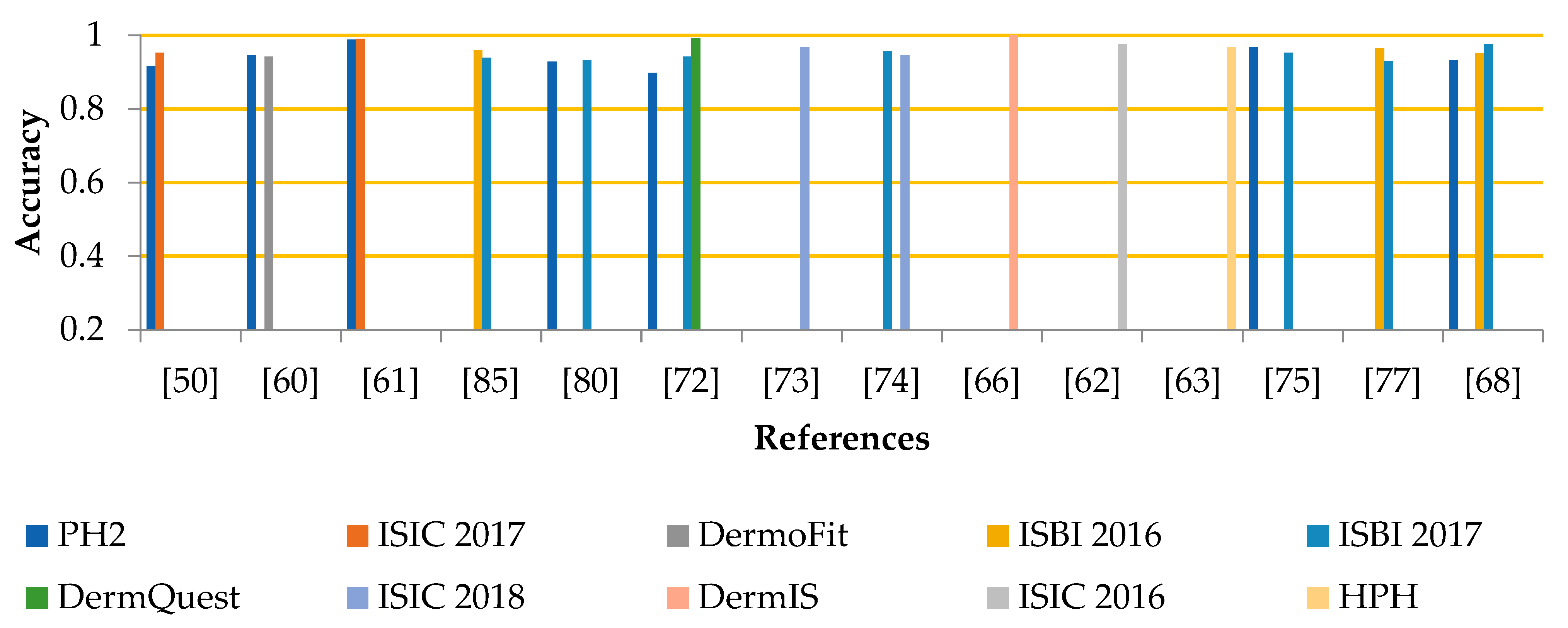
| Ref # | Year | Methods | Datasets | Results |
|---|---|---|---|---|
| [50] | 2019 | Local and global contrast starching, Dull razor, Region based active contour JSEG fusion | PH2 ISIC 2017 | 0.917 (ACC) 0.953 (ACC) |
| [60] | 2021 | Histogram equalization, max filter, morphological operations, hierarchical k-means with level set | PH2 DermoFit | 0.946 (ACC) 0.942 (ACC) |
| [61] | 2021 | Threshold and morphological operations, K-means clustering, firefly algorithm | ISIC 2017 PH2 | 0.991 (ACC) 0.989 (ACC) |
| [85] | 2018 | Adam approach, a Dense deconvolutional network | ISBI 2016 ISBI 2017 | 0.959 (ACC) 0.939 (ACC) |
| [80] | 2019 | Dull razor, morphological operations, YOLO, GrabCut | PH2 ISBI 2017 | 0.929 (ACC) 0.933 (ACC) |
| [72] | 2021 | Retina-DeepLab, R-CNN, and graph-related approaches | ISBI 2017 PH2 DermQuest | 0.942 (ACC) 0.898 (ACC) 0.992 (ACC) |
| [73] | 2021 | Data augmentation (flipping, rotation, translation, etc.), dense encoder-decoder based framework | ISIC 2018 | 0.969 (ACC) |
| [74] | 2020 | Bicubic interpolation, data augmentation, Adaptive dual attention module | ISBI 2017 ISIC 2018 | 0.957 (ACC) 0.947 (ACC) |
| [68] | 2020 | Median filter, histogram, Auxiliary function, global optimization algorithm | PH2 ISBI 2016 ISBI 2017 | 0.932 (ACC) 0.952 (ACC) 0.976 (ACC) |
| [66] | 2020 | Median filter, contrast stretching, ABCD, Threshold-based segmentation | DermIS DermQuest | 1.00 (ACC) |
| [62] | 2020 | GA, OCE-NGC | ISIC 2016 | 0.976 (ACC) |
| [63] | 2017 | Fuzzy clustering mean | HPH | 0.968 (ACC) |
| [75] | 2020 | Ifcn | PH2 ISBI 2017 | 0.969 (ACC) 0.953 (ACC) |
| [77] | 2020 | Resizing, augmentation, ResNet34, Scale-Att-ASPP, PPM, GAN | ISBI 2016 ISBI 2017 PH2 | 0.964 (ACC) 0.931 (ACC) 0.112 (DV) |
| [79] | 2020 | LabelMe, R-CNN | ISIC | 0.910 (recall) |
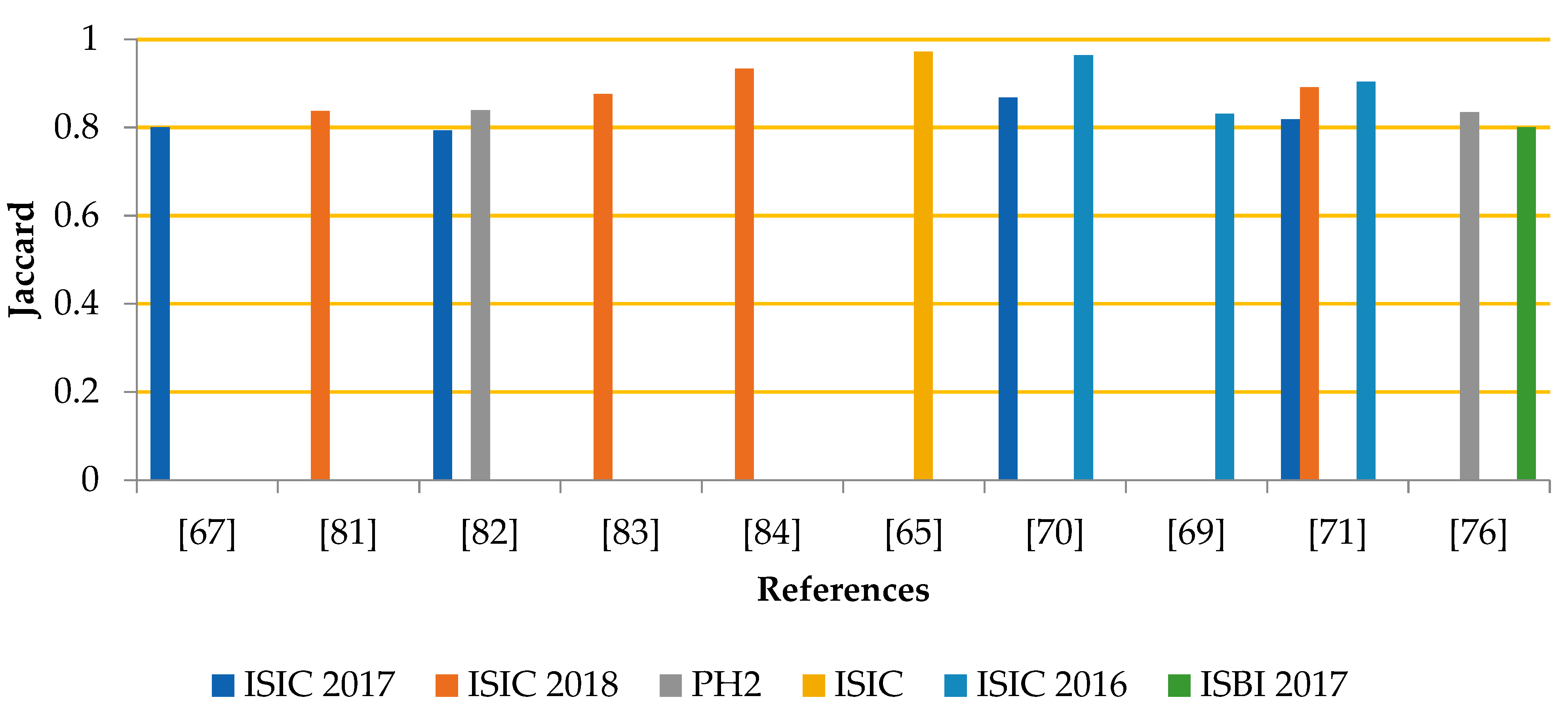
| [67] | 2019 | Gaussian filter, OTSU, SegRNorm, SegXNorm | ISIC 2017 | 0.800 (JAC) |
| [81] | 2019 | Resizing, encoder-decoder deep convolutional with aggregate multi-resolution skip connections | ISIC 2018 | 0.837 (JAC) |
| [82] | 2019 | Morphological operations, DeeplabV3+ and Mask R-CNN | ISIC 2017 PH2 | 0.793 (JAC) 0.839 (JAC) |
| [83] | 2019 | Data augmentation, VGG16 encoder, DeeplabV3, SegNet, threshold with dilations | ISIC 2018 | 0.876 (JAC) |
| [84] | 2019 | Linear filter, restoration, enhancement, U-Net 46 layered, U-Net 32 layered | ISIC 2018 | 0.933 (JAC) |
| [64] | 2017 | Illumination correction, histogram calculation, Frangi vesselness, K- means clustering | ISIC 2017 | 0.548 (validation set) |
| [65] | 2017 | Dull razor, ABCD, Shift mean algorithm | ISIC | 0.972 (JAC) |
| [70] | 2022 | Image Resize, FCEDN, EN-GWO | ISIC 2016 17 | 0. 964 (JAC) 0.868 (JAC) |
| [69] | 2021 | Dull razor, Initial contour optimization, GA | ISIC 2016 | 0.831 (JAC) |
| [71] | 2021 | Downsampling, translation, rotation and scaling, Atrous dilation CNN | ISIC 2016 17 18 | 0.904 (JAC) 0.818 (JAC) 0.891 (JAC) |
| [76] | 2020 | Augmentation, resizing, FC-DPN | ISBI 2017 PH2 | 0.800 (JAC) 0.835 (JAC) |
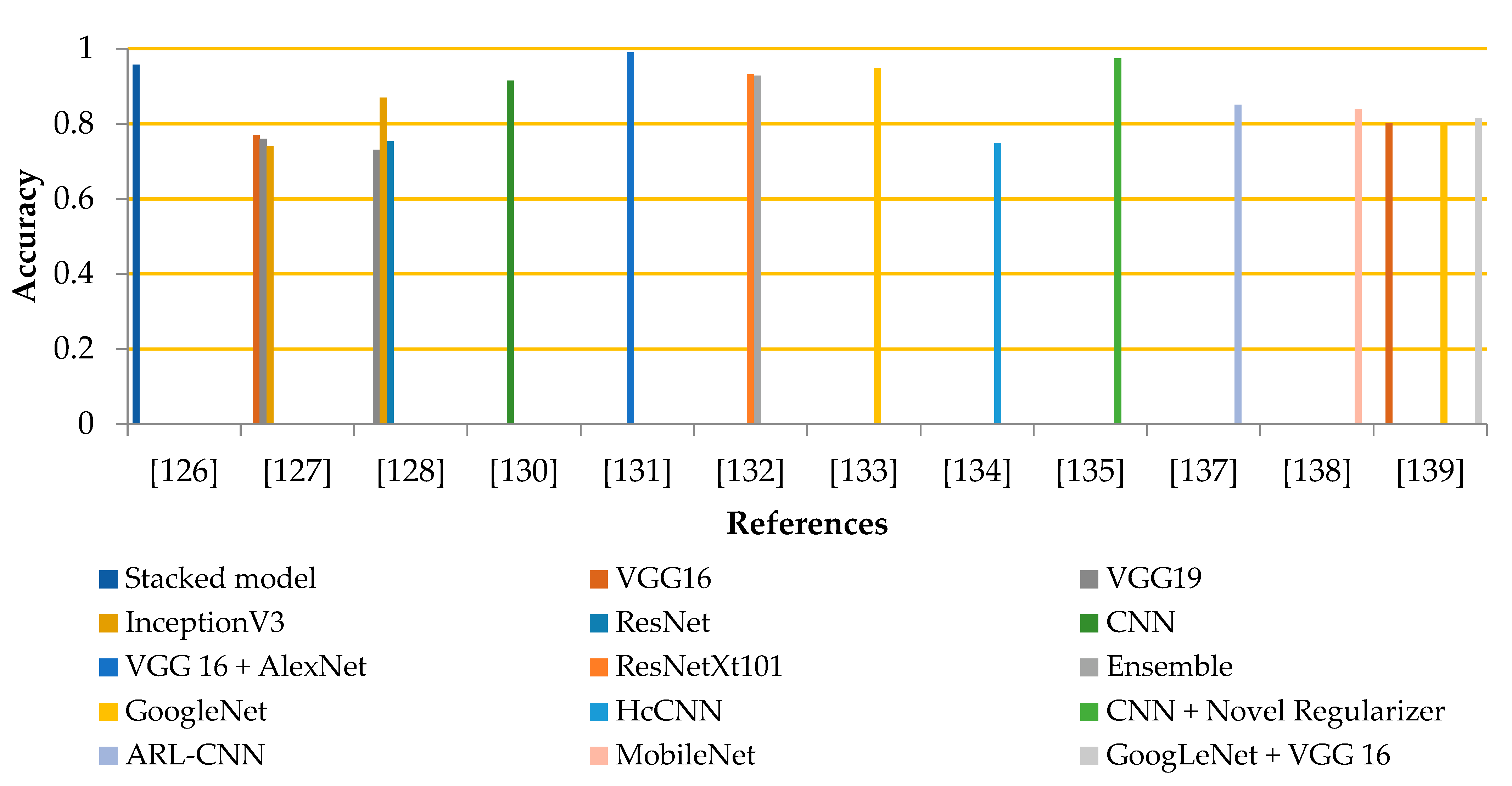
| Ref # | Year | CNN Models | Datasets | No. of Classes | Results (ACC) |
|---|---|---|---|---|---|
| [126] | 2022 | Stacked model | ISIC 2020 | 2 | 0.957 |
| [127] | 2021 | VGG16, VGG19, InceptionV3 | ISIC 2018 | 7 | 0.770 0.760 0.740 |
| [128] | 2021 | ResNet, InceptionV3, VGG19 | ISIC archive | 2 | 0.753 0.869 0.731 |
| [129] | 2021 | EfficientNet B6 models ensemble and EfficientNet B5 | ISIC 2020 | 2 | 0.941(ROC curve) |
| [130] | 2021 | CNN architecture | HAM-10000 | 7 | 0.915 |
| [131] | 2020 | VGG 16 + AlexNet | PH2+ +ISBI 2016 +ISBI 2017 | 2 | 0.990 |
| [132] | 2020 | ResNetXt101, InceptionResNetV2+ ResNetXt101 | HAM-10000 | 7 | 0.932 0.928 |
| [133] | 2020 | GoogleNet | ISIC 2019 | 8 | 0.949 0.798(SEN) 0.970 (SPE) |
| [134] | 2020 | HcCNN | 7 point check | 7 | 0.749 |
| [135] | 2019 | CNN + Novel Regularizer | ISIC archive | 2 | 0.974 |
| [136] | 2019 | PNASNeT-5-Large | ISIC 2018 | 7 | 0.76 (V.score) |
| [137] | 2019 | ARL-CNN | ISIC 2017 | 3 | 0.850 |
| [138] | 2019 | MobileNet | HAM-10000 | 7 | 0.839 |
| [139] | 2018 | GoogleNet, VGG 16, and their ensemble | ISIC 2018 | 7 | 0.797 0.801 0.815 |
Disclaimer/Publisher’s Note: The statements, opinions and data contained in all publications are solely those of the individual author(s) and contributor(s) and not of MDPI and/or the editor(s). MDPI and/or the editor(s) disclaim responsibility for any injury to people or property resulting from any ideas, methods, instructions or products referred to in the content. |
© 2023 by the authors. Licensee MDPI, Basel, Switzerland. This article is an open access article distributed under the terms and conditions of the Creative Commons Attribution (CC BY) license (https://creativecommons.org/licenses/by/4.0/).
Share and Cite
Zafar, M.; Sharif, M.I.; Sharif, M.I.; Kadry, S.; Bukhari, S.A.C.; Rauf, H.T. Skin Lesion Analysis and Cancer Detection Based on Machine/Deep Learning Techniques: A Comprehensive Survey. Life 2023, 13, 146. https://doi.org/10.3390/life13010146
Zafar M, Sharif MI, Sharif MI, Kadry S, Bukhari SAC, Rauf HT. Skin Lesion Analysis and Cancer Detection Based on Machine/Deep Learning Techniques: A Comprehensive Survey. Life. 2023; 13(1):146. https://doi.org/10.3390/life13010146
Chicago/Turabian StyleZafar, Mehwish, Muhammad Imran Sharif, Muhammad Irfan Sharif, Seifedine Kadry, Syed Ahmad Chan Bukhari, and Hafiz Tayyab Rauf. 2023. "Skin Lesion Analysis and Cancer Detection Based on Machine/Deep Learning Techniques: A Comprehensive Survey" Life 13, no. 1: 146. https://doi.org/10.3390/life13010146