
Oromaxillofacial Surgery: Both a Treatment and a Possible Cause of Obstructive Sleep Apnea—A Narrative Review
 , , ,
, , ,  and
and 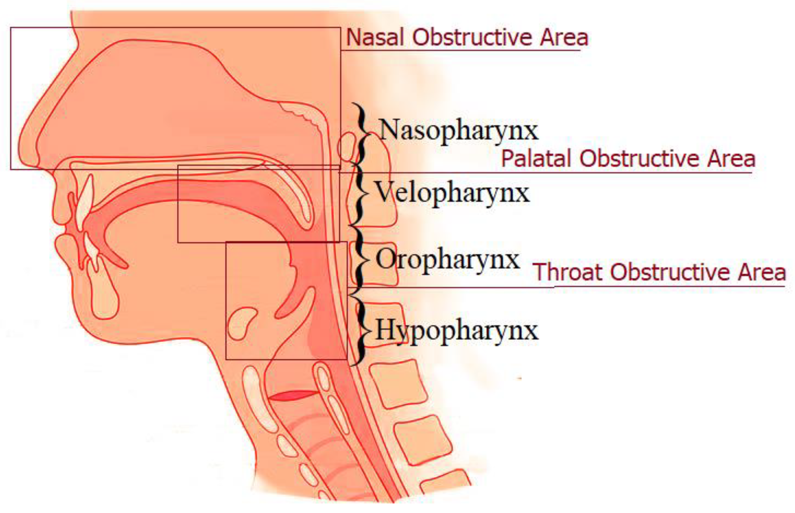
Abstract
:1. Introduction
2. Oromaxillofacial Surgery as a Cause of Obstructive Sleep Apnea
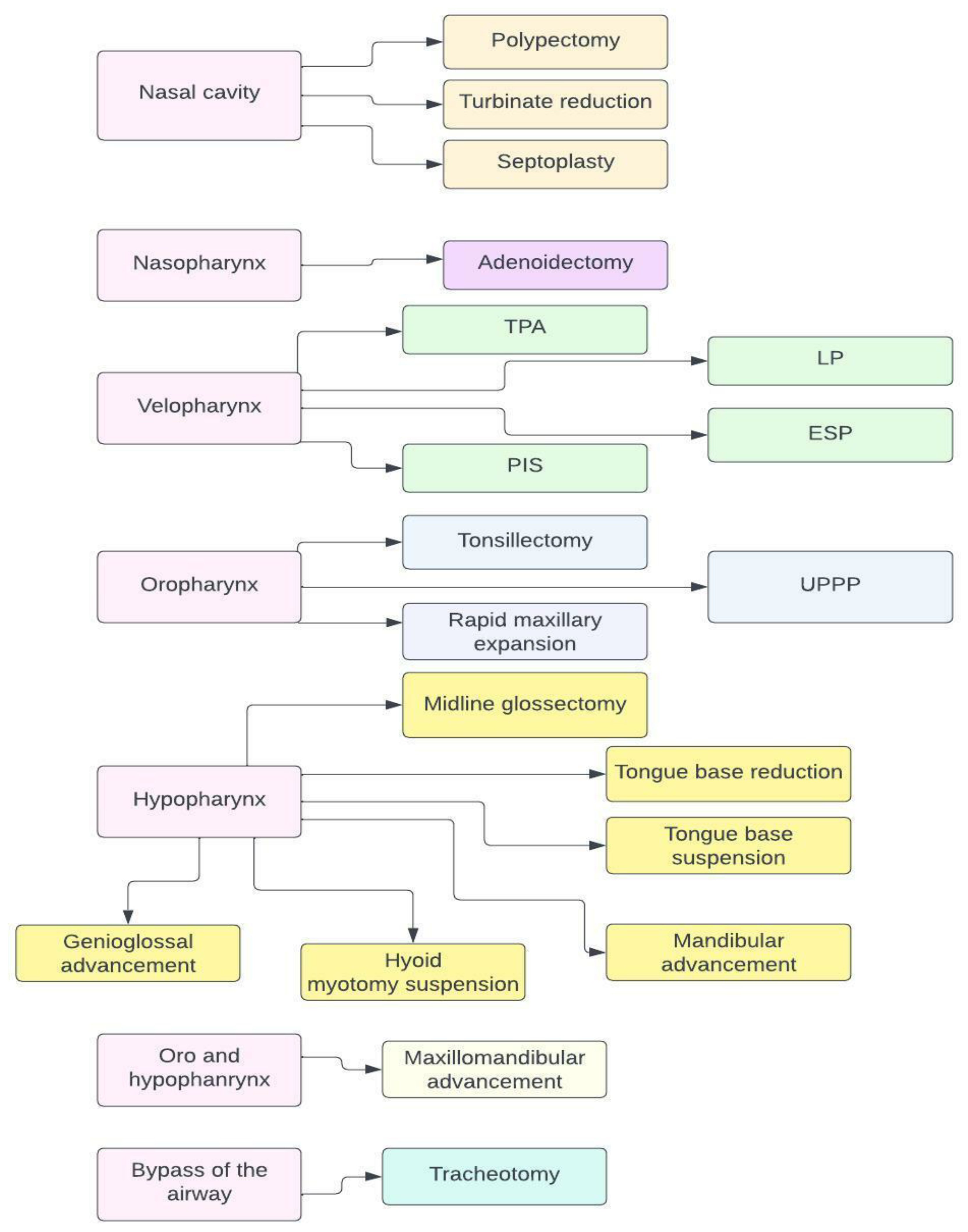
3. Surgical Therapy for Obstructive Sleep Apnea
3.1. Maxillomandibular Advancement
3.2. Maxillary Expansion and Maxillomandibular Expansion
3.3. Septoplasty, Turbinate Reduction, and Polypectomy
3.4. Surgeries of the Oropharynx
3.5. Genioglossal Advancement
3.6. Hyoid Suspension
3.7. Tongue Base Reduction
3.8. Tongue Base Suspension
3.9. Adenoidectomy and Tonsillectomy
3.10. Tracheostomy
4. Conclusions
Author Contributions
Funding
Institutional Review Board Statement
Informed Consent Statement
Data Availability Statement
Conflicts of Interest
References
- Bozic, J.; Galic, T.; Supe-Domic, D.; Ivkovic, N.; Ticinovic Kurir, T.; Valic, Z.; Lesko, J.; Dogas, Z. Morning cortisol levels and glucose metabolism parameters in moderate and severe obstructive sleep apnea patients. Endocrine 2016, 53, 730–739. [Google Scholar] [CrossRef] [PubMed]
- Osman, A.M.; Carter, S.G.; Carberry, J.C.; Eckert, D.J. Obstructive sleep apnea: Current perspectives. Nat. Sci. Sleep 2018, 10, 21–34. [Google Scholar] [CrossRef] [PubMed] [Green Version]
- Borovac, J.A.; Dogas, Z.; Supe-Domic, D.; Galic, T.; Bozic, J. Catestatin serum levels are increased in male patients with obstructive sleep apnea. Sleep Breath. 2019, 23, 473–481. [Google Scholar] [CrossRef] [PubMed]
- Bozic, J.; Borovac, J.A.; Galic, T.; Kurir, T.T.; Supe-Domic, D.; Dogas, Z. Adropin and Inflammation Biomarker Levels in Male Patients with Obstructive Sleep Apnea: A Link With Glucose Metabolism and Sleep Parameters. J. Clin. Sleep Med. 2018, 14, 1109–1118. [Google Scholar] [CrossRef] [Green Version]
- Slouka, D.; Honnerova, M.; Hosek, P.; Gal, B.; Trcka, O.; Kostlivy, T.; Landsmanova, J.; Havel, D.; Baneckova, M.; Kucera, R. Improved prediction of CPAP failure using T90, age and gender. J. Appl. Biomed. 2019, 17, 10. [Google Scholar] [CrossRef] [Green Version]
- Ralli, M.; Campo, F.; Angeletti, D.; Allegra, E.; Minni, A.; Polimeni, A.; Greco, A.; de Vincentiis, M. Obstructive Sleep Apnoea in Patients Treated for Head and Neck Cancer: A Systematic Review of the Literature. Medicina 2020, 56, 399. [Google Scholar] [CrossRef]
- Kim, J.W.; Kwon, T.G. Why most patients do not exhibit obstructive sleep apnea after mandibular setback surgery? Maxillofac. Plast. Reconstr. Surg. 2020, 42, 7. [Google Scholar] [CrossRef]
- Gavidia, R.; Dunietz, G.L.; O’Brien, L.; Shannon, C.; Schuetz, S.; Spector, M.; Swiecicki, P.; Chervin, R.D. Obstructive sleep apnea in patients with head and neck cancer: A systematic review. J. Clin. Sleep Med. 2021, 17, 1109–1116. [Google Scholar] [CrossRef]
- Liao, F.H.; Chang, C.C.; Lu, Y.C.; Lin, C.Y.; Lai, W.S. Impact of free flap reconstruction on obstructive sleep apnea in patients with oral and oropharyngeal cancer. Asia-Pac. J. Oncol. Nurs. 2022, 9, 100136. [Google Scholar] [CrossRef]
- Gavidia, R.; Dunietz, G.L.; O’Brien, L.M.; Schütz, S.G.; Spector, M.E.; Swiecicki, P.L.; Chervin, R.D. Risk of obstructive sleep apnea after treatment of head and neck squamous cell carcinoma: A cross-sectional study. J. Clin. Sleep Med. 2022, 18, 1681–1686. [Google Scholar] [CrossRef]
- Lupi-Ferandin, S.; Galic, T.; Ivkovic, N.; Pecotic, R.; Dogas, Z. Prevalence of obstructive sleep apnea in male patients with surgically treated maxillary and zygomatic fractures. Can. J. Surg. 2019, 62, 105–110. [Google Scholar] [CrossRef] [PubMed] [Green Version]
- Loth, A.; Michel, J.; Giorgi, R.; Santini, L.; Rey, M.; Elbaum, J.M.; Roux, N.; Giovanni, A.; Dessi, P.; Fakhry, N. Prevalence of obstructive sleep apnoea syndrome following oropharyngeal cancer treatment: A prospective cohort study. Clin. Otolaryngol. 2017, 42, 1281–1288. [Google Scholar] [CrossRef] [PubMed]
- Friedman, M.; Landsberg, R.; Pryor, S.; Syed, Z.; Ibrahim, H.; Caldarelli, D.D. The occurrence of sleep-disordered breathing among patients with head and neck cancer. Laryngoscope 2001, 111, 1917–1919. [Google Scholar] [CrossRef] [PubMed] [Green Version]
- Qian, W.; Haight, J.; Poon, I.; Enepekides, D.; Higgins, K.M. Sleep apnea in patients with oral cavity and oropharyngeal cancer after surgery and chemoradiation therapy. Otolaryngol. Head Neck Surg. 2010, 143, 248–252. [Google Scholar] [CrossRef]
- Payne, R.J.; Hier, M.P.; Kost, K.M.; Black, M.J.; Zeitouni, A.G.; Frenkiel, S.; Naor, N.; Kimoff, R.J. High prevalence of obstructive sleep apnea among patients with head and neck cancer. J. Otolaryngol. 2005, 34, 304–311. [Google Scholar] [CrossRef]
- Gómez-Merino, E.; Arriero, J.M.; Chiner, E.; Signes-Costa, J.; Marco, J. Obstructive sleep apnea syndrome as first manifestation of pharyngeal non-Hodgkin’s lymphoma. Respiration 2003, 70, 107–109. [Google Scholar] [CrossRef]
- Hockstein, N.G.; Anderson, T.A.; Moonis, G.; Gustafson, K.S.; Mirza, N. Retropharyngeal lipoma causing obstructive sleep apnea: Case report including five-year follow-up. Laryngoscope 2002, 112, 1603–1605. [Google Scholar] [CrossRef]
- Koopmann, C.F., Jr.; Feld, R.A.; Coulthard, S.W. Sleep apnea syndrome associated with a neck mass. Otolaryngol. Head Neck Surg. 1981, 89, 949–952. [Google Scholar] [CrossRef]
- Pellanda, A.; Zagury, S.; Pasche, P. Parapharyngeal lipoma causing obstructive sleep apnea syndrome. Otolaryngol. Head Neck Surg. 2003, 128, 301–302. [Google Scholar] [CrossRef]
- Abdullah, B.J.; Liam, C.K.; Kaur, H.; Mathew, K.M. Parapharyngeal space lipoma causing sleep apnoea. Br. J. Radiol. 1997, 70, 1063–1065. [Google Scholar] [CrossRef]
- Werlinger, F.; Villalón, M.; Duarte, V.; Acevedo, R.; Aguilera, R.; Alcocer, D.; Badillo, O.; Briones, R.; Condal, C.; Del Río, M.; et al. Trends of maxillofacial trauma: An update from the prospective register of a multicenter study in emergency services of Chile. Med. Oral Patol. Oral Cir. Bucal 2019, 24, e588–e594. [Google Scholar] [CrossRef] [PubMed]
- Kaul, R.P.; Sagar, S.; Singhal, M.; Kumar, A.; Jaipuria, J.; Misra, M. Burden of maxillofacial trauma at level 1 trauma center. Craniomaxillofac. Trauma Reconstr. 2014, 7, 126–130. [Google Scholar] [CrossRef] [PubMed] [Green Version]
- El-Anwar, M.W.; Askar, S.; Abou Shab, Y.A.; Abou Sharkh, A.A.M. Could mandibular fractures lead to obstructive sleep apnea? Cranio 2021, 8, 1–4. [Google Scholar] [CrossRef] [PubMed]
- Khechoyan, D.Y. Orthognathic surgery: General considerations. Semin. Plast. Surg. 2013, 27, 133–136. [Google Scholar] [CrossRef] [PubMed] [Green Version]
- Hasebe, D.; Kobayashi, T.; Hasegawa, M.; Iwamoto, T.; Kato, K.; Izumi, N.; Takata, Y.; Saito, C. Changes in oropharyngeal airway and respiratory function during sleep after orthognathic surgery in patients with mandibular prognathism. Int. J. Oral Maxillofac. Surg. 2011, 40, 584–592. [Google Scholar] [CrossRef] [PubMed]
- Kobayashi, T.; Funayama, A.; Hasebe, D.; Kato, Y.; Yoshizawa, M.; Saito, C. Changes in overnight arterial oxygen saturation after mandibular setback. Br. J. Oral Maxillofac. Surg. 2013, 51, 312–318. [Google Scholar] [CrossRef]
- Fernández-Ferrer, L.; Montiel-Company, J.M.; Pinho, T.; Almerich-Silla, J.M.; Bellot-Arcís, C. Effects of mandibular setback surgery on upper airway dimensions and their influence on obstructive sleep apnoea—A systematic review. J. Craniomaxillofac. Surg. 2015, 43, 248–253. [Google Scholar] [CrossRef]
- Dahy, K.; Takahashi, K.; Saito, K.; Kiso, H.; Rezk, I.; Oga, T.; Uozumi, R.; Chin, K.; Bessho, K. Gender differences in morphological and functional outcomes after mandibular setback surgery. J. Craniomaxillofac. Surg. 2018, 46, 887–892. [Google Scholar] [CrossRef]
- Isono, S.; Remmers, J.E.; Tanaka, A.; Sho, Y.; Sato, J.; Nishino, T. Anatomy of pharynx in patients with obstructive sleep apnea and in normal subjects. J. Appl. Physiol. 1985, 82, 1319–1326. [Google Scholar] [CrossRef]
- Martin, S.E.; Mathur, R.; Marshall, I.; Douglas, N.J. The effect of age, sex, obesity and posture on upper airway size. Eur. Respir. J. 1997, 10, 2087–2090. [Google Scholar] [CrossRef]
- Young, T.; Evans, L.; Finn, L.; Palta, M. Estimation of the clinically diagnosed proportion of sleep apnea syndrome in middle-aged men and women. Sleep 1997, 20, 705–706. [Google Scholar] [CrossRef] [PubMed]
- Rowley, J.A.; Zhou, X.; Vergine, I.; Shkoukani, M.A.; Badr, M.S. Influence of gender on upper airway mechanics: Upper airway resistance and Pcrit. J. Appl. Physiol. 1985, 91, 2248–2254. [Google Scholar] [CrossRef] [PubMed]
- Trinder, J.; Kay, A.; Kleiman, J.; Dunai, J. Gender differences in airway resistance during sleep. J. Appl. Physiol. 1985, 83, 1986–1997. [Google Scholar] [CrossRef] [PubMed]
- Popovic, R.M.; White, D.P. Influence of gender on waking genioglossal electromyogram and upper airway resistance. Am. J. Respir. Crit. Care Med. 1995, 152, 725–731. [Google Scholar] [CrossRef] [PubMed]
- Won, C.H.; Li, K.K.; Guilleminault, C. Surgical treatment of obstructive sleep apnea: Upper airway and maxillomandibular surgery. Proc. Am. Thorac. Soc. 2008, 5, 193–199. [Google Scholar] [CrossRef]
- Goyal, M.; Johnson, J. Obstructive Sleep Apnea Diagnosis and Management. Mo. Med. 2017, 114, 120–124. [Google Scholar]
- Boyd, S.B.; Chigurupati, R.; Cillo, J.E., Jr.; Eskes, G.; Goodday, R.; Meisami, T.; Viozzi, C.F.; Waite, P.; Wilson, J. Maxillomandibular Advancement Improves Multiple Health-Related and Functional Outcomes in Patients with Obstructive Sleep Apnea: A Multicenter Study. J. Oral Maxillofac. Surg. 2019, 77, 352–370. [Google Scholar] [CrossRef] [Green Version]
- Zaghi, S.; Holty, J.E.; Certal, V.; Abdullatif, J.; Guilleminault, C.; Powell, N.B.; Riley, R.W.; Camacho, M. Maxillomandibular Advancement for Treatment of Obstructive Sleep Apnea: A Meta-analysis. JAMA Otolaryngol. Head Neck Surg. 2016, 142, 58–66. [Google Scholar] [CrossRef]
- John, C.R.; Gandhi, S.; Sakharia, A.R.; James, T.T. Maxillomandibular advancement is a successful treatment for obstructive sleep apnoea: A systematic review and meta-analysis. Int. J. Oral Maxillofac. Surg. 2018, 47, 1561–1571. [Google Scholar] [CrossRef]
- Friedman, M.; Wilson, M. Re-redefining success in airway surgery for obstructive sleep apnea. Sleep 2009, 32, 17. [Google Scholar]
- Liao, Y.F.; Chiu, Y.T.; Lin, C.H.; Chen, Y.A.; Chen, N.H.; Chen, Y.R. Modified maxillomandibular advancement for obstructive sleep apnoea: Towards a better outcome for Asians. Int. J. Oral Maxillofac. Surg. 2015, 44, 189–194. [Google Scholar] [CrossRef] [PubMed]
- Schendel, S.A.; Broujerdi, J.A.; Jacobson, R.L. Three-dimensional upper-airway changes with maxillomandibular advancement for obstructive sleep apnea treatment. Am. J. Orthod. Dentofac. Orthop. 2014, 146, 385–393. [Google Scholar] [CrossRef] [PubMed]
- Boyd, S.B.; Walters, A.S.; Waite, P.; Harding, S.M.; Song, Y. Long-Term Effectiveness and Safety of Maxillomandibular Advancement for Treatment of Obstructive Sleep Apnea. J. Clin. Sleep Med. 2015, 11, 699–708. [Google Scholar] [CrossRef] [PubMed]
- Blumen, M.B.; Buchet, I.; Meulien, P.; Hausser, H.C.; Neveu, H.; Chabolle, F. Complications/adverse effects of maxillomandibular advancement for the treatment of OSA in regard to outcome. Otolaryngol. Head Neck Surg. 2009, 141, 591–597. [Google Scholar] [CrossRef] [PubMed]
- Vinha, P.P.; Faria, A.C.; Xavier, S.P.; Christino, M.; de Mello-Filho, F.V. Enlargement of the Pharynx Resulting From Surgically Assisted Rapid Maxillary Expansion. J. Oral Maxillofac. Surg. 2016, 74, 369–379. [Google Scholar] [CrossRef]
- Abdullatif, J.; Certal, V.; Zaghi, S.; Song, S.A.; Chang, E.T.; Gillespie, M.B.; Camacho, M. Maxillary expansion and maxillomandibular expansion for adult OSA: A systematic review and meta-analysis. J. Craniomaxillofac. Surg. 2016, 44, 574–578. [Google Scholar] [CrossRef]
- Camacho, M.; Zaghi, S.; Certal, V.; Abdullatif, J.; Means, C.; Acevedo, J.; Liu, S.; Brietzke, S.E.; Kushida, C.A.; Capasso, R. Inferior turbinate classification system, grades 1 to 4: Development and validation study. Laryngoscope 2015, 125, 296–302. [Google Scholar] [CrossRef]
- Guilleminault, C.; Quo, S.; Huynh, N.T.; Li, K. Orthodontic expansion treatment and adenotonsillectomy in the treatment of obstructive sleep apnea in prepubertal children. Sleep 2008, 31, 953–957. [Google Scholar]
- Camacho, M.; Chang, E.T.; Song, S.A.; Abdullatif, J.; Zaghi, S.; Pirelli, P.; Certal, V.; Guilleminault, C. Rapid maxillary expansion for pediatric obstructive sleep apnea: A systematic review and meta-analysis. Laryngoscope 2017, 127, 1712–1719. [Google Scholar] [CrossRef]
- Machado-Júnior, A.J.; Zancanella, E.; Crespo, A.N. Rapid maxillary expansion and obstructive sleep apnea: A review and meta-analysis. Med. Oral Patol. Oral Cir. Bucal 2016, 21, 21073. [Google Scholar] [CrossRef]
- Gokce, G.; Basoglu, O.K.; Veli, I. Polygraphic evaluation of the effects of different rapid maxillary expansion appliances on sleep quality: A randomized clinical trial. Sleep Breath. 2022, 5. [Google Scholar] [CrossRef] [PubMed]
- Vinha, P.P.; Eckeli, A.L.; Faria, A.C.; Xavier, S.P.; de Mello-Filho, F.V. Effects of surgically assisted rapid maxillary expansion on obstructive sleep apnea and daytime sleepiness. Sleep Breath. 2016, 20, 501–508. [Google Scholar] [CrossRef] [PubMed]
- Koudstaal, M.J.; Poort, L.J.; van der Wal, K.G.; Wolvius, E.B.; Prahl-Andersen, B.; Schulten, A.J. Surgically assisted rapid maxillary expansion (SARME): A review of the literature. Int. J. Oral Maxillofac. Surg. 2005, 34, 709–714. [Google Scholar] [CrossRef] [PubMed]
- Liu, S.Y.-C.; Guilleminault, C.; Huon, L.-K.; Yoon, A. Distraction Osteogenesis Maxillary Expansion (DOME) for Adult Obstructive Sleep Apnea Patients with High Arched Palate. Otolaryngol. Head Neck Surg. 2017, 157, 345–348. [Google Scholar] [CrossRef] [PubMed]
- Loriato, L.; Ferreira, C.E. Surgically-assisted rapid maxillary expansion (SARME): Indications, planning and treatment of severe maxillary deficiency in an adult patient. Dent. Press J. Orthod. 2020, 25, 73–84. [Google Scholar] [CrossRef] [PubMed]
- Yoon, A.; Guilleminault, C.; Zaghi, S.; Liu, S.Y. Distraction Osteogenesis Maxillary Expansion (DOME) for adult obstructive sleep apnea patients with narrow maxilla and nasal floor. Sleep Med. 2020, 65, 172–176. [Google Scholar] [CrossRef] [PubMed]
- Cakarer, S.; Keskin, B.; Isler, S.C.; Cansiz, E.; Uzun, A.; Keskin, C. Complications associated with surgically assisted rapid palatal expansion without pterygomaxillary separation. J. Stomatol. Oral Maxillofac. Surg. 2017, 118, 279–282. [Google Scholar] [CrossRef]
- Tanna, N.; Smith, B.D.; Zapanta, P.E.; Karanetz, I.; Andrews, B.T.; Urata, M.M.; Bradley, J.P. Surgical Management of Obstructive Sleep Apnea. Plast. Reconstr. Surg. 2016, 137, 1263–1272. [Google Scholar] [CrossRef]
- Mickelson, S.A. Nasal Surgery for Obstructive Sleep Apnea Syndrome. Otolaryngol. Clin. N. Am. 2016, 49, 1373–1381. [Google Scholar] [CrossRef]
- Bloom, J.D.; Kaplan, S.E.; Bleier, B.S.; Goldstein, S.A. Septoplasty complications: Avoidance and management. Otolaryngol. Clin. N. Am. 2009, 42, 463–481. [Google Scholar] [CrossRef]
- Bin Lajdam, G.; Alaryani, K.; Ghaddaf, A.A.; Aljabri, A.; Halawani, A.; Alshareef, M.; Algarni, M.; Al-Hakami, H. Septoplasty versus septoplasty with turbinate reduction for nasal obstruction due to deviated nasal septum: A systematic review and meta-analysis. Rhinology 2022, 60, 411–420. [Google Scholar] [CrossRef] [PubMed]
- Cai, Y.; Goldberg, A.N.; Chang, J.L. The Nose and Nasal Breathing in Sleep Apnea. Otolaryngol. Clin. N. Am. 2020, 53, 385–395. [Google Scholar] [CrossRef] [PubMed]
- Takahashi, R.; Ohbuchi, T.; Hohchi, N.; Takeuchi, S.; Ohkubo, J.; Ikezaki, S.; Suzuki, H. Effect of septoplasty and turbinectomy on obstructive sleep apnea syndrome. Nihon Jibiinkoka Gakkai Kaiho 2013, 116, 789–792. [Google Scholar] [CrossRef] [PubMed]
- Migueis, D.P.; Thuler, L.C.; Lemes, L.N.; Moreira, C.S.; Joffily, L.; Araujo-Melo, M.H. Systematic review: The influence of nasal obstruction on sleep apnea. Braz. J. Otorhinolaryngol. 2016, 82, 223–231. [Google Scholar] [CrossRef] [PubMed] [Green Version]
- Kalam, I. Objective assessment of nasal obstruction in snoring and obstructive sleep apnea patients: Experience of a Police Authority Hospital. Ann. Saudi Med. 2002, 22, 158–162. [Google Scholar] [PubMed]
- Hisamatsu, K.; Kudo, I.; Makiyama, K. The effect of compound nasal surgery on obstructive sleep apnea syndrome. Am. J. Rhinol. Allergy 2015, 29, e192–e196. [Google Scholar] [CrossRef]
- Weder, S.; Landis, B.N.; Banz, Y.; Caversaccio, M.; Dubach, P. Paediatric traffic accident and obstructive sleep apnoea by antrochoanal polyps: Case report and literature review. Int. J. Pediatr. Otorhinolaryngol. 2011, 75, 1359–1363. [Google Scholar] [CrossRef]
- Fujita, S.; Conway, W.; Zorick, F.; Roth, T. Surgical correction of anatomic azbnormalities in obstructive sleep apnea syndrome: Uvulopalatopharyngoplasty. Otolaryngol. Head Neck Surg. 1981, 89, 923–934. [Google Scholar] [CrossRef]
- Holty, J.E.; Guilleminault, C. Surgical options for the treatment of obstructive sleep apnea. Med. Clin. N. Am. 2010, 94, 479–515. [Google Scholar] [CrossRef]
- Collop, N.A. Advances in treatment of obstructive sleep apnea syndrome. Curr. Treat. Options Neurol. 2009, 11, 340–348. [Google Scholar] [CrossRef]
- Chandrashekariah, R.; Shaman, Z.; Auckley, D. Impact of upper airway surgery on CPAP compliance in difficult-to-manage obstructive sleep apnea. Arch. Otolaryngol. Head Neck Surg. 2008, 134, 926–930. [Google Scholar] [CrossRef] [PubMed] [Green Version]
- Pang, K.P.; Vicini, C.; Montevecchi, F.; Piccin, O.; Chandra, S.; Yang, H.C.; Agrawal, V.; Chung, J.C.K.; Chan, Y.H.; Pang, S.B.; et al. Long-term Complications of Palate Surgery: A Multicenter Study of 217 Patients. Laryngoscope 2020, 130, 2281–2284. [Google Scholar] [CrossRef] [PubMed]
- Stuck, B.A.; Ravesloot, M.J.L.; Eschenhagen, T.; de Vet, H.C.W.; Sommer, J.U. Uvulopalatopharyngoplasty with or without tonsillectomy in the treatment of adult obstructive sleep apnea—A systematic review. Sleep Med. 2018, 50, 152–165. [Google Scholar] [CrossRef] [PubMed]
- Friedman, M.; Ibrahim, H.; Bass, L. Clinical staging for sleep-disordered breathing. Otolaryngol. Head Neck Surg. 2002, 127, 13–21. [Google Scholar] [CrossRef] [PubMed]
- Caples, S.M.; Rowley, J.A.; Prinsell, J.R.; Pallanch, J.F.; Elamin, M.B.; Katz, S.G.; Harwick, J.D. Surgical modifications of the upper airway for obstructive sleep apnea in adults: A systematic review and meta-analysis. Sleep 2010, 33, 1396–1407. [Google Scholar] [CrossRef] [PubMed] [Green Version]
- Bäck, L.J.; Hytönen, M.L.; Roine, R.P.; Malmivaara, A.O. Radiofrequency ablation treatment of soft palate for patients with snoring: A systematic review of effectiveness and adverse effects. Laryngoscope 2009, 119, 1241–1250. [Google Scholar] [CrossRef]
- Li, K.K.; Powell, N.B.; Riley, R.W.; Troell, R.J.; Guilleminault, C. Radiofrequency volumetric reduction of the palate: An extended follow-up study. Otolaryngol. Head Neck Surg. 2000, 122, 410–414. [Google Scholar] [CrossRef]
- Finkelstein, Y.; Stein, G.; Ophir, D.; Berger, R.; Berger, G. Laser-assisted uvulopalatoplasty for the management of obstructive sleep apnea: Myths and facts. Arch. Otolaryngol. Head Neck Surg. 2002, 128, 429–434. [Google Scholar] [CrossRef]
- Ryan, C.F.; Love, L.L. Unpredictable results of laser assisted uvulopalatoplasty in the treatment of obstructive sleep apnoea. Thorax 2000, 55, 399–404. [Google Scholar] [CrossRef] [Green Version]
- Emara, T.A.; Ibrahim, H.A.; Elmalt, A.E.; Dahy, K.G.; Rashwan, M.S. Upper airway multilevel radiof-requency under local anesthesia can improve CPAP adherence for severe OSA patients. Am. J. Otolaryngol. 2023, 44, 103671. [Google Scholar] [CrossRef]
- Cammaroto, G.; Stringa, L.M.; Iannella, G.; Meccariello, G.; Zhang, H.; Bahgat, A.Y.; Calvo-Henriquez, C.; Chiesa-Estomba, C.; Lechien, J.R.; Barillari, M.R.; et al. Manipulation of Lateral Pharyngeal Wall Muscles in Sleep Surgery: A Review of the Literature. Int. J. Environ. Res. Public Health 2020, 17, 5315. [Google Scholar] [CrossRef] [PubMed]
- Woodson, B.T.; Toohill, R.J. Transpalatal advancement pharyngoplasty for obstructive sleep apnea. Laryngoscope 1993, 103, 269–276. [Google Scholar] [CrossRef] [PubMed]
- Volner, K.; Dunn, B.; Chang, E.T.; Song, S.A.; Liu, S.Y.; Brietzke, S.E.; O’Connor, P.; Camacho, M. Transpalatal advancement pharyngoplasty for obstructive sleep apnea: A systematic review and meta-analysis. Eur. Arch. Otorhinolaryngol. 2017, 274, 1197–1203. [Google Scholar] [CrossRef] [PubMed]
- Cahali, M.B.; Formigoni, G.G.; Gebrim, E.M.; Miziara, I.D. Lateral pharyngoplasty versus uvulopalatopharyngoplasty: A clinical, polysomnographic and computed tomography measurement comparison. Sleep 2004, 27, 942–950. [Google Scholar] [CrossRef] [Green Version]
- Cahali, M.B. Lateral pharyngoplasty: A new treatment for obstructive sleep apnea hypopnea syndrome. Laryngoscope 2003, 113, 1961–1968. [Google Scholar] [CrossRef]
- Pang, K.P.; Woodson, B.T. Expansion sphincter pharyngoplasty: A new technique for the treatment of obstructive sleep apnea. Otolaryngol. Head Neck Surg. 2007, 137, 110–114. [Google Scholar] [CrossRef]
- Pang, K.P.; Pang, E.B.; Win, M.T.; Pang, K.A.; Woodson, B.T. Expansion sphincter pharyngoplasty for the treatment of OSA: A systemic review and meta-analysis. Eur. Arch. Otorhinolaryngol. 2016, 273, 2329–2333. [Google Scholar] [CrossRef]
- Elbassiouny, A.M. Modified barbed soft palatal posterior pillar webbing flap palatopharyngoplasty. Sleep Breath. 2016, 20, 829–836. [Google Scholar] [CrossRef]
- El-Ahl, M.A.; El-Anwar, M.W. Expansion Pharyngoplasty by New Simple Suspension Sutures without Tonsillectomy. Otolaryngol. Head Neck Surg. 2016, 155, 1065–1068. [Google Scholar] [CrossRef]
- Pianta, L.; Bertazzoni, G.; Morello, R.; Perotti, P.; Nicolai, P. Barbed expansion sphincter pharyngoplasty for the treatment of oropharyngeal collapse in obstructive sleep apnoea syndrome: A retrospective study on 17 patients. Clin. Otolaryngol. 2018, 43, 696–700. [Google Scholar] [CrossRef] [Green Version]
- Salamanca, F.; Costantini, F.; Mantovani, M.; Bianchi, A.; Amaina, T.; Colombo, E.; Zibordi, F. Barbed anterior pharyngoplasty: An evolution of anterior palatoplasty. Acta Otorhinolaryngol. Ital. 2014, 34, 434–438. [Google Scholar] [PubMed]
- Mantovani, M.; Carioli, D.; Torretta, S.; Rinaldi, V.; Ibba, T.; Pignataro, L. Barbed snore surgery for concentric collapse at the velum: The Alianza technique. J. Craniomaxillofac. Surg. 2017, 45, 1794–1800. [Google Scholar] [CrossRef] [PubMed]
- Mantovani, M.; Rinaldi, V.; Torretta, S.; Carioli, D.; Salamanca, F.; Pignataro, L. Barbed Roman blinds technique for the treatment of obstructive sleep apnea: How we do it? Eur. Arch. Otorhinolaryngol. 2016, 273, 517–523. [Google Scholar] [CrossRef]
- Barbieri, M.; Missale, F.; Incandela, F.; Fragale, M.; Barbieri, A.; Roustan, V.; Canevari, F.R.; Peretti, G. Barbed suspension pharyngoplasty for treatment of lateral pharyngeal wall and palatal collapse in patients affected by OSAHS. Eur. Arch. Otorhinolaryngol. 2019, 276, 1829–1835. [Google Scholar] [CrossRef]
- Vicini, C.; Meccariello, G.; Montevecchi, F.; De Vito, A.; Frassineti, S.; Gobbi, R.; Pelucchi, S.; Iannella, G.; Magliulo, G.; Cammaroto, G. Effectiveness of barbed repositioning pharyngoplasty for the treatment of obstructive sleep apnea (OSA): A prospective randomized trial. Sleep Breath. 2020, 24, 687–694. [Google Scholar] [CrossRef]
- Cammaroto, G.; Montevecchi, F.; D’Agostino, G.; Zeccardo, E.; Bellini, C.; Meccariello, G.; Vicini, C. Palatal surgery in a transoral robotic setting (TORS): Preliminary results of a retrospective comparison between uvulopalatopharyngoplasty (UPPP), expansion sphincter pharyngoplasty (ESP) and barbed repositioning pharyngoplasty (BRP). Acta Otorhinolaryngol. Ital. 2017, 37, 406–409. [Google Scholar] [CrossRef] [PubMed]
- Friedman, M.; Schalch, P.; Lin, H.C.; Kakodkar, K.A.; Joseph, N.J.; Mazloom, N. Palatal implants for the treatment of snoring and obstructive sleep apnea/hypopnea syndrome. Otolaryngol. Head Neck Surg. 2008, 138, 209–216. [Google Scholar] [CrossRef]
- Zhang, X.M.; Tham, C.J.; Yin, Y.L.; Sun, Y.Q.; Zhou, X. A novel palatal implant surgery combined with uvulopalatopharyngoplasty and inferior turbinate radiofrequency for the treatment of moderate to severe obstructive sleep apnea: A pilot study. Eur. Arch. Otorhinolaryngol. 2015, 272, 1195–1202. [Google Scholar] [CrossRef]
- Lee, N.R.; Madani, M. Genioglossus muscle advancement techniques for obstructive sleep apnea. Atlas Oral Maxillofac. Surg. Clin. N. Am. 2007, 15, 179–192. [Google Scholar] [CrossRef]
- García Vega, J.R.; de la Plata, M.M.; Galindo, N.; Navarro, M.; Díez, D.; Láncara, F. Genioglossus muscle advancement: A modification of the conventional technique. J. Craniomaxillofac. Surg. 2014, 42, 239–244. [Google Scholar] [CrossRef]
- Emara, T.A.; Omara, T.A.; Shouman, W.M. Modified genioglossus advancement and uvulopalatopharyngoplasty in patients with obstructive sleep apnea. Otolaryngol. Head Neck Surg. 2011, 145, 865–871. [Google Scholar] [CrossRef] [PubMed]
- Emara, T.A.; Elhamshary, A.A.S.; Elkady, A.S.; Elhewity, A.M.M.; Abdelsamee, H.A. Modified genioglossus advancement with radiofrequency tongue base reduction for retroglossal collapse in Obstructive sleep apnea patients. Am. J. Otolaryngol. 2022, 43, 2. [Google Scholar] [CrossRef] [PubMed]
- Goh, Y.H.; Abdullah, V.; Kim, S.W. Genioglossus Advancement and Hyoid Surgery. Sleep Med. Clin. 2019, 14, 73–81. [Google Scholar] [CrossRef] [PubMed]
- Li, K.K. Surgical therapy for obstructive sleep apnea syndrome. Semin. Respir. Crit. Care Med. 2005, 26, 80–88. [Google Scholar] [CrossRef] [PubMed]
- Kezirian, E.J.; Goldberg, A.N. Hypopharyngeal surgery in obstructive sleep apnea: An evidence-based medicine review. Arch. Otolaryngol. Head Neck Surg. 2006, 132, 206–213. [Google Scholar] [CrossRef] [PubMed] [Green Version]
- Lee, J.W.; Lee, D.W.; Ohe, J.Y. Accurate genial tubercle capturing method using computer-assisted virtual surgery for genioglossus advancement. Br. J. Oral Maxillofac. Surg. 2017, 55, 92–93. [Google Scholar] [CrossRef]
- Riley, R.W.; Powell, N.B.; Guilleminault, C.; Nino-Murcia, G. Maxillary, mandibular, and hyoid advancement: An alternative to tracheostomy in obstructive sleep apnea syndrome. Otolaryngol. Head Neck Surg. 1986, 94, 584–588. [Google Scholar] [CrossRef]
- Riley, R.W.; Powell, N.B.; Guilleminault, C. Inferior sagittal osteotomy of the mandible with hyoid myotomy-suspension: A new procedure for obstructive sleep apnea. Otolaryngol. Head Neck Surg. 1986, 94, 589–593. [Google Scholar] [CrossRef]
- den Herder, C.; van Tinteren, H.; de Vries, N. Hyoidthyroidpexia: A surgical treatment for sleep apnea syndrome. Laryngoscope 2005, 115, 740–745. [Google Scholar] [CrossRef]
- Canzi, P.; Berardi, A.; Tinelli, C.; Montevecchi, F.; Pagella, F.; Vicini, C.; Benazzo, M. Thirteen Years of Hyoid Suspension Experience in Multilevel OSAHS Surgery: The Short-Term Results of a Bicentric Study. Int. J. Otolaryngol. 2013, 263043, 20. [Google Scholar] [CrossRef]
- Hormann, K.; Baisch, A. The hyoid suspension. Laryngoscope 2004, 114, 1677–1679. [Google Scholar] [CrossRef] [PubMed]
- Piccin, O.; Scaramuzzino, G.; Martone, C. Modified hyoid suspension technique in the treatment of multilevel related obstructive sleep apnea. Otolaryngol. Head Neck Surg. 2014, 150, 321–324. [Google Scholar] [CrossRef]
- Hamans, E.; Stuck, B.A.; de Vries, N. Hyoid expansion as a treatment for obstructive sleep apnea: A pilot study. Sleep Breath. 2013, 17, 195–201. [Google Scholar] [CrossRef] [PubMed]
- Chen, J.H.; Luo, Z.H.; Yang, R.; Kang, J.; Wang, Y.P.; Yang, X.L.; Zhu, M.W.; Tao, Z.Z. Complications of hyoid suspension with Repose system on obstructive sleep apnea hypopnea syndrome. Zhonghua Er Bi Yan Hou Tou Jing Wai Ke Za Zhi 2012, 47, 449–453. [Google Scholar]
- Song, S.A.; Wei, J.M.; Buttram, J.; Tolisano, A.M.; Chang, E.T.; Liu, S.Y.; Certal, V.; Camacho, M. Hyoid surgery alone for obstructive sleep apnea: A systematic review and meta-analysis. Laryngoscope 2016, 126, 1702–1708. [Google Scholar] [CrossRef]
- Vicini, C.; Dallan, I.; Canzi, P.; Frassineti, S.; La Pietra, M.G.; Montevecchi, F. Transoral robotic tongue base resection in obstructive sleep apnoea-hypopnoea syndrome: A preliminary report. ORL J. Otorhinolaryngol. Relat. Spec. 2010, 72, 22–27. [Google Scholar] [CrossRef]
- Li, K.K.; Powell, N.B.; Riley, R.W.; Guilleminault, C. Temperature-controlled radiofrequency tongue base reduction for sleep-disordered breathing: Long-term outcomes. Otolaryngol. Head Neck Surg. 2002, 127, 230–234. [Google Scholar] [CrossRef]
- Fujita, S.; Woodson, B.T.; Clark, J.L.; Wittig, R. Laser midline glossectomy as a treatment for obstructive sleep apnea. Laryngoscope 1991, 101, 805–809. [Google Scholar] [CrossRef]
- Maturo, S.C.; Mair, E.A. Submucosal minimally invasive lingual excision: An effective, novel surgery for pediatric tongue base reduction. Ann. Otol. Rhinol. Laryngol. 2006, 115, 624–630. [Google Scholar] [CrossRef]
- Murphey, A.W.; Kandl, J.A.; Nguyen, S.A.; Weber, A.C.; Gillespie, M.B. The Effect of Glossectomy for Obstructive Sleep Apnea: A Systematic Review and Meta-analysis. Otolaryngol. Head Neck Surg. 2015, 153, 334–342. [Google Scholar] [CrossRef]
- Süslü, A.E.; Katar, O.; Jafarov, S.; Özer, S.; Önerci, M. Results of coblation midline glossectomy for obstructive sleep apnea. Auris Nasus Larynx 2021, 48, 697–703. [Google Scholar] [CrossRef]
- Hay, A.; Migliacci, J.; Karassawa Zanoni, D.; Boyle, J.O.; Singh, B.; Wong, R.J.; Patel, S.G.; Ganly, I. Complications following transoral robotic surgery (TORS): A detailed institutional review of complications. Oral Oncol. 2017, 67, 160–166. [Google Scholar] [CrossRef] [PubMed] [Green Version]
- Friedman, M.; Soans, R.; Gurpinar, B.; Lin, H.C.; Joseph, N. Evaluation of submucosal minimally invasive lingual excision technique for treatment of obstructive sleep apnea/hypopnea syndrome. Otolaryngol. Head Neck Surg. 2008, 139, 378–384. [Google Scholar] [CrossRef] [PubMed]
- Hwang, C.S.; Kim, J.W.; Lee, E.J.; Kim, C.H.; Yoon, J.H.; Cho, H.J. Comparison of robotic and coblation tongue base resection for obstructive sleep apnoea. Clin. Otolaryngol. 2018, 43, 249–255. [Google Scholar] [CrossRef]
- Lechien, J.R.; Chiesa-Estomba, C.M.; Fakhry, N.; Saussez, S.; Badr, I.; Ayad, T.; Chekkoury-Idrissi, Y.; Melkane, A.E.; Bahgat, A.; Crevier-Buchman, L.; et al. Surgical, clinical, and functional outcomes of transoral robotic surgery used in sleep surgery for obstructive sleep apnea syndrome: A systematic review and meta-analysis. Head Neck 2021, 43, 2216–2239. [Google Scholar] [CrossRef] [PubMed]
- Tsou, Y.A.; Huang, C.W.; Wu, T.F.; Hung, L.W.; Chang, W.D. The effect of tongue base suspension with uvulopalato-pharyngoplasty on sleep quality in obstructive sleep apnea. Sci. Rep. 2018, 8, 8788. [Google Scholar] [CrossRef]
- Kocdor, P.; Froymovich, O.; Mickelson, S. Tongue base suspension procedures for obstructive sleep apnea syndrome. Oper. Tech. Otolaryngol.-Head Neck Surg. 2015, 26, 187–192. [Google Scholar] [CrossRef]
- Hsin, L.J.; Lee, Y.C.; Lin, W.N.; Lu, Y.A.; Lee, L.A.; Tsai, M.S.; Cheng, W.N.; Chiang, Y.T.; Li, H.Y. Transoral Tongue Suspension for Obstructive Sleep Apnea-A Preliminary Study. J. Clin. Med. 2022, 11, 4960. [Google Scholar] [CrossRef]
- Toh, S.T.; Hsu, P.P. Robotic Obstructive Sleep Apnea Surgery. Adv. Otorhinolaryngol. 2017, 80, 125–135. [Google Scholar]
- Bostanci, A.; Turhan, M. A systematic review of tongue base suspension techniques as an isolated procedure or combined with uvulopalatopharyngoplasty in obstructive sleep apnea. Eur. Arch. Otorhinolaryngol. 2016, 273, 2895–2901. [Google Scholar] [CrossRef]
- Omur, M.; Ozturan, D.; Elez, F.; Unver, C.; Derman, S. Tongue base suspension combined with UPPP in severe OSA patients. Otolaryngol. Head Neck Surg. 2005, 133, 218–223. [Google Scholar] [CrossRef]
- Cahali, M.B.; Soares, C.F.; Dantas, D.A.; Formigoni, G.G. Tonsil volume, tonsil grade and obstructive sleep apnea: Is there any meaningful correlation? Clinics 2011, 66, 1347–1352. [Google Scholar] [CrossRef] [Green Version]
- Smith, M.M.; Peterson, E.; Yaremchuk, K.L. The Role of Tonsillectomy in Adults with Tonsillar Hypertrophy and Obstructive Sleep Apnea. Otolaryngol. Head Neck Surg. 2017, 157, 331–335. [Google Scholar] [CrossRef]
- Tan, L.T.; Tan, A.K.; Hsu, P.P.; Loh, I.C.; Yuen, H.W.; Chan, Y.H.; Lu, P.K. Effects of tonsillectomy on sleep study parameters in adult patients with obstructive sleep apnea--a prospective study. Sleep Breath. 2014, 18, 265–268. [Google Scholar] [CrossRef]
- Holmlund, T.; Franklin, K.A.; Levring Jäghagen, E.; Lindkvist, M.; Larsson, T.; Sahlin, C.; Berggren, D. Tonsillectomy in adults with obstructive sleep apnea. Laryngoscope 2016, 126, 2859–2862. [Google Scholar] [CrossRef]
- Senchak, A.J.; McKinlay, A.J.; Acevedo, J.; Swain, B.; Tiu, M.C.; Chen, B.S.; Robitschek, J.; Ruhl, D.S.; Williams, L.L.; Camacho, M.; et al. The effect of tonsillectomy alone in adult obstructive sleep apnea. Otolaryngol. Head Neck Surg. 2015, 152, 969–973. [Google Scholar] [CrossRef]
- Verse, T.; Kroker, B.A.; Pirsig, W.; Brosch, S. Tonsillectomy as a treatment of obstructive sleep apnea in adults with tonsillar hypertrophy. Laryngoscope 2000, 110, 1556–1559. [Google Scholar] [CrossRef]
- Camacho, M.; Li, D.; Kawai, M.; Zaghi, S.; Teixeira, J.; Senchak, A.J.; Brietzke, S.E.; Frasier, S.; Certal, V. Tonsillectomy for adult obstructive sleep apnea: A systematic review and meta-analysis. Laryngoscope 2016, 126, 2176–2186. [Google Scholar] [CrossRef]
- Tetter, N.; Tschopp, K. Contribution of Hyoid and Tonsillar Procedures to Outcome in Multilevel Surgery for Obstructive Sleep Apnea Syndrome. ORL J. Otorhinolaryngol. Relat. Spec. 2016, 78, 353–360. [Google Scholar] [CrossRef]
- Sommer, U.J.; Heiser, C.; Gahleitner, C.; Herr, R.M.; Hörmann, K.; Maurer, J.T.; Stuck, B.A. Tonsillectomy with Uvulopalatopharyngoplasty in Obstructive Sleep Apnea. Dtsch. Arztebl. Int. 2016, 113, 1–8. [Google Scholar] [CrossRef] [Green Version]
- Nikisha, G.N.; Mohana Karthikeyan, S. Modified Uvulopalatopharyngoplasty with Tonsil-lectomy in Treatment of Obstructive Sleep Apnoea. Indian J. Otolaryngol. Head Neck Surg. 2022, 74, 272–278. [Google Scholar] [CrossRef]
- Decuzzi, J.; Redline, S.; Isaiah, A. Secondary Analyses of the Childhood Adenotonsillectomy Trial: A Narrative Review. JAMA Otolaryngol. Head Neck Surg. 2022, 148, 779–784. [Google Scholar] [CrossRef]
- Gowda, S.; Leong, W.S.; Edafe, O. Day-case discharge criteria and safety of children undergoing adenoidectomy and tonsillectomy for obstructive symptoms—A systematic review. Clin. Otolaryngol. 2022, 47, 553–560. [Google Scholar] [CrossRef]
- Kuhlo, W.; Doll, E.; Franck, M.C. Successful management of Pickwickian syndrome using long-term tracheostomy. Dtsch. Med. Wochenschr. 1969, 94, 1286–1290. [Google Scholar] [CrossRef]
- Guilleminault, C.; Simmons, F.B.; Motta, J.; Cummiskey, J.; Rosekind, M.; Schroeder, J.S.; Dement, W.C. Obstructive sleep apnea syndrome and tracheostomy. Long-term follow-up experience. Arch. Intern. Med. 1981, 141, 985–988. [Google Scholar] [CrossRef]
- Kim, S.H.; Eisele, D.W.; Smith, P.L.; Schneider, H.; Schwartz, A.R. Evaluation of patients with sleep apnea after tracheotomy. Arch. Otolaryngol. Head Neck Surg. 1998, 124, 996–1000. [Google Scholar] [CrossRef] [Green Version]
- Camacho, M.; Certal, V.; Brietzke, S.E.; Holty, J.E.; Guilleminault, C.; Capasso, R. Tracheostomy as treatment for adult obstructive sleep apnea: A systematic review and meta-analysis. Laryngoscope 2014, 124, 803–811. [Google Scholar] [CrossRef]
- Rizzi, C.J.; Amin, J.D.; Isaiah, A.; Valdez, T.A.; Jeyakumar, A.; Smart, S.E.; Pereira, K.D. Tracheostomy for Severe Pediatric Obstructive Sleep Apnea: Indications and Outcomes. Otolaryngol. Head Neck Surg. 2017, 157, 309–313. [Google Scholar] [CrossRef]
- Haapaniemi, J.J.; Laurikainen, E.A.; Halme, P.; Antila, J. Long-term results of tracheostomy for severe obstructive sleep apnea syndrome. ORL J. Otorhinolaryngol. Relat. Spec. 2001, 63, 131–136. [Google Scholar] [CrossRef]


Disclaimer/Publisher’s Note: The statements, opinions and data contained in all publications are solely those of the individual author(s) and contributor(s) and not of MDPI and/or the editor(s). MDPI and/or the editor(s) disclaim responsibility for any injury to people or property resulting from any ideas, methods, instructions or products referred to in the content. |
© 2023 by the authors. Licensee MDPI, Basel, Switzerland. This article is an open access article distributed under the terms and conditions of the Creative Commons Attribution (CC BY) license (https://creativecommons.org/licenses/by/4.0/).
Share and Cite
Martinovic, D.; Tokic, D.; Puizina-Mladinic, E.; Kadic, S.; Lesin, A.; Lupi-Ferandin, S.; Kumric, M.; Bozic, J. Oromaxillofacial Surgery: Both a Treatment and a Possible Cause of Obstructive Sleep Apnea—A Narrative Review. Life 2023, 13, 142. https://doi.org/10.3390/life13010142
Martinovic D, Tokic D, Puizina-Mladinic E, Kadic S, Lesin A, Lupi-Ferandin S, Kumric M, Bozic J. Oromaxillofacial Surgery: Both a Treatment and a Possible Cause of Obstructive Sleep Apnea—A Narrative Review. Life. 2023; 13(1):142. https://doi.org/10.3390/life13010142
Chicago/Turabian StyleMartinovic, Dinko, Daria Tokic, Ema Puizina-Mladinic, Sanja Kadic, Antonella Lesin, Slaven Lupi-Ferandin, Marko Kumric, and Josko Bozic. 2023. "Oromaxillofacial Surgery: Both a Treatment and a Possible Cause of Obstructive Sleep Apnea—A Narrative Review" Life 13, no. 1: 142. https://doi.org/10.3390/life13010142