
Thyroid Eye Disease

Abstract
:1. Introduction
2. Background
2.1. Epidemiology
2.2. Pathophysiology
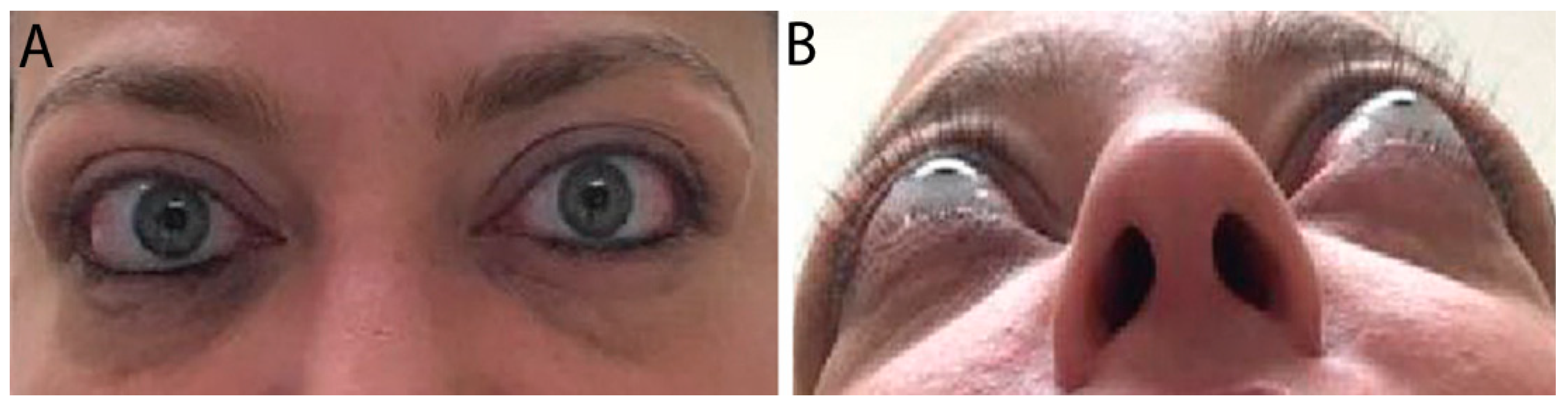
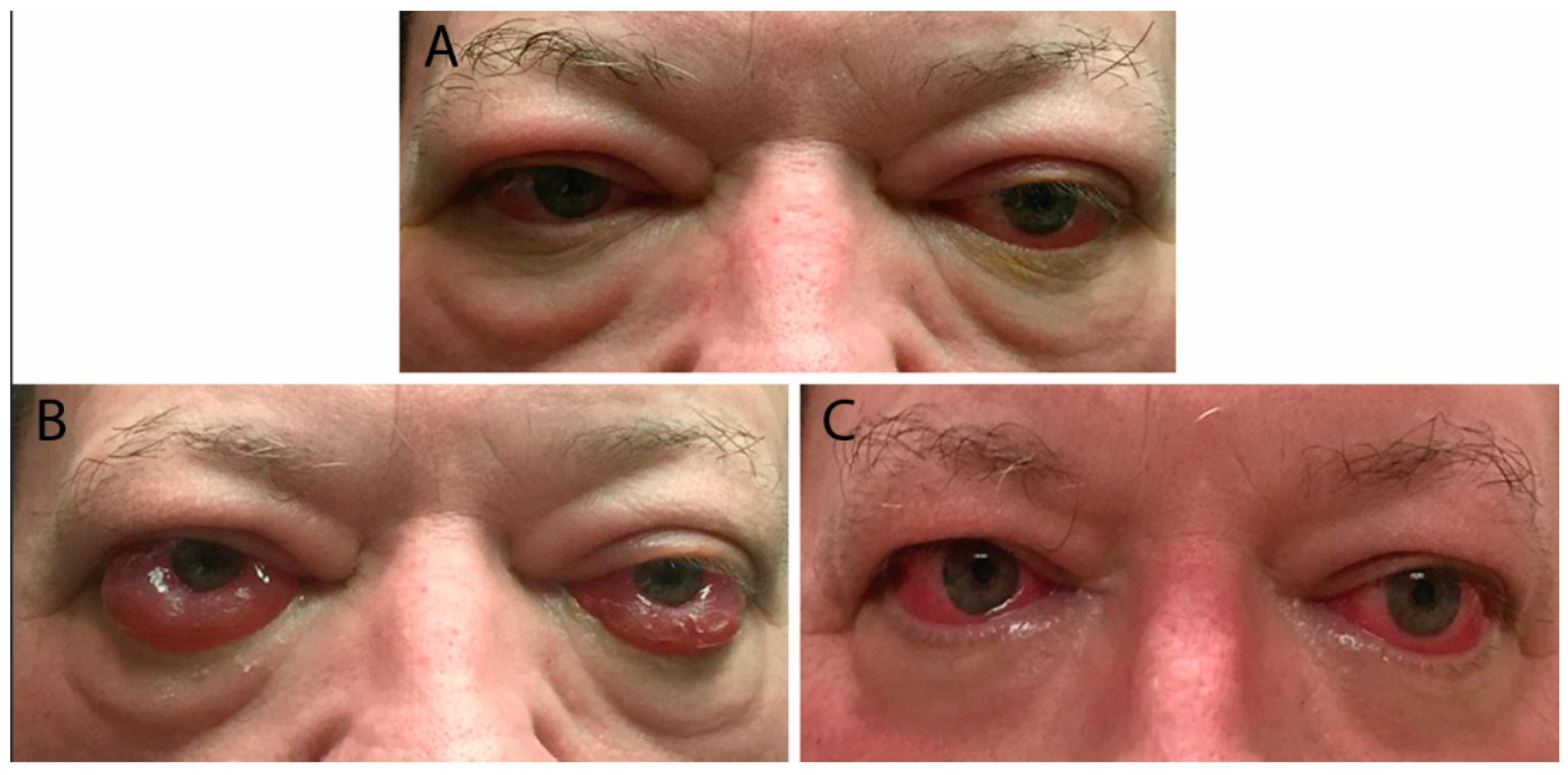
3. Clinical Evaluation
3.1. Mild Disease
3.2. Moderate Disease
3.3. Severe Disease
3.4. Natural Course
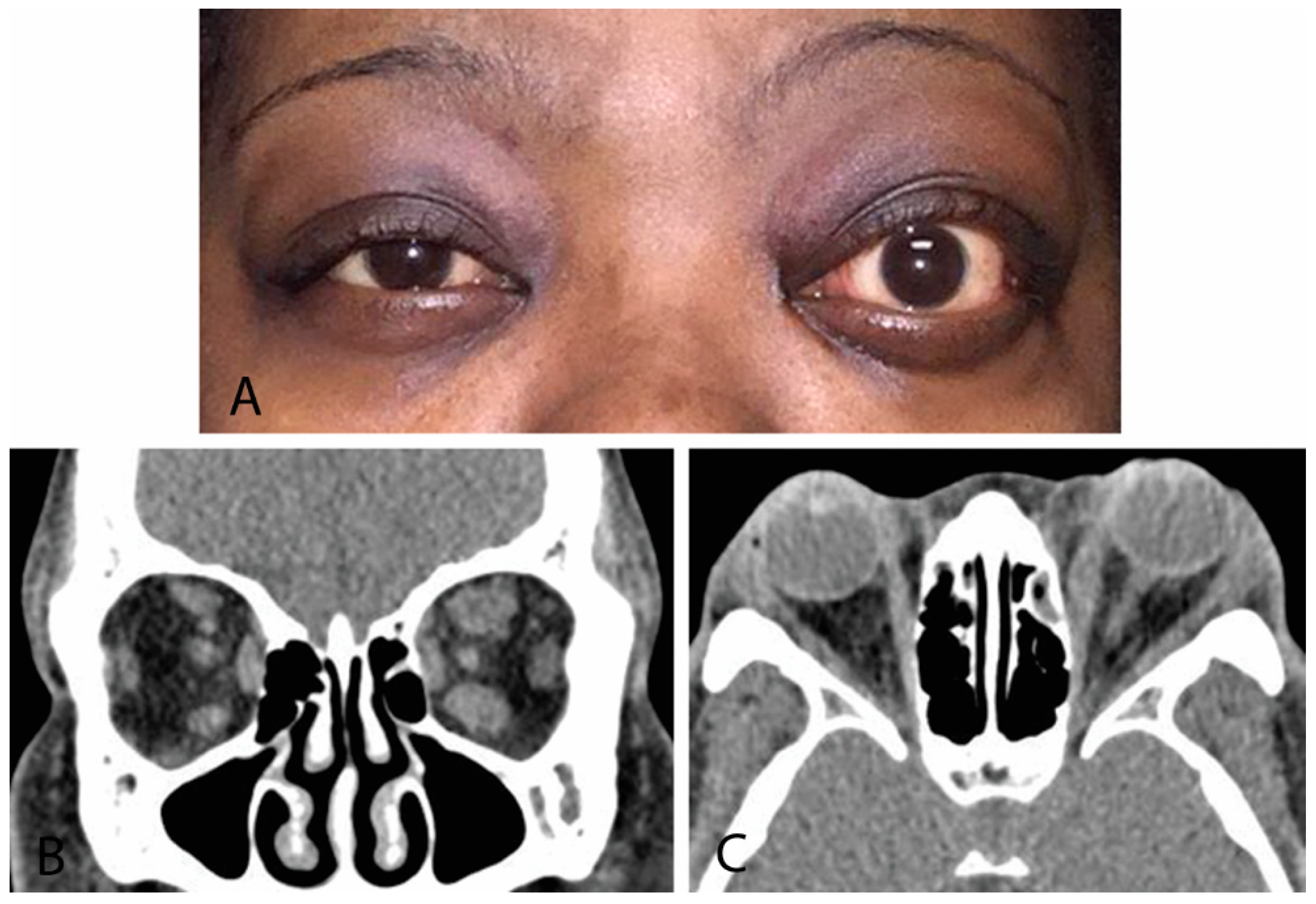
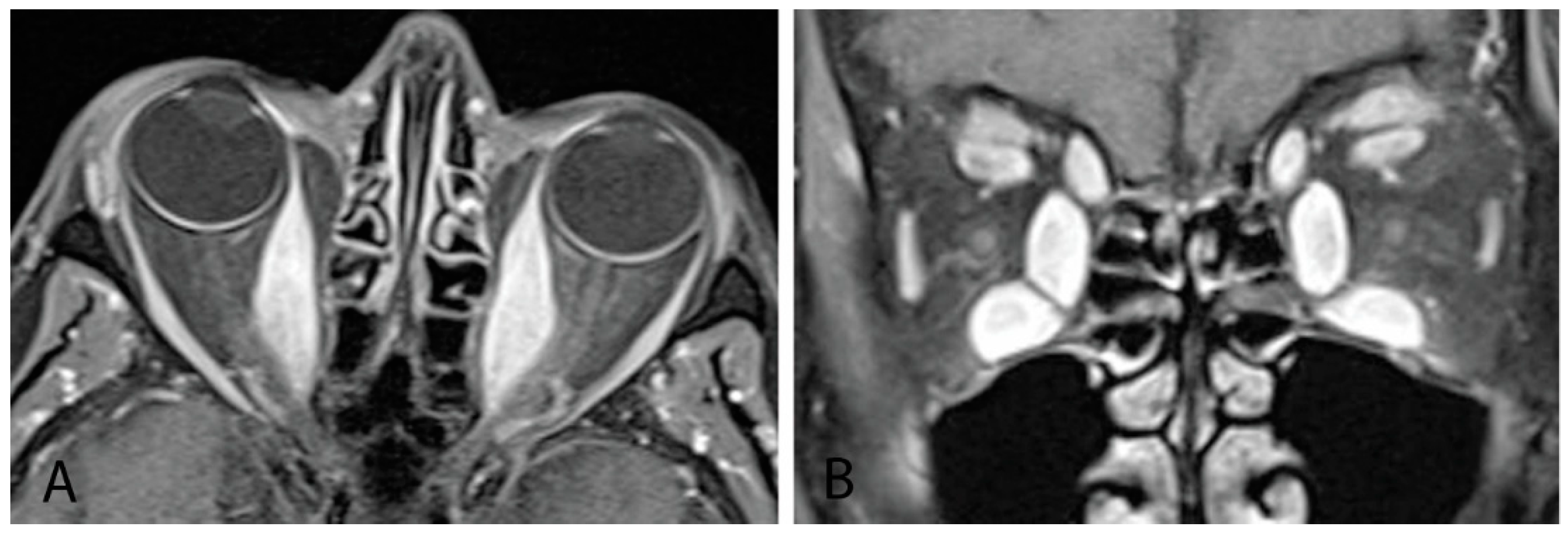
4. Diagnosis
4.1. Differential
4.2. Laboratory Testing
4.3. Imaging
4.3.1. Orbital Ultrasound
4.3.2. Computed Tomography
4.3.3. Magnetic Resonance Imaging
4.3.4. Octreoscan
4.3.5. Optical Coherence Tomography (OCT)
4.4. Biopsy
5. Additional Considerations
5.1. Pediatric Thyroid Eye Disease
5.1.1. Epidemiology
5.1.2. Clinical Considerations
5.1.3. Management
5.2. Pregnancy
6. Treatment
6.1. Management of Hyperthyroidism
6.2. Smoking
6.3. Mild Thyroid Eye Disease
6.4. Moderate-to-Severe Thyroid Eye Disease
6.4.1. Acute Disease
Steroids
Radiation
Steroid-Sparing Agents
6.4.2. Chronic Disease
6.5. Vision-Threatening Thyroid Eye Disease
6.5.1. Optic Neuropathy
6.5.2. Severe Corneal Exposure
6.6. Special Considerations
6.6.1. Pediatric Thyroid Eye Disease Treatment
6.6.2. Pregnancy
7. Conclusions
Author Contributions
Funding
Institutional Review Board Statement
Informed Consent Statement
Conflicts of Interest
Nomenclature
| CT | Computed tomography |
| EUGOGO | European Group on Graves’ Orbitopathy |
| IGF-1 | Insulin-like growth factor-1 |
| IL | Interleukin |
| IV | Intravenous |
| IVIG | Intravenous immunoglobulin |
| MRI | Magnetic resonance imaging |
| NOSPECS | N: no signs or symptoms; O: only signs; S: soft-tissue involvement; P: proptosis; E: extraocular muscle involvement; C: corneal involvement; S: sight loss due to optic nerve compression |
| TBII | Thyroid-binding inhibitory immunoglobulin |
| TED | Thyroid eye disease |
| TPO | Thyroid-peroxidase |
| TSAb | Thyrotropin receptor antibody |
| TSH | Thyroid-stimulating hormone |
| TSHR | Thyroid-stimulating hormone receptor |
| TSI | Thyroid-stimulating immunoglobulin |
| VISION | V: vision, optic neuropathy; I: inflammation, congestion; S: strabismus, motility restriction; A: appearance, exposure |
References
- Szczapa-Jagustyn, J.; Gotz-Wieckowska, A.; Kociecki, J. An update on thyroid-associated ophthalmopathy in children and adolescents. J. Pediatr. Endocrinol. Metab. 2016, 29, 1115–1122. [Google Scholar] [CrossRef] [PubMed] [Green Version]
- McAlinden, C. An overview of thyroid eye disease. Eye Vis. 2014, 1, 9. [Google Scholar] [CrossRef] [PubMed] [Green Version]
- Yin, X.; Latif, R.; Bahn, R.; Davies, T.F. Genetic profiling in Graves’ disease: Further evidence for lack of a distinct genetic contribution to Graves’ ophthalmopathy. Thyroid 2012, 22, 730–736. [Google Scholar] [CrossRef] [PubMed]
- Stan, M.N.; Bahn, R.S. Risk factors for development or deterioration of Graves’ ophthalmopathy. Thyroid 2010, 20, 777–783. [Google Scholar] [CrossRef]
- Stan, M.N. Natural History, Risk Factors, and Management of Patients with Mild GO. In Graves’ Disease—A Comprehensive Guide for Clinicians; Bahn, R.S., Ed.; Springer Science+Business Media: New York, NY, USA, 2015; pp. 241–255. [Google Scholar]
- Bahn, R.S. Pathogenesis of Graves’ Orbitopathy. In Graves’ Disease—A Comprehensive Guide for Clinicians; Bahn, R.S., Ed.; Springer Science+Business Media: New York, NY, USA, 2015; pp. 179–185. [Google Scholar]
- Wall, J.R.; Lahooti, H. Pathogenesis of thyroid eye disease—Does autoimmunity against the TSH receptor explain all cases? Endokrynol. Pol. 2010, 61, 222–227. [Google Scholar]
- Perros, P.; Dickinson, A.J.; Kendall-Taylor, P. Clinical Presentation and Natural History of Graves’ Ophthalmopathy. In Thyroid Eye Disease; Bahn, R.S., Ed.; Springer Science+Business Media: New York, NY, USA, 2001; pp. 119–136. [Google Scholar]
- Yamada, M.; Li, A.W.; Wall, J.R. Thyroid-associated ophthalmopathy: Clinical features, pathogenesis, and management. Crit. Rev. Clin. Lab. Sci. 2000, 37, 523–549. [Google Scholar] [CrossRef]
- Stagnaro-Green, A. Graves’ Disease and Pregnancy. In Graves’ Disease—A Comprehensive Guide for Clinicians; Bahn, R.S., Ed.; Springer Science+Business Media: New York, NY, USA, 2015; pp. 167–178. [Google Scholar]
- Kahaly, G.J.; Muller-Forell, W.; Forster, G.J.; Pitz, S.; Rosier, H.P.; Mann, W.J. Imaging in Graves’ Ophthalmopathy. In Thyroid Eye Disease; Bahn, R.S., Ed.; Springer Science+Business Media: New York, NY, USA, 2001; pp. 137–162. [Google Scholar]
- Merino, P.; de Liano, P.G.; Ruiz, Y.; Franco, G. Atypical restrictive strabismus secondary to an anomalous orbital structure: Differential diagnosis. Strabismus 2012, 20, 162–165. [Google Scholar] [CrossRef]
- Muller-Forell, W.; Kahaly, G.J. Neuroimaging of Graves’ orbitopathy. Best Pract. Res. Clin. Endocrinol. Metab. 2012, 26, 259–271. [Google Scholar] [CrossRef]
- Guo, J.; Li, X.; Ma, R.; Gan, L.; Qian, J. The changes of retinal nerve fibre layer and ganglion cell layer with different severity of thyroid eye disease. Eye 2022, 36, 129–134. [Google Scholar] [CrossRef]
- Jamshidian Tehrani, M.; Mahdizad, Z.; Kasaei, A.; Fard, M.A. Early macular and peripapillary vasculature dropout in active thyroid eye disease. Graefes. Arch. Clin. Exp. Ophthalmol. 2019, 257, 2533–2540. [Google Scholar] [CrossRef]
- Kurt, M.M.; Akpolat, C.; Evliyaoglu, F.; Yilmaz, M.; Ordulu, F. Evaluation of Retinal Neurodegeneration and Choroidal Thickness in Patients with Inactive Graves’ Ophthalmopathy. Klin. Monbl. Augenheilkd. 2021, 238, 797–802. [Google Scholar] [PubMed]
- Rivkees, S.A. Graves’ Disease in Childhood. In Graves’ Disease—A Comprehensive Guide for Clinicians; Bahn, R.S., Ed.; Springer Science+Business Media: New York, NY, USA, 2015; pp. 147–166. [Google Scholar]
- Wall, J.R.; Lahooti, H.; Hibbert, E.J.; Champion, B. Relationship between Clinical and Immunological Features of Thyroid Autoimmunity and Ophthalmopathy during Pregnancy. J. Thyroid Res. 2015, 2015, 698470. [Google Scholar] [CrossRef] [PubMed] [Green Version]
- Abbouda, A.; Trimboli, P.; Bruscolini, A. A mild Grave’s ophthalmopathy during pregnancy. Semin Ophthalmol. 2014, 29, 8–10. [Google Scholar] [CrossRef]
- Bartalena, L.; Kahaly, G.J.; Baldeschi, L.; Dayan, C.M.; Eckstein, A.; Marcocci, C.; Marinò, M.; Vaidya, B.; Wiersinga, W.M.; EUGOGO. The 2021 European Group on Graves’ orbitopathy (EUGOGO) clinical practice guidelines for the medical management of Graves’ orbitopathy. Eur. J. Endocrinol. 2021, 185, G43–G67. [Google Scholar] [CrossRef] [PubMed]
- Taylor, P.N.; Zhang, L.; Lee, R.W.J.; Muller, I.; Ezra, D.G.; Dayan, C.M.; Kahaly, G.J.; Ludgate, M. New insights into the pathogenesis and nonsurgical management of Graves orbitopathy. Nat. Rev. Endocrinol. 2020, 16, 104–116. [Google Scholar] [CrossRef] [PubMed]
- Khong, J.J.; McNab, A. Medical treatment in thyroid eye disease in 2020. Br. J. Ophthalmol. 2021, 105, 299–305. [Google Scholar] [CrossRef]
- Hall, A.J.H.; Topliss, D.J. Medical and surgical treatment of thyroid eye disease. Intern Med. J. 2022, 52, 14–20. [Google Scholar] [CrossRef]
- Thornton, J.; Kelly, S.P.; Harrison, R.A.; Edwards, R. Cigarette smoking and thyroid eye disease: A systematic review. Eye 2007, 21, 1135–1145. [Google Scholar] [CrossRef]
- Marcocci, C.; Kahaly, G.J.; Krassas, G.E.; Bartalena, L.; Prummel, M.; Stahl, M.; Altea, M.A.; Nardi, M.; Pitz, S.; Boboridis, K.; et al. Selenium and the course of mild Graves’ orbitopathy. N. Engl. J. Med. 2011, 364, 1920–1931. [Google Scholar] [CrossRef] [Green Version]
- Niskar, A.S.; Paschal, D.C.; Kieszak, S.M.; Flegal, K.M.; Bowman, B.; Gunter, E.W.; Pirkle, J.L.; Rubin, C.; Sampson, E.J.; McGeehin, M. Serum selenium levels in the US population: Third National Health and Nutrition Examination Survey, 1988–1994. Biol. Trace Elem. Res. 2003, 91, 1–10. [Google Scholar] [CrossRef]
- Verity, D.H.; Rose, G.E. Acute thyroid eye disease (TED): Principles of medical and surgical management. Eye 2013, 27, 308–319. [Google Scholar] [CrossRef]
- Perumal, B.; Meyer, D.R. Treatment of severe thyroid eye disease: A survey of the American Society of Ophthalmic Plastic and Reconstructive Surgery (ASOPRS). Ophthal. Plast. Reconstr. Surg. 2015, 31, 127–131. [Google Scholar] [CrossRef]
- Shams, P.N.; Ma, R.; Pickles, T.; Rootman, J.; Dolman, P.J. Reduced risk of compressive optic neuropathy using orbital radiotherapy in patients with active thyroid eye disease. Am. J. Ophthalmol. 2014, 157, 1299–1305. [Google Scholar] [CrossRef] [PubMed]
- Chundury, R.V.; Weber, A.C.; Perry, J.D. Orbital Radiation Therapy in Thyroid Eye Disease. Ophthal. Plast. Reconstr. Surg. 2016, 32, 83–89. [Google Scholar] [CrossRef] [PubMed]
- Stiebel-Kalish, H.; Robenshtok, E.; Hasanreisoglu, M.; Ezrachi, D.; Shimon, I.; Leibovici, L. Treatment modalities for Graves’ ophthalmopathy: Systematic review and metaanalysis. J. Clin. Endocrinol. Metab. 2009, 94, 2708–2716. [Google Scholar] [CrossRef] [PubMed] [Green Version]
- Bartalena, L.; Marcocci, C.; Bogazzi, F.; Panicucci, M.; Lepri, A.; Pinchera, A. Use of corticosteroids to prevent progression of Graves’ ophthalmopathy after radioiodine therapy for hyperthyroidism. N. Engl. J. Med. 1989, 321, 1349–1352. [Google Scholar] [CrossRef]
- Ginter, A.; Migliori, M.E. The Role of Biological Agents and Immunomodulators in Treatment Strategies for Thyroid Eye Disease: An Evidence-based Review. RI Med. J. 2016, 99, 26–29. [Google Scholar]
- Ayabe, R.; Rootman, D.B.; Hwang, C.J.; Ben-Artzi, A.; Goldberg, R. Adalimumab as steroid-sparing treatment of inflammatory-stage thyroid eye disease. Ophthal. Plast. Reconstr. Surg. 2014, 30, 415–419. [Google Scholar] [CrossRef]
- Stan, M.N.; Garrity, J.A.; Carranza Leon, B.G.; Prabin, T.; Bradley, E.A.; Bahn, R.S. Randomized controlled trial of rituximab in patients with Graves’ orbitopathy. J. Clin. Endocrinol. Metab. 2015, 100, 432–441. [Google Scholar] [CrossRef] [Green Version]
- Salvi, M.; Vannucchi, G.; Curro, N.; Campi, I.; Covelli, D.; Dazzi, D.; Simonetta, S.; Guastella, C.; Pignataro, L.; Avignone, S.; et al. Efficacy of B-cell targeted therapy with rituximab in patients with active moderate to severe Graves’ orbitopathy: A randomized controlled study. J. Clin. Endocrinol. Metab. 2015, 100, 422–431. [Google Scholar] [CrossRef]
- Perez-Moreiras, J.V.; Alvarez-Lopez, A.; Gomez, E.C. Treatment of active corticosteroid-resistant graves’ orbitopathy. Ophthal. Plast. Reconstr. Surg. 2014, 30, 162–167. [Google Scholar] [CrossRef] [PubMed]
- Smith, T.J.; Kahaly, G.J.; Ezra, D.G.; Fleming, J.C.; Dailey, R.A.; Tang, R.A.; Harris, G.J.; Antonelli, A.; Salvi, M.; Goldberg, R.A.; et al. Teprotumumab for Thyroid-Associated Ophthalmopathy. N. Engl. J. Med. 2017, 376, 1748–1761. [Google Scholar] [CrossRef] [PubMed]
- Douglas, R.S.; Kahaly, G.J.; Patel, A.; Sile, S.; Thompson, E.H.Z.; Perdok, R.; Fleming, J.C.; Fowler, B.T.; Marcocci, C.; Marinò, M.; et al. Teprotumumab for the treatment of active thyroid eye disease. N. Engl. J. Med. 2020, 382, 341–352. [Google Scholar] [CrossRef] [PubMed]
- Douglas, R.S.; Dailey, R.; Subramanian, P.S.; Barbesino, G.; Ugradar, S.; Batten, R.; Qadeer, R.A.; Cameron, C. Proptosis and Diplopia Response with Teprotumumab and Placebo vs the Recommended Treatment Regimen with Intravenous Methylprednisolone in Moderate to Severe Thyroid Eye Disease: A Meta-analysis and Matching-Adjusted Indirect Comparison. JAMA Ophthalmol. 2022, 140, 328–335. [Google Scholar] [CrossRef] [PubMed]
- Li, H.; Yang, L.; Song, Y.; Zhao, X.; Sun, C.; Zhang, L.; Zhao, H.; Pan, Y. Comparative effectiveness of different treatment modalities for active, moderate-to-severe Graves’ orbitopathy: A systematic review and network meta-analysis. Acta Ophthalmol. 2021, 100, e1189–e1198. [Google Scholar] [CrossRef]
- Kang, J.; Lechuga, M.; Braun, J.; Kossler, A.; Douglas, R.; Cockerham, K. Infusion Center Guidelines for Teprotumumab Infusions: Informed Consent, Safety, and Management of Side Effects. J. Infus. Nurs. 2021, 44, 331–338. [Google Scholar] [CrossRef]
- Sears, C.M.; Wang, Y.; Bailey, L.A.; Turbin, R.; Subramanian, P.S.; Douglas, R.; Cockerham, K.; Kossler, A.L. Early efficacy of teprotumumab for the treatment of dysthyroid optic neuropathy: A multicenter study. Am. J. Ophthalmol. Case Rep. 2021, 23, 101111. [Google Scholar] [CrossRef]
- Chiou, C.A.; Reshef, E.R.; Freitag, S.K. Teprotumumab for the treatment of mild compressive optic neuropathy in thyroid eye disease: A report of two cases. Am. J. Ophthalmol. Case Rep. 2021, 22, 101075. [Google Scholar] [CrossRef]
- Douglas, R.S.; Kahaly, G.J.; Ugradar, S.; Elflein, H.; Ponto, K.A.; Fowler, B.T.; Dailey, R.; Harris, G.J.; Schiffman, J.; Tang, R.; et al. Teprotumumab Efficacy, Safety, and Durability in Longer-Duration Thyroid Eye Disease and Re-treatment: OPTIC-X Study. Ophthalmology 2022, 129, 438–449. [Google Scholar] [CrossRef]
- Ugradar, S.; Kang, J.; Kossler, A.L.; Zimmerman, E.; Braun, J.; Harrison, A.R.; Bose, S.; Cockerham, K.; Douglas, R.S. Teprotumumab for the treatment of chronic thyroid eye disease. Eye 2022, 36, 1553–1559. [Google Scholar] [CrossRef]
- Ozzello, D.J.; Kikkawa, D.O.; Korn, B.S. Early experience with teprotumumab for chronic thyroid eye disease. Am. J. Ophthalmol. Case Rep. 2020, 19, 100744. [Google Scholar] [CrossRef] [PubMed]
- Ozzello, D.J.; Dallalzadeh, L.O.; Liu, C.Y. Teprotumumab for chronic thyroid eye disease. Orbit 2022, 41, 539–546. [Google Scholar] [CrossRef] [PubMed]
- Roopenian, D.C.; Akilesh, S. FcRn: The neonatal Fc receptor comes of age. Nat. Rev. Immunol. 2007, 7, 715–725. [Google Scholar] [CrossRef] [PubMed]
- Prat, M.C.; Braunstein, A.L.; Dagi Glass, L.R.; Kazim, M. Orbital fat decompression for thyroid eye disease: Retrospective case review and criteria for optimal case selection. Ophthal. Plast. Reconstr. Surg. 2015, 31, 215–218. [Google Scholar] [CrossRef] [Green Version]
- Graham, S.M.; Brown, C.L.; Carter, K.D.; Song, A.; Nerad, J.A. Medial and lateral orbital wall surgery for balanced decompression in thyroid eye disease. Laryngoscope 2003, 113, 1206–1209. [Google Scholar] [CrossRef]
- Boboridis, K.G.; Uddin, J.; Mikropoulos, D.G.; Bunce, C.; Mangouritsas, G.; Voudouragkaki, I.C.; Konstas, A.G. Critical Appraisal on Orbital Decompression for Thyroid Eye Disease: A Systematic Review and Literature Search. Adv. Ther. 2015, 32, 595–611. [Google Scholar] [CrossRef] [Green Version]
- Wakelkamp, I.M.; Baldeschi, L.; Saeed, P.; Mourits, M.P.; Prummel, M.F.; Wiersinga, W.M. Surgical or medical decompression as a first-line treatment of optic neuropathy in Graves’ ophthalmopathy? A randomized controlled trial. Clin. Endocrinol. 2005, 63, 323–328. [Google Scholar] [CrossRef]
- Uddin, J.M.; Davies, P.D. Treatment of upper eyelid retraction associated with thyroid eye disease with subconjunctival botulinum toxin injection. Ophthalmology 2002, 109, 1183–1187. [Google Scholar] [CrossRef]
- Fraser, F.C.; Sajoo, A. Teratogenic potential of corticosteroids in humans. Teratology 1995, 51, 45–46. [Google Scholar] [CrossRef]
- Stafford, I.P.; Dildy, G.A., 3rd; Miller, J.M. Severe Graves’ ophthalmopathy in pregnancy. Obstet. Gynecol. 2005, 105, 1221–1223. [Google Scholar] [CrossRef]


Publisher’s Note: MDPI stays neutral with regard to jurisdictional claims in published maps and institutional affiliations. |
© 2022 by the authors. Licensee MDPI, Basel, Switzerland. This article is an open access article distributed under the terms and conditions of the Creative Commons Attribution (CC BY) license (https://creativecommons.org/licenses/by/4.0/).
Share and Cite
Rashad, R.; Pinto, R.; Li, E.; Sohrab, M.; Distefano, A.G. Thyroid Eye Disease. Life 2022, 12, 2084. https://doi.org/10.3390/life12122084
Rashad R, Pinto R, Li E, Sohrab M, Distefano AG. Thyroid Eye Disease. Life. 2022; 12(12):2084. https://doi.org/10.3390/life12122084
Chicago/Turabian StyleRashad, Ramy, Raquel Pinto, Emily Li, Mahsa Sohrab, and Alberto G. Distefano. 2022. "Thyroid Eye Disease" Life 12, no. 12: 2084. https://doi.org/10.3390/life12122084