Article

Two Novel Functional Mutations in Promoter Region of SCN3B Gene Associated with Atrial Fibrillation

1
Department of Clinical Laboratory, Liyuan Hospital, Tongji Medical College, Huazhong University of Science and Technology, Wuhan 430077, China
2
Key Laboratory of Molecular Biophysics of the Ministry of Education, Cardio-X Institute, College of Life Science and Technology, Huazhong University of Science and Technology, Wuhan 430074, China
3
Human Genome Research Center, College of Life and Science and Technology, Huazhong University of Science and Technology, Wuhan 430074, China
4
Liyuan Cardiovascular Center, Liyuan Hospital, Tongji Medical College, Huazhong University of Science and Technology, Wuhan 430077, China
*
Authors to whom correspondence should be addressed.
These authors contributed equally to this work.
Life 2022, 12(11), 1794; https://doi.org/10.3390/life12111794
Submission received: 28 September 2022 / Revised: 1 November 2022 / Accepted: 3 November 2022 / Published: 5 November 2022
(This article belongs to the Special Issue The Molecular Mechanism of Cardiovascular Disease)

Abstract

:
The sodium voltage-gated channel beta subunit 3 (SCN3B) plays a crucial role in electrically excitable cells and conduction tissue in the heart. Some previous studies have established that genetic modification in sodium voltage-channel genes encoding for the cardiac β-subunits, such as SCN1B, SCN2B, SCN3B and SCN4B, can result in atrial fibrillation (AF). In the current study, we identified two rare variants in 5′UTR (NM_018400.4: c.-324C>A, rs976125894 and NM_018400.4: c.-303C>T, rs1284768362) of SCN3B in two unrelated lone AF patients. Our further functional studies discovered that one of them, the A allele of c.-324C>A (rs976125894), can improve transcriptional activity and may raise SCN3B expression levels. The A allele of c.-324C>A (rs976125894) has higher transcriptional activity when it interacts with GATA4, as we confirmed transcription factor GATA4 is a regulator of SCN3B. To the best of our knowledge, the current study is the first to demonstrate that the gain-of-function mutation of SCN3B can produce AF and the first to link a mutation occurring in the non-coding 5′UTR region of SCN3B to lone AF. The work also offers empirical proof that GATA4 is a critical regulator of SCN3B gene regulation. Our findings may serve as an encyclopedia for AF susceptibility variants and can also provide insight into the investigation of the functional mechanisms behind AF variants discovered by genetic methods.

1. Introduction

Atrial Fibrillation (AF), characterized by a rapid and chaotic rhythm in atria, including the absence of P waves, irregular RR intervals and a fast-atrial rate of up to 300 beats per minute, is the most common clinically sustained cardiac arrhythmia [1]. AF occurs when the normal cycle of electrical impulses is interrupted, resulting in a disorganized quiver of the atria and an irregular contraction of the ventricles. With a 0.7% population prevalence, AF affects about 8 million people in China, 2.2 million people in the United States and 4.5 million people in the European Union. The estimated frequency of AF in people over 65 is 8.6% and its incidence has remained much higher in the elderly population [2]. The risk of stroke is increased five-fold by AF, while the risk of congestive heart failure is increased three-fold. AF is also a well-known independent risk factor for stroke and can aggravate heart failure. According to recent data, AF doubles to triples the chance of sudden cardiac death [3].
AF is often associated with complications, such as coronary artery disease (CAD), hypertension, valvular heart disease, hyperthyroidism, heart failure and structural heart diseases. In the Framingham cohort and after age adjustment, hypertension, diabetes mellitus, coronary artery disease and smoking were the main risk factors for non-valvular AF [4]. The combined effects of these factors could explain 44% of the burden of non-valvular AF in men and 58% in women. In some of the cases, AF occurs in the absence of the complications of hypertension, heart failure, coronary heart diseases or other types of cardiac diseases, and is, hence, clinically diagnosed as lone AF [3].
Epidemiological studies revealed that while numbers of prevalent cardiovascular disorders are risk factors for developing AF, genetic components have also been proven to be involved, particularly in the case of lone AF [5]. According to a study of Danish twins, the heritability of AF was as high as 62% [6]. The discovery of a few private, AF-causing mutations in ion channel genes highlights the significant contribution of genetic factors to the etiology of AF. The S140G mutation in the ion channel gene KCNQ1 was first reported by Chen et al. in 2003 [7]. Later, mutations in non-ion channel genes, such as NPPA and NUP155, were found in some patients with lone AF [8,9]. Mutations or rare variants in more than 40 genes are believed to be the cause of familial or sporadic AF and these genes were mapped to pathways of ion channels, ion channel-associated genes, transcription factors, cardiac structural components, signaling regulators and others [10,11]. Loss-of-function rare variants in the TTN gene are enriched among individuals with early-onset AF (2.1% prevalence) versus control subjects (1.1% prevalence; odds ratio, 1.76 (95% CI, 1.04–2.97)) [12]. In addition to the mutations and rare variants, genome-wide association studies (GWAS) have been performed for AF in several populations, which successfully identified about 300 common variants associated with AF [11].
Studies have linked cardiac arrhythmogenesis to mutations in the genes encoding the cardiac sodium channel (NaV1.5) and AF-causing variants have been reported in both the α-subunit (encoded by the SCN5A gene) and associated β-subunits (Navβ) (encoded by the genes SCN1B–SCN4B) [13,14,15,16,17]. SCN1B, the gene encoding for the β1-subunit and SCN2B, the gene encoding for the β2-subunit, have been identified as susceptibility genes for AF [14]. SCN1B and SCN3B that encode for the β3-subunit have been identified as susceptibility genes of Brugada Syndrome (BrS), while SCN4B has been linked to the congenital long-QT syndrome (LQTS) 3, ventricular tachycardia and familial AF [15,16,17,18]. In our previous study, A103V mutation in SCN3B was found to induce lone AF and Olesen et al. also showed that R6K, L10P and M161T mutation of SCN3B was associated with early-onset lone AF [19,20]; however, there are no more reports that link SCN3B mutation to AF.
In the current study, we carried out mutation screening of the SCN3B gene in 355 lone AF patients and found that two patients carried rare variants in 5′UTR (NM_018400.4: c.-324C>A, rs976125894 and NM_018400.4: c.-303C>T, rs1284768362) of SCN3B and our further functional studies revealed that the rare variant c.-324C>A (rs976125894), which is located in the promoter region and enhanced the transcriptional activity, may regulate the expression level of SCN3B through interacting with the GATA4 transcription factor.

2. Materials and Methods

2.1. Study Population

The study subjects included in the present study were enrolled from the GeneID database. All the study patients are of Han ethnic origin by self-description.
AF diagnosis was performed by a panel of cardiologists based on the data from electrocardiograms (ECG) and/or Holter ECG and based on the criteria of the ACC/AHA/ESC AF guidelines. The ECG characteristics of AF include the absence of P waves, replacement of P waves by rapid oscillations of fibrillation waves that varied in size, shape and timing (referred to as “f” waves) and irregular ventricular response (irregular RR intervals) with abnormal atrioventricular conduction. Patients with other types of cardiac arrhythmias (e.g., ventricular arrhythmias), thyroid dysfunction, congenital heart disease, cardiomyopathies and valvulopathies were excluded. An AF patient was diagnosed as being affected with “lone AF” if he/she had no CAD, congestive heart failure, hypertension or diabetes and was below the age of 55 years at the first diagnostics of AF.
Demographic and clinical information on age, gender, CAD, hypertension and diabetes mellitus was obtained from medical records. CAD was defined as ongoing therapy of CAD, 70% luminal narrowing in at least one vessel by coronary angiography, percutaneous coronary angioplasty, coronary artery bypass graft and MI. Hypertension was defined as ongoing medication for hypertension, systolic blood pressure of ≥140 mmHg or diastolic blood pressure of ≥90 mmHg. Diabetes mellitus was defined as ongoing therapy of diabetes or a fasting plasma glucose level of ≥7.0 mmol/L.
The study protocol was approved by Ethics Committee on Human Subject Research at Huazhong University of Science and Technology (HUST) and Liyuan Hospital Affiliated to Tongji Medical College of HUST and also conforms to the principles outlined in the Declaration of Helsinki. Written informed consent was obtained from all subjects.

2.2. Mutation Analysis of SCN3B

Genomic DNA was isolated from peripheral blood samples using standard protocols with the Wizard Genomic DNA Purification Kit (Promega, Madison, WI, USA). Direct Sanger DNA sequencing was used for screening mutations as described previously. All the six exons of SCN3B (NM_018400), including the coding region, 5′ untranslated region, 3′ untranslated region and the boundaries of intron–exon were amplified by polymerase chain reactions (PCR) from AF patient DNA samples and sequenced. The primers for PCR are shown in Table 1. Mutation analysis of the SCN3B gene was performed in all study subjects to identify a novel mutation responsible for AF. A variant was defined as a mutation if present in less than 0.01% in the general population according to the public database.
The sequencing of SCN3B in unrelated controls was checked by high-resolution melt analysis (HRM) utilizing a Rotor-Gene 6000 High-Resolution Melt system (Corbett Life Science, Sydney, NSW, Australia), as previously disclosed by us, to validate the mutation frequency in the Chinese common population. Primers for PCR of HRM are shown in Table 1. Ten samples were randomly selected for direct Sanger sequencing to verify the accuracy of HRM genotyping.

2.3. Bioinformatics-Based Prediction

The deleterious potential of the identified mutations was measured using Functional Analysis through Hidden Markov Models (FATHMM) (http://fathmm.biocompute.org.uk/ accessed on 5 August 2022), GERP++ (http://mendel.stanford.edu/SidowLab/downloads/gerp/index.html accessed on 5 August 2022) and regulatory Mendelian mutation (ReMM) (https://charite.github.io/software-remm-score.html accessed on 5 August 2022).
The possible regulatory features of the variants in the non-coding region were predicted by Ensembl database (http://asia.ensembl.org/ accessed on 5 August 2022) and ENCODE data of UCSC genome browser (https://genome.ucsc.edu accessed on 5 August 2022). The Human Transcription Factor Database (http://bioinfo.life.hust.edu.cn/HumanTFDB#!// accessed on 15 August 2022) and JASPAR (https://jaspar.genereg.net accessed on 15 August 2022) were applied to analyze and predict the potential transcription factors binding to the area of the SNP.

2.4. Clones and Site-Directed Mutagenesis

To further explore whether the genomic region overlapping > (rs976125894) or c.-303C>T (rs1284768362) has regulatory activity and is a potential promoter, the wild-type 858 bp length genomic fragment overlapping rs976125894 and rs1284768362 was generated by PCR analysis using human genomic DNA as a template. The Prime STAR HS DNA polymerase (Takara, Dalian, China) was applied and PCR primers were SCN3B-Kpn I-F 5′-GATTCCCAGGGCTGACAGCACACACGG-3′ and SCN3B-HindⅢ-R 5′-AAGCTTGAAGCCGCCAGCCCCAGAAG-3′. The wild-type 858 bp length PCR products were digested with restriction enzyme KpnI and HindIII (Takara, Dalian, China) and then sub-cloned into the multiple cloning site of the pGL3-basic luciferase vector (Promega, Madison, WI, USA), resulting in pGL3-SCN3B-WT.
The two variants, c.-324 C>A (rs976125894) and c.-303 C>T (rs1284768362), were introduced into the pGL3-SCN3B-WT plasmid by using PCR-based site-directed mutagenesis, referred to as pGL3-Mut-324A and pGL3-Mut-303T, respectively. The primer sequences for c.-324 C>A were 5′-GAGCGAGGCAGGGGAGCGAGTGGAAGCTGG-3′ (forward) and 5′-CCAGCTTCCACTCGCTCCCCTGCCTCGCTC (reverse). The primer sequences for c.-303 C>T were 5′-AGTGGAAGCTGGAGTTCTGGGGTGGGCGGGGAGGC-3′ (forward) and 5′-GCCTCCCCGCCCACCCCAGAACTCCAGCTTCCACT-3′ (reverse). The PCR products were digested by DpnⅠ (Takara, Dalian, China) in a 37 °C incubator for 6 h.
The 5′ UTR contained wild-type c.-324C or mutant A, plus the whole CDS (delete the stop codon) of SCN3B was amplified and digested by EcoRI and BamHI (Takara, Dalian, China) and then sub-cloned into pEGFP-N1 vector (named pEGFP-N1-WT-324C and pEGFP-N1-Mut-324A) before the ORF of EGFP to validate whether the mutant can regulate the expression of SCN3B. The primer sequences are 5′-GGCGACCGGTGGATCCCGTTCCTCCACTGGTACCGCAGAG-3′ (forward) and 5′-CTCAAGCTTCGAATTCTGCAGGGCTGACAGCACAC-3′ (reverse).
pcDNA3.1, pcDNA3.1-GATA4, pcDNA3.1-IKZF1 and pcDNA3.1-WT1 were reserved in the plasmid library from our laboratory. All constructions were confirmed by Sanger sequencing.

2.5. Dual Luciferase Reporter Assays

Human embryonic kidney 293T cells (HEK293T) and rat cardiomyoblast cells (H9C2) were cultured in 24-well plates, at a density of 105 cells per well and transfected at 37 °C with 5% CO2 of 24 h seeding in Dulbecco’s modified Eagle’s medium (DMEM, Gibco, USA) supplemented with 10% fetal bovine serum (FBS, Gibco, USA). Further, 25ng Renilla luciferase control vector was co-transfected with (1) 200ng pGL3-basic, (2) 200ng pGL3-WT, (3) 200ng pGL3-Mut-324A or 200ng pGL3-Mut-303T. For transcription factor co-transfection experiments, 25ng Renilla luciferase control vector was co-transfected with (1) 100ng pcDNA3.1 + 100ng pcDNA3.1-GATA4 or pcDNA3.1-IKZF1 or pcDNA3.1-WT1, (2) 100ng pGL3-WT + 100ng pcDNA3.1-GATA4 or pcDNA3.1-IKZF1 or pcDNA3.1-WT1, (3) 100ng pGL3-Mut-324A + 100ng pcDNA3.1-GATA4 or pcDNA3.1-IKZF1 or pcDNA3.1-WT1. Lipofectamine 2000 (Invitrogen, Thermo Fisher Scientific, Waltham, MA, USA) was used. Cells were harvested in 48 h. Firefly luciferase signal intensities and Renilla luciferase activities were measured by using the Dual-Glo Luciferase Assay System (Promega, Madison, WI, USA) on GloMax 20/20 (Promega, Madison, WI, USA). Renilla luciferase activity was normalized by relative luciferase intensity. Independent experiments were performed in triplicate at minimum for wild types and mutant types.

2.6. Western Blotting

HEK293T cells were plated in 12-well plates in a humidified incubator at 37 °C and 5% CO2. After 24 h seeding in DMEM with 10% of FBS, at a density of 105 cells per well, cells were transfected with (1) 1 μg pEGFP-N1, (2) 1 μg pEGFP-N1-WT-324C, (3) 1 μg pEGFP-N1-Mut-324A, (4) 500 ng pEGFP-N1 + 500 ng pcDNA3.1-GATA4, (5) 500 ng pEGFP-N1-WT-324-C + 500 ng pcDNA3.1-GATA4 and (6) 500 ng pEGFP-N1-Mut-324A + 500 ng pcDNA3.1-GATA4. Lipofectamine 2000 was used. Cells were collected using cell lysis buffer after 48 h feeding. SCN3B (Novus, Littleton, CO, USA) and GAPDH (Abcam, Cambridge, UK) antibodies were used for Western blotting.

2.7. RNA Isolation and Quantitative RT-PCR

Total RNA was isolated with RNAiso Plus TRIZOL (Takara, Dalian, China) and was reverse transcribed using HiScriptⅢ RT SuperMix for qPCR (Vazyme, Nanjing, China), according to the manufacturer’s protocol. qRT-PCR analysis was conducted using MonAmp qPCR SYBR Green Mix (Monad, Suzhou, China) in an Applied Biosystems instrument (Thermo Fisher, Waltham, MA, USA). qPCR-primers are listed in Table 2.

2.8. Statistical Analysis

For analysis of promoter activity, data were presented as means ± standard deviations from at least three independent experiments. Statistical multiple-group comparisons were evaluated with a one-way ANOVA test with Turkey’s post hoc test and the difference between two groups was tested using Student’s unpaired t-test. A value of p < 0.05 was considered statistically significant. Data analysis was performed using GraphPad Prism software version 6 (GraphPad 6, San Diego, CA, USA).

3. Results

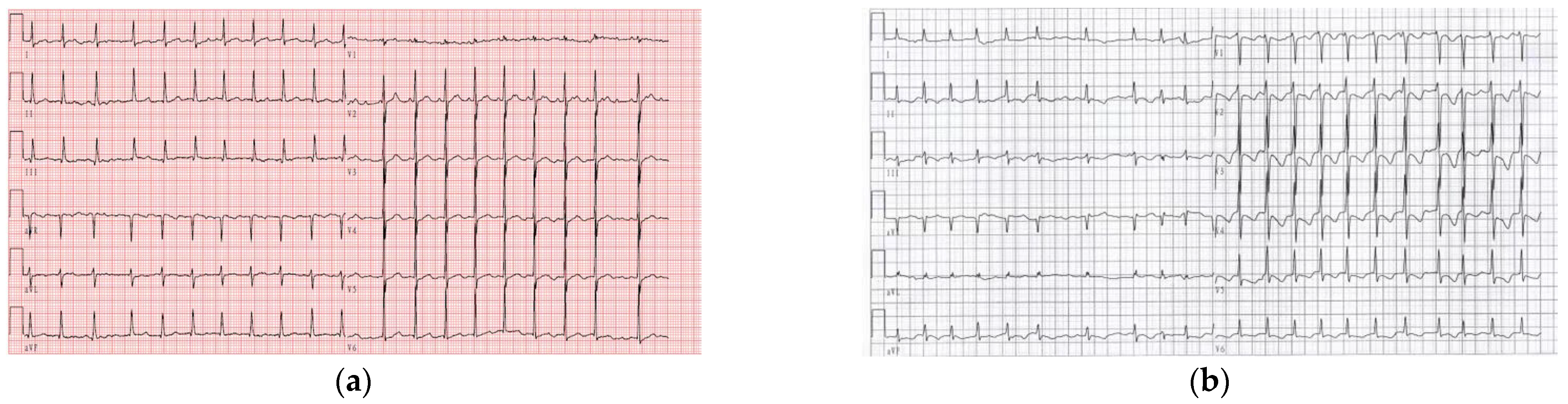
3.1. Identification of Two Novel Susceptibility Mutations in SCN3B

We performed a mutation screening of SCN3B in 355 lone AF sporadic patients selected from the GeneID database to confirm whether SCN3B is the causative gene for AF. The basic clinical characteristics of the study patients are shown in Table 3.
After filtering by the allele frequency (<0.01% in the general population according to the public database), we identified two susceptibility rare variants, c.-324C>A (NM_018400.4, rs976125894) and c.-303C>T (NM_018400.4, rs1284768362), in the first exon and the 5′UTR region of SCN3B gene in two unrelated patients clinically diagnosed as lone AF (Figure 1a,b). The patient who carried the c.-324C>A mutation (rs976125894) is a female who was first discovered to have lone AF at the age of 41 and the patient who carried the c.-303C>T mutation (rs1284768362) is a man who was first found to have lone AF at the age of 46. We tried to obtain genomic DNA samples from each parent of these two SCN3B mutation carriers in order to determine if the mutations were de novo or germline inherited. While DNA from the parents of subjects who carried s c.-303C>T mutation (rs1284768362) was not accessible, we were successful in getting genomic DNA from the parents of the subject who carried the c.-324C>A (rs976125894) mutation. The results demonstrated that the mutation c.-324C>A (rs976125894) originated de novo as the parents showed an homozygous wild-type genotype and without AF (Figure 1c). The ECGs of two patients are shown in Figure 2.
According to the public database, both variants were found in exon 1 and were incredibly rare in the general population. In the public databases, such as the gnomAD database (1 allele in mutation carriers in a total of 152,180 alleles, with a frequency of 6.57 × 10⁻⁶) and the TOPMED database (1/125568, 7.96 × 10⁻⁶), the frequency of the mutation A allele of the c.-324C>A (rs976125894) variant was incredibly low. For the c.-303C>T (rs1284768362) variant, there was no record in the gnomAD database and we found only two carriers in the TOPMED database (2/125568). We also performed validation of the frequency of the two variants in the Chinese Han population and the results indicated that none of the mutations were present in control group containing 500 subjects without AF.
Using software that can predict the effects of non-coding variants, we conducted in silico analyses to determine whether the mutations are deleterious. The results indicated that the mutation c.-324C>A (rs976125894) may be deleterious based on the fathmm MKL non-coding score (0.993), GERP++ RS rankscore (0.997) and ReMM score of 0.984. Only fathmm MKL score revealed deleterious effects (0.915) for the c.-303C>T (rs1284768362) variant, but not in others.

3.2. Transcriptional Activity and Expression Level Enhanced in the Alternative a Allele of Rare Variant c.-324C>A (rs976125894)

Both the identified AF-related variants, rs1284768362 and rs976125894, are in the 5′UTR of SCN3B mRNA. The regulatory feature records in the Ensembl database indicated that both rs1284768362 and rs976125894 were active promoters in several tissues and cell types, including cardiac muscle (Ensembl regulatory feature ENSR00000445310). We next assess whether the mutant alleles of these two 5′-UTR rare variants alter the SCN3B promoter activity by in vitro cell transfection studies using luciferase reporter assays in HEK293T cells and H9C2 cells. We cloned the whole exon 1 of SCN3B (858 bp) into the pGL3-basic plasmid and then it was used as a template to construct a c.-303 C>T (rs1284768362) plasmid (Figure 3a) and a c.-324 C>A (rs976125894) plasmid (Figure 3b) by PCR-based site-directed mutagenesis.
Dual luciferase reporter assays were applied to validate the functionality of the two variants. Results of the dual luciferase reporter assays confirmed that c.-324C>A variant (rs976125894), carrying a reference C allele, has promoter activity relative to the pGL3-basic vector. As to the mutant A allele carriers, the relative luciferase intensity is significantly enhanced, compared to that of the wild-type C carriers. For the c.-303 C>T (rs1284768362) variant, there is no significant discrepancy between the reference C allele carriers and the alternative T allele carriers (Figure 4).
Further Western blot analysis was then performed to evaluate whether the c.-324C>A (rs976125894) regulates the expression level of SCN3B in HEK293T cells. Compared with the wild-type allele C, the promoter region containing the mutant A allele is able to promote higher protein expression level of SCN3B (Figure 5), which is in line with the results of luciferase reporter assays we obtained. These findings imply that the mutation at position-324 from C to A has an elevated effect on the transcriptional activity of the SCN3B promoter, suggesting that the variant c.-324C>A variant (rs976125894) is further suspected as disease inducing.

3.3. c.-324C>A Variant (rs976125894) Regulates the Expression of SCN3B through Interacting with Transcription Factor GATA4

To investigate whether the discrepancy of transcriptional activity between the wild type and the variant c.-324 C>A (rs976125894) is affected by the transcription factors that bind on the surrounding of the c.-324 C>A, bioinformatics-based algorithms were applied to analyze and predict the potential transcription factors. The prediction results suggest that the alteration of C to A may change the motif of GATA4, WT1 and IKZF1. We overexpressed the above-mentioned three transcription factors in HEK293T cells and H9C2 cells, as shown in Figure 6a,b, and the relative luciferase activity was measured. We observed that among the three transcription factors, GATA4 manifested a relatively high luciferase activity in comparison with IKZF1 and WT1 in HEK293T cells and H9C2 cells (Figure 6c,d). To further validate the regulatory effects of IKZF1, WT1 and GATA4 on SCN3B, we overexpressed these three transcription factors in HEK293T cells. As shown in Figure 6e, the mRNA levels of SCN3B were upregulated by GATA4 (p < 0.05), while the other two transcription factors, IKZF1 and WT1, did not result in a significant difference. The putative binding site of GATA4 was predicted by JASPAR (Figure 6f). To determine whether the GATA4 would be capable of affecting the protein level of SCN3B, we overexpressed GATA4 in HEK293T cells. Interestingly, the expression level of both SCN3B protein and mRNA was increased by the overexpression of GATA4 (Figure 6g–i). These results showed that the GATA4 is a transcription factor that regulates the transcription of the SCN3B gene.

4. Discussion

In the present investigation, we carried out mutational screening for all exons and exon–intron boundaries of SCN3B and in a total of 355 AF patients, we found two rare variations, c.-324C>A (rs976125894) and c.-303C>T (rs1284768362), in the 5′UTR. We next examined whether the mutant alleles altered the regulatory features using bioinformatics and in vitro functional analyses. We discovered that variant c.-324C>A (rs976125894) in the SCN3B promoter region interacted with the transcription factor GATA4. The mutant A allele of the c.-324C>A (rs976125894) variant increased the promoter activity and may have resulted in abnormal overexpression of SCN3B when compared to the wild-type C allele. The most recent ACMG (American College of Medical Genetics) guideline classifies the c.-324C>A (rs976125894) variant as pathogenic when taken as a whole based on the extremely low frequency in the population database (PM2 Supporting), de novo via trio analysis (PS2), harmful prediction (PP3) and functional analysis (PS3). The disease-causing potential of the variant c.-303C>T (rs1284768362) is uncertain according to its ACMG categorization of variant of uncertain significance (VUS).
Four non-synonymous mutations (A130V, R6K, L10P and M161T) showed a decrease in the cardiac sodium current density and were discovered to be related to lone AF in previous studies [19,20]. However, the findings of the present investigation showed that a mutation in the 5′UTR of SCN3B is linked to lone AF and may be related to an increase in the expression level of the β3-subunit of the cardiac sodium ion channel (NaV1.5). Together, these results provide strong genetic support for SCN3B’s role as an AF susceptibility gene. The data also support the idea that rare mutations with major impacts play a significant role in the genetic underpinnings of AF.
More than 100 rare variants have been reported in patients with lone AF. All four previously described rare non-synonymous mutations in SCN3B—A130V, R6K, L10P and M161T—were electrophysiologically classified as loss-of-function mutations because they all decreased the cardiac sodium channel peak current density and impaired sodium current function. SCN3B A130V mutation might prevent the sodium channel Nav1.5 from trafficking to plasma membranes, R6K exerted a negative influence on steady-state inactivation, M161T caused a decrease in the peak current density and L10P affected both the steady-state inactivation and the sodium peak current density [15,19,20]. All these electrophysiological results support the hypothesis that the refractory period may be shortened by the decreased sodium current, thereby being attributed to AF susceptibility. In 2009, an SCN3B gene knockout mice model (SCN3B−/−) was applied to study atrial electrophysiology by Hakim et al. Compared with WT mice, the SCN3B−/− mice exhibited abnormal ECG characteristics and irregular spontaneous atrial activity. Furthermore, SCN3B−/− mice demonstrated a conduction abnormality and dysfunction of sinus node since the hearts of SCN3B−/− mice show significantly longer sinus node recovery time than that of the WT hearts [21]. Valdivia and colleagues identified an SCN3B mutation V54G in a 20-year-old patient suffering from idiopathic ventricular fibrillation (IVF) and the mutation then caused a functional defect of SCN3B and reduced the sodium current, possibly via the mechanism of interfering with the normal localization of chaperone and cell membrane of SCN3B, thus, confirming that SCN3B, indeed, performs a significantly important role in maintaining the electric stability of the heart [22]. The above findings further accentuate the importance of SCN3B for the atrial function and conduction. Taken together, loss-of-function mutations in SCN3B may cause AF via shortening the cardiac refractory period or affecting the electric stability of the heart, indicating that SCN3B is AF causing.
Our findings may indicate, for the first time, that the variant increases the expression level of SCN3B or that gain of function can increase vulnerability to AF. The mutant A allele of the c.-324C>A variant (rs976125894) displayed an increased promoter activity and was positively controlled by GATA4. Together, the findings of the current study’s variant in the 5′UTR and the previously reported rare non-synonymous mutations showed that both gain-of-function and loss-of-function variants can provide a proarrhythmogenic substrate and increase the risk of AF. The same circumstances were seen in the SCN5A and SCN1B studies. In 2008, Ellinor and colleagues discovered that the SCN5A N1986K mutation led to a significant loss of sodium channel function and a prolongation of the atrial APD [23]. Although the most commonly postulated mechanism for the initiation of AF is a shortening of the atrial APD, an alternative mechanism may involve a prolongation of the atrial APD. Numerous studies linking SCN5A gain-of-function mutations with heightened vulnerability to AF lend credence to this theory. An SCN5A gain-of-function mutation (K1493R) that can increase cellular excitability and reduce the AP threshold has been found in AF patients, according to Li and colleagues [24]. Another gain-of-function mutation in SCN5A, M1851V, has been linked to ventricular arrhythmias and early-onset atrial fibrillation [25]. It has been found to reduce sodium channel current inactivation and accelerate recovery from inactivation. One gain-of-function SCN1B T189M mutation was found in patients with paroxysmal lone AF and when NaV1.5 and the T189M mutant NaVβ1 subunit were co-expressed, electrophysiological analysis revealed that the expressed current density was significantly higher than that seen with the WT NaVβ1 subunit [26]. Pilsicainide, a sodium-channel blocker, proved successful in treating AF in probands with gain-of-function variations in SCN1B. Additionally, it was discovered that the PITX2c M207V mutation caused familial AF and reporter gene assays showed that M207V increased PITX2c’s transactivation activity by 3.1-fold in comparison to its wild-type counterpart. This gain of function increased the mRNA levels of KCNH2 (2.6-fold), SCN1B (1.9-fold), GJA5 (3.1-fold), GJA1 (2.1-fold) and KCNQ1 in the homozygous form (1.8-fold) in the HL-1-immortalized mouse atrial cell line when the variant was co-expressed with wild-type PITX2c [23]. Taken together, gain-of-function mutations in sodium channels can cause AF, according to all our findings in the current study and other reports.
Genetic analysis, such as GWAS and candidate gene-based analysis, has identified a panel of SNPs associated with the risk of AF and most of them were found located within intergenic or intronic genomic regions [27]. Previous studies have demonstrated four mutations in the coding region of SCN3B; however, functional evidence about the mechanisms linking to non-coding variants with SCN3B or the incidence of AF is limited [19,20]. Recently, studies have suggested that SNPs predispose individuals to diseases via gradual and subtle functional differences rather than through significant biological alterations in functionality [28]. Therefore, for SNP rs976125894, a variant in the non-coding region we identified, we attempted to predict its potential functions through predicting the potential binding transcription factors, for instance, GATA4 and other functional studies, including promoter activity, enhancer activity and transcription factor binding activity.
Growing data suggest that a number of transcription factors, including GATA4, TBX5 and NKX2.5, play critical roles in cardiogenesis, recently conferring risk to AF or lone AF [29,30,31]. In our research, we found that the c.-324C>A mutation (rs976125894) in the 5′UTR of SCN3B caused an increase in transcriptional activity via interacting with transcription factor GATA4. GATA4 expression is seen throughout cardiac morphogenesis and is essential for normal heart development [32,33]. Patients with a wide range of congenital cardiovascular abnormalities have had the GATA4 gene mutation. GATA4 variants, such as G16C and p.H28D Y38D, P103A, M247T and A411V, have also been found in patients with lone AF, which is an interesting finding [34,35,36]. In addition, GWAS in large populations also discovered common variant rs35620480 in GATA4, conferring risk to common AF [37]. These findings suggest that a genetic predisposition to AF is conferred through impaired GATA4. Our research revealed a unique AF pathway by suggesting that one potential GATA4-linked mechanism of AF is the regulation of SCN3B expression levels.
The current study has some limitations. First, it is unknown if the variants in the current investigation impact the level of SCN3B expression in the myocardium because we were unable to obtain the patients’ human tissue. We examined man eQTL databases, including GTEX and eQTLGen, which can find the correlation between genetic variants and gene expression level in human tissues and obtained no results because of the extremely low frequency of these two variations and the lack of records for these two variants in these databases. Another limitation is that our functional work is preliminary and further investigation, such as functional electrophysiological analysis in induced pluripotent stem cells (iPS) or Crispr-Cas9 base-edited cells, will be required to establish the mechanism by which mutations in the 5′UTR of SCN3B cause atrial fibrillation.
In conclusion, we identified two rare variants in the 5′UTR and promoter region of SCN3B in patients with lone AF and discovered that one of them, the A allele of c.-324C>A (rs976125894), can improve transcriptional activity and may raise SCN3B expression levels. The A allele of c.-324C>A (rs976125894) has higher transcriptional activity when it interacts with GATA4, as we confirmed that the transcription factor GATA4 is a regulator of SCN3B. To the best of our knowledge, the current study is the first to demonstrate that the gain-of-function mutation of SCN3B can produce AF and the first to link a mutation occurring in the non-coding 5′UTR region of SCN3B to lone AF. The work also offers empirical proof that GATA4 is a critical regulator of SCN3B gene regulation. Our findings may serve as an encyclopedia for AF susceptibility variants and can also provide insight into the investigation of the functional mechanisms behind AF variants discovered by genetic methods.

Author Contributions

Conceptualization, P.W. and Y.Z.; methodology, L.L., K.L., B.T. and M.J.; writing—original draft preparation, L.L., K.L. and Y.Z.; writing—review and editing, Q.W. (Qianyan Wang), L.X., C.X. and Q.W. (Qing Wang); supervision, Q.W. (Qing Wang) and L.X.; funding acquisition, P.W. and Y.Z. All authors have read and agreed to the published version of the manuscript.

Funding

This work was funded by the China National Natural Science Foundation, grants number: 82070355, 32070581 and 32270662.

Institutional Review Board Statement

The study protocol was approved by the Ethics Committee on Human Subject Research at Huazhong University of Science and Technology (HUST) and Liyuan Hospital Affiliated to Tongji Medical College of HUST.

Informed Consent Statement

Written informed consent was obtained from all subjects.

Data Availability Statement

Not applicable.

Conflicts of Interest

The authors declare no conflict of interest.

References

  1. Zimetbaum, P. Atrial Fibrillation. Ann. Intern. Med. 2017, 166, ITC33–ITC48. [Google Scholar] [CrossRef] [PubMed]
  2. Piccini, J.P.; Hammill, B.G.; Sinner, M.F.; Jensen, P.N.; Hernandez, A.F.; Heckbert, S.R.; Benjamin, E.J.; Curtis, L.H. Incidence and prevalence of atrial fibrillation and associated mortality among Medicare beneficiaries, 1993–2007. Circ. Cardiovasc. Qual. Outcomes 2012, 5, 85–93. [Google Scholar] [CrossRef] [PubMed] [Green Version]
  3. Fuster, V.; Rydén, L.; Cannom, D.S.; Crijns, H.J.; Curtis, A.B.; Ellenbogen, K.A.; Halperin, J.L.; Le, H.J.; Kay, G.N.; Lowe, J.E. ACC/AHA/ESC 2006 Guidelines for the Management of Patients with Atrial Fibrillation: A report of the American College of Cardiology/American Heart Association Task Force on Practice Guidelines and the European Society of Cardiology Committee for Practice Guidelines (Writing Committee to Revise the 2001 Guidelines for the Management of Patients With Atrial Fibrillation): Developed in collaboration with the European Heart Rhythm Association and the Heart Rhythm Society. Eur. Heart J. 2006, 38, 1231–1265. [Google Scholar] [CrossRef] [Green Version]
  4. Tsao, C.W.; Vasan, R.S. Cohort Profile: The Framingham Heart Study (FHS): Overview of milestones in cardiovascular epidemiology. Int. J. Epidemiol. 2015, 44, 1800–1813. [Google Scholar] [CrossRef] [PubMed] [Green Version]
  5. Roberts, J.D.; Gollob, M.H. Impact of genetic discoveries on the classification of lone atrial fibrillation. J. Am. Coll. Cardiol. 2010, 55, 705–712. [Google Scholar] [CrossRef] [Green Version]
  6. Christophersen, I.E.; Ravn, L.S.; Budtz-Joergensen, E.; Skytthe, A.; Haunsoe, S.; Svendsen, J.H.; Christensen, K. Familial aggregation of atrial fibrillation: A study in Danish twins. Circ. Arrhythm. Electrophysiol. 2009, 2, 378–383. [Google Scholar] [CrossRef] [Green Version]
  7. Chen, Y.H.; Xu, S.J.; Bendahhou, S.; Wang, X.L.; Wang, Y.; Xu, W.Y.; Jin, H.W.; Sun, H.; Su, X.Y.; Zhuang, Q.N.; et al. KCNQ1 gain-of-function mutation in familial atrial fibrillation. Science 2003, 299, 251–254. [Google Scholar] [CrossRef]
  8. Xiang, R.; Xu, C.; Zhan, C.; Yang, Y.; Shi, L.; Fan, W.; Wang, C.; Xia, Y.; Yang, B.; Gang, W. Identification of NPPA variants associated with atrial fibrillation in a Chinese GeneID population. Clin. Chim. Acta 2010, 411, 481–485. [Google Scholar] [CrossRef]
  9. Zhang, X.; Chen, S.; Yoo, S.; Chakrabarti, S.; Zhang, T.; Ke, T.; Oberti, C.; Yong, S.L.; Fang, F.; Li, L. Mutation in Nuclear Pore Component NUP155 Leads to Atrial Fibrillation and Early Sudden Cardiac Death. Cell 2008, 135, 1017–1027. [Google Scholar] [CrossRef] [Green Version]
  10. Olesen, M.S.; Nielsen, M.W.; Haunso, S.; Svendsen, J.H. Atrial fibrillation: The role of common and rare genetic variants. Eur. J. Hum. Genet. 2014, 22, 297–306. [Google Scholar] [CrossRef]
  11. Chengqi, X.; Rongfeng, Z.; Yunlong, X.; Liang, X.; Wei, Y.; Pengyun, W. Annotation of susceptibility SNPs associated with atrial fibrillation. Aging 2020, 12, 16981–16998. [Google Scholar] [CrossRef]
  12. Choi, S.H.; Weng, L.C.; Roselli, C.; Lin, H.; Haggerty, C.M.; Shoemaker, M.B.; Barnard, J.; Arking, D.E.; Chasman, D.I.; Albert, C.M.; et al. Association Between Titin Loss-of-Function Variants and Early-Onset Atrial Fibrillation. JAMA 2018, 320, 2354–2364. [Google Scholar] [CrossRef] [PubMed]
  13. Olson, T.M.; Michels, V.V.; Ballew, J.D.; Reyna, S.P.; Karst, M.L.; Herron, K.J.; Horton, S.C.; Rodeheffer, R.J.; Anderson, J.L. Sodium Channel Mutations and Susceptibility to Heart Failure and Atrial Fibrillation. JAMA 2005, 293, 447–454. [Google Scholar] [CrossRef] [PubMed]
  14. Watanabe, H.; Darbar, D.; Kaiser, D.W.; Jiramongkolchai, K.; Chopra, S.; Donahue, B.S.; Kannankeril, P.J.; Roden, D.M. Mutations in sodium channel β1- and β2-subunits associated with atrial fibrillation. Circ. Arrhythmia Electrophysiol. 2009, 2, 268–275. [Google Scholar] [CrossRef] [Green Version]
  15. Hu, D.; Barajas-Martinez, H.; Burashnikov, E.; Springer, M.; Wu, Y.; Varro, A.; Pfeiffer, R.; Koopmann, T.T.; Cordeiro, J.M.; Guerchicoff, A. A Mutation in the β3 Subunit of the Cardiac Sodium Channel Associated With Brugada ECG Phenotype. Circ. Cardiovasc. Genet. 2009, 2, 270. [Google Scholar] [CrossRef]
  16. Medeiros-Domingo, A.; Kaku, T.; Tester, D.J.; Iturralde-Torres, P.; Itty, A.; Ye, B.; Valdivia, C.; Ueda, K.; Canizales-Quinteros, S.; Tusié-Luna, M.T. SCN4B-Encoded Sodium Channel β4 Subunit in Congenital Long-QT Syndrome. Circ. Cardiovasc. Genet. 2007, 116, 134. [Google Scholar] [CrossRef] [Green Version]
  17. Li, R.G.; Wang, Q.; Xu, Y.J.; Zhang, M.; Qu, X.K.; Liu, X.; Fang, W.Y.; Yang, Y.Q. Mutations of the SCN4B-encoded sodium channel beta4 subunit in familial atrial fibrillation. Int. J. Mol. Med. 2013, 32, 144–150. [Google Scholar] [CrossRef] [Green Version]
  18. Watanabe, H.; Koopmann, T.T.; Scouarnec, S.L.; Tao, Y.; Bezzina, C.R. Sodium channel β1 subunit mutations associated with Brugada syndrome and cardiac conduction disease in humans. J. Clin. Investig. 2008, 118, 2260–2268. [Google Scholar] [CrossRef] [Green Version]
  19. Wang, P.; Yang, Q.; Wu, X.; Yang, Y.; Shi, L.; Wang, C.; Wu, G.; Xia, Y.; Yang, B.; Zhang, R.; et al. Functional dominant-negative mutation of sodium channel subunit gene SCN3B associated with atrial fibrillation in a Chinese GeneID population. Biochem. Biophys. Res. Commun. 2010, 398, 98–104. [Google Scholar] [CrossRef] [Green Version]
  20. Olesen, M.S.; Jespersen, T.; Nielsen, J.B.; Bo, L.; Møller, D.V.; Hedley, P.; Christiansen, M.; Varró, A.; Olesen, S.P.; Haunsø, S. Mutations in sodium channel β-subunit SCN3B are associated with early-onset lone atrial fibrillation. Cardiovasc. Res. 2011, 89, 786–793. [Google Scholar] [CrossRef]
  21. Hakim, P.; Brice, N.; Thresher, R.; Lawrence, J.; Zhang, Y.; Jackson, A.P.; Grace, A.A.; Huang, C.L. Scn3b knockout mice exhibit abnormal sino-atrial and cardiac conduction properties. Acta Physiol. 2010, 198, 47–59. [Google Scholar] [CrossRef] [PubMed] [Green Version]
  22. Valdivia, C.R.; Medeiros-Domingo, A.; Ye, B.; Shen, W.K.; Algiers, T.J.; Ackerman, M.J.; Makielski, J.C. Loss-of-function mutation of the SCN3B-encoded sodium channel β3 subunit associated with a case of idiopathic ventricular fibrillation. Cardiovasc. Res. 2010, 86, 392–400. [Google Scholar] [CrossRef] [PubMed] [Green Version]
  23. Ellinor, P.T.; Nam, E.G.; Shea, M.A.; Milan, D.J.; Ruskin, J.N.; MacRae, C.A. Cardiac sodium channel mutation in atrial fibrillation. Heart Rhythm. 2008, 5, 99–105. [Google Scholar] [CrossRef] [PubMed]
  24. Li, Q.; Huang, H.; Liu, G.; Lam, K.; Rutberg, J.; Green, M.S.; Birnie, D.H.; Lemery, R.; Chahine, M.; Gollob, M.H. Gain-of-function mutation of Nav1.5 in atrial fibrillation enhances cellular excitability and lowers the threshold for action potential firing. Biochem. Biophys. Res. Commun. 2009, 380, 132–137. [Google Scholar] [CrossRef] [PubMed]
  25. Lieve, K.V.; Verkerk, A.O.; Podliesna, S.; van der Werf, C.; Tanck, M.W.; Hofman, N.; van Bergen, P.F.; Beekman, L.; Bezzina, C.R.; Wilde, A.A.M.; et al. Gain-of-function mutation in SCN5A causes ventricular arrhythmias and early onset atrial fibrillation. Int. J. Cardiol. 2017, 236, 187–193. [Google Scholar] [CrossRef] [Green Version]
  26. Hayashi, K.; Konno, T.; Tada, H.; Tani, S.; Liu, L.; Fujino, N.; Nohara, A.; Hodatsu, A.; Tsuda, T.; Tanaka, Y.; et al. Functional Characterization of Rare Variants Implicated in Susceptibility to Lone Atrial Fibrillation. Circ. Arrhythm. Electrophysiol. 2015, 8, 1095–1104. [Google Scholar] [CrossRef]
  27. Zhu, Y.; Tazearslan, C.; Suh, Y. Challenges and progress in interpretation of non-coding genetic variants associated with human disease. Exp. Biol. Med. 2017, 242, 1325–1334. [Google Scholar] [CrossRef] [Green Version]
  28. Kraft, P.; Wacholder, S.; Cornelis, M.C.; Hu, F.B.; Hayes, R.B.; Thomas, G.; Hoover, R.; Hunter, D.J.; Chanock, S. Beyond odds ratios—Communicating disease risk based on genetic profiles. Nat. Rev. Genet. 2009, 10, 264–269. [Google Scholar] [CrossRef]
  29. Sampsa, P.; Heikki, T.; Risto, K.; Heikki, R.J. GATA transcription factors in the developing and adult heart. Cardiovasc. Res. 2004, 2, 196–207. [Google Scholar] [CrossRef]
  30. Bruneau, B.G. The developmental genetics of congenital heart disease. Nature 2008, 451, 943–948. [Google Scholar] [CrossRef]
  31. Ang, Y.S.; Rivas, R.N.; Ribeiro, A.J.S.; Srivas, R.; Rivera, J.; Stone, N.R.; Pratt, K.; Mohamed, T.M.A.; Fu, J.D.; Spencer, C.I.; et al. Disease Model of GATA4 Mutation Reveals Transcription Factor Cooperativity in Human Cardiogenesis. Cell 2016, 167, 1734–1749.e22. [Google Scholar] [CrossRef] [PubMed] [Green Version]
  32. Eric, N.O. Gene regulatory networks in the evolution and development of the heart. Science 2006, 313, 1922–1927. [Google Scholar] [CrossRef] [Green Version]
  33. Song, K.; Nam, Y.J.; Luo, X.; Qi, X.; Tan, W.; Huang, G.N.; Acharya, A.; Smith, C.L.; Tallquist, M.D.; Neilson, E.G.; et al. Heart repair by reprogramming non-myocytes with cardiac transcription factors. Nature 2012, 485, 599–604. [Google Scholar] [CrossRef] [PubMed] [Green Version]
  34. Jiang, J.Q.; Shen, F.F.; Fang, W.Y.; Liu, X.; Yang, Y.Q. Novel GATA4 mutations in lone atrial fibrillation. Int. J. Mol. Med. 2011, 28, 1025–1032. [Google Scholar] [CrossRef] [PubMed]
  35. Yang, Y.Q.; Wang, M.Y.; Zhang, X.L.; Tan, H.W.; Shi, H.F.; Jiang, W.F.; Wang, X.H.; Fang, W.Y.; Liu, X. GATA4 loss-of-function mutations in familial atrial fibrillation. Clin. Chim. Acta 2011, 412, 1825–1830. [Google Scholar] [CrossRef]
  36. Posch, M.G.; Boldt, L.H.; Polotzki, M.; Richter, S.; Rolf, S.; Perrot, A.; Dietz, R.; Ozcelik, C.; Haverkamp, W. Mutations in the cardiac transcription factor GATA4 in patients with lone atrial fibrillation. Eur. J. Med. Genet. 2010, 53, 201–203. [Google Scholar] [CrossRef]
  37. Nielsen, J.B.; Thorolfsdottir, R.B.; Fritsche, L.G.; Zhou, W.; Skov, M.W.; Graham, S.E.; Herron, T.J.; McCarthy, S.; Schmidt, E.M.; Sveinbjornsson, G.; et al. Biobank-driven genomic discovery yields new insight into atrial fibrillation biology. Nat. Genet. 2018, 50, 1234–1239. [Google Scholar] [CrossRef]
Figure 1. The sequence electropherograms of SCN3B in two lone AF patients and the parents of subject who carried c.-324>A (rs976125894) mutation. The arrow indicates the heterozygous SCN3B mutations of (a) c.-303C>T (rs1284768362, down) and (b) c.-324C>A (rs976125894, down), compared with control sequences (up), respectively; (c) the results of Sanger sequencing of the parents of subject with c.-324>A (rs976125894) mutation (mother, up; father, down).
Life 12 01794 g001aLife 12 01794 g001b
Figure 2. The ECGs of the two lone AF patients. (a) ECG of the patient who carried c.-303C>T (rs1284768362); (b) ECG of the patient who carried c.-324C>A (rs976125894).
Life 12 01794 g002
Figure 3. The 858 bp exon1 of SCN3B containing (a) rs1284768362 C/T and (b) rs976125894 C/A was directionally cloned into pGL3-basic vector between the KpnI and HindIII sites.
Life 12 01794 g003
Figure 4. Dual luciferase assay manifests that the wild-type construction has promoter activity and rs976125894 with A allele has a significantly higher transcriptional activity when compared to the wild-type one. A one-way ANOVA test with Turkey’s post hoc test was used to compare difference among groups. **** p < 0.0001, ns, not significant.
Life 12 01794 g004
Figure 5. (a) Immunoblot shows the expression level of SCN3B between the wild type and the mutant type; (b) qualification of SCN3B expression level from (a). Three repeats of each experiment were conducted and analyzed by Student’s unpaired t-test. *** p < 0.001.
Life 12 01794 g005
Figure 6. GATA4 positively regulates the expression of SCN3B. (a) Western Blot shows the overexpression degree of transcription factors IKZF1, WT1 and GATA4 in HEK293T cells; (b) above three transcription factors were overexpressed in H9C2 cells; (c) dual luciferase assay was performed when three filtered transcription factors including GATA4 overexpressed in HEK293T cells, respectively; (d) relative luciferase activities were measured when the above transcription factors were overexpressed in H9C2 cells; (e) SCN3B mRNA level among wild type, transcription factor WT1, IKZF1 and GATA4; (f) the putative binding site of GATA4 per human TFDB database, (g) Western blot exhibits the expression level of SCN3B when overexpressed GATA4; (h) qualification of SCN3B expression level from (g); (i) RT-PCR result shows SCN3B mRNA level when overexpressed transcription factor GATA4. (ce) Analyzed by one-way ANOVA with Turkey’s post hoc test. (h,i) Analyzed by Student’s unpaired t-test. ** p < 0.01, *** p < 0.001, **** p < 0.0001, ns, not significan.
Life 12 01794 g006aLife 12 01794 g006b
Table 1. Primers for Sanger DNA sequencing and HRM.
Primer Sequence
rs976125894 (Sanger)FCCCCACTGGACCTCCCCAGT
RGATTCCAGTCGGAACGCAAC
rs976125894 (HRM)FCCTCCCCAGTTCGAGGGAGC
RCCACAGCCTGGCTGCTAGGC
rs1284768362 (Sanger)FCCCAGGGGGCGACTTTCTGA
RGAACGCAACCGATCCTGGGGA
rs1284768362 (HRM)FCCGATCAGCCGCTCCGCGCC
RAGCAGTGCGACTCCCTTCCGA
Table 2. Primers for qRT-PCR.
Target Sequence
18S rRNAFCTCACTGAGGATGAGGTGG
RGTTCAAGAACCAGTCTGGGA
β-actinFGAAGATCAAGATCATTGCTCCTC
RATCCACATCTGCTGGAAGG
SCN3BFTCTACTGGGTCAGTGTCTG
RCTTCATGCAGGAGATGCAG
GATA4FAGATGCGTCCCATCAAGAC
RCAGAGGCATTCAGGATGTG
WT1FCGTTTCTCACTGGTCTCAG
RATCCTCCGAACCCAAAAGCC
IKZF1FAAGTTTCAGGGAAGGAAAGC
RCTCTTGGAGCTTTGCTGTC
Table 3. Characteristics of the GeneID lone AF population used for screening mutations in SCN3B.
Demographic and Clinical FeatureAF Population
Number of study subjects355
Gender (number of females and %)145 (40.85%)
Age (mean ± SD years)44.05 ± 12.85
Publisher’s Note: MDPI stays neutral with regard to jurisdictional claims in published maps and institutional affiliations.

