
Europium-Doped Carbonated Apatites

Department of Chemistry, Franklin and Marshall College, Lancaster, PA 17604, USA
*
Author to whom correspondence should be addressed.
Minerals 2022, 12(5), 503; https://doi.org/10.3390/min12050503
Submission received: 3 March 2022
/
Revised: 6 April 2022
/
Accepted: 14 April 2022
/
Published: 19 April 2022
(This article belongs to the Topic Study of Minerals by Molecular Spectroscopy)
1. Introduction
The applications of the apatite mineral family are well known: orthopedic bone and tooth restoration, remediation of heavy metals, fertilizer production, and radioactive waste encapsulation. The use of apatite in bone and tooth restoration often relies on a close analog of the mineral portion of bones and teeth—carbonated apatite. The structure of this biomaterial [1] has been studied since the 1950s when it was recognized [2,3,4] that the carbonate in apatite can take the place of either phosphate (B-type substitution) or hydroxide (A-type substitution). B-type substitution was believed to be dominant in low temperature apatite, both in bones and teeth, and in synthetic apatite prepared in aqueous solution at T < 100 °C.
The structure of apatite permits the substitution of many types of ions [5]. Cations with charges of +1 to +3 substitute for calcium ions, while anions, varying considerably in their charge and structure, substitute for either phosphate or hydroxide. When the charge on the substituting ion is different from that of the ion that it replaces, the local charge changes. For example, if carbonate takes the place of phosphate, the matrix is left with excess positive charge, which must then be removed to restore electrical neutrality. This balance generally must take place close to the site of substitution.
Among the cations that substitute for calcium in apatites, the rare earth elements are among the most interesting, partly because their f-electrons permit electronic transitions that result in properties such as luminescence as well useful magnetic properties. For example, europium, a rare earth element, has a common oxidation state of +3 with six unpaired f-electrons. The Eu3+ ion has been used as a phosphor, and, as a result of its paramagnetism, has been useful as an NMR shift reagent (see for example [6]). Europium-containing apatites have also been explored for applications in biological imaging [7,8].
As part of our ongoing attempts to influence the stability of the channel carbonate ions, we have prepared seven Eu3+-doped carbonated apatites. Our hypothesis is that the Eu3+ ion with its greater charge than Ca2+ and possible Lewis acid–base interactions with the carbonate ion might stabilize carbonate in the channel. Moreover, its relatively strong magnetic field might provide a greater dispersion in 13C chemical shift, thereby making A-type carbonate easier to distinguish in the NMR spectrum.
1.1. Charge-Balance Mechanisms
When B-type substitution of carbonate occurs, the −3 phosphate ion is replaced by a −2 carbonate ion, which requires a decrease in the positive charge remaining in the matrix. This charge-balance can occur by several mechanisms [5,9]: co-substitution of Na+ along with carbonate, with the sodium replacing a calcium +2 ion in the channel (Equation (1)), or, when the concentration of Na+ is low, removal of a calcium ion and a hydroxide ion from the channel (Equation (2)).
Na+ + CO32− → PO43− + Ca2+
CO32− → Ca2+ + OH- + PO43−
Regardless of which mechanism dominates, the channel composition is changed by B-type substitution: co-substitution changes the channel cations in one unit cell from a configuration of Ca6 to Ca5Na, whereas Equation (2) produces vacancies for calcium and produces the configuration Ca5 ![Minerals 12 00503 i001]() (
( ![Minerals 12 00503 i001]() represents a vacancy).
represents a vacancy).
 ( represents a vacancy).
When A-type substitution occurs, the carbonate ion replaces two hydroxide ions in the apatite channel. If carbonated apatites are prepared in aqueous solution both A- and B-type substitutions occur and, as a result of the B-type substitution, channel carbonate can exist in channels that have configurations of Ca6, Ca5Na, Ca5 ![Minerals 12 00503 i001]() , Ca4Na2, and so on. Therefore, a channel carbonate ion may be surrounded by six calcium ions that provide a total surrounding charge of +12, by five calcium ions and one sodium ion for a charge of +11, and so on. Because of the difference in the surrounding charge, carbonate ions in different environments have different vibrational frequencies. Fleet [10] utilized the difference in these channel environments to explain the IR spectrum of apatites synthesized at high temperature and pressure. This channel environment model has also been used to explain the IR spectra of calcium and strontium apatites prepared in aqueous solution [11,12].
, Ca4Na2, and so on. Therefore, a channel carbonate ion may be surrounded by six calcium ions that provide a total surrounding charge of +12, by five calcium ions and one sodium ion for a charge of +11, and so on. Because of the difference in the surrounding charge, carbonate ions in different environments have different vibrational frequencies. Fleet [10] utilized the difference in these channel environments to explain the IR spectrum of apatites synthesized at high temperature and pressure. This channel environment model has also been used to explain the IR spectra of calcium and strontium apatites prepared in aqueous solution [11,12].
, Ca4Na2, and so on. Therefore, a channel carbonate ion may be surrounded by six calcium ions that provide a total surrounding charge of +12, by five calcium ions and one sodium ion for a charge of +11, and so on. Because of the difference in the surrounding charge, carbonate ions in different environments have different vibrational frequencies. Fleet [10] utilized the difference in these channel environments to explain the IR spectrum of apatites synthesized at high temperature and pressure. This channel environment model has also been used to explain the IR spectra of calcium and strontium apatites prepared in aqueous solution [11,12].1.2. IR Spectra
Deconvolution of the generally complex carbonate asymmetric stretching region (ν3) is done by assuming [10,11] that for this region (1350 cm−1 to 1560 cm−1): (a) there is a doublet for every structurally and environmentally distinct carbonate ion in the apatite structure, (b) the doublet for A-type carbonate appears at a higher frequency than that of B-type carbonate, (c) the distance (∆ν) between the members of the doublets is greater for A-type doublets, and (d) the appearance of the ν3 region of an AB carbonated apatite can be estimated by summing the spectra of A- and B-type apatites.
The IR spectrum of carbonate ion also has a distinctive out-of-plane bending (ν2) region at about 860–885 cm−1, which is also indicative of the different types of carbonate ions [13,14] and each structurally and environmentally different carbonate ion gives rise to only one peak in this region
Fleet [10] found evidence for three different A-type channel environments and one B-type environment in apatites prepared at high temperatures and pressures. For hydroxylapatites prepared in aqueous solution, carbonate ion in both A- (Ca6) and A’- (Ca5Na or Ca5 ![Minerals 12 00503 i001]() ) environments have been proposed [11].
) environments have been proposed [11].
) environments have been proposed [11].1.3. Substitution of Eu3+
Although there is evidence for substitution of Eu3+ at the Ca(I) site, substitution at the channel site, Ca(II), is dominant at higher concentrations of europium [5,15,16,17,18]. A number of charge-balance strategies have been reported [5,19]. In Equation (3) below, a Eu3+ ion replaces Ca2+ in the channel, while a hydroxide ion is deprotonated to form an oxide ion. Although this deprotonation seems thermodynamically unlikely, the process is facilitated energetically by the interaction of the Eu3+ with the oxide ion [17,18,19]. In Equation (4) two Eu3+ ions replace three calcium ions, leaving a vacancy in the calcium “triangles” that constitute the channel walls in the unit cell. In addition to the substitution of Eu3+ and hydroxide for a calcium ion (Equation (5)), there is also the possibility of co-substitution of Eu3+ with carbonate (Equation (6)) or of Eu3+ with a cation such as Na+ (Equation (7)).
Eu3+ + O2− → Ca2+ + OH−
2 Eu3+ + ☐→ 3 Ca2+
Eu3+ + OH− → Ca2+
Eu3+ + CO32− → Ca2+ + OH−
Eu3+ + Na+ → 2 Ca2+
2. Materials and Methods
2.1. Synthesis of Apatites
All samples were prepared using Milli-Q deionized water and ACS reagent grade reagents with purities above 98%. 13C labeled NaHCO3 (99% purity), was obtained from Sigma-Aldrich. Triammonium phosphate was obtained from City Chemical Co. (New York, NY, USA). Yields were >90%. Samples were prepared using either the one-step [20] or the direct addition method [21].
One-step method: All reagents (Table 1) were combined in a 125-mL Erlenmeyer flask with a 14/20 outer joint. The reagents were Ca(NO3)2·4H2O, Eu(NO3)3·5H2O, either (NH4)3PO4 or Na2HPO4, and NaH13CO3. About 70 mL of water was added to the flask and the mixture was stirred magnetically. The pH was adjusted to 9 with 6M NH3, and the mixture was maintained at 80 °C using a hot plate. The mixture was digested for 24 h at a pH of 9 and temperature of 80 °C and the precipitate was then vacuum filtered and washed four times with a total of 120-mL of water. Samples were dried in a 120 °C oven for 12 h and then ground with a mortar and pestle before characterization.
Direct addition method: A 30-mL bicarbonate solution of 0.17M NaH13CO3 in the bottom of a 250-mL three-necked, 14/20 round-bottom flask was heated to 80 °C and stirred magnetically. A 30-mL solution of 0.28M Ca(NO3)2·4H20 with the desired ratio of Eu(NO3)3·5H2O and 30-mL of 0.17M (NH4)3PO4 were added simultaneously at a rate of about 1 drop per second. The mixture was digested for 24 h at a pH of 9 and temperature of 80 °C and the precipitate was then vacuum filtered and washed four times with a total of 120-mL of water. Samples were dried in a 120 °C oven for 12 h and then ground with a mortar and pestle before characterization.
2.2. Characterization
Products were characterized using X-ray powder diffraction with a PANalytical X’Pert PRO Multipurpose diffractometer Theta-Theta System with Cu-Kα radiation (λ = 1.54060 Å). The samples were prepared on a cavity slide and were analyzed using the PANalytical program X’Pert Highscore Plus in a range from 5 to 70° 2θ using a step size of 0.0167°/step and a dwell time of 3.34 s/step. All products were free of impurities such as calcium phosphate (Ca3(PO4)2) and calcium carbonate as indicated by XRD analyses. Lattice parameters were obtained with the program UnitCell [22] using hexagonal symmetry. Results were analyzed by removing peaks indicated as potentially deleterious and uncertainties were determined using the statistical measure sigmafit. The program has been previously found to give good agreement with Rietveld analyses [23].
A Bruker Tensor 37 IR Spectrometer with a Ge ATR mount was used to obtain the IR spectra of products using 256 scans at a resolution of 2 cm−1. The uncertainty in peak positions obtained from multiple scans of the same sample is ±0.1 cm−1. For all samples peak-fitting was performed on spectra not modified by smoothing or base-line correction using Thermo Scientific GRAMS/AI Spectroscopy Software Suite. Peak-fitting of the carbonate asymmetric stretch region (ν3) was based on the model [10,11] that the spectral envelope is a sum of intensity due to two to four underlying doublets, the members of which are nearly equally intense (though the A-doublet generally has a more intense low-frequency member). In the case of carbonate ions of less than D3h symmetry, each structurally different ion gives rise to two asymmetric stretch and one out-of-plane bend peaks [10,14]. The use of Gaussian functions for the carbonate asymmetric stretch region (ν3) and either Gaussian or Lorentzian functions for the out-of-plane bend region (ν2) accounted for at least 96% of the spectral intensity of most samples. The average standard error for the peak-fitting was 0.0011. Populations of A, A’, and B- carbonate environments were obtained from band areas assuming that the extinction coefficients for each band were the same.
Elemental composition--weight percent sodium, calcium, phosphorus, and potassium-- was obtained using X-ray fluorescence spectroscopy (XRF) with a Panalytical PW 2404 Vacuum Spectrometer equipped with a 4 kW Rh X-ray tube. An anhydrous powder of each sample was prepared by ignition at 1200 °C, and then used to prepare a glass disc with one part anhydrous sample material and 9 parts lithium tetraborate. The uncertainty in the determination of the percentages of Ca and P is ±0.02%.
Carbonate was determined by combustion analysis by Galbraith laboratories (Knoxville, TN, USA), using combustion at 950 °C. The relative error in the carbonate percentage is 5%.
NMR spectra were obtained on an Agilent Unity 500 MHz NMR spectrometer equipped with a 3.2 mm solids probe capable of spin speeds of 24 kHz. 13C spectra were obtained at 125.500 MHz using a delay time of 100 s and referenced to adamantane at 37.4 ppm. Errors in the 13C chemical shifts are approximately ±0.3 ppm.
3. Results
3.1. Composition
Each of the europium-doped carbonated apatite samples was identified as containing only an apatite phase by XRD and analyzed by XRF for elemental composition and by combustion analysis for carbonate. Compositional data are given in Table 2. Most of the europium-doped carbonated apatites have a Eu/(Eu + Ca) mole ratio of about 10% and do not contain europium impurities [24] for europium-doped apatite with a greater than 2.5% ratio. Both synthetic methods produced the desired apatite, though the slow addition method resulted in samples with somewhat sharper XRD patterns (Figure 1 shows a typical XRD pattern for the Eu-containing apatites).
Many of the syntheses utilized triammonium phosphate ((NH4)3PO4, TAP), obtained from City Chemical, which, based on elemental analyses, appears to be identical to diammonium hydrogen phosphate [25]. However, in our syntheses, triammonium phosphate appeared to produce apatites that contain a higher percentage of A-type carbonate (see below) than that obtained by use of (NH4)2HPO4. The compounds synthesized using TAP contained only small amounts of ammonium ion, consistent with previous work [26]. Sample KS-87 contained 0.10% N by Kjeldahl analysis, which is equivalent to approximately 7 × 10⁻⁵ mole of NH4+ relative to 7 × 10⁻¹ mole of Ca2+ in the sample.
For the incorporation of both carbonate and Eu in the presence of Na+ with a mole ratio of 1:1 carbonate to phosphate and a 1 to 9 mole ratio of Eu to Ca, one can construct the formula Ca8EuNa(PO4)5(CO3)(O)(OH). This assumes that 1 mole of carbonate is incorporated by co-substitution with sodium (Equation (1)) and that europium substitutes for calcium using the charge-balance mechanism given by Equation (3). The average experimental Ca/Eu ratio is 6.96 in the prepared europium-doped apatites with reactant ratios of one mole of Eu(III) to nine mole of Ca. This suggests that an alternative charge-balance mechanism should be considered for the incorporation of the europium(III). The average Ca/P ratio for the apatites, except KS-87, with this same starting ratio of Eu/Ca is 1.42, also consistent with a formula that involves a Ca/P ratio of 7 to 5, which provides a Ca/P of 1.4. Table 3 shows the experimental molar amounts of Ca, Eu, PO4, and CO3 in the products calculated based on an average unit cell formula mass of 1029 g/mol, which is the average formula mass of four formulas encompassing the use of the charge-balance schemes expressed by Equations (1)–(4). These formula masses vary from 1004 to 1046, and the molar amounts of constituents in Table 3 must be considered to be approximate and are expressed with an appropriate number of significant figures.
3.2. IR and NMR Carbonate Spectra
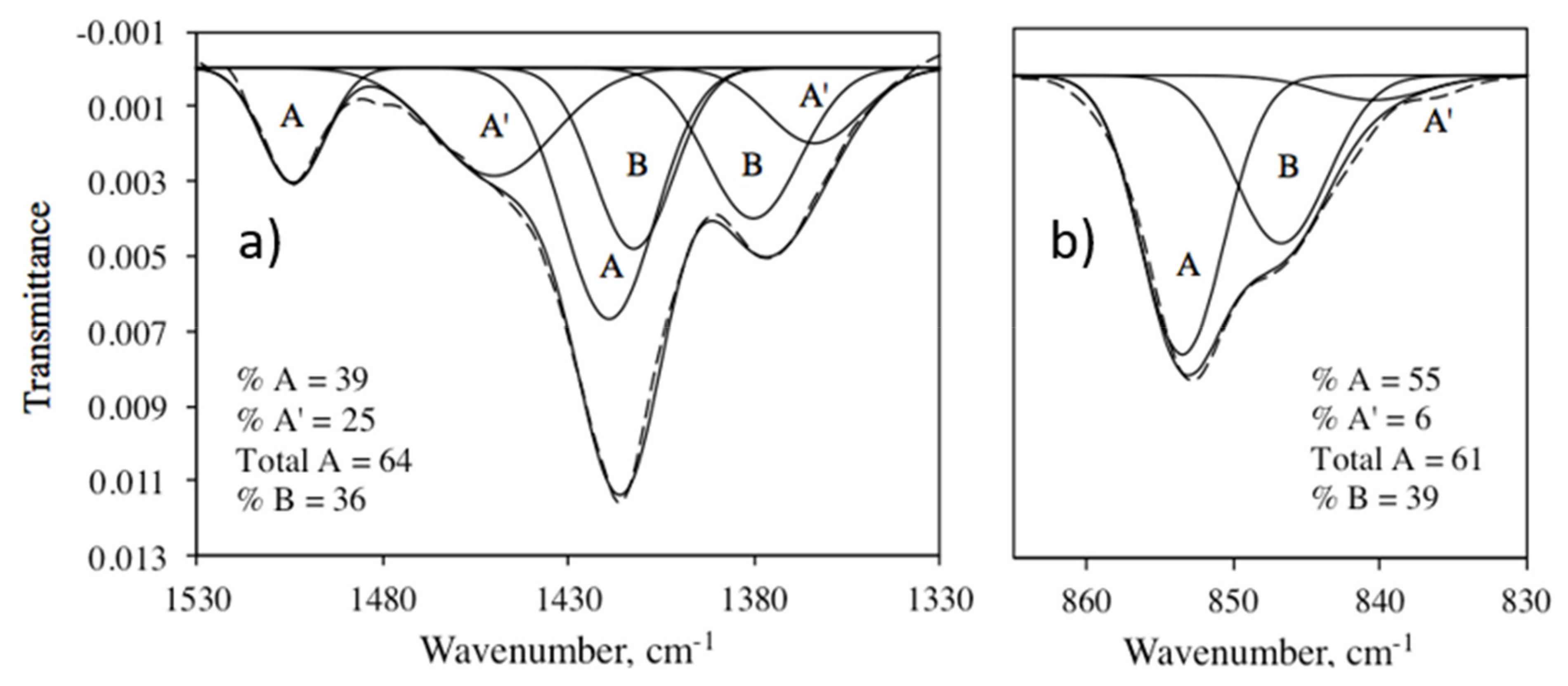
Figure 2 contains the IR and NMR spectra of the carbonate region of europium-containing KS-87. The carbonate asymmetric stretch (ν3) and out-of-plane bending (ν2) IR regions were fit with Gaussian peaks following the model [11] demonstrating that carbonate ions in three different environments are necessary to fit the IR intensities. Although both regions can be fit assuming only two carbonate species, the band widths are unacceptably large, and more of the intensity of the overall region is left unfit. Perhaps more importantly, six peaks (three carbonate environments) provide a more satisfying explanation for the overall shape of ν3, in particular the “peak” at about 1450 cm−1, the low frequency shoulder at about 1350 cm−1, and the low frequency shoulder in the ν2 region.
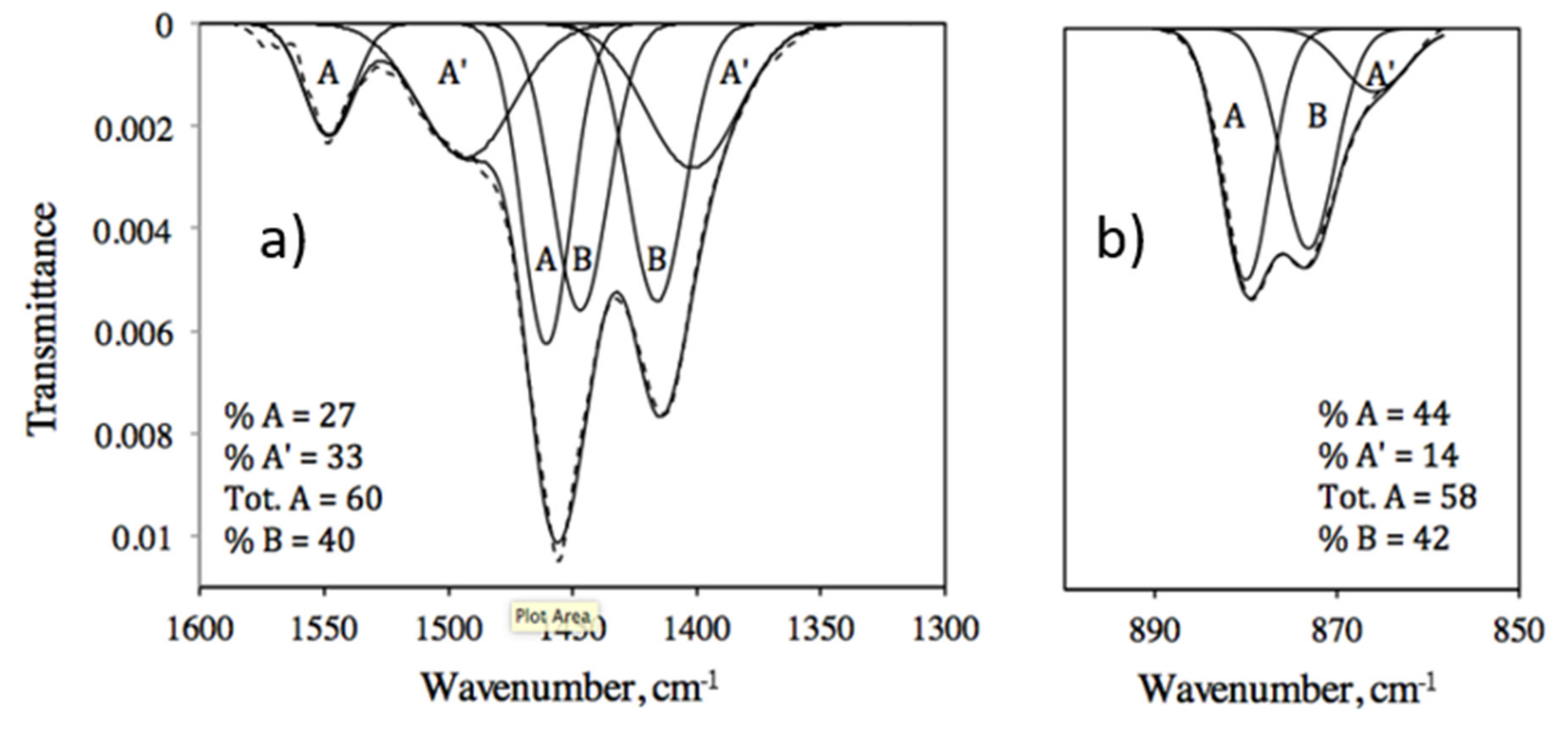
The feature at 1450 cm−1 is assigned to the high frequency member of the A’ doublet, while its low frequency member accounts for the low frequency tail to the ν3 region. A comparison of the spectrum of KS-136, prepared using the same procedure as KS-87 but without 13C labeling and without Eu [25], with that of KS-87 shows a more obvious peak at about 1500 cm−1 in KS-136, comparable to the 1450 cm−1 shoulder in KS-87 (Figure 3). This comparison also provides a way to adjust the band positions in those samples prepared with NaH13CO3: the spectral difference between the 12C and 13C isotopomers in ν3 is ca. 44 cm−1 and in ν2 is ca. 27 cm−1. The band in the ν2 region at 840 cm−1 in KS-87 (866 cm−1 in KS-136) has been the subject of a number of papers which assign it to A2 carbonate obtained at high pressure and temperature [27] and to “labile carbonate” [28] partly because of its response to maturation and heating. This band, observed in all of the compounds studied here, is not affected by heating [25].
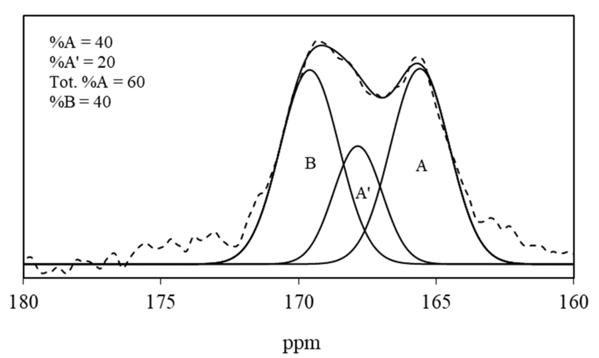
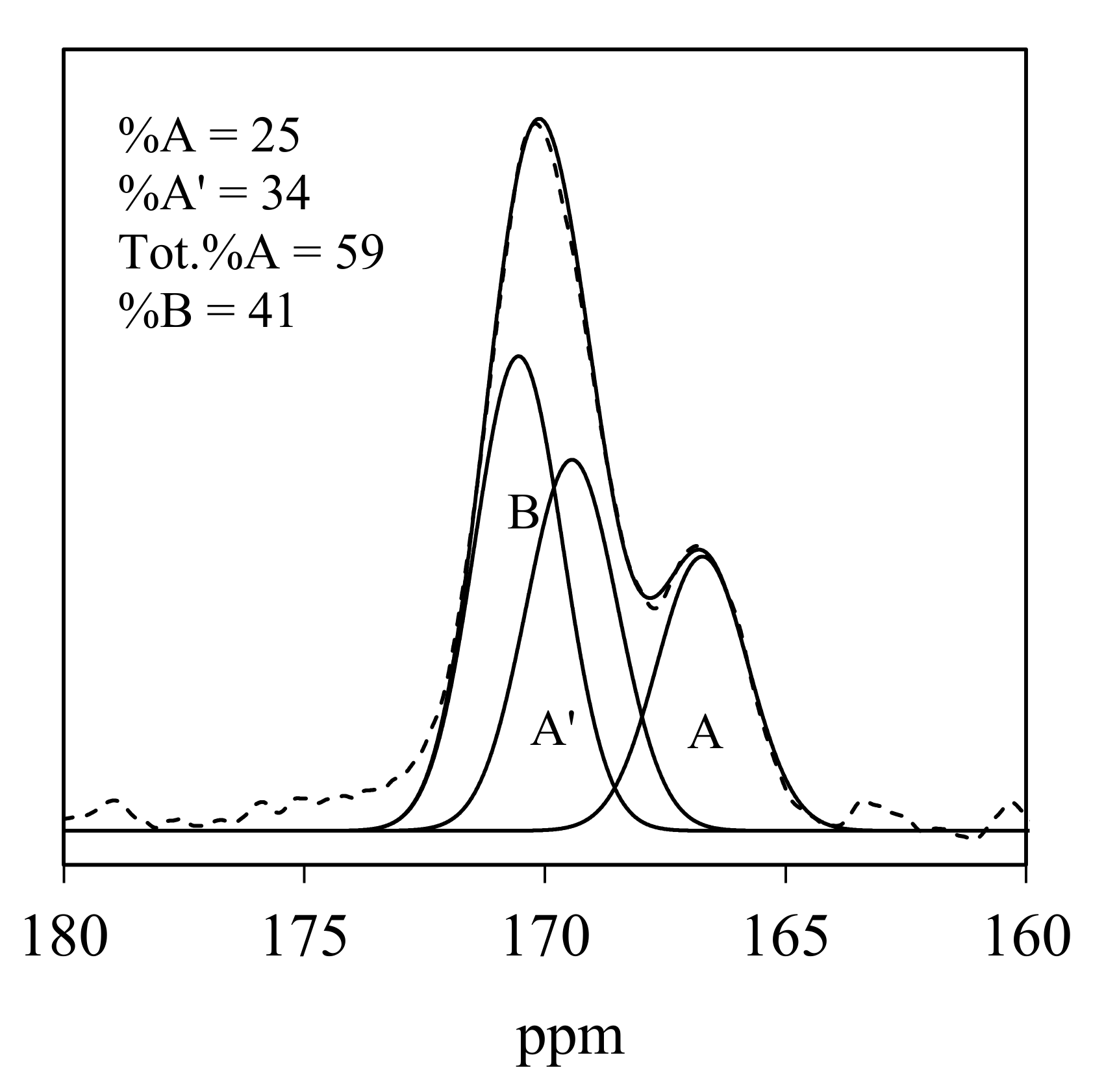
The 13C NMR spectrum of KS-87 (Figure 4) contains two peaks, one at 165.6 ppm, and the other at 169.6 ppm. The higher frequency peak is considerably broader and, as shown in the deconvolution, consists of both the A’ and B peaks. The assignments of the A and B peaks are consistent with previous work [29,30]. The agreement of the percentage of A-type carbonate as obtained from the IR analysis (39%) with that from the NMR analysis (40%) is good. The distribution percentages among the A-, A’-, and B-type carbonate were obtained from the areas under the peaks, assuming that the extinction coefficients are the same for each type of carbonate [10]). All spectroscopic data are given in Table 4.
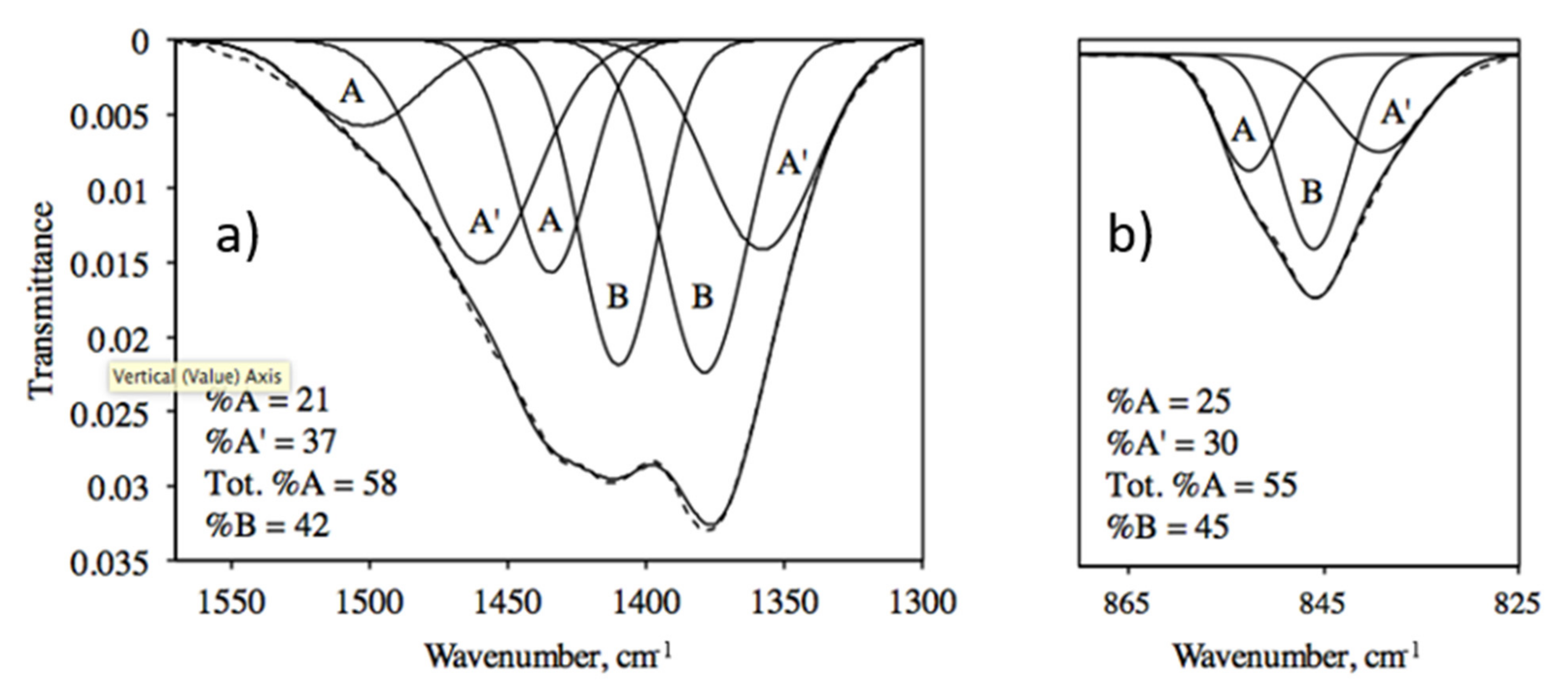
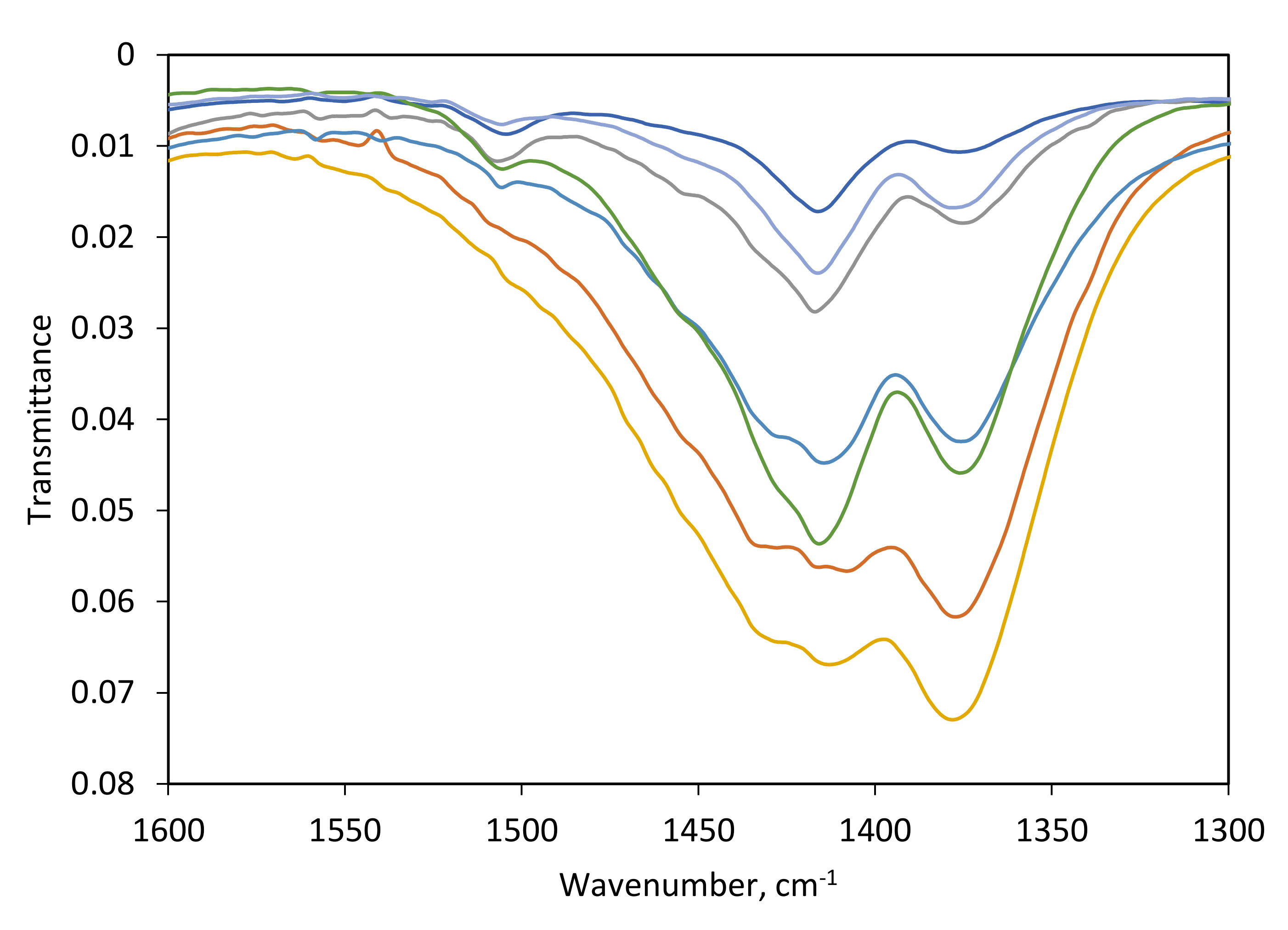
The IR and NMR spectra of the other apatites indicate considerably less A-type carbonate, though KS-92 and -152 contain approximately 30% A-type carbonate. The majority of the apatites that contain less A-type carbonate have a carbonate (ν3) IR spectrum that, due to the smaller, lower frequency A-type peak, has a different shape (the carbonate regions of KS-114 are shown in Figure 5 and an overlay of the ν3 region of all of the apatites is shown in Figure 6). Table 4 also contains the data for apatites KS-117 and -150 prepared using the same method, but without europium.
3.3. Lattice Parameters
4. Discussion
In one of the first explorations of europium-substituted carbonated apatites, we find evidence for (a) an enhanced effect of Eu(III) on the stability of carbonate in the apatite channel, (b) the substitution of one Eu3+ for 1.5 Ca2+, (c) the weak effect of the greater size of Eu3+ on the lattice parameters, which correlate modestly with the percentage of A-type carbonate (not total A-type), and (d) the validity of the environment model for the description of carbonate in apatites containing a variety of different substituents.
4.1. Charge-Balance Mechanism
Substitution of Eu3+ in the apatite channel (M(II) position) has been established [5,15,16,17,18]. Of the possible charge-balance mechanisms [5,19] for the substitution of a +3 ion for a +2 ion [24], the most common is the deprotonation of OH− to produce the oxide ion O2− [19]. This mechanism has been invoked in europium oxy apatites [18], europium- and copper-doped apatites [15], and in europium-doped apatite nanocrystals [31].
The experimental stoichiometry of the europium-doped carbonated apatites in this study is summarized in Table 3. The theoretical amounts of the constituents of the apatites can be predicted by first using IR and NMR data (Table 4) to determine the percentage of B-type carbonate according to the environment model. The presence of Na would lead to co-substitution of carbonate (Equation (1)) and the remaining B-type carbonate would be produced by the use of Equation (2). Finally, the use of Equation (3) or Equation (4) for the substitution of Eu3+ generated a unit-cell formula by substitution of calcium, phosphate, and hydroxide in the parent formula Ca10(PO4)6(OH)2. The predicted, theoretical molar coefficients of Ca, PO4, and OH obtained in this way are reported in Table 6. When these coefficients are compared with the actual, experimental coefficients (Table 3) for the obtained products it is apparent that the experimental coefficients for Ca are better matched by Eu substitution brought about by the substitution of one Eu3+ for 1.5 Ca2+ (Equation (4)). This mechanism has also been proposed by Han et al. [32]. It is tempting therefore to conclude that the substitution of europium proceeds by Equation (4), but it is important to note that the substitution may occur via several mechanisms, especially Equation (7) (Eu3+ + Na+ → 2 Ca2+) which also provides for the replacement of a higher mole percentage of Ca2+. Presumably the actual mix of mechanisms is determined primarily by the relative thermodynamics of each process.
4.2. Distribution of Carbonate
Our data indicate that both Eu3+ and (NH4)3PO4 are effective in producing large amounts of A-type carbonate. Even though KS-87, prepared using both Eu3+ and (NH4)3PO4, has the largest amount of A-type carbonate in our series of 17 compounds (the present study and [25]), other apatites, prepared with (NH4)3PO4 but not Eu3+, also have considerable A-type carbonate. A comparison of the 13C solid state NMR spectrum of KS-117 (Figure 7), which contains no europium, with that of KS-87 (Figure 2), shows that the presence of Eu3+ does not produce a greater dispersion of chemical shifts (the difference between the shifts of A- and B-types carbonate is 4.0 ppm for both KS-87 and KS-117), but does appear to increase the percentage of A-type carbonate. For the seven europium-containing apatites, the carbonate is distributed in approximately a 60 to 40 ratio for channel occupancy versus replacement of phosphate.
4.3. Lattice Parameters
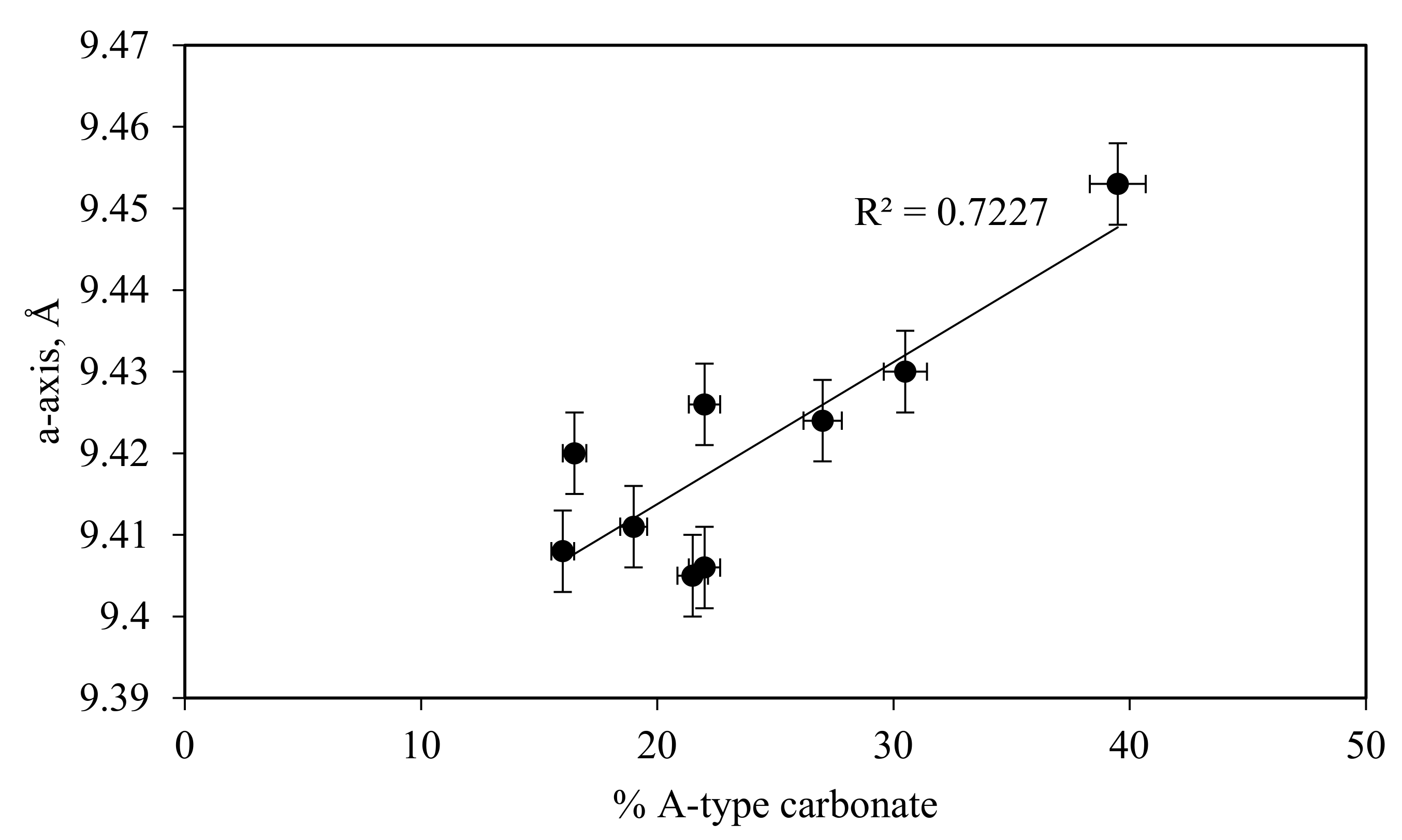
The lattice parameters (Table 5) for the europium-doped compounds show a decrease of the a-axial length with increasing percentage of carbonate (see [33,34]). This decrease has generally been attributed to an increase in B-type carbonate substitution (see for example [4,33]), while an increase in the a-axial length is rationalized by increased A-type substitution [12,25]. For example, the higher value of the a-axis for KS-87 compared to the other apatites prepared using a 1 to 9 ratio of Eu3+ to Ca2+ can be attributed to the larger percentage of A-type carbonate (see Table 4 for type A, A’, and B), and, indeed, the modest correlation (Figure 8) between the a-axial length and the percentage of A-type carbonate (Table 4) reveals that most of the variation in this parameter is a result of the variation in the percentage of A-type carbonate. A comparison of the a-axial lengths for KS-117, 148, 87, and 92, which contain 0, 5, 15, and 27% Eu shows no relationship with the amount of Eu3+, which has a slightly larger ionic radius than Ca2+.
5. Conclusions
In one of the first explorations of europium-doped carbonated apatite we have demonstrated that: (a) europium-doped carbonated apatites contain a considerable percentage of carbonate in the apatite channel, (b) both triammonium phosphate and europium (III) appear to facilitate the population of the channel environment (both A- and A’- environments), (c) the variation of the lattice parameters of these apatites can be partly accounted for by the distribution of A-type (not total A-type) carbonate, as calculated by the environment model, and (d) the environment model proposed by Fleet [10] for apatites prepared at high temperature and pressure is also valid and useful for a variety of substituted apatites produced synthetically at ambient pressures and low temperatures. The location of carbonate in the structure of carbonated apatite, the closest analog of the inorganic portion of bone and teeth, is of importance in the biological function of this compound, and especially in the possibility of the involvement of structural carbonate in acid–base regulation [10]. Considerable effort has been expended over several decades on the identification of different orientations of carbonate in apatite, but the environment model utilizes the different structural surroundings to describe the different types of carbonate observed in IR and NMR spectra. The model results in the conclusion that considerably more carbonate is sequestered in the apatite channel than was previously thought, and that the distribution of this substituent may be affected by cations such as Eu3+ and the use of synthesis reagents such as triammonium phosphate. The roughly 3 to 2 ratio of channel to matrix carbonate brings a greater focus on the role of both types of carbonate in the biological function of carbonate.
Author Contributions
Conceptualization, C.H.Y. and K.R.S.; methodology, K.R.S. and C.H.Y.; software, K.R.S.; validation, K.R.S. and C.H.Y.; formal analysis, C.H.Y.; investigation, K.R.S. and C.H.Y.; resources, C.H.Y.; data curation, K.R.S.; writing—original draft preparation, C.H.Y.; writing—review and editing, C.H.Y. and K.R.S.; visualization, C.H.Y. and K.R.S.; supervision, C.H.Y.; project administration, C.H.Y.; funding acquisition, C.H.Y. All authors have read and agreed to the published version of the manuscript.
Funding
This research was funded by the Yoder Research fund at Franklin and Marshall College.
Institutional Review Board Statement
Not applicable.
Informed Consent Statement
Not applicable.
Data Availability Statement
Not applicable.
Acknowledgments
The authors are indebted to Robyn N. Dudrick, Stanley Mertzman and Karen Mertzman of Franklin and Marshall College for technical assistance. The authors are also indebted to donors to the Yoder research fund.
Conflicts of Interest
The authors declare no conflict of interest. The funders had no role in the design of the study; in the collection, analyses, or interpretation of data; in the writing of the manuscript; or in the decision to publish the results.
References
- Pasteris, J.D. A mineralogical view of apatitic biomaterials. Am. Mineral. 2016, 101, 2594–2610. [Google Scholar] [CrossRef]
- McConnell, D. The crystal chemistry of carbonate apatites and their relationship to the composition of calcified tissues. J. Dental Res. 1952, 31, 53–63. [Google Scholar] [CrossRef]
- LeGeros, R.Z.; Trautz, O.R.; LeGeros, J.P.; Klein, E.J. Spectral properties of carbonate in carbonate-apatites. Appl. Spectrosc. 1968, 22, 357–358. [Google Scholar]
- LeGeros, R.Z.; Trautz, O.R.; Klein, E.; LeGeros, J.P. Two Types of Carbonate Substitution in the apatite structure. Experientia 1969, 25, 5–7. [Google Scholar] [CrossRef] [PubMed]
- Pan, Y.; Fleet, M.E. Compositions of the apatite-group minerals: Substitution mechanisms and controlling factors. In Reviews in Mineralogy and Geochemistry; Kohn, M., Rakovan, J., Hughes, J.M., Eds.; Mineralogical Society of America: Washington, DC, USA, 2002; Volume 48, pp. 13–49. [Google Scholar]
- Demarco, P.V.; Elzey, T.K.; Lewis, R.B.; Wenkert, E. Tri(dipivalomethanato)europium (III). Shift reagent for use in the proton magnetic resonance analysis of steroids and terpenoids. J. Am. Chem. Soc. 1970, 92, 5737–5739. [Google Scholar] [CrossRef]
- Morales, J.G.; Escamilla, C.V.; Penas, R.F.; Parra-Milla, C.M.; Drouet, C.; Maube-Bosc, F.; Oltolina, F.; Prat, M.; Ffernandez-Sanchez, J.F. Luminescent biomimetic citrate-coated europium-doped carbonated apatite nanoparticles for use in bioimaging: Physico-chemistry and cytocompatibility. RSC Adv. 2018, 9, 2385–2397. [Google Scholar] [CrossRef] [Green Version]
- Perera, T.S.H.; Han, Y.; Lu, X.; Wang, X.; Dai, H.; Li, S. Rare earth doped apatite nanomaterials for biological application. J. Nanomater. 2015, 2015, 705390. [Google Scholar] [CrossRef] [Green Version]
- De Maeyer, E.A.P.; Verbeeck, R.M.H.; Pieters, I.Y. Carbonate and alkalimetal incorporation in calciumhydroxyapatite. Trends Inorg. Chem. 1996, 4, 157–171. [Google Scholar]
- Fleet, M.E. Infrared spectra of carbonate apatites: Evidence for a connection between bone mineral and body fluids. Am. Mineral. 2017, 102, 149–157. [Google Scholar] [CrossRef]
- Yoder, C.H.; Bollmeyer, M.M.; Stepien, K.R.; Dudrick, R.N. The effect of incorporated carbonate and sodium on the IR spectra of A-, B-, and AB-type carbonate apatites. Am. Mineral. 2019, 104, 869–877. [Google Scholar] [CrossRef]
- Bollmeyer, M.M.; Carney, M.C.; Yoder, C.H. A-type carbonate in strontium apatites. Am. Mineral. 2019, 104, 438–446. [Google Scholar] [CrossRef]
- Fleet, M.E. Infrared spectra of carbonate apatites: ν2-Region bands. Biomaterials 2009, 30, 1473–1481. [Google Scholar] [CrossRef] [PubMed]
- Fleet, M.E. Carbonated Hydroxyapatite: Materials, Synthesis, and Application; CRC Press: Boca Raton, FL, USA, 2015. [Google Scholar]
- Pogosova, M.A.; Eliseev, A.A.; Kazin, P.E.; Azarmi, F. Synthesis, structure, luminescence, and color features of the Eu- and Cu-doped calcium apatite. Dye. Pigment. 2017, 141, 209–216. [Google Scholar] [CrossRef]
- Pazik, R.; Nedelec, J.-M.; Wiglusz, R.J. Preferential site substitution of Eu3+ ions in Ca10(PO4)6Cl2 nanoparticles obtained using a microwave stimulated wet chemistry technique. CrystEngComm 2014, 16, 5308–5318. [Google Scholar] [CrossRef]
- Ternane, R.; Trabelsi-Ayedi, M.; Jbir-Ariguib, N.; Piriou, B.L. Luminescent properties of Eu3+ in calcium hydroxyapatite. J. Lumin. 1999, 81, 165–170. [Google Scholar] [CrossRef]
- Piriou, B.; Fahmi, D.; Dexpert-Ghys, J.; Taitai, A.; Lacout, J.L. Unusual fluorescent properties of Eu3+ in oxyapatites. J. Lumin. 1987, 29, 97–103. [Google Scholar] [CrossRef]
- Hosseini, S.M.; Drouet, C.; Al-Kattan, A.; Navrotsky, A. Energetics of lanthanide-doped calcium phosphate apatite. Am. Mineral. 2014, 99, 2320–2327. [Google Scholar] [CrossRef] [Green Version]
- Yoder, C.H.; Havlusch, M.D.; Dudrick, R.N.; Shermerhorn, J.T.; Tran, L.K.; Deymier, A.C. The synthesis of phosphate and vanadate apatites using an aqueous one-step method. Polyhedron 2017, 127, 403–409. [Google Scholar] [CrossRef] [Green Version]
- Vignoles, M.; Bonel, G.; Holcomb, D.W.; Young, R.A. Influence of preparation conditions on the composition of type B carbonated hydroxyapatite and on the localization of the carbonate ions. Calcif. Tissue Int. 1988, 43, 33–40. [Google Scholar] [CrossRef]
- Holland, T.J.B.; Redfern, S.A.T. Unit cell refinement from powder diffraction data: The use of regression diagnostics. Mineral. Mag. 1997, 61, 65–77. [Google Scholar] [CrossRef]
- Bollmeyer, M.M.; Yoder, C.H. Incorporation of fluorophosphate in apatite. Polyhedron 2018, 145, 176–181. [Google Scholar] [CrossRef]
- Al-Kattan, A.; Dufour, P.; Dexpert-Ghys, I.; Drouet, C. Preparation and physicochemical characteristics of luminescent apatite-based colloids. J. Phys. Chem. C. 2010, 113, 2918–2924. [Google Scholar] [CrossRef]
- Yoder, C.H.; Stepien, K.R.; Dudrick, R.N. The distribution of carbonate in apatite: The environment model. Am. Mineral. 2022, 3, 1713–1728. [Google Scholar]
- Vignoles, M.; Bonel, G.; Young, R.A. Occurrence of nitrogenous species in precipitated B-type carbonated hydroxyapatites. Calcif. Tissue Int. 1987, 40, 64–70. [Google Scholar] [CrossRef] [PubMed]
- Fleet, M.E.; Liu, X.; Liu, X. Orientation of channel carbonate ions in apatite: Effect of pressure and composition. Am. Mineral. 2011, 96, 1148–1157. [Google Scholar] [CrossRef]
- Rey, C.; Collins, B.; Goehl, T.; Dickson, I.R.; Glimcher, M.J. The carbonate environment in bone mineral: A resolution-enhanced fourier transform infrared spectroscopy study. Calcif. Tissue Int. 1989, 45, 157–164. [Google Scholar] [CrossRef] [PubMed]
- Bashah, K.; Rey, C.; Gllmcher, M.J.; Schimizu, M.; Griffin, R.G. Solid state carbon-13 and proton NMR studies of carbonate-containing phosphate and enamel. J. Solid State Chem. 1990, 84, 71–81. [Google Scholar] [CrossRef]
- Babonneau, F.; Bonhomme, C.; Hayakawa, S.; Osaka, A. Solid state NMR characterization of nano-crystalline hydroxyl-carbonate apatite using 1H-31P-13C triple resonance experiments. Mater. Res. Soc. Symp. Proc. 2007, 984, 39–44. [Google Scholar]
- Silva, F.R.O.; de Lima, N.B.; Bressiani, A.H.A.; Courrol, L.C.; Gomes, L. Synthesis, characterization and luminescence properties of Eu3+-doped hydroxyapatite nanocrystals and thermal treatment effects. Opt. Mater. 2015, 47, 135–142. [Google Scholar] [CrossRef]
- Han, Y.; Wang, X.; Dai, H.; Li, S. Synthesis and luminescence of Eu3+ doped hydroxyapatite nanocrystallines: Effect of calcinations and Eu3+ content. J. Lumin. 2013, 135, 281–287. [Google Scholar] [CrossRef]
- LeGeros, R.Z. Effect of carbonate on the lattice parameters of apatite. Nature 1965, 206, 403–404. [Google Scholar] [CrossRef] [PubMed]
- Deymier, A.C.; Nair, A.K.; Dapalie, B.; Qin, Z.; Arcot, K.; Drouet, C.; Yoder, C.H.; Buehler, M.J.; Thomopoulos, S.; Genin, G.M.; et al. Protein-free Formation of bone-like apatite; New insights into the key role of carbonation. Biomaterials 2017, 127, 75–88. [Google Scholar] [CrossRef] [PubMed] [Green Version]
Figure 1.
The XRD pattern of KS-152.

Figure 2.
The carbonate asymmetric stretch (ν3) (a) and out-of-plane bend (ν2) (b) regions of the IR spectrum of KS--87.
Figure 2.
The carbonate asymmetric stretch (ν3) (a) and out-of-plane bend (ν2) (b) regions of the IR spectrum of KS--87.

Figure 3.
The carbonate asymmetric stretch (ν3) (a) and out-of-plane bend (ν2) (b) regions of the IR spectrum of KS-136 (unlabeled and with no Eu) [23].
Figure 3.
The carbonate asymmetric stretch (ν3) (a) and out-of-plane bend (ν2) (b) regions of the IR spectrum of KS-136 (unlabeled and with no Eu) [23].

Figure 4.
The solid-state MAS 13C NMR spectrum of KS-87.

Figure 5.
The carbonate ν3 (a) and ν2 (b) regions of the IR spectrum of KS-114.

Figure 6.
The carbonate ν3 regions of the IR spectra of (in order of increasing intensity in the figure)): KS-87, 90, 92, 114, 116, 148, 152.
Figure 6.
The carbonate ν3 regions of the IR spectra of (in order of increasing intensity in the figure)): KS-87, 90, 92, 114, 116, 148, 152.

Figure 7.
The 13C MAS solid-state NMR spectrum of KS-117, containing no Eu3+.

Figure 8.
The correlation between the a-axial lengths of the unit cells of the apatites in Table 4 with the spectroscopically-determined percentage of A-type carbonate.
Figure 8.
The correlation between the a-axial lengths of the unit cells of the apatites in Table 4 with the spectroscopically-determined percentage of A-type carbonate.


Table 1.
Conditions for the synthesis of prepared apatites.
| ID | CO3:PO4 Ratio | Eu:Ca Ratio | PO4 Reagent * | CO3 Reagent * | Synthesis Method |
|---|---|---|---|---|---|
| KS-87 | 1 | 1:9 | T | 13NaH | One-step |
| KS-90 | 2 | 1:9 | T | 13NaH | One-step |
| KS-92 | 1 | 2:8 | T | 13NaH | One-step |
| KS-114 | 1 | 1:9 | Na2HPO4 | 13NaH | One-step |
| KS-116 | 1 | 1:9 | T | 13NaH | One-step + 0.5 g NaNO3 |
| KS-148 | 1 | 0.3:9.7 | T | 13NaH | Direct |
| KS-152 | 0.2 | 1:9 | T | 13NaH | Direct |
* T = (NH4)3PO4, 13NaH = NaH13CO3.
Table 2.
Composition (weight percent) and element ratios.
| ID | % CO3 | % Na | % Eu | Ca/Eu | Ca/P | (Ca + Eu)/P |
|---|---|---|---|---|---|---|
| KS-87 | 4.8 | 0.47 | 15.17 | 7.35 | 1.31 | 1.49 |
| KS-90 | 10.9 | 1.35 | 16.49 | 6.91 | 1.44 | 1.65 |
| KS-92 | 9.9 | 0.64 | 27.18 | 3.11 | 1.12 | 1.56 |
| KS-114 | 8.25 | 1.48 | 16.81 | 6.80 | 1.50 | 1.73 |
| KS-116 | 3.9 | 1.4 | 15.97 | 6.95 | 1.37 | 1.56 |
| KS-117 * | 2.5 | 0.66 | 0 | - | 1.59 | - |
| KS-148 | 4.6 | 0.82 | 4.8 | 28.7 | 1.55 | 1.61 |
| KS-152 | 2 | 0.36 | 16.03 | 7.03 | 1.36 | 1.55 |
* For comparison; does not contain Eu.
Table 3.
Experimental molar amounts of constituents in products.
| Starting Mole Ratios | Moles in ca. 1029 g | ||||
|---|---|---|---|---|---|
| ID | Ca: Eu, X: 9 | CO3:PO4 | Ca | PO4 | Eu |
| KS-148 | 0.3 | 1 | 9.3 | 6 | 0.33 |
| KS-87 | 1 | 1 | 7.5 | 5.7 | 1 |
| KS-114 | 1 | 1 | 7.5 | 5 | 1.1 |
| KS-116 | 1 | 1 | 7.3 | 5 | 1.1 |
| KS-152 | 1 | 0.2 | 7.6 | 5.6 | 1.1 |
| KS-90 | 1 | 2 | 7.6 | 5.3 | 1.1 |
| KS-92 | 2 | 1 | 5.7 | 5.1 | 1.8 |
| KS-117 * | - | 1 | 10 | 6.3 | - |
* For comparison; does not contain Eu.
Table 4.
Spectroscopic data.
| ID | CO32− Environment | IR ν3 | IR ν2 | NMR | Average (IR ν3 + NMR) | |||
|---|---|---|---|---|---|---|---|---|
| Position (cm−1) | % | Position (cm−1) | % | Position (ppm) | % | |||
| 87 | A | 1419, 1504 | 39 | 853 | 55 | 165.6 | 40 | 39.5 |
| A’ | 1366, 1451 | 25 | 840 | 39 | 167.9 | 20 | 22.5 | |
| B | 1380, 1413 | 36 | 847 | 6 | 169.6 | 40 | 38 | |
| 90 | A | 1431, 1506 | 13 | 854 | 22 | 165.8 | 20 | 16.5 |
| A’ | 1363, 1454 | 46 | 840 | 33 | 168.4 | 39 | 42.5 | |
| B | 1377, 1409 | 41 | 847 | 45 | 170.2 | 41 | 41 | |
| 92 | A | 1422, 1504 | 36 | 853 | 44 | 166.5 | 25 | 30.5 |
| A’ | 1362, 1455 | 34 | 841 | 20 | 169.2 | 38 | 36 | |
| B | 1380, 1409 | 30 | 846 | 36 | 170.4 | 37 | 33.5 | |
| 114 | A | 1435, 1503 | 21 | 853 | 25 | 165.3 | 23 | 22 |
| A’ | 1358, 1460 | 37 | 839 | 30 | 167.7 | 38 | 37.5 | |
| B | 1379, 1410 | 42 | 846 | 45 | 169.3 | 39 | 40.5 | |
| 116 | A | 1431, 1500 | 23 | 853 | 32 | 166.5 | 20 | 21.5 |
| A’ | 1353, 1457 | 32 | 840 | 19 | 168.8 | 36 | 34 | |
| B | 1376, 1410 | 45 | 846 | 48 | 170.3 | 43 | 44 | |
| 117 * | A | 1422, 1505 | 29 | 853 | 46 | 166.7 | 25 | 27 |
| A’ | 1360, 1450 | 32 | 842 | 14 | 169.4 | 34 | 33 | |
| B | 1377, 1414 | 39 | 847 | 39 | 170.5 | 41 | 40 | |
| 148 | A | 1426, 1507 | 20 | 853 | 34 | 166.4 | 18 | 19 |
| A’ | 1365, 1454 | 45 | 841 | 24 | 168.9 | 44 | 44.5 | |
| B | 1378, 1411 | 35 | 847 | 42 | 170.1 | 38 | 36.5 | |
| 150 * | A | 1429, 1499 | 19 | 853 | 35 | 166.1 | 13 | 16 |
| A’ | 1363, 1454 | 39 | 841 | 28 | 168.3 | 41 | 40 | |
| B | 1379, 1409 | 42 | 847 | 38 | 169.6 | 46 | 44 | |
| 152 | A | 1421, 1505 | 30 | 853 | 41 | 165.1 | 14 | 22 |
| A’ | 1365, 1451 | 33 | 842 | 16 | 167.3 | 38 | 35.5 | |
| B | 1380, 1411 | 37 | 847 | 44 | 169.1 | 47 | 42 | |
* KS-117 and -150 do not contain Eu, but are included for comparison.
Table 5.
Lattice parameters.
| ID | % CO3 | % Na | a-axis (Å) | c-axis (Å) | Unit cell Volume (Å3) |
|---|---|---|---|---|---|
| 87 | 4.8 | 0.47 | 9.453(7) | 6.894(7) | 533.6(7) |
| 90 | 10.9 | 1.35 | 9.42(1) | 6.89(2) | 529.(1) |
| 92 | 9.9 | 0.64 | 9.430(6) | 6.870(6) | 529.0(6) |
| 114 | 8.25 | 1.48 | 9.406(1) | 6.879(4) | 527.1(4) |
| 116 | 3.9 | 1.4 | 9.405(5) | 6.894(4) | 528.1(4) |
| 117 * | 2.5 | 0.66 | 9.424(1) | 6.885(2) | 529.5(2) |
| 148 | 4.6 | 0.82 | 9.411(1) | 6.876(4) | 527.5(4) |
| 150 * | 6.05 | 0.58 | 9.408(8) | 6.888(8) | 527.0(8) |
| 152 | 2 | 0.36 | 9.426(6) | 6.889(6) | 530.0(6) |
* KS-117 and 150 do not contain Eu, but are included for comparison.
Table 6.
Theoretical mole amounts of Ca, PO4, and OH in products after estimation of substitution consequences of carbonate using IR/NMR data and Equations (1) and (2), and europium substitution mechanism based on Equation (3) or Equation (4).
Table 6.
Theoretical mole amounts of Ca, PO4, and OH in products after estimation of substitution consequences of carbonate using IR/NMR data and Equations (1) and (2), and europium substitution mechanism based on Equation (3) or Equation (4).
| Charge-Balance Mechanism (Equation (3)) | Charge-Balance Mechanism (Equation (4)) | |||||
|---|---|---|---|---|---|---|
| ID | Ca | PO4 | OH | Ca | PO4 | OH |
| KS-148 | 9.38 | 5.71 | 0.67 | 9.21 | 5.71 | 1 |
| KS-87 | 8.69 | 5.69 | -0.02 | 8.19 | 5.69 | 0.89 |
| KS-114 | 8.33 | 5.43 | -0.7 | 7.78 | 5.43 | 0.4 |
| KS-152 | 8.76 | 5.86 | 0.5 | 8.21 | 5.86 | |
| KS-90 | 8.12 | 5.22 | -1.48 | 7.57 | 5.22 | 1.6 |
| KS-92 | 7.63 | 5.43 | -2.38 | 6.73 | 5.43 | -0.38 |
| KS-117 * | 9.83 | 5.83 | 1.48 | -0.58 | ||
* Does not contain Eu, presented only for comparison.
Publisher’s Note: MDPI stays neutral with regard to jurisdictional claims in published maps and institutional affiliations. |
© 2022 by the authors. Licensee MDPI, Basel, Switzerland. This article is an open access article distributed under the terms and conditions of the Creative Commons Attribution (CC BY) license (https://creativecommons.org/licenses/by/4.0/).
Share and Cite
MDPI and ACS Style
Stepien, K.R.; Yoder, C.H. Europium-Doped Carbonated Apatites. Minerals 2022, 12, 503. https://doi.org/10.3390/min12050503
AMA Style
Stepien KR, Yoder CH. Europium-Doped Carbonated Apatites. Minerals. 2022; 12(5):503. https://doi.org/10.3390/min12050503
Chicago/Turabian StyleStepien, Kathleen R., and Claude H. Yoder. 2022. "Europium-Doped Carbonated Apatites" Minerals 12, no. 5: 503. https://doi.org/10.3390/min12050503
Note that from the first issue of 2016, this journal uses article numbers instead of page numbers. See further details here.