Article

Microbial Mat Stratification in Travertine Depositions of Greek Hot Springs and Biomineralization Processes

by
Christos Kanellopoulos
1,2,*,
Vasiliki Lamprinou
3,
Artemis Politi
3,
Panagiotis Voudouris
1,
Ioannis Iliopoulos
2,
Maria Kokkaliari
2,
Leonidas Moforis
1 and
Athena Economou-Amilli
3,*
1
Faculty of Geology and Geoenvironment, National and Kapodistrian University of Athens, Panepistimiopolis, Ano Ilissia, 15784 Athens, Greece
2
Department of Geology, University of Patras, 26500 Rio, Greece
3
Faculty of Biology, Department of Ecology and Systematics, National and Kapodistrian University of Athens, Panepistimiopolis, Ano Ilissia, 15784 Athens, Greece
*
Authors to whom correspondence should be addressed.
Minerals 2022, 12(11), 1408; https://doi.org/10.3390/min12111408
Submission received: 23 September 2022 / Revised: 30 October 2022 / Accepted: 3 November 2022 / Published: 4 November 2022
(This article belongs to the Special Issue Bio-Geochemistry of Heavy Metals/Metalloids)

Abstract

:
The study of microbial mats in extreme environments is of high scientific interest from geological, ecological, and geomicrobiological aspects. These mats represent multilayer bio-structures where each taxonomic group dominates a specific vertical layering distribution resulting from its growth and metabolic activity. In the present study, microbial mats in a hot spring environment from Aedipsos (Euboea Island, Greece) resulting in the creation of thermogenic travertine, were studied through an interdisciplinary approach. The mineralogical composition was determined by optical microscopy, XRD, and SEM-EDS microanalysis, and the identification of Cyanobacteria was made primarily on morphological characteristics. The main mineral phase in the studied samples is calcite and, to a less extent, aragonite, with several trace elements in the mineral-chemistry composition, i.e., up to 1.93 wt. % MgO, up to 0.52 wt. % SrO, up to 0.44 wt. % Na2O, up to 0.17 wt. % K2O, and up to 3.99 wt. % SO3. The dominant facies are lamination and shrubs, which are the most common among the facies of thermogenic travertines of the area. Several layers were identified, (i) a top mainly abiotic layer consisting of calcium carbonate micritic crystals, (ii) a second biotic layer–the Cyanobacteria layer, dominated by the species Leptolyngbya perforans, (iii) a third biotic layer where Leptolyngbya perforans, Chloroflexus and other bacteria occur, and (iv) a deeper abiotic part with several layers where no photosynthetic microorganisms occur. In the upper layers, nineteen (19) species of Cyanobacteria were identified, classified in the orders Chroococcales (37%), Synechococcales (31%), Oscillatoriales (16%), and Spirulinales (6%). Among the identified Cyanobacteria, there are typical thermophilic and limestone substrate species. These Cyanobacteria are found to participate in the biomineralization and biologically-influenced processes, i.e., (i) filamentous Cyanobacteria are trapping calcium carbonate crystals, and diatoms, (ii) extracellular polymeric substances (EPS) create crystal retention lattice contributing to the biomineralization process, and (iii) filamentous sheaths of Cyanobacteria are calcified, resulting in the creation of calcium carbonate tubes.

Graphical Abstract

1. Introduction

One of the most interesting structures of microorganisms (i.e., bacteria, fungi, and archaea) occurring in the environment of hot springs is that of microbial mats of several layers, whose thickness counts up to a few centimeters [1]. Microbial mats are generally significant parts of earth ecosystems; they were found as fossils 3500 Ma old [2,3,4,5], and they commonly occur in extreme environments such as hot springs, alkaline, and hypersaline environments [6,7,8,9]; they are also related to biomineralization processes, such as calcite, aragonite, iron-oxides, i.e., the typical mineral compounds of thermal springs depositions. Microbial mats differ from microbial biofilms, which are much thinner, i.e., tens to hundreds of μm, and display different architecture [10]. Geomicrobiological studies in extreme environments offer a unique view of the biodiversity structure in conjunction with the geological environment e.g., [11,12,13,14,15,16,17].
Even if every microbial mat comprises several layers, the composition of each mat differs depending on the environmental conditions. Although different microorganisms dominate in each layer, relationships among them exist based on competition and cooperation [1,18,19]. In environments with sunlight and water access, aerobic photosynthetic microorganisms, such as Cyanobacteria, normally dominate the top layer. The deeper layers are usually dominated by anaerobic non-photosynthetic microorganisms, such as sulfate-reducing bacteria and archaea. The extracellular polymeric substances (EPSs) play an essential role in microbial mats since they keep the whole structure together [20,21]. Filamentous microorganisms also contribute to structure-binding, especially if they are equipped with sheaths [1].
Additionally, it should be noted that thermal springs, balneotherapy (i.e., treatment of disease by bathing in thermal mineral waters), and pelotherapy (i.e., mud therapy), have been of particular interest even from ancient times, and still today there is a firm belief in their curative powers. Pelotherapy is the therapeutic use of heated peloids, i.e., common depositions of thermal springs, whose effect is attributed to the properties and action of water, minerals, and their biological fraction, which may be made up of Cyanobacteria, and other microalgae (Bacillariophyta and Chlorophyta) present in water and clays e.g., [22]. Research is demonstrating that natural peloids can be considered a therapeutic mud and a dynamic “living system” [23] due to their curative properties dependent on the diversity and survival properties of the microorganisms inhabiting and shaping the peloids [17,24], thus permitting potential biotechnological exploitation. It is in the last decade that microalgae became the focus of extensive research efforts, aiming at novel compounds that might lead to therapeutically useful agents of great interest in the pharmaceutical industry [25,26,27].
Previous studies have been conducted in the study of the thermogenic travertine of Northern Euboea (Greece) [28,29,30,31,32,33,34]. The present study is the first attempt to examine the stratification of microbial mats in the hot springs of the Aedipsos area (i.e., biodiversity, mineralogical composition, and biomineralization processes of calcium carbonate minerals taking place at the top layers).

2. Materials and Methods

2.1. Sample Collection and Microbial Identification

Physicochemical parameters of the water, i.e., temperature, pH, salinity, total dissolved solids (T.D.S.), and electrical conductivity (E.C.), were measured for each sampling site in situ during the fieldwork using portable devices. The pH-meter was calibrated with standard buffer solutions of pH 4.0 and 7.0 before any measurements were taken. The pH measurement error, including accuracy and reproducibility, was less than ±0.05 pH units. The temperature was measured with a probe connected to the pH-meter, and the error was estimated to be less than ±0.3 °C.
During the sampling process, sterile metal tweezers and chisels were used. The physicochemical water parameters, i.e., temperature, salinity, and pH, were measured in situ using portable apparatus (WTW 315i, Xylem Inc., Washington, DC, USA).
From each sampling site, two sub-samples were collected and investigated. The first sub-sample was incubated in sterile transparent vials in the field. The second sub-sample was stored in a formaldehyde solution (2.5%). Enriched cultures were obtained in flasks and Petri dishes with BG11 and BG110 culture media [35]. Cultures were maintained in an incubator (Sanyo, Gallenkamp) under stable conditions, i.e., temperature and humidity, and a natural diurnal cycle (north-facing window) at room temperature.
The samples were studied under an optical microscope and a stereo-microscope. For species identification, morphological determination was made based on classical and recent literature [36,37,38,39] and references within. Although the morphological determination of Cyanobacteria presents some limitations, in some cases it is the most appropriate approach, such as in the case of microbial mats, where the dominating species of each layer ought to be identified.
The ArcGIS software was used to modify the geological map presented by Kanellopoulos et al. [40].

2.2. Mineralogical Study

The mineralogical study was conducted on polished sections under an optical microscope and powders using X-ray diffraction (Bruker D8 Advanced Diffractometer, using Ni filtered Cu-Kα radiation, operating at 40 kV and 40 mA and employing a Bruker Lynx Eye fast detector). The XRD results were evaluated using the DIFFRACplus EVA software (Bruker) and the ICDD Powder Diffraction File (2006 version). In addition, selected dehydrated samples in an alcohol series (30%–100%), critical point dried, gold-coated, and studied under SEM (Jeol JSM 5600; Jeol USA, Inc.: Peabody, MA, USA), with operation contusions: accelerating voltage 20 kV and beam current 0.5 nA. SEM-EDS analyses were carried out in carbon-coated thin sections using a Jeol JSM-IT500 (Jeol JSM 5600; Jeol USA, Inc.: Peabody, MA, USA) instrument equipped with an Oxford 100 Ultramax microanalytical analytical device, with beam diameter 1–2 mm and with operation contusions: accelerating voltage 20 kV, beam current 1.5 nA, time of measurement 50 s.

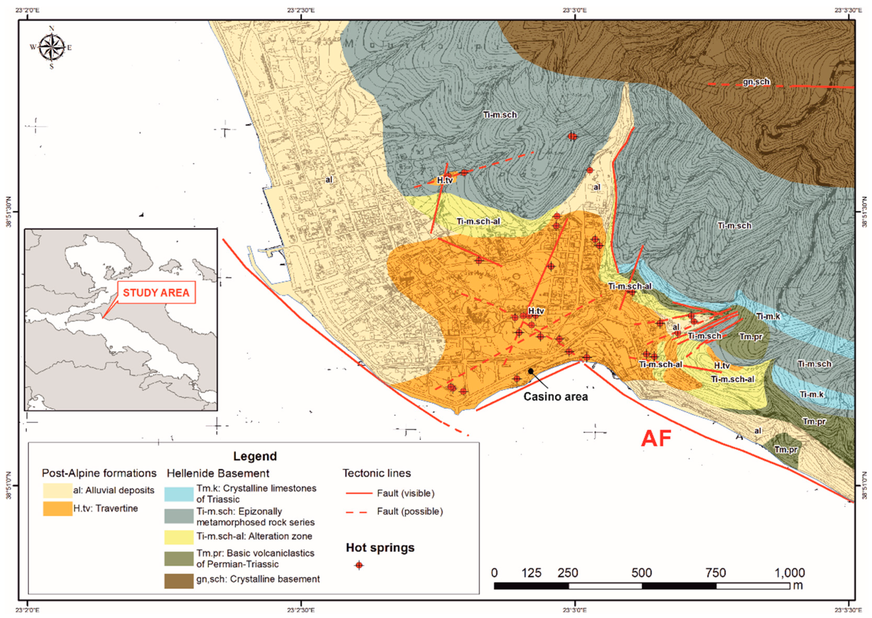
3. Geological Setting

Euboea Island is located in the western part of the Aegean Sea, near the Greek mainland, and Aedipsos is located in the northwestern part of the Island, near the seaside.
The geological formations in the greater Aedipsos area belong to the Pelagonian geotectonic unit of the Hellenides [41,42,43,44] (Figure 1), and consist of: a metamorphic crystalline basement (pre-middle to middle Carboniferous age), a basic volcanoclastic complex series (Permian–Triassic age), shallow marine carbonate and clastic rocks (middle Triassic age) with volcanic rocks intercalations [45,46], alluvial deposits and thermogenic travertine depositions.
As Kanellopoulos et al. [28,40,47] described, all the hot springs of NW Euboea (i.e., located at Aedipsos, Ilia, and Gialtra) are a combined result of active tectonics and the recent volcanism of the Lichades volcanic center. This volcanic center consists of trachyandesite lava, of 0.5 Ma age (dated by the K-Ar method; [48]). The main hot spring manifestations occur in the Aedipsos area, with temperatures up to 84 °C. The fluids are of Na-Cl geochemical type, near-neutral pH, and they present chemical similarities [29,41].
In Aedipsos, the hot springs have been known since ancient times, depositing thermogenic travertine [28,29,30,31]. The travertines present a large variety of facies [28,30,31,49] with strong indications of biomineralization processes [28,29]. Recent studies prove the contribution of Cyanobacteria to the formation of travertines [32], even in the very early stages of Cyanobacteria settlement [33].

4. Results and Discussion

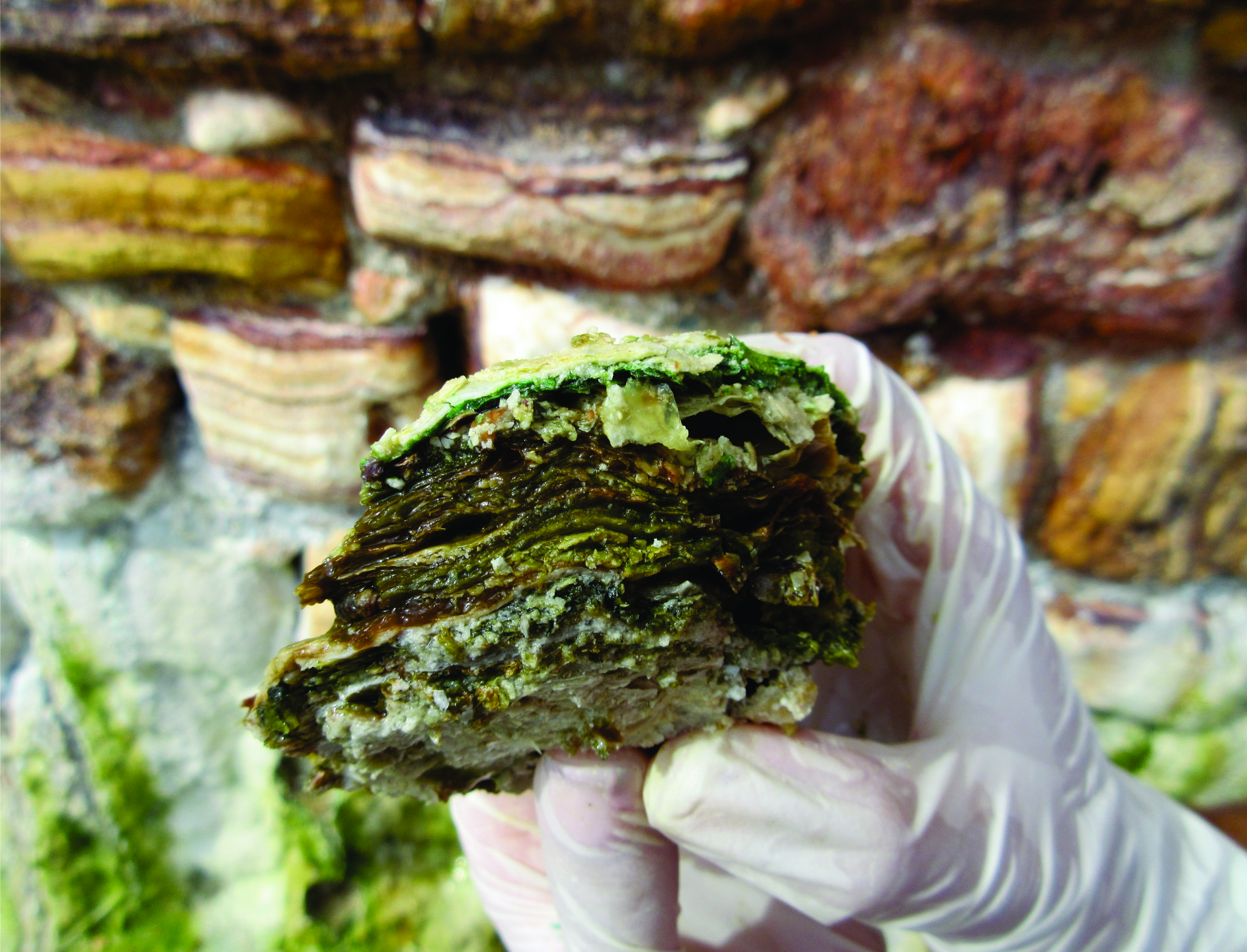
4.1. Sample Description and In-Situ Analysis

Samples were extracted from one of the Aedipsos thermal springs, i.e., in the Casino area (Figure 1). The study site is a vertical wall near the coast, a few meters above sea level. It is built with travertine blocks, and hot water flows through them (Figure 2A–C). The water temperature in all sampling sites was 55.8 °C, the pH was 6.3, and the salinity was 14‰.
At the study sites, successive layers of different microorganisms, i.e., microbial mats a few centimeters thick, occurred (Figure 2D–F). The microbial mats consisted of microorganisms that belonged to different taxonomic groups. These organisms follow a specific vertical layering distribution resulting from their growth and metabolic activity (cf. [50]). In the vertical stratification of the microbial mats, Cyanobacteria occupy the upper layer since they are photosynthetic organisms and need access to sunlight. The lower layer consists of other photosynthetic bacteria, such as green non-sulfur bacteria, e.g., Chloroflexus. Finally, in the even lower layers, where the necessary sunlight is insufficient, non-photosynthetic bacteria are thriving, usually sulfur bacteria (cf. [51]).
In the studied samples, a top layer was observed just above the Cyanobacteria layer (Figure 2E,G,H). This top layer represents an abiotic area since it contains no microorganisms. Furthermore, this layer is composed of micritic calcium carbonate crystals, which are bound with the help of EPS produced by the cyanobacterial layer, which occurs just below (cf. [51,52]).
In the Cyanobacteria layer, further vertical micro-stratification was recognized which might be attributed to the different light requirements of the various Cyanobacteria species. For example, Leptolyngbya perforans is a species characteristic of calcareous substrates, appearing in the upper layers of the vertical stratification (cf. [38]). Ward et al. [53] suggested that there are differences in the optimal amount of sunlight energy even in species belonging to the same genus. In the Cyanobacteria layer, diurnal micro-stratification also occurs due to the daily movement of Cyanobacteria upwards depending on the light availability. Takashima and Kano [54] suggested that, in the morning, Cyanobacteria remain under the carbonate layer (top abiotic layer), while in the afternoon, when the light is limited, Cyanobacteria enter the carbonate layer to absorb as much sunlight as possible.
No Cyanobacteria were found in the deeper layers as not enough sunlight is reaching there to enable photosynthesis. However, other bacteria (e.g., Chloroflexus sp.) were detected.
Based on the findings mentioned above, it is suggested that the collected samples present the following vertical micro-stratification (Figure 2D–F):
(i) Top (first) layer-White-colored surface layer. It is abiotic since no microorganisms occur. It consists of micritic calcium carbonate crystals, which are bound with the help of EPS produced by the below layer, i.e., the cyanobacterial layer.
(ii) Second layer-Blue-green-colored Cyanobacteria layer. It is dominated by the species Leptolyngbya perforans.
(iii) Third layer-Oil-green-colored layer, where Leptolyngbya perforans, Chloroflexus, and other bacteria occur.
(iv) Deeper layers, where no photosynthetic microorganisms occur.

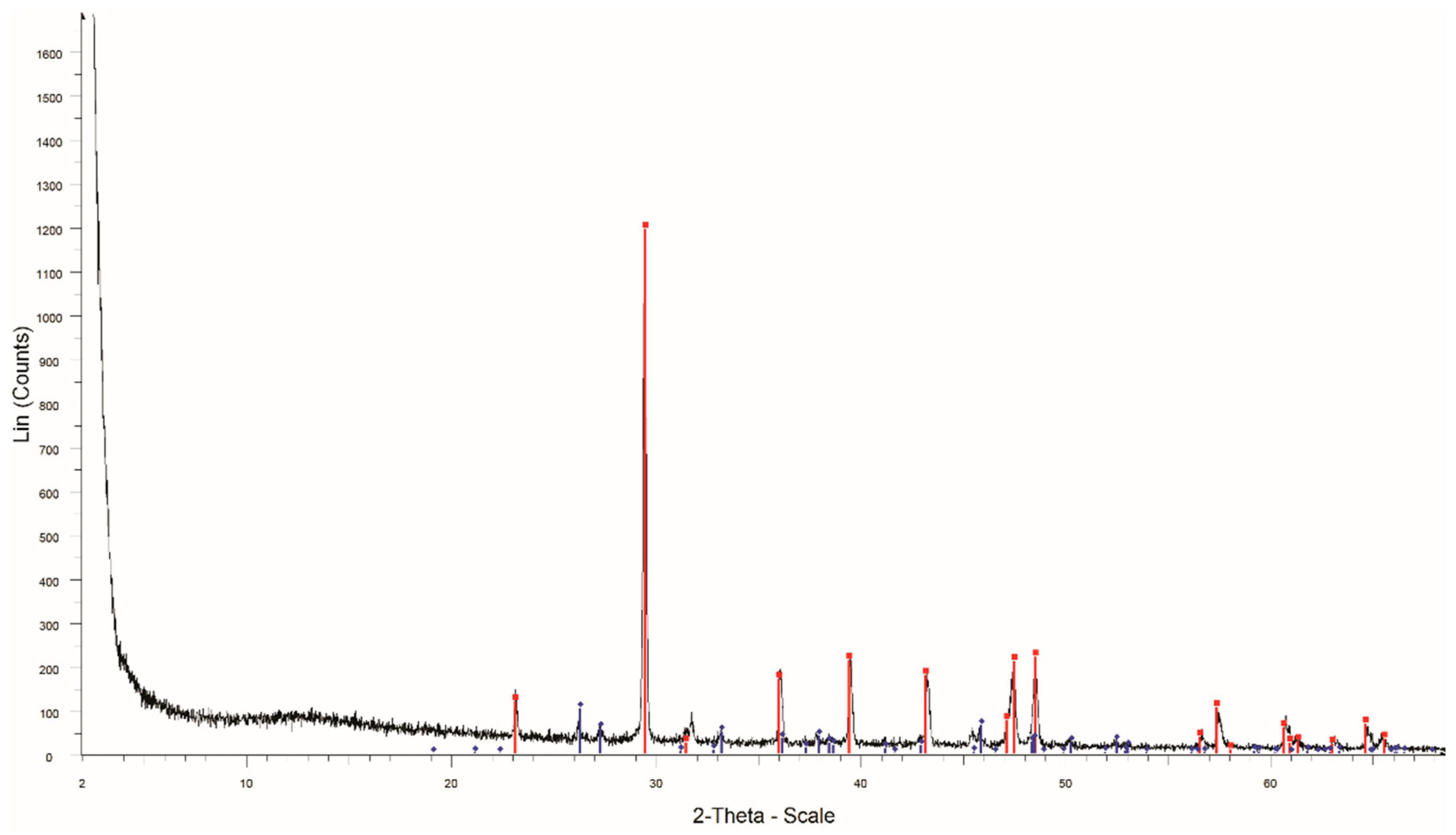
4.2. Mineralogical Characterization and Facies

Based on the XRD analyses, the studied samples consist mainly of calcite and some aragonite (Figure 3). According to SEM-EDS microanalyses, the calcium carbonate mineral phases, except for CaCO3, contain additional trace elements (Table 1), such as Mg (up to 1.93 wt. % MgO), Sr (up to 0.52 wt. % SrO), Na (up to 0.44 wt. % Na2O), and K (up to 0.17 wt. % K2O). The substitution of Ca by these elements is not something new e.g., [55,56]; still, it ought to be mentioned that Mg-calcites and Na-calcite/aragonite have been related to biogenic origin in marine organisms [57,58]. In addition to the elements mentioned above, in several cases in the studied carbonate mineral phases, S was identified (up to 3.99 wt. % SO3, Table 1IV–VI). The S-containing calcites were recently verified. Okumura et al. [59] proved the presence of sulfate-containing calcites in the hot spring LaDuke Yellowstone, USA. Applying several analytical techniques, such as XPS, XRD, and TEM analysis, it was proved that the mean atomic ratio of S/Ca was ca. 5% and sulfur was present in the form of sulfate (⁠SO2−4⁠). Sulfate could be incorporated at the carbonate site of the calcite structure (structural substitute [60,61]). Besides the calcium carbonate mineral phases, fluorite was also found inside calcite crystals (Figure 4A). Kanellopoulos et al. [29,35] have also identified the presence of fluorite in the thermogenic travertines of Northern Euboea.
Aragonite crystals present hexagonal prisms, usually creating characteristic radial spheres (Figure 4B,C), while calcite usually creates rhombohedral crystals (Figure 4D). Similar shapes of the aragonite crystals have been described in the thermogenic travertine depositions of the area [30,31].
The studied samples present lamination consisting of micritic crystals, visible to the naked eye (millimeters) to a few micrometers thick (Figure 4B,D,E). In most cases, the laminas consist of micritic calcite crystals and alternate with other laminas, which could be similar in terms of mineralogical composition but could differ in terms of crystal size and density (Figure 4D). In other cases, calcite laminae alter with aragonite laminae (Figure 4B). The presence of shrubs (Figure 4B) is common to the studied samples. Their thicknesses could reach up ca. over 100 μm (Figure 4G). They are mainly dense and stubby crystalline masses, which consist of calcite crystals that expand upward by irregular branching. These faces have also been identified as the most common among the thermogenic travertine deposition of Northern Euboea [24,27,35], and in several cases have been related to biomineralization processes [37,38].
In a few cases, in the upper parts of the samples, laminas of a loosely connected line of single calcite crystals were identified. In the cases when the samples were not critical point dried, and the samples were condensed as the biological material was dehydrated, these structures present a characteristic condensed form, implying their close connection with biological material, such as EPS (Figure 4F), i.e., biologically-influenced processes. It is known that the EPS play the role of a template favoring mineral precipitation for crystal nucleation [62,63,64] and a low-energy substrate for crystal nucleation (EPS-mediated mineralization [64]). In hot springs, where carbonate precipitation usually occurs, organic mucilaginous substances could usually play a significant role in the biomineralization processes leading to the creation of several facies, i.e., mats consisting of various microbial communities are embedded within a 3D network of EPS secreted by the microorganisms, even if the EPS could be acting as passive substrates [65]. In some other cases, over that loosely connected laminae, shrubs have developed (Figure 4G). It is interesting that the calcite crystals of that loosely connected laminae, as well as the aragonitic radial spheres, are enriched in Sr and present depletion to S (Table 1VII–VIII), in contrast with the other laminas (Table 1IV–VI; Figure 4H,I).

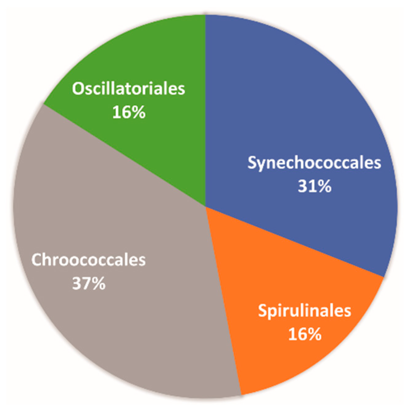
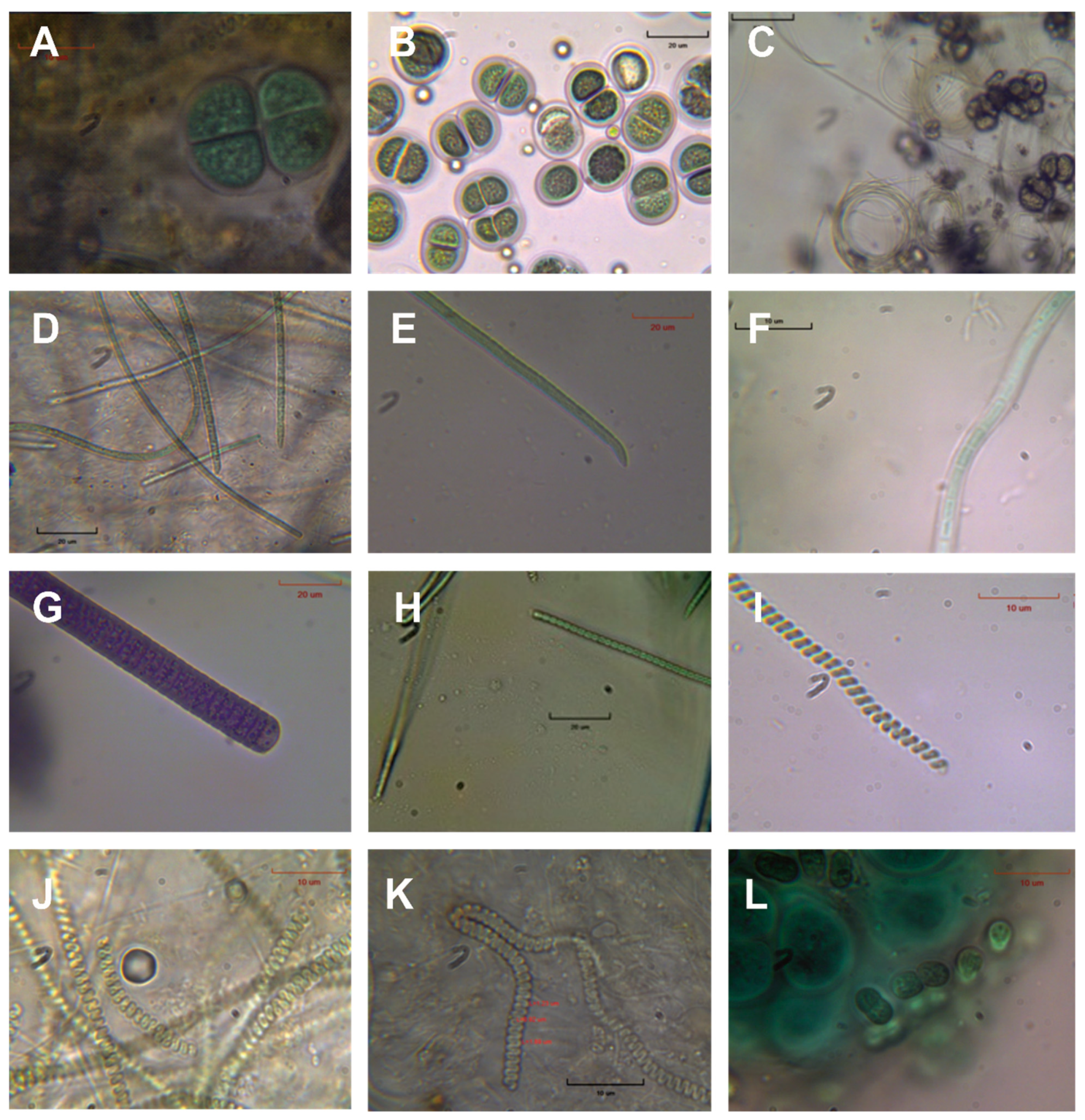
4.3. Cyanobacteria Diversity and Possible Applications

In order to assess the Cyanobacteria biodiversity, pie diagrams of the orders were made (Figure 5), and the latest classification system [66] was used. Based on the studied samples, Chroococcales (37%) and Synechococcales (31%) are the dominant orders, followed by Oscillatoriales (16%) and Spirulinales (16%). Previous studies [31] demonstrated that the most commonly dominating Cyanobacteria orders in the hot springs of Aedipsos were Oscillatoriales (35.7%) and Synechococcales (31.4%), followed by Chroococcales (15.9%), Spirulinales (10.1%), Nostocales (6.6%), and Chroococcidiopsidales (0.3%).
Based on the microscopic study of fresh and cultured material, a total number of 19 Cyanobacteria species, plus diatoms, were observed (Table 2; Figure 6). Among them, typical thermophilic species were identified, such as Spirulina subtilissima (Figure 6J) and Chroococcus thermalis, and typical limestone substrate Cyanobacteria species such as Leptolyngbya perforans (Figure 6F). The species Spirulina subtilissima (Figure 6J), according to Kanellopoulos et al. [32], is the most common Cyanobacteria species in the Aedipsos hot springs. Most of the identified Cyanobacteria species are filamentous.
As mentioned above, the peloids biological fraction is highly important for applied uses. A characteristic example is pelotherapy. During peloid maturation, organic substances are provided by the developing microalgae. Therefore, studying microalgal and bacterial community composition in peloids along the maturation process is of paramount importance since products of their metabolism may transfuse a healing effect. Furthermore, an important recent issue regarding the dominant mechanism that shapes the medicinal properties has to do with the diversity of the microorganisms inhabiting and shaping the peloids [67,68]. Microalgae communities in thermal springs have long attracted the attention of microbial ecologists because of their unique capacity to adapt to high temperatures [69]. Moreover, their long phylogenetic history may justify properties for survival in harsh conditions and, therefore, unique properties for potential biotechnological exploitation. In addition, new therapeutically active compounds could be formed during peloid maturation by the metabolic activity of living organisms such as diatoms, Cyanobacteria (and protozoa). It is in the last decade that microalgae became the focus of extensive research efforts, aiming at compounds that previously have not been considered acting agents in the pharmaceutical industry [22,25,26,27].

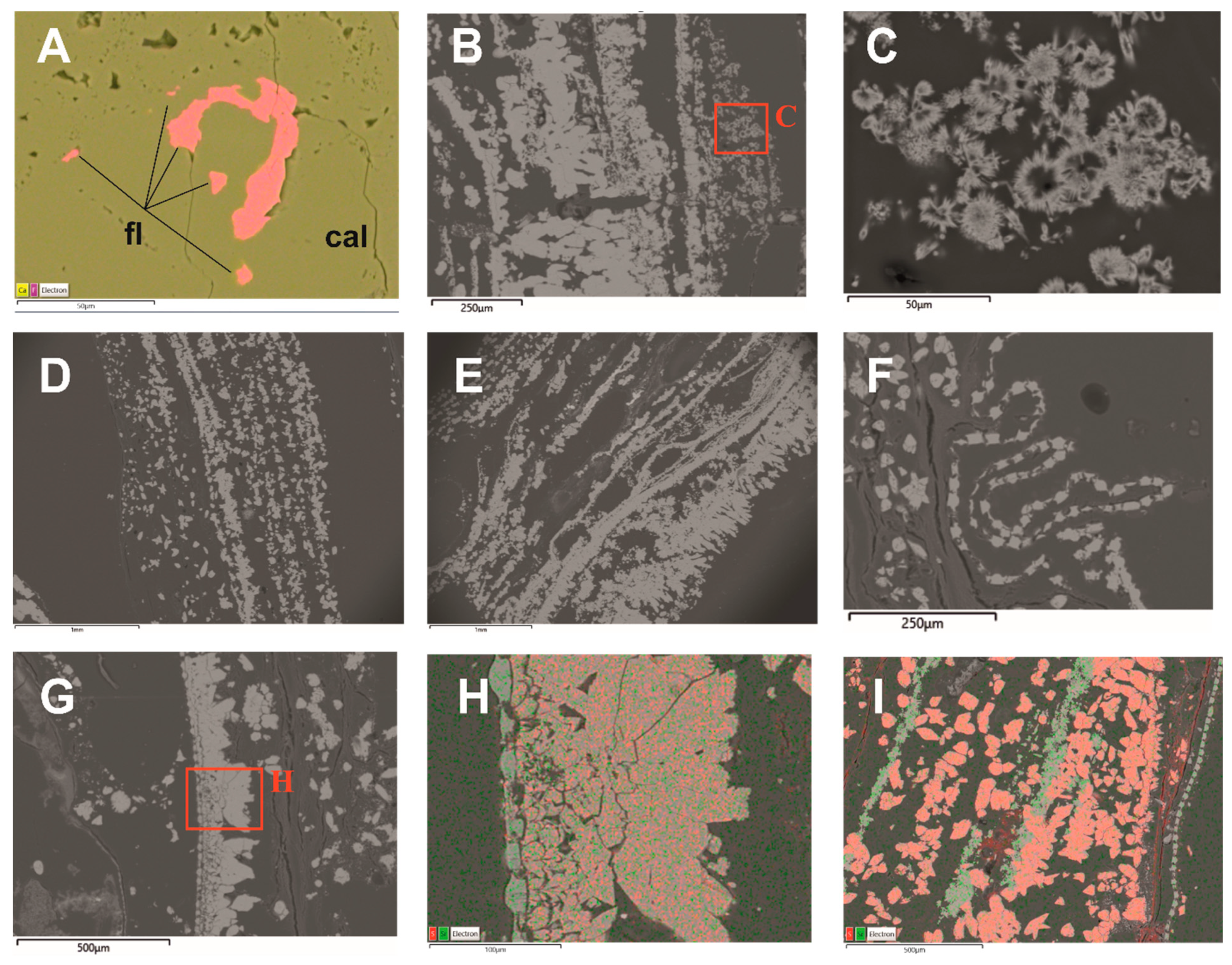
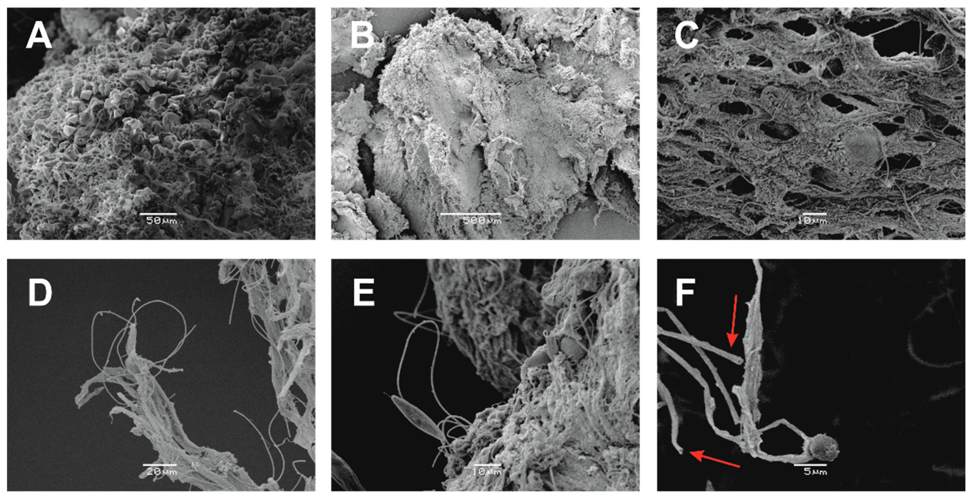
4.4. Biomineralisation

Based mainly on the SEM study, biomineralization processes were identified (Figure 7). The upper abiotic layer consists of micritic calcium carbonate crystals without any trace of microorganisms (Figure 7A). In that layer, only some filamentous Cyanobacteria that develop upwards, originating from the lower layer (Cyanobacteria layer) were found, and also EPS. This development model could be explained due to the daily movement of Cyanobacteria to reach sunlight, as Takashima and Kano [54] suggested, and at the same time, the EPS could be acting as passive substrates [65] leading to biologically-influenced processes.
In the second layer, i.e., the Cyanobacteria layer, the presence of EPS and filamentous Cyanobacteria were dominant (Figure 7B–E). The EPSs, mainly consisting of polysaccharides, polymers, nucleic acids, humic acids, lipids, and proteins [70], form a crystal retention lattice, and small crystals stick to it. It is also known that the EPSs act as a template favoring mineral precipitation for crystal nucleation [63,64]. At the same time, the presence of EPS is crucial for constructing the microbial mat. The exact composition of the EPSs in the study site is the subject of future research.
The filamentous Cyanobacteria favor the trapping of micritic mineral phases or diatoms (Figure 7C–E).
At the same time, some filamentous Cyanobacteria present properties of sheath calcification (Figure 7F), resulting in the formation of dense, thick micritic encrustation around the sheaths by calcium carbonate minerals. These processes could be related either to photosynthesis, resulting in a local increase of pH and subsequent carbonate oversaturation and precipitation [71,72], or to the presence of nucleating molecules. [73].

5. Conclusions

Microbial mats are multilayer bio-structures invaluable for geology, ecology, and geomicrobiological studies. In Aedipsos, microbial mats were studied, several centimeters thick, which occur in a vertical wall where hot water is coming out.
The main mineral phase is calcite, and some aragonite is also present. These mineral phases contain several trace elements, i.e., up to 1.93 wt. % MgO, up to 0.52 wt. % SrO, up to 0.44 wt. % Na2O, up to 0.17 wt. % K2O and up to 3.99 wt. % SO3, in the mineral-chemistry composition. The main facies of the studied samples are lamination and shrubs, which are the most common among the facies of the thermogenic travertines of the area.
The mat consists of several layers, i.e., (i) a top mainly abiotic layer of calcium carbonate micritic crystals, (ii) a second layer–the Cyanobacteria layer, which is dominated by the species Leptolyngbya perforans, (iii) a third layer where Leptolyngbya perforans, Chloroflexus, and other bacteria occur, and (iv) several deeper layers, where no photosynthetic microorganisms were found.
Nineteen Cyanobacteria species (plus diatoms) were identified belonging to Chroococcales (37%) and Synechococcales (31%), which are the dominant orders, followed by Oscillatoriales (16%) and Spirulinales (16%). Among the identified Cyanobacteria, typical thermophilic species, i.e., Spirulina subtilissima and Chroococcus thermalis, and typical limestone substrate species, i.e., Leptolyngbya perforans, have been noticed.
Based mainly on SEM observations, several biomineralization and biologically-influenced processes were recognized, mainly in the second layer, i.e., the Cyanobacteria layer. Filamentous Cyanobacteria are trapping calcium carbonate crystals and diatoms. The extracellular polymeric substances (EPSs) form a crystal retention lattice contributing to the biomineralization process and also to the structure of microbial mats. Finally, calcified filamentous Cyanobacteria sheaths were also identified, contributing to the biomineralization processes.
Among the future plans is a detailed geological (mineralogy and water characteristics) and biological study of each layer separately, as well as metagenomic analysis, in an attempt to identify all the species involved and further understand the processes taking place in each layer.
The present study confirmed the importance of microbial mats in the microbial ecology of extreme environments via understanding biomineralization and biologically-influenced processes. Further research is needed, including phylogenetic analysis and additional study sites to comprehend the biodiversity of all layers of the microbial mats in these or relevant areas. Future selection of local strains of Cyanophyta with optimized biomass development in cultures would further allow biotechnological uses, i.e., isolation of high-value co-products with scientific, medical, or tentative commercial value (e.g., in balneotherapy).

Author Contributions

Conceptualization C.K., V.L. and A.E.-A.; Methodology, C.K., V.L. and A.E.-A.; Sampling C.K. and V.L.; Biological experiments and assessment A.P., V.L. and A.E.-A.; Geological experiments and assessment A.P., C.K., P.V., M.K., I.I. and L.M.; SEM analysis and geobiological assessment A.P., C.K. and V.L.; Statistical analysis A.P., C.K. and V.L.; GIS C.K.; Visualization C.K., V.L. and A.P.; Writing—original draft C.K. and V.L.; Writing—review and editing A.E.-A., C.K. and V.L., P.V., I.I., M.K. and L.M.; Supervision A.E.-A. All authors have read and agreed to the published version of the manuscript.

Funding

This research received no external funding.

Data Availability Statement

Not applicable.

Acknowledgments

The authors would like to thank the local population and authorities, and especially the Director of the Public Properties Company-Aedipsos branch, Ilias Siakantaris, for their cooperation during the fieldwork. The corresponding author would like to thank the two Special Issue Editors, i.e., Maria Economou-Eliopoulos and Ifigeneia Megremi, as well as George Vougioukalakis from the Greek Geological Survey (IGME, present name Hellenic Survey of Geology and Mineral Exploration, HSGME) for his support and encouragement during this research.

Conflicts of Interest

The authors declare no conflict of interest.

References

  1. Reitner, J.; Thiel, V. Encyclopedia of Geobiology; Springer Science and Business Media: Berlin/Heidelberg, Germany, 2011; ISBN 978-1-4020-9211-4. [Google Scholar]
  2. Allwood, A.C.; Walter, M.R.; Kamber, B.S.; Marshal, C.P.; Burch, I.W. Stromatolite reef from the Early Archaean era of Australia. Nature 2006, 441, 714–717. [Google Scholar] [CrossRef]
  3. Van Kranendonk, M.J. A review of the evidence for putative Paleoarchean life in the Pilbara Craton. In Earth’s Oldest Rocks—Developments in Precambrian Geology; Van Kranendonk, M.J., Smithies, R.H., Bennet, V., Eds.; Elsevier: Amsterdam, The Netherlands, 2007; pp. 855–896. [Google Scholar]
  4. Homann, M. Earliest life on earth: Evidence from the Barberton Greenstone Belt, South Africa. Earth-Sci. Rev. 2019, 196, 102888. [Google Scholar] [CrossRef]
  5. Westall, F.; de Vries, S.T.; Nijman, W.; Rouchon, V.; Orberger, B.; Pearson, V.; Watson, J.; Verchovski, A.; Wright, I.; Rouzaud, J.N.; et al. The 3.466 Ga “Kitty’s gap Chert” an early Archean microbial ecosystem. In Processes on the Early Earth; Reimod, W.U., Gibson, R.L., Eds.; Geological Society of America: Boulder, CO, USA, 2006; Volume 405, pp. 105–131. [Google Scholar] [CrossRef]
  6. Van Dover, C.L. The Ecology of Deep-Sea Hydrothermal Vents; Princeton University Press: Princeton, NJ, USA, 2000; 424p. [Google Scholar]
  7. Riding, R.; Awramik, S.M. (Eds.) Microbial Sediments; Springer: Berlin/Heidelberg, Germany, 2000; 331p. [Google Scholar]
  8. Arp, G.; Reimer, A.; Reitner, J. Microbialite formation in seawater of increased alkalinity, Satonda Crater Lake, Indonesia. J. Sediment. Res. 2003, 73, 105–127. [Google Scholar] [CrossRef]
  9. Pedersen, K. Microbial life in deep granitic rock. FEMS Microbiol. Rev. 2006, 20, 399–414. [Google Scholar] [CrossRef]
  10. Characklis, W.G.; Wilderer, P.A. Structure and Function of Biofilms; Wiley: Hoboken, NJ, USA, 1989; 387p. [Google Scholar]
  11. Fouke, B.W.; Bonheyo, G.T.; Sanzenbacher, B.; Frias-Lopez, J. Partitioning of bacterial communities between travertine depositional facies at Mammoth Hot Springs, Yellowstone National Park, USA. Can. J. Earth Sci. 2003, 40, 1531–1548. [Google Scholar] [CrossRef] [Green Version]
  12. Meyer-Dombard, D.R.; Shock, E.L.; Amend, J.P. Archaeal and bacterial communities in geochemically diverse hot springs of Yellowstone National Park, USA. Geobiology 2005, 3, 211–227. [Google Scholar] [CrossRef]
  13. Fouke, B.W. Hot-spring systems geobiology: Abiotic and biotic influences on travertine formation at mammoth hot springs, Yellowstone National Park, USA. Sedimentology 2011, 58, 170–199. [Google Scholar] [CrossRef]
  14. Thomas, C. Investigating the Subsurface Biosphere of a Hypersaline Environment—The Dead Sea (Levant). Ph.D. Thesis, Université de Genève, Geneva, Switzerland, 2015. [Google Scholar]
  15. Thomas, C.; Ariztegui, D. Fluid inclusions from the deep Dead Sea sediment provide new insights on Holocene extreme microbial life. Quat. Sci. Rev. 2019, 212, 18–27. [Google Scholar] [CrossRef] [Green Version]
  16. Merino, N.; Aronson, H.S.; Bojanova, D.P.; Feyhl-Buska, J.; Wong, M.L.; Zhang, S.; Giovannelli, D. Living at the Extremes: Extremophiles and the Limits of Life in a Planetary Context. Front. Microbiol. 2019, 10, 780. [Google Scholar] [CrossRef] [PubMed]
  17. Bech, J. Les Terres Medicinales. Discurs per Reial Academia de Farmàcia de Barcelona; Reial Acadèmia de Farmàcia de Barcelona-CIRIT (Generalitat de Calalunya): Barcelona, Spain, 1987; p. 105. [Google Scholar]
  18. Canfield, D.E.; Des Marais, D.J. Biogeochemical cycles of carbon, sulfur, and free oxygen in a microbial mat. GCA 1993, 57, 3971–3984. [Google Scholar] [CrossRef]
  19. Allison, D.G.; Gilbert, P.; Lappin-Scott, H.M.; Wilson, M. Community Structure and Co-Operation in Biofilms; Cambridge University Press: Cambridge, UK, 2000; 349p. [Google Scholar]
  20. Wingender, J.; Neu, T.R.; Flemming, H.-C. Microbial Extracellular Polymeric Substances; Characterization, Structure, and Function; Springer: Berlin/Heidelberg, Germany, 1999; 258p. [Google Scholar]
  21. Decho, A.W.; Visscher, P.T.; Reid, P. Production and cycling of natural microbial exopolymers (EPS) within a marine stromatolite. Palaeogeogr. Palaeoclimatol. Palaeoecol. 2005, 219, 71–86. [Google Scholar] [CrossRef]
  22. Mourelle, M.L.; Gómez, C.P.; Legido, J.L. Microalgal Peloids for Cosmetic and Wellness Uses. Mar. Drugs 2021, 19, 666. [Google Scholar] [CrossRef] [PubMed]
  23. Gomes, C.; Carretero, M.I.; Pozo, M.; Maraver, F.; Cantista, P.; Armijo, F.; Legido, J.L.; Teixeira, F.; Rautureau, M.; Delgado, R. Peloids and pelotherapy: Historical evolution, classification and glossary. Appl. Clay Sci. 2013, 75–76, 28–38. [Google Scholar] [CrossRef]
  24. Yilmaz, B.; Goktepe, A.S.; Alaca, R.; Mohur, H.; Kayar, A.H. Comparison of a generic and a disease specific quality of life scale to assess a comprehensive spa therapy program for knee osteoarthritis. Jt. Bone Spine 2004, 71, 563–566. [Google Scholar] [CrossRef]
  25. Abedin, R.M.A.; Taha, H.M. Antibacterial and antifungal activity of cyanobacteria and green microalgae. Evaluation of medium components by Plackett-Burman design for antimicrobial activity of Spirulina platensis. GJBBR 2008, 3, 22–31. [Google Scholar]
  26. Gademann, K.; Portmann, C. Secondary metabolites from cyanobacteria: Complex structures and powerful bioactivities. Curr. Org. Chem. 2008, 12, 326–341. [Google Scholar] [CrossRef]
  27. Martins, R.F.; Ramos, M.F.; Herfindal, L.; Sousa, J.A.; Skærven, K.; Vasconcelos, V.T. Antimicrobial and cytotoxic assessment of marine cyanobacteria Synechocystis & Synechococcus. Mar. Drugs 2008, 6, 1–11. [Google Scholar] [PubMed]
  28. Kanellopoulos, C.; Mitropoulos, P.; Valsami-Jones, E.; Voudouris, P. A new terrestrial active mineralizing hydrothermal system associated with ore-bearing travertines in Greece (northern Euboea Island and Sperchios area). J. Geochem. Explor. 2017, 179, 9–24. [Google Scholar] [CrossRef]
  29. Kanellopoulos, C. Geochemical Research on the Distribution of Metallic and Other Elements in the Cold and Thermal Groundwater, Soils and Plants in Fthiotida Prefecture and N. Euboea. Environmental Impact. Ph.D. Thesis, National and Kapodistrian University of Athens, Athens, Greece, 2011. (In Greek with English abstract). [Google Scholar]
  30. Kanellopoulos, C. Distribution, lithotypes and mineralogical study of newly formed thermogenic travertines in Northern Euboea and Eastern Central Greece. Open Geosci. (former Cent. Eur. Geol.) 2012, 4, 545–560. [Google Scholar] [CrossRef]
  31. Kanellopoulos, C. Various morphological types of thermogenic travertines in northern Euboea and Eastern Central Greece. Bull. Geol. Soc. Greece 2013, 47, 1929–1938. [Google Scholar] [CrossRef]
  32. Kanellopoulos, C.; Lamprinou, V.; Politi, A.; Voudouris, P.; Economou-Amilli, A. Insights on the biomineralization processes and related diversity of cyanobacterial microflora in thermogenic travertine deposits in Greek hot springs (North-West Euboea Island). Depos. Rec. 2022, 8, 1055–1078. [Google Scholar] [CrossRef]
  33. Kanellopoulos, C.; Lamprinou, V.; Politi, A.; Voudouris, P.; Economou-Amilli, A. Pioneer species of Cyanobacteria in hot springs and their role to travertine formation: The case of Aedipsos hot springs, Euboea (Evia), Greece. Depos. Rec. 2022, 8, 1079–1092. [Google Scholar] [CrossRef]
  34. Kanellopoulos, C.; Thomas, C.; Xirokostas, N.; Ariztegui, D. Banded Iron Travertines at the Ilia Hot Spring (Greece): An interplay of biotic and abiotic factors leading to a modern BIF analog? Depos. Rec. 2019, 5, 109–130. [Google Scholar] [CrossRef]
  35. Stanier, R.Y.; Kunisawa, R.; Mandel, M.; Cohen-Bazire, G. Purification and properties of unicellular blue-green algae (order Chroococcales). Bacteriol. Rev. 1971, 35, 171. [Google Scholar] [CrossRef]
  36. Komárek, J.; Anagnostidis, K. Modern approach to the classification system of Cyanophytes 4-Nostocales. Arch. Hydrobiol. Suppl. Bd. Algol. Stud. 1989, 56, 247–345. [Google Scholar]
  37. Komárek, J.; Anagnostidis, K. Cyanoprokaryota, Part 1: Chroococcales. In Süßwasserflora von Mitteleuropa, Bd. 19 (1); Büdel, B., Gärtner, G., Krienitz, L., Schagerl, M., Eds.; Elsevier GmbH: München, Germany, 1999; pp. 1–548. [Google Scholar]
  38. Komárek, J.; Anagnostidis, K. Cyanoprokaryota, Part 2: Oscillatoriales. In Süßwasserflora von Mitteleuropa, Bd. 19 (2); Büdel, B., Gärtner, G., Krienitz, L., Schagerl, M., Eds.; Elsevier GmbH: München, Germany, 2005; pp. 1–759. [Google Scholar]
  39. Komárek, J.; Kastovsky, J.; Mares, J.; Johansen, J.R. Taxonomic classification of cyanoprokaryotes (cyanobacterial genera), using a polyphasic approach. Preslia 2014, 86, 295–335. [Google Scholar]
  40. Kanellopoulos, C.; Xenakis, M.; Vakalopoulos, P.; Kranis, H.; Christopoulou, M.; Vougioukalakis, G. Seawater-dominated, tectonically controlled and volcanic related geothermal systems: The case of the geothermal area in the northwest of the Island of Euboea (Evia), Greece. Int. J. Earth Sci. 2020, 109, 2081–2112. [Google Scholar] [CrossRef]
  41. Aubouin, J. Contribution a l’ étude géologique de la Grèce septentrionale: Les confins de l’Epire et de la Thessalie. Ann. Geol. Pays Hellen. 1959, 10, 1–483. [Google Scholar]
  42. Mountrakis, D. The Pelagonian zone in Greece: A polyphase-deformed fragment of the Cimmerian continent and its role in the geotectonic evolution of the eastern Mediterranean. J. Geol. 1986, 94, 335–347. [Google Scholar] [CrossRef]
  43. Vavassis, I. Geology of the Pelagonian Zone in Northern Evia Island (Greece): Implications for the Geodynamic Evolution of the Hellenides. Ph.D. Thesis, Universite de Lausanne, Lausanne, Switzerland, 2001. [Google Scholar]
  44. Jolivet, L.; Faccenna, C.; Huet, B.; Labrousse, L.; Le Pourhiet, L.; Lacombe, O.; Lecomte, E.; Burov, E.; Denèle, Y.; Brun, J.-P.; et al. Aegean tectonics: Strain localization, slab tearing and trench retreat. Tectonophysics 2013, 597–598, 1–33. [Google Scholar] [CrossRef] [Green Version]
  45. Katsikatsos, G.; Mettos, A.; Vidakis, M.; Bavay, P.; Panagopoulos, A.; Basilaki, A.; Papazeti, E. Geological study of Aedipos Area—Euboea; Geothemal Studies (P.E.C.); IGME: Athens, Greece, 1982. (In Greek) [Google Scholar]
  46. Scherreiks, R. Platform margin and oceanic sedimentation in a divergent and convergent plate setting (Jurassic, Pelagonian Zone, NE Evvoia, Greece). Int. J. Earth Sci. 2000, 89, 90–107. [Google Scholar] [CrossRef]
  47. Kanellopoulos, C.; Christopoulou, M.; Xenakis, M.; Vakalopoulos, P. Hydrochemical characteristics and geothermometry applications of hot groundwater in Edipsos area, NW Euboea (Evia), Greece. Bull. Geol. Soc. Greece 2016, 50, 720–729. [Google Scholar] [CrossRef] [Green Version]
  48. Fytikas, M.; Giuliani, O.; Innocenti, F.; Marinelli, G.; Mazzuoli, R. Geochronological data on recent magmatism of the Aegean Sea. Tectonophysics 1976, 31, T29–T34. [Google Scholar] [CrossRef]
  49. De Boever, E.; Brasier, A.T.; Foubert, A.; Kele, S. What do we really know about early diagenesis of non-marine carbonates? Sediment. Geol. 2017, 361, 25–51. [Google Scholar] [CrossRef] [Green Version]
  50. Jorgensen, B.B.; Revsbech, N.P.; Cohen, Y. Photosynthesis and structure of benthic microbial mats: Microelectrode and SEM studies of four cyanobacterial communities. Limnol. Oceanogr. 1983, 28, 1075–1093. [Google Scholar] [CrossRef]
  51. Stal, L.J. Cyanobacterial mats and stromatolites. In The Ecology of Cyanobacteria; Whitton, B.A., Potts, M., Eds.; Kluwer Academic Publishers: Dordrecht, The Netherlands, 2000; pp. 61–120. [Google Scholar]
  52. Pentecost, A. Travertine; Springer: Berlin/Heidelberg, Germany, 2005. [Google Scholar]
  53. Ward, D.M.; Ferris, M.J.; Nold, S.C.; Bateson, M.M.; Kopczynski, E.D.; Ruff-Roberts, A.L. Species diversity in hot spring microbial mats as revealed by both molecular and enrichment culture approaches—Relationship between biodiversity and community structure. In Microbial Mats; Stal, J.L., Caumette, P., Eds.; Springer: Berlin/Heidelberg, Germany, 1994; pp. 33–44. [Google Scholar]
  54. Takashima, C.; Kano, A. Microbial processes forming daily lamination in a stromatolitic travertine. Sediment. Geol. 2008, 208, 114–119. [Google Scholar] [CrossRef]
  55. Mucci, A.; Mucci, J.W. The incorporation of Mg2+ and Sr2+ into calcite overgrowths: Influences of growth rate and solution composition. Geochim. Cosmochim. Acta 1983, 47, 217–233. [Google Scholar] [CrossRef]
  56. Ishikawa, M.; Ichikuni, M. Uptake of sodium and potassium by calcite. Chem. Geol. 1984, 42, 137–146. [Google Scholar] [CrossRef]
  57. Long, X.; Ma, Y.; Qi, L. Biogenic and synthetic high magnesium calcite—A review. J. Struct. Biol. 2014, 185, 1–14. [Google Scholar] [CrossRef]
  58. Yoshimura, T.; Tamenori, Y.; Suzuki, A.; Kawahata, H.; Iwasaki, N.; Hasegawa, H.; Nguyen, L.T.; Kuroyanagi, A.; Yamazaki, T.; Kuroda, J.; et al. Altervalent substitution of sodium for calcium in biogenic calcite and aragonite. Geochim. Cosmochim. Acta 2017, 202, 21–38. [Google Scholar] [CrossRef]
  59. Okumura, T.; Kim, H.-J.; Kim, J.-W.; Kogure, T. Sulfate-containing calcite: Crystallographic characterization of natural and synthetic materials. Eur. J. Mineral. 2018, 30, 929–937. [Google Scholar] [CrossRef]
  60. Kampschulte, A.; Strauss, H. The sulfur isotopic evolution of Phanerozoic seawater based on the analysis of structurally substituted sulfate in carbonates. Chem. Geol. 2004, 204, 255–286. [Google Scholar] [CrossRef]
  61. Balan, E.; Aufort, J.; Pouillé, S.; Dabos, M.; Blanchard, M.; Lazzeri, M.; Rollion-Bard, C.; Blamart, D. Infrared spectroscopic study of sulfate-bearing calcite from deep-sea bamboo coral. Eur. J. Mineral. Copernic. 2017, 29, 397–408. [Google Scholar] [CrossRef]
  62. Reitner, J. Modern cryptic microbialite/metazoan facies from Lizard Island (Great Barrier Reef, Australia): Formation and Concept. Facies 1993, 29, 3–40. [Google Scholar] [CrossRef] [Green Version]
  63. Görgen, S.; Benzerara, K.; Skouri-Panet, F.; Gugger, M.; Chauvat, F.; Cassier-Chauvat, C. The diversity of molecular mechanisms of carbonate biomineralization by bacteria. Discov. Mater. 2021, 1, 2. [Google Scholar] [CrossRef]
  64. Della Porta, G.; Hoppert, M.; Hallmann, C.; Schneider, D.; Reitner, J. The influence of microbial mats on travertine precipitation in active hydrothermal systems (Central Italy). Depos. Rec. 2021, 8, 165–209. [Google Scholar] [CrossRef]
  65. Della Porta, G. Carbonate build-ups in lacustrine, hydrothermal and fluvial settings: Comparing depositional geometry, fabric types and geochemical signature. Geol. Soc. Lond. Spec. Publ. 2015, 418, 17–68. [Google Scholar] [CrossRef]
  66. Hauer, T.; Komárek, J. CyanoDB 2.0—On-line Database of Cyanobacterial Genera; World-Wide Electronic Publication; University of South Bohemia & Institute of Botany AS CR: České Budějovice, Czechia, 2021; Available online: https://www.cyanodb.cz (accessed on 19 December 2021).
  67. Pesciaroli, C.; Viseras, C.; Aguzzi, C.; Rodelas, B.; Gonzalez-Lopez, J. Study of bacterial community structure and diversity during the maturation process of a therapeutic peloid. Appl. Clay Sci. 2016, 132, 59–67. [Google Scholar] [CrossRef]
  68. Vadlja, D.; Bujak, M.; Čož-Rakovac, R.; Roje, M.; Čižmek, L.; Horvatić, A.; Svetličić, E.; Diminić, J.; Rakovac, S.; Oros, D.; et al. Bioprospecting for Microorganisms in Peloids—Extreme Environment Known for Its Healing Properties. Front. Mar. Sci. 2022, 9, 822139. [Google Scholar] [CrossRef]
  69. Sompong, U.; Hawkins, P.R.; Besley, C.; Peerapornpisal, Y. The distribution of cyanobacteria across physical and chemical gradients in hot springs in Northern Thailand. FEMS 2005, 52, 365–376. [Google Scholar] [CrossRef] [Green Version]
  70. Flemming, H.-C.; Wingender, J. The biofilm matrix. Nat. Rev. Microbiol. 2010, 8, 623–633. [Google Scholar] [CrossRef] [PubMed]
  71. Riding, R. Cyanobacterial calcification, carbon dioxide concentrating mechanisms, and Proterozoic-Cambrian changes in atmospheric composition. Geobiology 2006, 4, 299–316. [Google Scholar] [CrossRef]
  72. Jansson, C.; Northen, T. Calcifying cyanobacteria—The potential of biomineralization for carbon capture and storage. Curr. Opin. Biotechnol. 2010, 21, 365–371. [Google Scholar] [CrossRef] [PubMed]
  73. Merz-Preiss, M.; Riding, R. Cyanobacterial tufa calcification in two freshwater streams: Ambient environment, chemical thresholds and biological processes. Sediment. Geol. 1999, 126, 103–124. [Google Scholar] [CrossRef]
Figure 1. Geological map of the Aedipos area, NW of Euboea (AF = Aedipsos Fault); modified after Kanellopoulos et al. [40]. The sampling site is located in the Casino area, which is marked with a black dot. The geographical coordinates system is EGSA87.
Minerals 12 01408 g001
Figure 2. Field photos and sample photos. (A,B) Paired views of the study site. Normal image (A) and corresponding thermal image (B). On the right side of the thermal picture, a column shows the temperature scale (°C). (C) Detailed image of the hot water flow and the microbial mats. (D) A vertical cut of the microbial mats, displaying the different vertical layering. (E) A complete piece of microbial mats displaying the vertical stratification: (1) top layer-the abiotic layer of travertine (white color), (2) intense blue-green layer of Cyanobacteria, and (3) olive-green layer where there is a reduction of Cyanobacteria and presence of other bacteria. (F) Detailed image of the hot water flow and the microbial mats. (G) Parts of the top abiotic layer where dense white parts can be observed (arrows). (H) The top abiotic layer of micritic calcium carbonate minerals under a stereoscope.
Minerals 12 01408 g002
Figure 3. Evaluated XRD pattern of a characteristic sample. The dominant mineral phase is calcite (red columns), whereas aragonite is also present (blue columns).
Minerals 12 01408 g003
Figure 4. Back-scattered electron images (BSEI). (A) False color BSEI results of the mapping, displaying the distribution of Ca (yellow) and F (purple) demonstrating the presence of fluorite in calcite. (B,C) Lamination with calcite crystals creating shrubs and aragonite crystals usually creating characteristic radial spheres. (D,E) Laminas are created by micritic crystals of calcium carbonate mineral phases. (F) Laminas of a loosely connected line of single calcite crystals, which in some cases are presenting a characteristic condensed form. (G) In some cases, over previously presented loosely connected laminae, shrubs have developed. (H,I) False color BSEI results of the mapping, displaying the distribution of S (red) and Sr (green). In (H) it is shown that the loosely connected base laminae present S depletion and Sr enrichment, in contrast to the shrubs that have developed over it and present Sr depletion and S enrichment. In (I) it is shown that the aragonite-dominated (aggregates of radial spheres) laminas present S depletion and Sr enrichment, in contrast to the calcite-dominated laminas, which present Sr depletion and S enrichment.
Minerals 12 01408 g004
Figure 5. Pie diagrams presenting the percentage of each Cyanobacteria order.
Minerals 12 01408 g005
Figure 6. Cyanobacterial microflora under an optical microscope. (A) Chroococcus minutus (Scale bar: 10 μm), (B) Chroococcus turgidus (Scale bar: 20 μm), (C) Johannesbaptistia pellucida (Scale bar: 50 μm), (D) Kamptonema chlorinum (Scale bar: 20 μm), (E) Kamptonema formosum (Scale bar: 20 μm), (F) Leptolyngbya perforans (Scale bar: 10 μm), (G) Oscillatoria crassa (Scale bar: 20 μm), (H) Pseudanabaena minima (Scale bar: 20 μm), (I) Spirulina cf. labyrinthiformis (Scale bar: 10 μm), (J) Spirulina subtilissima (Scale bar: 10 μm), (K) Spirulina tenuior (Scale bar: 10 μm), (L) Synechocystis salina (Scale bar: 10 μm).
Minerals 12 01408 g006
Figure 7. Biomineralizing processes by Cyanobacteria under SEM. (A) A top abiotic layer consisting of calcium carbonate crystals (scale bar: 50 μm). (B,C) EPSs surfaces along with filamentous Cyanobacteria. (B) Overview of a dense EPSs layer (scale bar: 500 μm). (C) Closer view in another position, where the EPSs form a net along with filamentous Cyanobacteria, favoring the trapping of calcium carbonate crystals and diatoms (scale bar: 10 μm). (D,E) Details of filamentous Cyanobacteria which are developing upwards above the EPSs layer, favoring their trapping. In (E), a diatom is trapped (scale bar: 10 μm). (F) Calcified sheaths of filamentous Cyanobacteria (scale bar: 5 μm).
Minerals 12 01408 g007
Table 1. Representative microanalyses of calcium carbonate mineral phases.
IIIIIIIVVVIVIIVIII
Na2O-0.16-0.410.230.19--
MgO0.560.590.110.870.520.450.70.96
SO3---3.263.763.99--
K2O-------0.14
CaO53.5851.4354.7152.3251.6253.8152.5151.69
SrO------0.280.25
Total54.1352.3754.8256.8656.1358.4453.4953.64
Table 2. Identified Cyanobacteria species.
Aphanothece cf. minutissima (West) J.Komárková-Legnerová and G.Cronberg 1994
Aphanothece sp.A
Chondrocystis dermochroa (Nägeli) Komárek and Anagnostidis, 1995
Chroococcus minutus (Kützing) Nägeli 1849
Chroococcus minutus var. thermalis J.J.Copeland
Chroococcus thermalis (Meneghini) Nägeli 1849
Chroococcus turgidus (Kützing) Nägeli 1849
Leptolyngbya cf. angusta (Skuja) Anagnostidis 2001
Johannesbaptistia pellucida (Dickie) W.R.Taylor and Drouet in Drouet 1938
Kamptonema chlorinum (Kützing ex Gomont) Strunecký, Komárek and J.Smarda 2014
Kamptonemaformosum (Bory ex Gomont) Strunecký, Komárek and J.Smarda 2014
Leptolyngbya perforans (Geitler) Anagnostidis and Komárek 1988
Limnothrix redekei (Goor) Meffert 1988
Oscillatoria crassa (C.B.Rao) Anagnostidis 2001
Pseudanabaena minima (G.S.An) Anagnostidis 2001
Spirulina cf. labyrinthiformis Gomont 1892
Spirulina subtilissima Kützing ex Gomont 1892
Spirulina tenuior (Lagerheim) Kirchner 1900
Synechocystis salina Wislouch 1924
Diatoms
Publisher’s Note: MDPI stays neutral with regard to jurisdictional claims in published maps and institutional affiliations.

