Article

A Study of Thermal Stability of Hydroxyapatite

by
Natalia V. Bulina
1,*,
Svetlana V. Makarova
1,
Sergey G. Baev
2,
Alexander A. Matvienko
1,
Konstantin B. Gerasimov
1,
Olga A. Logutenko
1 and
Vladimir S. Bystrov
3
1
Institute of Solid State Chemistry and Mechanochemistry, Siberian Branch of the Russian Academy of Sciences, 630128 Novosibirsk, Russia
2
Institute of Automation and Electrometry, Siberian Branch of Russian Academy of Sciences, 630090 Novosibirsk, Russia
3
Institute of Mathematical Problems of Biology, The Branch of Keldysh Institute of Applied Mathematics, Russian Academy of Sciences, 142290 Pushchino, Russia
*
Author to whom correspondence should be addressed.
Minerals 2021, 11(12), 1310; https://doi.org/10.3390/min11121310
Submission received: 20 October 2021 / Revised: 14 November 2021 / Accepted: 20 November 2021 / Published: 24 November 2021
(This article belongs to the Special Issue Recent Advances of Hydroxyapatite and Its Applications)

Abstract

:
High-temperature powder sintering is an integral part of the dense ceramic manufacturing process. In order to find the optimal conditions for producing a ceramic product, the information about its behavior at high temperatures is required. However, the data available in the literature are very contradictory. In this work, the thermal stability of hydroxyapatite prepared by a solid-state mechanochemical method and structural changes occurring during sintering were studied. Stoichiometric hydroxyapatite was found to remain as a single-phase apatite structure with the space group P63/m up to 1300 °C inclusively. A further increase in the sintering temperature leads to its partial decomposition, a decrease in the crystallite size of the apatite phase, and the appearance of significant structural strains. It was shown that small deviations from stoichiometry in the Ca/P ratio upward or downward during the hydroxyapatite synthesis lead to a significant decrease in the thermal stability of hydroxyapatite. An apatite containing almost no hydroxyl groups, which is close to the composition of oxyapatite, was prepared. It was shown that the congruent melting of stoichiometric hydroxyapatite upon slow heating in a high-temperature furnace does not occur. At the same time, the fast heating of hydroxyapatite by laser radiation allows, under certain conditions, its congruent melting with the formation of a recrystallized monolayer of oxyhydroxyapatite. The data obtained in this study can be used when choosing sintering conditions to produce hydroxyapatite-based ceramics.

1. Introduction

Hydroxyapatite (HA), also known as hydroxylapatite in earlier literature, is a rare, naturally occurring mineral with the chemical formula Ca5(PO4)3(OH). HA formula is commonly written as Ca10(PO4)6(OH)2 in order to show that the crystal unit cell comprises two entities. HA is the hydroxyl end member of the apatite group with general formula M10(RO4)6X2. The hydroxyl ion in the HA structure can be replaced by fluoride, chloride, or carbonate producing fluorapatite, chlorapatite, or carbonateapatite, respectively [1]. Apart from that, the substitution of calcium cations and the tetrahedral phosphate ion in HA is also possible [2]. In the case of partial replacement of the ions, the corresponding solid solutions are formed. Replacement in the HA structure significantly changes both its structural characteristic [3,4] and physicochemical and biological properties [2].
Synthetic HA can be prepared by different methods, such as precipitation from an aqueous solution, by the sol–gel process, hydrothermal, mechanochemical, and solid-state reaction methods, among others [5,6,7].
HA is produced in the form of an ionic crystal, either of hexagonal (P63/m) or monoclinic (P21/b) symmetry, depending on the conditions [1,8,9]. The main difference between the hexagonal and the monoclinic HA is the orientations of the hydroxyl groups located in the channel formed by calcium cations [9]. In monoclinic HA, all of the OH groups in a given column are pointed in the same direction, and the direction reverses in the next column. In hexagonal HA, the adjacent OH groups point in opposite directions. The most common case is crystallization of the HA with the formation of a less ordered hexagonal structure.
The values of lattice parameters for hexagonal HA reported in the literature differ (Table 1). This can be explained by the fact that to prepare a stoichiometric HA, Ca- and PO4-containing compounds should be mixed with a strict Ca/P ratio of 1.667. However, maintaining the Ca/P ratio of 1.667 is a difficult task, since various synthesis conditions, in particular the pH of the solution, significantly affect it [1,10,11]. Thus, if the proper pH is not maintained, the so-called calcium-deficient apatite or calcium-rich HA (nonstoichiometric HAs) is formed [1,11]. In addition, the carbonate ions or other impurity ions used for preparing HA can be incorporated into its structure during the precipitation process [1,12], thus affecting the values of the lattice constants [4].
Due to its chemical similarity to the mineral component of human bone and dental tissue, hydroxyapatite is widely used in medicine, particularly, as filler material to repair bone defects, to produce ceramic implants or coatings on metal implants, for the preparation of bone cements, as drug delivery systems, and as an additive in toothpastes and cleaning means [11,13,14]. In addition, synthetic HA is used as a filler for column chromatography, as gas sensors, catalysts, and sorbents [11,15]. However, in order to achieve the required characteristics of the final product, such as density, porosity, morphology, mechanical, and electrical/dielectrical properties, heat treatment of the synthetic HA is necessary [16,17].
The thermal behavior of HA during heat treatment is affected by the composition, structure, morphology, dispersibility, and some other properties of HA, which depend on its synthesis conditions. HA samples synthesized by precipitation, hydrothermal, sol–gel, and mechanochemical methods usually contain water incorporated into its structure. The evolution of adsorbed water is observed at low temperatures. It does not affect the lattice parameters of HA [18]. Water in pores, cracks, and inter-crystallite locations is stabilized by capillary effects and requires higher temperatures for release. The removal of lattice water from HA is observed with a further increase in temperature and leads to a contraction of the lattice parameter a [19]. Moreover, the surface phosphorus is reactive and readily forms the P-OH groups which are dehydroxylated when heated to form surface P-O-P groups [20].
The process of HA dehydroxylation with the release of water starts at a noticeable rate at 850–900 °C [18] and leads to the formation of vacancies in the position of the hydroxyl groups in the HA structure. As a result, the formation of oxyhydroxyapatite (OHA) takes place by the following reaction:
Ca10(PO4)6(OH)2 → Ca10(PO4)6(OH)2−2xOxx + xH2O
where ☐ is the OH-group vacancy.
The dehydroxylation process is affected by the kinetic factors, the heating rate, and the water vapor partial pressure. The slower the heating rate, the more hydroxyl groups leave the structure during heating to a certain temperature [1,21].
The limiting threshold for reaction (1) is x = 1 when the apatite structure has no hydroxyl groups and an oxyapatite (OA) Ca10(PO4)6O11 is formed. In air, OA is metastable. The restoration of the lost hydroxyl groups, i.e., rehydroxylation, occurs upon its cooling in an air environment. This is due to the fact that reaction (1) is reversible, so an increase in the water vapor pressure and a decrease in the temperature lead to the incorporation of water into the OA structure [22]. The concentration of the restored hydroxyl groups depends on the heating temperature, the density of the substance, and water partial pressure.
An increase in the temperature above 1300 °C leads to decomposition of the OA with the formation of α-tricalcium and tetracalcium phosphates, and calcium oxide by the following reactions [18,22]:
Ca10(PO4)6O → 2Ca3(PO4)2 + Ca4O(PO4)2
Ca10(PO4)6O → 3Ca3(PO4)2 + CaO.
The OA decomposition also depends on the heating rate and water partial pressure. In study [23], the dependence of the thermal stability of OA on the water vapor pressure is reported. OA decomposes in vacuum at temperatures above 1050 °C. An increase in the vapor pressure up to 30 mm Hg leads to an increase in its stability up to 1420 °C.
With a further increase in temperature, incongruent melting of the material can be achieved. As reported by Riboud, incongruent melting of HA occurs at 1570 °C [24]. According to [23], congruent melting of HA does not occur at a water partial pressure of less than 100 mm Hg. However, some authors reported that congruent melting of HA occurs. Thus, Bhatnagar [25] and Ma et al. [26] report that the melting points of HA are 1614 and 1500 °C respectively. In other studies [27,28], it was also shown that selective laser melting and detonation spraying allows melting of the HA powder to occur without its decomposition.
There is a huge number of studies concerning hydroxyapatite reported in the literature. However, little attention has been given to the investigation of the structure of OA, and those that have been reported in the literature are contradictory with regard to the lattice constants (Table 1). Thus, the authors of review articles [3,11] reported that OA has the hexagonal symmetry with space group P 6 ¯ . Elliott reported that OA retains the structure of HA, P63/m [1]. The determination of the lattice parameters of HA during heating in vacuum at 800 °C showed that the OA a parameter is less, while the OA c parameter is higher than the corresponding values a and c for HA [29].
In this work, the behavior of stoichiometric HA during heating in various ways up to the melting temperature was studied. The studies were carried out using an HA powder synthesized by the soft mechanochemical method [7]. Since this synthesis method is solid state, there is no need to maintain the required pH of the medium, while the Ca/P ratio in the synthesized product depends only on the mass ratios of the starting reagents, which facilitates the procedure for producing stoichiometric HA.

2. Materials and Methods

The HA powder was synthesized by the mechanochemical method via mechanical treatment of the reaction mixture in the planetary ball mill, AGO-2 (ISSCM SB RAS, Novosibirsk, Russia), which is equipped with two water-cooled steel vials according to the published procedure [7]. Anhydrous calcium hydrogen phosphate, CaHPO4, and just calcined calcium oxide, CaO, were the initial components in the synthesis. All reagents were of chemically pure grade manufactured by Vekton Company (St. Petersburg, Russia). The reagents were mixed in ratios according to the following equations:
Ca/P = 1.6   6CaHPO4 + 3.6CaO → Ca9.6(HPO4)0.4(PO4)5.6(OH)1.6 + 2H2O
Ca/P = 1.667   6CaHPO4 + 4CaO → Ca10(PO4)6(OH)2 + 2H2O
Ca/P = 1.7   6CaHPO4 + 4.2CaO + 0.315CO2→ 1.05Ca9.7(PO4)5.7(CO3)0.3(OH)1.7 + 2.11H2O.
Compacting the HA pellets with a diameter of 13 mm was carried out at a loading of 3 tons using a manual hydraulic press.
Sintering of the powders and pellets was carried out in a high-temperature electrical furnace, PVK-1.6-5 (Teplopribor, Ekaterinburg, Russia), with a chamber volume of 5 L at different temperatures (1000–1500 °C) using a heating rate of 5 °C/min. Powder (0.7 g) was sintered in a corundum crucible; pellets (0.7 g) were placed on the HA plate. The material was cooled in the furnace in air atmosphere and at a cooling rate of 5 °C/min. The experiments were carried out at 25% humidity and at 28 °C at room conditions.
The surface of the unsintered pellet was treated with a CO2 laser operating at a wavelength of 10.6 μm, using the experimental unit developed by the Institute of Automation and Electrometry SB RAS (Novosibirsk, Russia) [30]. The samples were irradiated by raster scanning of the surface with a focused laser beam in the program control mode with the following parameters: size of laser spot—0.2 mm, scan-line pitch—0.05 mm, scanning speed—500 or 200 mm/s, and laser power—4 W. During the experiments, a white glowing spot was observed on the surface of the exposed sample. The presence of a glassy coating after laser irradiation is an indirect visual indicator for achieving the exposure required to melt the sample surface.
Powder X-ray diffraction (PXRD) patterns of the samples obtained were recorded on a D8 Advance powder diffractometer (Bruker AXS, Karlsruhe, Germany) with Bragg–Brentano geometry using Cu–Kα radiation. PXRD patterns were collected in the interval 10° < 2Θ < 90° with a step size of Δ2Θ = 0.0195° and a counting time of 53 s per step. Heating chamber HTK 1200N with a volume of 1 L was used for high-temperature in situ diffraction measurements. The heating rate of the sample was 3 °C/min. The unsintered sample was heated to a temperature of 1100 °C in air; then, helium flow was supplied at a rate of 70 mL/min. Gaseous helium with a volume fraction of at least 99.995% was used. The volume fraction of water vapor was no more than 0.0005%.
X-ray phase analysis of the samples was carried out using a PDF-4 database (ICDD, Release 2011). The unit cell parameters and crystallite size were refined by the Rietveld method using Topas 4.2 software (Bruker AXS, Karlsruhe, Germany).
Simultaneous thermal analysis was carried out using a Netzsch STA 449 F1 Jupiter device (NETZSCH, Selb, Germany) equipped with a QMS 403 C Aeolos mass-spectrometer (NETZSCH, Selb, Germany). The measurements were performed in a Pt–10 wt% Rh covered crucible under an argon–oxygen mixture (80:20) at a heating rate of 10 °C/min. The mass of the analyzed sample was 30 mg.
Fourier transform infrared (FTIR) spectra were recorded on an Infralum–801 spectrometer (Simex, Novosibirsk, Russia) using the KBr pellet method.
The morphology of the ceramic surface was analyzed using a TM1000 scanning electron microscope (Hitachi, Tokyo, Japan).
The relative density of HA was evaluated from volumetric mass density of pellets, taking the theoretical density of hydroxyapatite as 3.156 g/cm3. The volumetric mass density of pellets was estimated by the gravimetric method. The pycnometric density of pellets was measured by a helium picnometer Ultrapyc 1200e (Quantachrome Instruments, Boynton Beach, FL, USA) at 20.2 °C. Gaseous helium with a volume fraction of at least 99.99% was used.

3. Results and Discussion

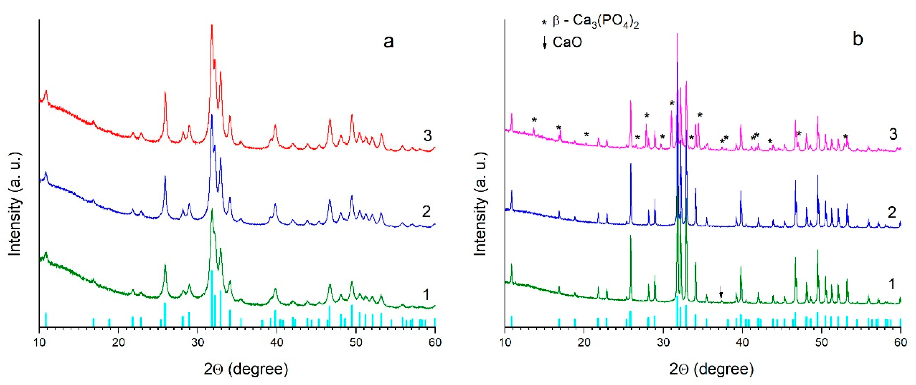
3.1. Effect of the Ca/P Ratio on the Thermal Stability of HA

Thermal stability of HA depends on the Ca/P ratio. Almost all conventional synthesis methods of HA, with the exception of those using high-temperature processing, give a fine HA powder with defective nanosized particles. According to phase analysis, it is a single-phase apatite. Deviations in the Ca/P ratio from the stochiometric value (1.667) upward or downward will not affect the analysis result due to the high defectiveness of the particles [31]. High-temperature treatment of this powder leads to annealing of the defects and an increase in the crystallite size with the formation of stoichiometric HA crystallites. With a lack of calcium or phosphate ions (Ca/P ≠ 1.667), both stoichiometric HA and impurity phases are formed.
As an example, Figure 1a shows the diffraction patterns of the HA samples prepared at different Ca/P ratios by reactions (4)–(6). As seen, at Ca/P ratios of 1.6, 1.667, and 1.7, the diffraction patterns look identical, and all reflections belong to the HA phase. However, after sintering of non-stoichiometric samples (Ca/P = 1.6 and 1.7), reflections of the impurity phases appear in the XRD patterns (Figure 1b). In the case of calcium-deficient HA (Ca/P = 1.6), the β-Ca3(PO4)2 phase is formed with a Ca/P ratio of 1.5, which is less than that of HA. With excess calcium cation (Ca/P = 1.7), the CaO reflection is observed in the XRD pattern. Prior to sintering, these impurity phases are not detected, because at a Ca/P ratio of 1.6, the calcium-deficient form of HA Ca10−x(HPO4)x(PO4)6−x(OH)2−x is produced (Equation (4)), in which the calcium cation vacancies are occupied by protons of the HPO42− group [11]. With an excess of calcium cations (Ca/P = 1.7), the lack of the phosphate groups is compensated by the carbonate anions, resulting in the formation of carbonated hydroxyapatite Ca10−x(PO4)6−x(CO3)x(OH)2−x [32]. In this case, the yield of the resultant product increases slightly (Equation (6)). Both of these non-stoichiometric apatites are unstable at high temperatures, and therefore, upon heating, their decomposition with the formation of stoichiometric HA and the corresponding impurity phases occurs (Table 2).
The presence of the new phases in the sample decreases the relative density of sintered material (Table 2).

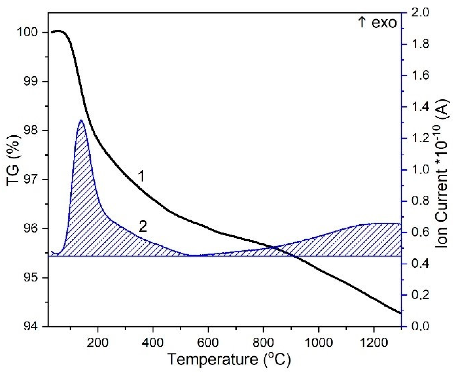
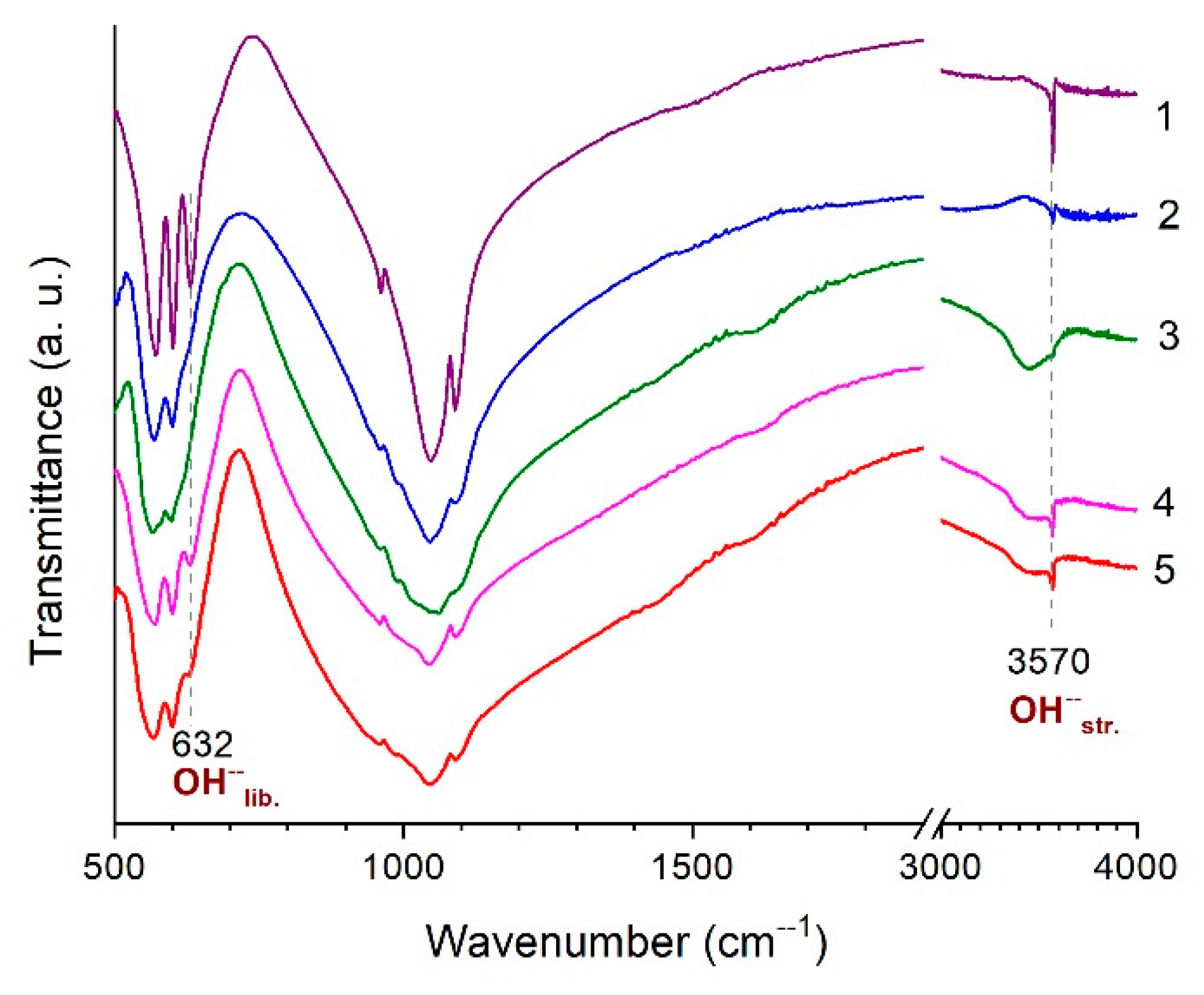
3.2. Thermal Analysis of HA

The results of thermal analysis of the stoichiometric HA powder are shown in Figure 2. According to Reaction (5), the mechanochemical synthesis of stoichiometric HA is accompanied by the release of two water molecules, which are adsorbed by HA particles. As seen from Figure 2, upon heating of the HA sample, adsorbed water and lattice water are released in a temperature range of 30–570 °C. According to the data reported by Tõnsuaadu et al. [18], the dehydroxylation of HA with the removal of water molecules by Reaction (1) in air atmosphere starts at 900 °C. In our case, this process probably starts at a lower temperature of 600 °C and proceeds up to temperatures above 1300 °C (Figure 2). The amount of released water increases as the temperature increases up to 1200 °C, and it almost does not change at high temperatures. Therefore, the reaction rate does not increase despite the increase in temperature. The kinetics of the reaction can be determined by the diffusion of water molecules inside the HA particles. During the reaction, the diffusion path increases, and the reaction is inhibited. Our estimations based on the data reported by Dorozhkin [33] suggest that the time required for water molecules to diffuse from a nanocrystal of 200 nm in size is about 20 s. When carrying out differential thermal analysis, the heating rate was 10 °C/min; therefore, it took 30 min to reach a temperature of 300 °C, which is much longer than the calculated diffusion time for a water molecule. It can be assumed that the kinetics of the reaction is determined by the diffusion of water molecules through the channels between particles in the sample. Probably, significant shrinkage of the sample, which is due to the densification caused by the elimination of pores contained in the initial powder compact, makes the process of HA dehydroxylation difficult. At a temperature of 1200 °C, the shrinkage is 20% of the initial volume [34]. In addition, the water molecule released during dehydroxylation is formed from a hydroxyl group and a proton from the adjacent hydroxyl group. Obviously, the probability of combining a proton and a hydroxyl group decreases as the number of hydroxyl groups in the channel decreases. All these factors should inhibit the process of HA dehydroxylation.

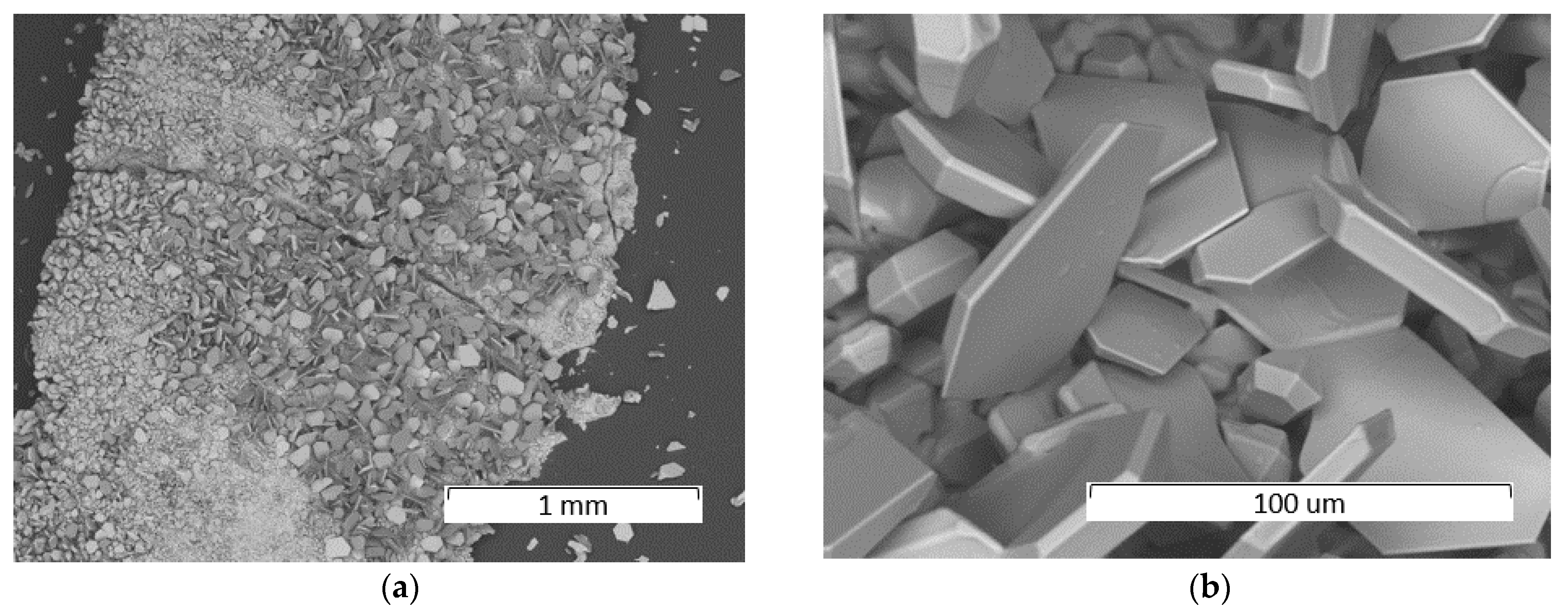
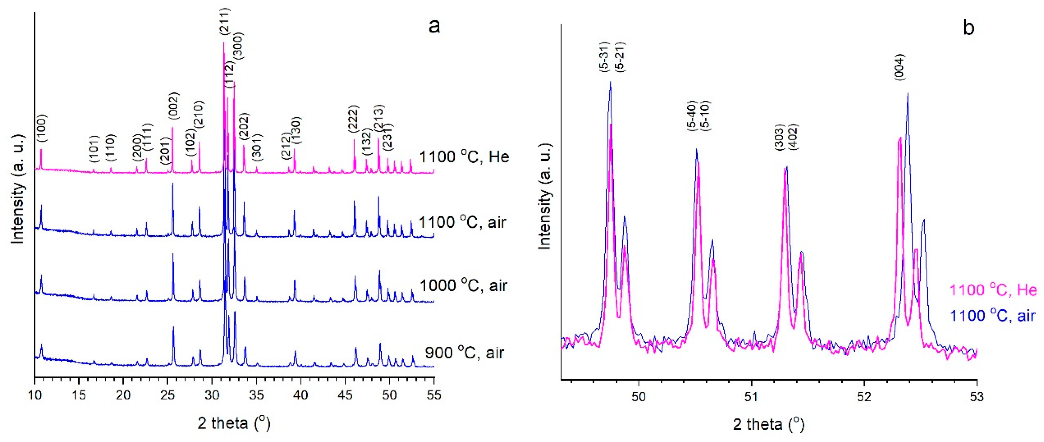
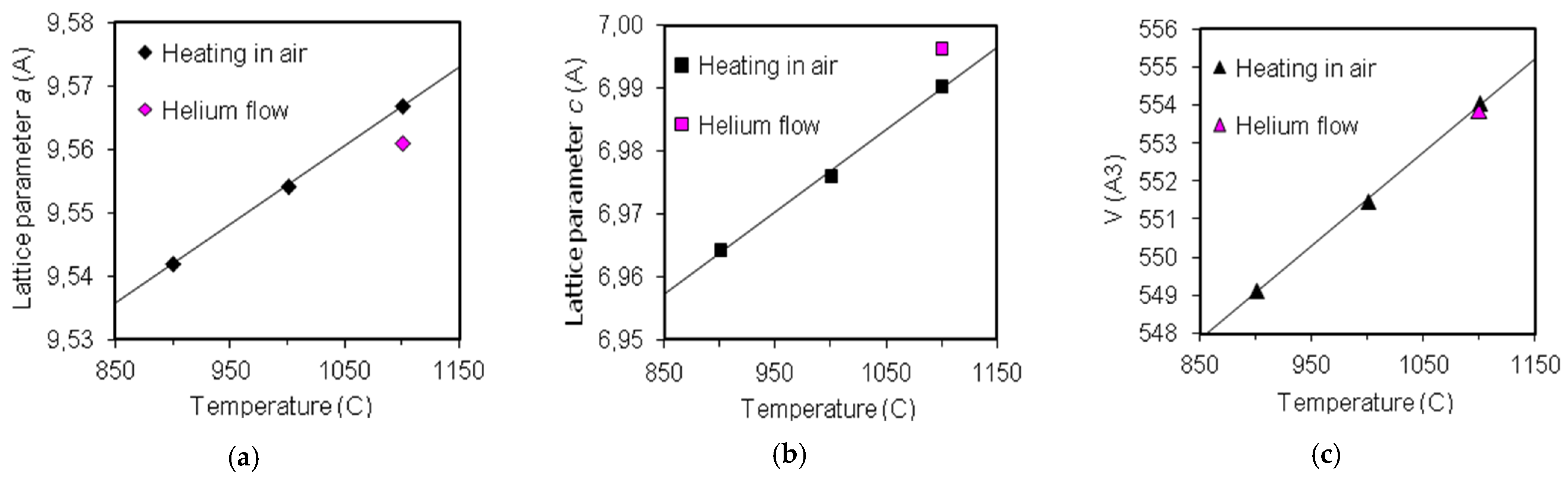
3.3. Sintering of HA in a High-Temperature Furnace

Stoichiometric HA samples, both as a powder and a compacted pellet, were heated in a high-temperature furnace. The results of the quantitative phase analysis of the samples and their lattice parameters after heating are shown in Table 3. As seen, the sample remains single phase up to a temperature of 1300 °C. According to the data presented in study [23], it can be assumed that the decomposition of HA with the formation of α-tricalcium and tetracalcium phosphates at a water vapor pressure of 7 mm Hg (25% humidity at 28 °C) should occur at temperatures above 1350 °C. Indeed, a small content of the α-Ca3(PO4)2 phase is observed at 1400 °C in our case. At a temperature of 1500 °C, the Ca4O(PO4)2 phase is also detected. Concentration of the decomposition products increases as the time of treatment increases, and it is accompanied by a decrease in the pycnometric density (Table 3). The pycnometric densities of the sintered pellets reach their maximum values at 1300 °C.
With regard to the structural parameters of the HA phase, it can be noted that when increasing the sintering temperature, a decrease in the lattice parameter a of the cooled sample occurs (Table 3). The lattice parameter c shows a very small increase with small fluctuations at decomposition temperatures of HA (1400, 1500 °C). A decrease in the a value is obviously related to a decrease in the diameter of the hydroxyl channel caused by a gradual increase in the OH vacancy concentration and the presence of the O2− ions (Equation (1)). A minimum value of the parameter a was achieved for a pellet treated at 1500 °C for 2 h. It may be assumed that this sample contains a minimum concentration of the OH groups, and the FTIR spectra support this (Figure 3). As seen from the spectrum, the intensity of the libration (632 cm−1) and stretching (3570 cm−1) vibrations of the OH group for this sample is close to zero. Therefore, it can be concluded that the pellet sintered at 1500 °C for 2 h contains both the decomposition products and an almost completely dehydroxylated OA phase. Accordingly, the lattice parameters a = 9.4094(12) Å and c = 6.8811(14) Å can be considered close to the values of stoichiometric OA. Atomic parameters of the OA phase cannot be refined by the Rietveld method due to its low content in the sample (only 35 wt%) and the overlapping of its reflections with those of the new phases. It can be assumed that when sintering HA in the pellet form, rehydroxylation upon cooling is difficult due to there being much less interpore space in the pellet as compared to that of the powder.
It should also be noted that the crystallite size increases when the sintering temperature increases to 1300 °C (Table 3). At higher temperatures, the crystallite size starts to decrease, while the concentration of decomposition products increases. Consequently, the HA decomposition takes place in the surface layer of the OHA particles.
Table 3 shows the strain effects observed at high temperatures both in the pellets and in the sintered powders. As seen, it starts from 1200 or 1300 °C and increases with increasing the temperature, making the structure more stressed. The strain value depends on the duration of the temperature treatment (the longer the treatment, the higher the strain) and on the sample density (the pellet has a higher strain rate).
The long treatment of the HA pellet during 10 h at a temperature of 1500 °C results in the formation of a large number of crystals on the surface of HA pellet (Figure 4a). As seen at higher magnification, they are monocrystalline with different crystal facets (Figure 4b). Apparently, these single crystals are the decomposition products, such as α-Ca3(PO4)2, Ca4O(PO4)2, and CaO.
The formation of the partial decomposition products of apatite at high temperatures indicates that the congruent melting of stoichiometric HA cannot be achieved by heating in an oven.

3.4. High-Temperature In Situ Diffraction

Figure 5a shows in situ diffraction patterns of the sample during the heating of unsintered stoichiometric HA pellet in air. As seen, the sample remains single phase in the temperature range 25–1100 °C. Reflections presented in the PXRD pattern indicated that the HA had a hexagonal structure with the space group P63/m (ICDD, PDF 40-11-9308). When changing the air medium to helium flow at 1100 °C, the phase composition does not change either; however, there is quite a noticeable shift in the positions of some reflections (Figure 5b).
The unit cell parameters and its volume increase linearly as the temperature increases (Figure 6). When changing the air medium to a helium flow, a decrease in the parameter a and an increase in the parameter c are observed, whereas the volume of the HA unit cell does not change significantly. The use of helium flow instead of the air medium accelerates the dehydroxylation process of HA. The pellets cooled in air and in helium media have comparable values of the lattice parameters (Table 4), while the a values are significantly different from those given in Table 3 for the samples heated in a high-temperature furnace to the same temperature in air. This can be explained by the different volumes and the design features of the high-temperature chambers, leading to different convection conditions inside the chambers and different water partial pressure. The volume of the chamber used for in situ experiments was five times smaller, and, consequently, the amount of water vapor therein was not enough for the rehydroxylation of OHA. Therefore, the a parameter of the pellet cooled in the in situ chamber is smaller. The FTIR spectroscopy data support this (Figure 7). As seen from the absorption spectra of the samples, after in situ heat treatment, the intensity of the stretching and libration vibrations of the OH group is much less than that after heating in the furnace.
It should also be noted that in the PXRD patterns of both the heated and cooled samples, there are no reflections indicating the change of the hexagonal space group to the monoclinic P21/b as was observed by other authors [9]. A splitting of reflections, which was reported by Alberius et al. and Gross et al. [35,36], was not observed, either.

3.5. Extremal Heating Conditions

As shown above, the congruent melting of HA by heating in a furnace at rates no more than 5 °C/min cannot be achieved. Given the fact that the process of dehydroxylation of HA, which eventually leads to its decomposition, proceeds rather slowly, it can be assumed that the stage of HA decomposition can be avoided by rapid heating. Therefore, it was of interest to find out whether the melting of HA at high heating rates would be congruent.
Ultrafast heating of HA can be achieved using plasma and detonation spraying or laser treatment [37]. In this case, heating to the melting point occurs in a fraction of a second. Under these treatment conditions, the possibility of overheating of HA due to very high processing temperatures is very high; therefore, incongruent melting of HA coupled with the partial dehydroxylation and partial decomposition of HA takes place.
However, under certain experimental conditions, using selective laser melting technology, HA can be melted without decomposition. For instance, under the CO2 laser treatment of the stoichiometric HA pellet surface at a spot size of 0.2 mm, at a power of 4 W, and at a scanning speed of 500 mm/s, a dense layer of recrystallized OHA is obtained (Figure 8a,c). In this case, melting occurs for 10−4 s. In such a short time, the melt does not lose a large number of OH groups, so the decomposition of HA is not observed. Obviously, the melt crystallization starts from the lower layer, and the unmelted particles of HA are the nuclei for crystallization. As a result of the competitive growth of crystallites with different orientations, the most rapidly growing crystallites have an advantage. Thus, a directed growth of the predominantly oriented crystallites is observed. As seen in the PXRD patterns of the recrystallized sample, the crystallites are predominantly oriented in the [002] direction (Figure 9a). The lattice parameters of this recrystallized layer correspond to the OHA crystalline phase (Table 4). Unfortunately, precisely measuring the melting point of the material under these conditions is difficult, since the area of the localized heat exposure is 0.2 mm, while the duration of the exposure is 10−4 s.
A decrease in the speed of the laser spot moving over the surface of the HA pellet to 200 mm/s leads to the formation of a thicker recrystallized layer (Figure 8b,d) with a more predominant orientation in the [002] direction (Figure 9b). However, in this case, the most intense reflection of the decomposition product α-Ca3(PO4)2 appears in the PXRD pattern of the recrystallized layer (Figure 9b). In this treatment mode, the time of intensive heat exposure is 10−3 s, which is still insufficient for significant water removal. However, a slower surface treatment leads to an increase in exposure and therefore to an increase in the transmitted energy, i.e., it leads to an increase in temperature, resulting in the partial decomposition of the material.

4. Conclusions

The crystal structure of HA is conducive to different types of cationic and anionic substitutions, while in the case of incomplete substitution, the formation of solid solutions takes place. Some types of substitutions significantly reduce the HA thermal stability. This study shows that if the proper synthesis conditions are not maintained, the calcium-deficient or carbonated HA are formed. These types of apatite are less stable than stoichiometric HA at high temperatures. Upon high-temperature treatment (≥900 °C), they are decomposed into stoichiometric HA and corresponding impurities, which can affect negatively the strength characteristics of the produced ceramics.
The stoichiometric HA produced by the solid-state mechanochemical method was shown to have a hexagonal structure with the space group P63/m. The sample remains single-phase up to a temperature of 1300 °C during sintering both as a powder and a pellet. At higher temperatures, the partial decomposition of HA occurs, which is preceded by the dehydroxylation of HA with the formation of OHA. Dehydroxylation does not lead to any phase transitions, but it is accompanied by changes in the size of the crystal lattice, such as a decrease in the a parameter and an increase in the c parameter. At temperatures above 1200 °C, microstrains appear in the structure. An increase in the density of the sample under sintering results in an increase in the microstrains.
A sample containing an apatite phase with a negligible amount of the hydroxyl groups, which could approximately be attributed to OA, was prepared. It may be assumed that OA is the end member in the Ca10(PO4)6(OH)2–2xOxx solid solution series with the space group P63/m and the lattice parameters a = 9.4094(12) Å and c = 6.8811(14) Å.
At temperatures above 1300 °C, the decomposition of OA occurs in the sample with the formation of more stable new phases of calcium phosphates and calcium oxide. The content of the decomposition products is affected by the duration of treatment of the sample and its density. The OA decomposition takes place in the upper layers of the particles due to which the size of the apatite crystallites decreases as the concentration of decomposition products increases. The decomposition products reduce the material density.
Thus, slow heating of stoichiometric HA in a high-temperature furnace (Vheating ≤ 5 °C/min) to a temperature of 1500 °C in air results in the following transformations:
HA → OHA → OHA + OA → OHA + OA + α-Ca3(PO4)2 → OHA + OA + α-Ca3(PO4)2 + Ca4O(PO4)2 → OA + α-Ca3(PO4)2 + Ca4O(PO4)2 + CaO.
Therefore, the congruent melting of stoichiometric HA upon heating in a high-temperature furnace does not occur.
On fast heating (Vheating >> 1000 °C/s) of stoichiometric HA by laser irradiation, under certain conditions, the congruent melting of OHA can be achieved. The fast heating and cooling, in this case, reduce the number of hydroxyl groups, leaving the OHA structure and thus allowing it to melt without decomposition. The recrystallized layer has a dense structure predominantly oriented in the [002] direction. The process of congruent melting of HA can be used to produce bulk ceramic products by layer-by-layer melting of apatite powder (3D printing). In addition, it can be assumed that congruent HA melting can also be achieved when using other methods, providing similar heating rates.
This study shows that the process of thermal treatment of HA depends on its synthesis conditions, on its morphology and dispersibility, heat treatment conditions, and how far they are from thermodynamic equilibrium. Since HA heating is accompanied by the removal of OH groups with the formation of water molecules, both temperature and the water vapor pressure affect the thermal stability of HA. These factors should be taken into account when selecting sintering modes to produce ceramic products.

Author Contributions

Conceptualization, N.V.B.; methodology, N.V.B.; investigation, S.V.M., S.G.B. and K.B.G.; writing—original draft preparation, N.V.B.; writing—review and editing, A.A.M., K.B.G., O.A.L. and V.S.B.; funding acquisition, V.S.B. All authors have read and agreed to the published version of the manuscript.

Funding

The study was supported by a grant Russian Science Foundation (RSF), No. 21-12-00251.

Data Availability Statement

The raw/processed data required to reproduce these results are included in the Materials and Methods section.

Conflicts of Interest

The authors declare no conflict of interest. The funders had no role in the design of the study; in the collection, analyses, or interpretation of data; in the writing of the manuscript; or in the decision to publish the results.

References

  1. Elliott, J.C. Structure and Chemistry of the Apatites and Other Calcium Orthophosphates; Studies in Inorganic Chemistry; Elsevier: Amsterdam, The Netherlands, 1994; pp. 1–404. [Google Scholar]
  2. Šupová, M. Substituted hydroxyapatites for biomedical applications: A review. Ceram. Int. 2015, 41, 9203–9231. [Google Scholar] [CrossRef]
  3. White, T.J.; Zhili, D. Structural derivation and crystal chemistry of apatites. Acta Crystallogr. B 2003, 59, 1–16. [Google Scholar] [CrossRef] [PubMed]
  4. Ratnayake, J.T.B.; Mucalo, M.; Dias, G.J. Substituted hydroxyapatites for bone regeneration: A review of current trends. J. Biomed. Mater. Res. B Appl. Biomater. 2017, 105, 1285–1299. [Google Scholar] [CrossRef] [PubMed]
  5. Sadat-Shojai, M.; Khorasani, M.-T.; Dinpanah-Khoshdargi, E.; Jamshidi, A. Synthesis methods for nanosized hydroxyapatite with diverse structures. Acta Biomater. 2013, 9, 7591–7621. [Google Scholar] [CrossRef] [PubMed]
  6. Lin, K.; Wu, C.; Chang, J. Advances in synthesis of calcium phosphate crystals with controlled size and shape. Acta Biomater. 2014, 10, 4071–4102. [Google Scholar] [CrossRef]
  7. Chaikina, M.V.; Bulina, N.V.; Vinokurova, O.B.; Prosanov, I.Y.; Dudina, D.V. Interaction of calcium phosphates with calcium oxide or calcium hydroxide during the “soft” mechanochemical synthesis of hydroxyapatite. Ceram. Int. 2019, 45, 16927–16933. [Google Scholar] [CrossRef]
  8. Ma, G.; Liu, X.Y. Hydroxyapatite: Hexagonal or monoclinic? Cryst. Growth Des. 2009, 9, 2991−2994. [Google Scholar] [CrossRef]
  9. Pastero, L.; Bruno, M.; Aquilano, D. About the Genetic Mechanisms of Apatites: A Survey on the Methodological Approaches. Minerals 2017, 7, 139. [Google Scholar] [CrossRef] [Green Version]
  10. Wilson, R.M.; Elliott, J.C.; Dowker, S.E.; Rodríguez-Lorenzo, L.M. Rietveld refinements and spectroscopic studies of the structure of Ca-deficient apatite. Biomaterials 2005, 26, 1317–1327. [Google Scholar] [CrossRef]
  11. Dorozhkin, S.V. Calcium orthophosphates (CaPO4): Occurrence and properties. Prog. Biomater. 2016, 5, 9–70. [Google Scholar] [CrossRef] [Green Version]
  12. Córdova-Udaeta, M.; Kim, Y.; Yasukawa, K.; Kato, Y.; Fujita, T.; Dodbiba, G. Study on the Synthesis of Hydroxyapatite under Highly Alkaline Conditions. Ind. Eng. Chem. Res. 2021, 60, 4385–4396. [Google Scholar] [CrossRef]
  13. Mucalo, M. Hydroxyapatite (HAp) for Biomedical Applications; Woodhead Publishing Limited: Waltham, MA, USA, 2015; pp. 1–364. [Google Scholar]
  14. Kenny, S.M.; Buggy, M. Bone cements and fillers: A review. J. Mater. Sci. Mater. Med. 2003, 14, 923–938. [Google Scholar] [CrossRef] [PubMed]
  15. Gruselle, M. Apatites: A new family of catalysts in organic synthesis. J. Organomet. Chem. 2015, 793, 93–101. [Google Scholar] [CrossRef]
  16. Dorozhkin, S.V. Current State of Bioceramics. J. Ceram. Sci. Technol. 2018, 9, 353–370. [Google Scholar] [CrossRef]
  17. Dorozhkin, S. Calcium Orthophosphate (CaPO4) Scaffolds for Bone Tissue Engineering Applications. J. Biotechnol. Biomed. Sci. 2018, 1, 25–93. [Google Scholar] [CrossRef]
  18. Tõnsuaadu, K.; Gross, K.A.; Plūduma, L.; Veiderma, M. A review on the thermal stability of calcium apatites. J. Therm. Anal. Calorim. 2012, 110, 647–659. [Google Scholar] [CrossRef]
  19. Liao, C.-J.; Lin, F.-H.; Chen, K.-S.; Sun, J.-S. Thermal decomposition and reconstitution of hydroxyapatite in air atmosphere. Biomaterials 1999, 20, 1807–1813. [Google Scholar] [CrossRef]
  20. Tanaka, H.; Chikazawa, M.; Kandori, K.; Ishikawa, T. Influence of thermal treatment on the structure of calcium hydroxyapatite. Phys. Chem. Chem. Phys. 2000, 2, 2647–2650. [Google Scholar] [CrossRef]
  21. Wang, T.; Dorner-Reisel, A. Thermo-analytical investigations of the decomposition of oxyhydroxyapatite. Mater. Lett. 2004, 58, 3025–3028. [Google Scholar] [CrossRef]
  22. Drouet, C. A comprehensive guide to experimental and predicted thermodynamic properties of phosphate apatite minerals in view of applicative purposes. J. Chem. Thermodyn. 2015, 81, 143–159. [Google Scholar] [CrossRef] [Green Version]
  23. Riboud, P.V. Comparaison de la stabilite de l’apatite d’oxyde de fer et de l’hydroxyapatite a haute temperature. Bull. Soc. Chim. Fr. 1968, 1701–1703. [Google Scholar]
  24. Riboud, P.V. Composition et stabilité des phases à structure d`apatite dans le système CaO-P2O5- de fer-H2O à haute température. Ann. Chim. 1973, 8, 381–390. [Google Scholar]
  25. Bhatnagar, V.M. The melting points of synthetic apatites. Miner. Mag. 1969, 37, 527–528. [Google Scholar] [CrossRef]
  26. Ma, J.; Ge, X.; Pelate, N.; Lei, S. Numerical investigation of two-dimensional thermally assisted ductile regime milling of nanocrystalline hydroxyapatite bioceramic material. Ceram. Int. 2015, 41, 3409–3419. [Google Scholar] [CrossRef]
  27. Bulina, N.V.; Baev, S.G.; Makarova, S.V.; Vorobyev, A.M.; Titkov, A.I.; Bessmeltsev, V.P.; Lyakhov, N.Z. Selective Laser Melting of Hydroxyapatite: Perspectives for 3D Printing of Bioresorbable Ceramic Implants. Materials 2021, 14, 5425. [Google Scholar] [CrossRef] [PubMed]
  28. Bulina, N.V.; Rybin, D.K.; Makarova, S.V.; Dudina, D.V.; Batraev, I.S.; Utkin, A.V.; Prosanov, I.Y.; Khvostov, M.V.; Ulianitsky, V.Y. Detonation Spraying of Hydroxyapatite on a Titanium Alloy Implant. Materials 2021, 14, 4852. [Google Scholar] [CrossRef]
  29. Trombe, J.; Montel, G. Some features of the incorporation of oxygen in different oxidation states in the apatitic lattice—I On the existence of calcium and strontium oxyapatites. J. Inorg. Nucl. Chem. 1978, 40, 15–21. [Google Scholar] [CrossRef]
  30. Bessmeltsev, V.P.; Goloshevsky, N.V.; Smirnov, K.K. Specific features of controlling laser systems for micromachining of moving carriers. Optoelectron. Instrum. Data Process. 2010, 46, 79–86. [Google Scholar] [CrossRef]
  31. Raynaud, S.; Champion, E.; Bernache-Assollant, D.; Thomas, P. Calcium phosphate apatites with variable Ca/P atomic ratio I. Synthesis, characterisation and thermal stability of powders. Biomaterials 2002, 23, 1065–1072. [Google Scholar] [CrossRef]
  32. Montel, G.; Bonel, G.; Heughebaert, J.; Trombe, J.; Rey, C. New concepts in the composition, crystallization and growth of the mineral component of calcified tissues. J. Cryst. Growth 1981, 53, 74–99. [Google Scholar] [CrossRef]
  33. Dorozhkin, S. Solid-Phase Conversion of Nonstoichiometric Hydroxoapatite into Two-Phase Calcium Phosphate. Russ. J. Appl. Chem. 2002, 75, 1897–1902. [Google Scholar] [CrossRef]
  34. Champion, E. Sintering of calcium phosphate bioceramics. Acta Biomater. 2013, 9, 5855–5875. [Google Scholar] [CrossRef]
  35. Alberius-Henning, P.; Adolfsson, E.; Grins, J.; Fitch, A. Triclinic oxy-hydroxyapatite. J. Mater. Sci. 2001, 36, 663–668. [Google Scholar] [CrossRef]
  36. Gross, K.; Berndt, C.; Stephens, P.; Dinnebier, R. Oxyapatite in hydroxyapatite coatings. J. Mater. Sci. 1998, 33, 3985–3991. [Google Scholar] [CrossRef]
  37. Dorozhkin, S.V. Calcium orthophosphate deposits: Preparation, properties and biomedical applications. Mater. Sci. Eng. C 2015, 55, 272–326. [Google Scholar] [CrossRef] [PubMed]
Figure 1. PXRD patterns of the as-synthesized HA powder (a) and samples obtained after 2 h of annealing at 1000 °C (b). Ca/P ratios are 1.7 (1), 1.667 (2), and 1.6 (3). Vertical bars in the lower graphs correspond to the HA phase from the ICDD database (PDF No. 40-11-9308).
Minerals 11 01310 g001
Figure 2. Simultaneous thermal analysis of the HA powder: 1—weight loss; 2—evolved water.
Minerals 11 01310 g002
Figure 3. FTIR spectra of HA samples after sintering in a high-temperature furnace: (1) powder after 1000 °C during 2 h; (2) pellet after 1500 °C during 10 min; (3) pellet after 1500 °C during 2 h; (4) powder after 1500 °C during 10 min; (5) powder after 1500 °C during 2 h.
Minerals 11 01310 g003
Figure 4. SEM images of the crystals obtained on the surface of HA pellet after treatment at 1500 °C during 10 h: (a) general view; (b) enlarged view.
Minerals 11 01310 g004
Figure 5. In situ PXRD patterns of the HA pellets heated at different temperatures in air and in helium flow; (a)—general view, (b)—enlarged view of the 49.4–53° 2θ region.
Minerals 11 01310 g005
Figure 6. Variation of the lattice parameters (a,b) and volume (c) as a function of temperature for the HA pellet at in situ experiment.
Minerals 11 01310 g006
Figure 7. IR spectra of HA samples after heating under different conditions: (1) pellet, heating in a PVK-1.6 furnace to 1100 °C, cooling in air; (2) pellet, heating in an in situ chamber HTK 1200N, cooling in air; (3) pellet, heating in an in situ chamber HTK 1200N, cooling in a flow of helium.
Minerals 11 01310 g007
Figure 8. SEM images of surface (a,b) and cross-section (c,d) of the pellets treated by a 0.2 mm laser spot at 4 W and at scanning speeds of 500 mm/s (a,c) and 200 mm/s (b,d).
Minerals 11 01310 g008
Figure 9. PXRD patterns of HA pellets after sintering at 1000 °C in a high-temperature furnace (1) in comparison with PXRD patterns of HA pellets after treatment by a 0.2 mm laser spot at 4 W and different scanning speeds: (a)—500 mm/s (2); (b)—200 mm/s (3).
Minerals 11 01310 g009
Table 1. Structural parameters of the hydroxyapatite (HA) and oxyapatite (OA) reported in the literature.
Crystal SystemSpace Groupa (Å)b (Å)c (Å)α (°)β (°)γ (°)Reference
HA
HexagonalP63/m9.43029.43026.89119090120[9,11]
9.41769.41766.88149090120[1]
MonoclinicP21/b9.41762a6.88049090120[1]
9.42142a6.8819090120[9]
9.842142a6.88149090120[11]
OA
HexagonalP63/m9.409.406.899090120[1]
P 6 ¯ 9.4329.4326.88190.390119.9[3,9,11]
Table 2. Phase composition, structural parameters of HA phase and relative density of the pellets with different Ca/P ratio before and after sintering in the high-temperature furnace.
Ca/P RatioContent (wt%)Structural Parameters of the HA PhaseRelative Density (%)
HAβ-Ca3(PO4)2CaOa (Å)c (Å)Crystallite Size (nm)
Before sintering
1.61009.4394(10)6.8867(7)25.1(2)63.9
1.6671009.4336(12)6.8932(9)29.0(4)64.9
1.71009.4374(16)6.8929(10)20.0(2)63.9
After sintering at 1000 °C, 2 h
1.672289.4249(6)6.8813(5)99(2)77.1
1.6671009.4240(2)6.8817(1)243(5)85.6
1.79919.4230(2)6.8814(2)209(4)76.9
Table 3. Phase composition and structural parameters of the stoichiometric HA sample after sintering in the high-temperature furnace at different temperatures and at a heating rate of 5°/min.
Temperature (°C)Content (wt%)Structural Parameters of the HA Phase Pycnometric
Density (g/cc)
α-Ca3(PO4)2Ca4O(PO4)2CaOHAa (Å)c (Å)Crystallite Size (nm)Strain
HA powder, duration of sintering 10 min
11001009.4250(1)6.8821(1)317(8)
12001009.4233(2)6.8831(1)551(20)
13001009.4196(1)6.8869(1)768(66)0.048(4)
14003979.4179(2)6.8867(2)252(12)0.057(7)
15002614609.4172(6)6.8861(6)139(8)0.19(1)
HA powder, duration of sintering 2 h
10001009.4232(2)6.8822(2)183(6)
11001009.4227(3)6.8845(1)405(20)
12001009.4215(1)6.8862(1)762(50)0.024(4)
13001009.4180(1)6.8885(1)425(26)0.053(5)
14008929.4182(1)6.8878(2)372(20)0.081(4)
15004322359.4181(1)6.8878(1)111(9)0.22(1)
HA pellet, duration of sintering 10 min
11001009.4237(1)6.8820(1)391(20)3.068(7)
12001009.4224(3)6.8828(2)349(24)0.122(6)3.126(19)
13001009.4181(2)6.8877(2)495(36)0.118(4)3.169(6)
14002989.4118(6)6.8903(4)441(58)0.330(9)3.117(19)
150019819.4170(8)6.8877(5)225(23)0.33(1)2.905(6)
HA pellet, duration of sintering 2 h
150050141359.4094(12)6.8811(14)304(70)0.34(4)2.909(6)
Table 4. Structural parameters for the HA phase treated under various conditions in in situ experiments. The data are given for the sample cooled at room temperature.
Heat ConditionsSample Statea (Å)c (Å)V (Å3)Crystallite Size (nm)Strain
Heating up to 1100 °C in air, cooling in airPellet9.41663(3)6.88542(3)528.752(4)542(10)0.025(1)
Heating up to 1100 °C in air, cooling in He flowPellet9.41497(6)6.88443(5)528.490(8)321(5)0.101(1)
Selective laser meltingPellet9.4139(8)6.8902(6)528.82(9)197(18)0.35(2)
Publisher’s Note: MDPI stays neutral with regard to jurisdictional claims in published maps and institutional affiliations.

