Article

Rippite, K2(Nb,Ti)2(Si4O12)O(O,F), a New K-Nb-Cyclosilicate from Chuktukon Carbonatite Massif, Chadobets Upland, Krasnoyarsk Territory, Russia

by
Victor V. Sharygin
1,2,3,*,
Anna G. Doroshkevich
1,4,
Yurii V. Seryotkin
1,3,
Nikolai S. Karmanov
1,
Elena V. Belogub
5,6,
Tatyana N. Moroz
1,
Elena N. Nigmatulina
1,
Alexander P. Yelisseyev
1,
Vitalii N. Vedenyapin
1 and
Igor N. Kupriyanov
1,3
1
V.S. Sobolev Institute of Geology and Mineralogy, Siberian Branch of the RAS, 3 Acad. Koptyuga pr., 630090 Novosibirsk, Russia
2
ExtraTerra Consortium, Institute of Physics and Technology, Ural Federal University, 19 Mira str., 620002 Ekaterinburg, Russia
3
Department of Geology and Geophysics, Novosibirsk State University, 1 Pirogov str., 630090 Novosibirsk, Russia
4
Geological Institute, Siberian Branch of the RAS, 6a Sakhyanova str., 670047 Ulan-Ude, Russia
5
South Urals Federal Research Center of Mineralogy and Geoecology, Uralian Branch of the RAS, 456317 Miass, Russia
6
Faculty of Geology, National Research South Ural State University, Miass Branch, 20 8-July str., Bldg. 10, 456304 Miass, Russia
*
Author to whom correspondence should be addressed.
Minerals 2020, 10(12), 1102; https://doi.org/10.3390/min10121102
Submission received: 9 November 2020 / Revised: 3 December 2020 / Accepted: 3 December 2020 / Published: 8 December 2020
(This article belongs to the Collection New Minerals)

Abstract

:
Rippite K2(Nb,Ti)2(Si4O12)(O,F)2, a new K-Nb-cyclosilicate, has been discovered in calciocarbonatites from the Chuktukon massif (Chadobets upland, SW Siberian Platform, Krasnoyarsk Territory, Russia). It was found in a primary mineral assemblage, which also includes calcite, fluorcalciopyrochlore, tainiolite, fluorapatite, fluorite, Nb-rich rutile, olekminskite, K-feldspar, Fe-Mn–dolomite and quartz. Goethite, francolite (Sr-rich carbonate–fluorapatite) and psilomelane (romanèchite ± hollandite) aggregates as well as barite, monazite-(Ce), parisite-(Ce), synchysite-(Ce) and Sr-Ba-Pb-rich keno-/hydropyrochlore are related to a stage of metasomatic (hydrothermal) alteration of carbonatites. The calcite–dolomite coexistence assumes crystallization temperature near 837 °C for the primary carbonatite paragenesis. Rippite is tetragonal: P4bm, a = 8.73885(16), c = 8.1277(2) Å, V = 620.69(2) Å3, Z = 2. It is closely identical in the structure and cell parameters to synthetic K2Nb2(Si4O12)O2 (or KNbSi2O7). Similar to synthetic phase, the mineral has nonlinear properties. Some optical and physical properties for rippite are: colorless; Mohs’ hardness—4–5; cleavage—(001) very perfect, (100) perfect to distinct; density (meas.)—3.17(2) g/cm3; density (calc.)—3.198 g/cm3; optically uniaxial (+); ω = 1.737-1.739; ε = 1.747 (589 nm). The empirical formula of the holotype rippite (mean of 120 analyses) is K2(Nb1.90Ti0.09Zr0.01)[Si4O12](O1.78OH0.12F0.10). Majority of rippite prismatic crystals are weakly zoned and show Ti-poor composition K2(Nb1.93Ti0.05Zr0.02)[Si4O12](O1.93F0.07). Raman and IR spectroscopy, and SIMS data indicate very low H2O content (0.09–0.23 wt %). Some grains may contain an outermost zone, which is enriched in Ti (+Zr) and F, up to K2(Nb1.67Ti0.32Zr0.01)[Si4O12](O1.67F0.33). It strongly suggests the incorporation of (Ti,Zr) and F in the structure of rippite via the isomorphism Nb5+ + O2− → (Ti,Zr)4+ + F1−. The content of a hypothetical end-member K2Ti2[Si4O12]F2 may be up to 17 mol. %. Rippite represents a new structural type among [Si4O12]-cyclosilicates because of specific type of connection of the octahedral chains and [Si4O12]8− rings. In structural and chemical aspects it seems to be in close with the labuntsovite-supergroup minerals, namely with vuoriyarvite-(K), K2(Nb,Ti)2(Si4O12)(O,OH)2∙4H2O.

1. Introduction

The natural analog of the tetragonal synthetic phase KNbSi2O7, a new structural type among natural K-Nb-cyclosilicates, named rippite, was discovered in calciocarbonatites of the Chuktukon massif, Chadobets upland, SW Siberian Platform, Krasnoyarsk Territory, Russia [1,2].
Synthetic K2Nb2(Si4O12)O2 (or KNbSi2O7) is actively studied since 1990 [3]. The melting point for K2Nb2(Si4O12)O2 is 1180 °C. No phase transition was found in K2Nb2(Si4O12)O2 between room temperature and melting point [4]. It is rather inert and chemically stable and is not dissolved in inorganic acids except HF. All studies on KNbSi2O7 are mainly focused on its nonlinear optical property [3,4,5,6,7,8,9,10,11,12,13,14,15]. Single crystals, ceramics, glass-ceramics, and glass of KNbSi2O7 and similar compositions in the systems SiO2(±GeO2)-Nb2O5-K2O have been fabricated [3,4,5,6,7,8,9,10,11,12]. The synthesis of glasses and glass-ceramics in the system SiO2-Nb2O5-K2O has shown that K2Nb2(Si4O12)O2 may be appeared in the 510–735 °C temperature range in glass/melt [4,6,8,9,10,13,14]. The KNbSi2O7 phase has very high optical nonlinearity with its second harmonic generation (SHG) signal greater than that for α-quartz [14,15]. The study of ferroelectric-dielectric properties has shown that K2Nb2(Si4O12)O2 is a ferroelectric material with low dielectric permittivity range {4,14]. The isostructural compound K2Ta2(Si4O12)O2, which may has nonlinear properties, was also synthesized [16].
Rippite, K2(Nb,Ti)2(Si4O12)O(O,F), was approved by the Commission on New Minerals, Nomenclature and Classification (CNMNC) of the International Mineralogical Association (IMA) as a new mineral species in June 2016 (IMA 2016-025) [1]. The mineral is named in the honor of German Samuilovich Ripp (1935 year of birth, Ulan-Ude, Buryatia, Russia). He is a leading expert on carbonatite petrogenesis, including carbonatite magmatic activity in Eastern Siberia, carbonatite mineralogy, geochemistry and stable isotope geochemistry and rare metal mineralization associated with carbonatites. He has performed detailed studies for carbonatite occurrences in Transbaikalia (Yuzhnoe, Khaluta, Arshan, Pogranichnoe, Veseloe), Eastern Sayan (Belaya Zima) and Mongolia (Mushugai-Khuduk) [17,18,19,20,21,22,23,24,25,26,27,28]. The holotype specimen of rippite-containing carbonatite from drillhole N 546 (193.5 m deep, author number 546–193.5) of the Chuktukon massif, Siberia, Russia has been deposited in the Central Siberian Geological Museum at the V.S. Sobolev Institute of Geology and Mineralogy (IGM), Siberian Branch of the RAS, Novosibirsk 630090, Russia, catalogue number XIII-347/1).
In this paper, we provide a detailed description of rippite. Some data on rippite-bearing calciocarbonatites of the Chuktukon massif were reported in the previous publications [1,2,29,30,31,32,33,34,35].

2. Brief Geological Background for the Chuktukon Massif

The Chadobets alkaline complex is located within the Chadobets upland (basin of the Chadobets River, Krasnoyarsk Territory, Russia) at the southwestern part of the Siberian craton (Figure 1) [36]. The upland is 2000 km2 in size and occurs at the intersection of two Neoproterozoic grabens being a part of the Angara-Kotuy large-scale rift system [29,37,38,39]. The Chadobets alkaline ultramafic carbonatite complex is conventionally subdivided into the Chuktukon alkaline ultramafic carbonatite massif (southern part of the upland) and the Terina alkaline massif (silicate rocks, northern part of the upland). In addition, the Chadobets upland also includes dikes of ultramafic lamprophyres (aillikite–damtjernite series). Carbonatites and their weathering species of the Chuktukon massif host major reserves of Nb and REE [40,41,42]. The U-Pb and Ar-Ar ages for the Chadobets ultramafic alkaline rocks and carbonatites are between 252 and 231 Ma [30,31,43]. For comparison, the Ilbokich aillikite–damtjernite lamprophyre series, which is located nearby the Chadobets upland, has 392 Ma age [43,44,45].
The detailed geology of the Chadobets alkaline ultramafic carbonatite complex was described by Kirichenko et al. [36]. The sequence of the Chuktukon rocks, from the earliest to the youngest, was established by field observations (drillholes description) and comprises alkaline ultramafic rocks, calciocarbonatites and, finally, damtjernites [36]. Alkaline ultramafic rocks (aillikites–damtjernites, olivine melilitites, peridotites and others) of the first emplacement phase form small plugs (up to 1–1.5 km2), dykes and sills ranging in thickness up to 100–120 m. Carbonatites also form small plugs (up to 4 km2), dykes and sills (up to 20 m thick). Damtjernites are presented by pipes (up to 0.5 km2), crosscutting early phases of alkaline ultramafic rocks and carbonatites and typically containing their xenoliths and fragments of the country sedimentary rocks. All rocks of the Chuktukon massif are altered in different degree by hydrothermal and weathering processes [36]. The detailed petrography and mineralogy of the Chadobets alkaline rocks were summarized in [29,30,32,36,43,46,47].

3. Analytical Methods

Double-polished sections of calciocarbonatites (~50–100 µm in thickness) and individual rippite grains from the Chuktukon massif were used for optical examination in transmitted and reflected light and for other studies. The individual rippite crystals were selected from residue after dissolution of carbonatite samples in the diluted HCl or acetic acid.
The identification of all minerals in the Chuktukon calciocarbonatites was based on energy-dispersive spectra (EDS), back-scattered electron (BSE) images and elemental mapping (EDS system), using a TESCAN MIRA 3MLU scanning electron microscope equipped with an INCA Energy 450 XMax 80 microanalysis system (Oxford Instruments Ltd., Abingdon, UK) at the IGM, Novosibirsk, Russia. The SEM-EDS analyses of minerals were operated at an accelerating voltage of 20 kV and a probe current of 1 nA in high-vacuum mode and at an accumulation time of 20–40 s. The synthetic compounds, pure metals and minerals were used as reference standards for the elements: SiO2 (Si and O), Al2O3 (Al), diopside (Mg and Ca), albite (Na), orthoclase (K), Ca2P2O7 (P), BaF2 (Ba and F), Cr2O3 (Cr), CsRe2Cl6 (Cl), LaPO4 (La), CePO4 (Ce), SrF2 (Sr), metallic Nb, Ti, Ta, Zr, Fe, Mn, Zn and V. Matrix effects were corrected using the XPP algorithm, implemented in the software of the microanalysis system. Metallic Co served for quantitative optimization (normalization to probe current and energy calibration of the spectrometer).
Electron microprobe analyses (EMPA-WDS) of rippite and related carbonatite minerals were done at the IGM, using a JEOL JXA-8100 electron microprobe (Jeol Ltd., Tokyo, Japan). Grains (sizes >5 μm) previously analyzed by SEM-EDS were selected for this purpose. The operating conditions were as follows: beam diameter of 1-2 µm, accelerating voltage of 20 kV and a beam current of 10–15 nA, counting time of 10 (5 + 5) s (for most elements). Accumulation time for analyzing of F (using LDE crystal) in rippite was 40 s (20 s—counting of background; 20 s—counting of peak for F), detection limit for F was 308 ppm (0.03 wt %). Synthetic fluorophogopite (Si, Al, F, Mg), orthoclase (K), LiNbO3 (Nb), zircon (Zr), rutile (Ti), Ta2O5 (Ta), diopside (Ca), Fe-rich pyrope (Fe), Ba-silicate glass (Ba), Sr-silicate glass (Sr) and albite (Na) were used as standards during measuring. The precision of analysis for major elements was better than 2% relative. The detection limits for elements were (in ppm): Si—181; Ti—132; Nb—295; Zr—405; Ta—400; Al—168; Fe—28; Mg—188; Ca—123; Sr—844; Na—128; K—115. Data reduction was performed using a PAP routine (Pouchou & Pichoir, 1985). Overlap corrections were done for the following elements: Si—Sr, Si—Ta and Fe—F.
The contents of some light trace elements (Be, Li, B), F and H2O in rippite of the Chuktukon carbonatite were analyzed by secondary-ion mass spectrometry (SIMS) on a Cameca IMS-4f ion probe at the Analytical Centre of the Yaroslavl Branch of the Institute of Physics and Technology (YBIPT), Yaroslavl, Russia. For the analysis, grains larger than 20 μm and previously analyzed by EMPA-WDS and SEM-EDS were selected. Analysis of trace elements was carried out by the energy filter method; the operating conditions were as follows: primary O2– beam—20 μm, I = 2–4 nA, energy offset—100 eV, and energy slit—50 eV. Concentrations of elements were determined from the ratios of their isotopes to 30Si, using calibration curves for standard samples [48]. The hydrogen content was determined from the 1H mass together with trace elements. Low background content of H2O (0.03 wt %) in the mass spectrometer was achieved by storing the samples for 24 h in high-vacuum. The NIST SRM610 glass was used as external standard [49].
The trace element composition of rippite was also determined by laser ablation ICP-MS at the Friedrich-Alexander University, Erlangen-Nürnberg, Germany. The instrumentation includes an Agilent 7500i inductively coupled plasma mass spectrometer combined with an ESI New Wave UP193-FX laser ablation system. Samples were ablated at plasma power of 1350 W in an atmosphere of pure He (0.65 L/min) and Ar (1.10 L/min) as gas carriers. In addition, Ar was used as the plasma (14.9 L/min) and auxiliary gas (0.9 L/min) plasma immediately after the ablation cell. Analyses were conducted in spot mode, each analysis lasting 80 s, including 20 s of background acquisition (laser off) followed by 60 s of ablation. A beam 10–50 μm in diameter (2.7 J/cm2) with a laser frequency of 16 Hz was used. The NIST SRM610 standard [49] was used to calibrate the weights of determined elements. The contents of minor elements were compared to those measured on an electron microprobe. Trace element concentrations were calculated with the GLITTER software [50].
The Raman spectra for rippite were recorded on a LabRAM HR 800 mm (HORIBA Scientific Ltd., Kyoto, Japan) spectrometer equipped with a 1024 pixel LN/CCD detector and coupled to an Olympus BX40 confocal microscope (objective ×100) at the IGM. A semiconductor laser emitting at 514.5 nm with a nominal power output of 50 mW was used for excitation. In each case, a 200 µm confocal hole and integrated. Most spectra were recorded between 100 and 1200 cm−1, and some spectra were made for the 100–4000 cm−1 and 3000–4000 cm−1 region. The monochromator was calibrated using the 520.7 cm−1 Raman line of elemental Si. A Bruker Vertex 70 FTIR spectrometer equipped with a Hyperon 2000 microscope at the IGM was used for measurements of rippite in the IR region (powder KBr pellets and individual crystals putted on KBr plate). Pellets for infrared absorbance measurements were obtained by pressing a mixture of about 1 mg of powdered rippite diluted in 400 mg of dried KBr. Spectra over 4000–370 cm−1 range were obtained by of 23 scans with a resolution of 2 cm−1. Interferences from KBr and air were cancelled by subtracting their spectra from the sample spectrum.
The second harmonic generation test (non-linear properties) were provided for rippite using diode pumped solid-state laser DTL-399QT (laser YLF:Nd3+; pulse duration—7–10 ns; energy—100 microjoule; focusing with 50 mm lens; basic frequency 1052 nm was separated by IRG-3 filter) at the IGM. A JSM 6510 LV scanning microscope equipped with Gatan Chrome CL 4UV at the IGM was used for the cathodoluminescence (CL) images (V = 10 kV, quality 1024 × 1024) of rippite.
Crystallographic studies of rippite were done at Novosibirsk State University, Novosibirsk, Russia. X-ray powder diffraction data were collected using Stoe IPDS 2T diffractometer (MoKα, λ = 0.71073 Å, Gandolfi geometry). Single-crystal X-ray studies were carried out using an Oxford Diffraction Xcalibur Gemini diffractometer (MoKα, λ = 0.71073 Å).
The main results are presented in Figure 1, Figure 2, Figure 3, Figure 4, Figure 5, Figure 6, Figure 7, Figure 8, Figure 9, Figure 10, Figure 11, Figure 12, Figure 13, Figure 14 and Figure 15 and Table 1, Table 2, Table 3, Table 4, Table 5, Table 6, Table 7, Table 8 and Table 9. Some data are given in Supplementary section (Table S1 and Figures S1–S7).

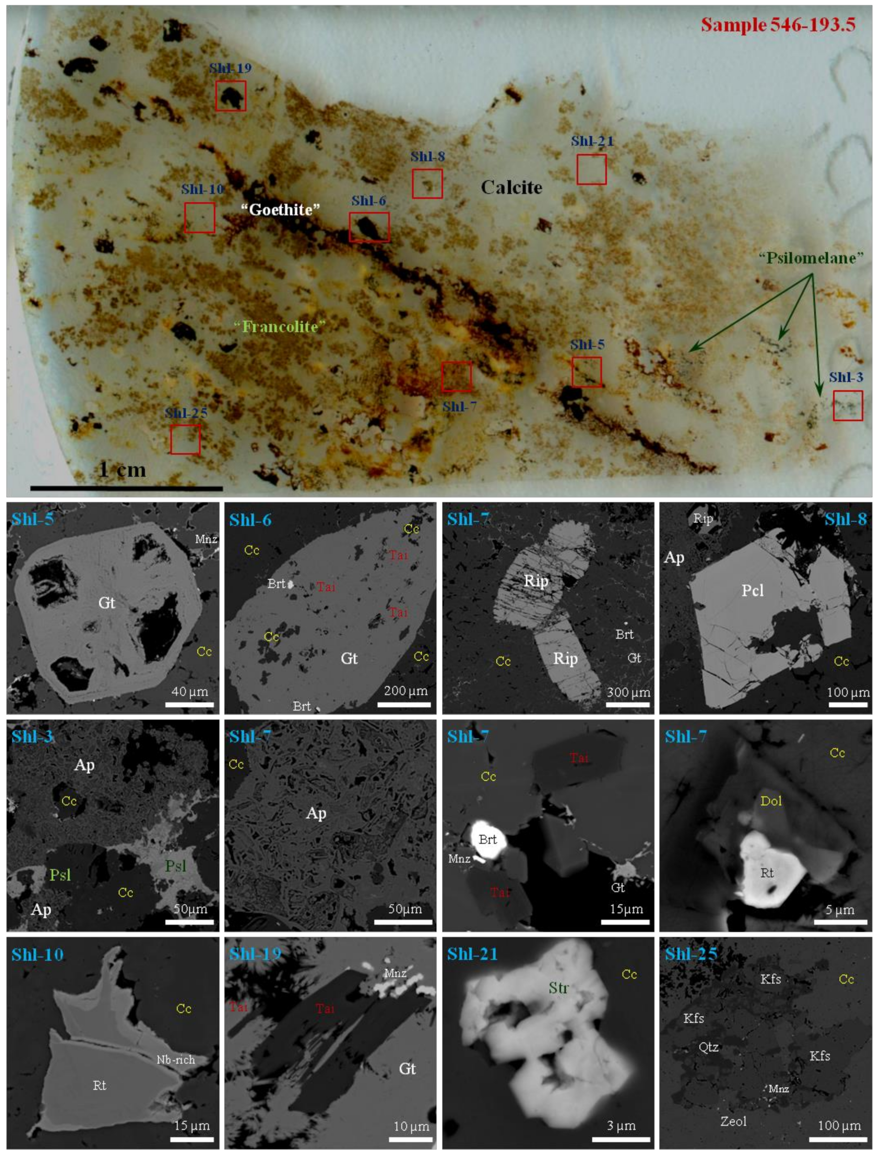
4. Mineralogy and Petrography of Calciocarbonatites from the Chuktukon Massif

The Chuktukon calciocarbonatites are fine- and medium-grained rocks with massive, spotty and banded textures (Figure 2), sometimes contain vugs which are filled with euhedral calcite, barite and/or quartz. Some species are medium to highly altered. The primary mineral assemblage includes calcite, fluorcalciopyrochlore, rippite, tainiolite, fluorapatite (up to 2.3 wt % SrO), fluorite, Nb-rich rutile (up to 17 wt % Nb2O5, up to 5.1 wt % Fe2O3), K-feldspar, dolomite, ancylite-(Ce), strontianite, olekminskite, sulfides, aegirine, quartz and zircon. Calcite is speckled with dolomite spots and fine disseminations of ancylite-(Ce), strontianite, and olekminskite. The SrO concentration in calcite is up to 0.8 wt % [29]. Minerals found in all species of calciocarbonatites are listed in Table 1. The chemical compositions of some minerals are considered in previous works [1,2,29,30,31,32,33,34,35]. In general, rippite is the second main mineral to concentrate Nb in the Chuktukon calciocarbonatites [1,2,29,32], and its content in some rocks may be equal and even higher rather than that of pyrochlore.
Strontianite, daqingshanite-(Ce), ferrohagendorfite, barite, burbankite–khanneshite, Na-Ca carbonates (nyerereite, shortite), thorite, anhydrite, hematite, baddeleyite, zirconolite-group minerals, unidentified Na-REE-Ba-Sr-Ca-rich and Na-Fe-rich phosphates, and Na-rich hydrated carbonate occasionally appear in multiphase or monomineralic inclusions in fluorapatite, fluorcalciopyrochlore, zircon and calcite of the Chuktukon calciocarbonatites [30,34,35]. The secondary multiphase inclusions from the cores of zircon are carbonate-rich in composition and multiphase: calcite + dolomite or alkali-rich carbonates (nyerereite + shortite + burbankite) [30,34].
Aggregates of “francolite” (Sr-rich carbonate-fluorapatite with up to 7 wt % SrO), “goethite” (goethite + “hydrogoethite” ± other Fe-oxides/hydroxides) and “psilomelane” (romanèchite ± hollandite) are common in various quantities for all calciocarbonatite species (Figure 2). Barite, quartz, Ca-REE-fluorcarbonates (parisite-(Ce), synchysite-(Ce)), monazite-(Ce), Sr-Ba-Pb-rich hydro-/kenopyrochlores and above aggregates are related to a stage of metasomatic (hydrothermal) alteration of the calciocarbonatites. They occur interstitially among grains of primary minerals and/or form networks of microveinlets. In addition, goethite forms complete pseudomorphs after euhedral pyrite and prismatic grains (up to 1 cm) of an unidentified mineral, which commonly contains inclusions of calcite and tainiolite (Figure 2) [33].
Detailed studies of the petrography and mineralogy of the highly altered calciocarbonatite are given in [32,40]. These rocks are composed of relict minerals (calcite, fluorcalciopyrochlore, rippite, rutile, and zircon) and minerals formed during alteration. The rocks are fine-grained, banded, and patchy-striped, which is expressed as alternating bands and spots of goethite and rare earth minerals. The groundmass is composed of an aggregate of “goethite” and kaolinite. Florencite-(Ce), monazite-(Ce), “psilomelane” aggregate, churchite-(Y), and daqingshanite-(Ce) are minor and accessory minerals.
“Goethite” aggregate from hydrothermally altered carbonatites contains up to 2.2 wt % of Nb2O5 [32]. Florencite-(Ce) and monazite-(Ce) occur as irregularly shaped grains, up to 0.3 cm in size, as well as aggregates of micron-sized grains of intricately shaped patches, as well as network of vein-like segregations. Typically, both minerals have a porous internal structure. Churchite-(Y) forms radially fibrous 2–5 mm aggregates and individual 2–3 mm crystals. A network of “psilomelane” (microveinlets) dissects the rocks [29]. The petrographic observations of different types of the Chuktukon calciocarbonatites have shown that rippite is very resistant to alteration/weathering processes. In addition to holotype sample 546–193.5 (Figure 2) this mineral was also found as minor component in calciocarbonatites (including highly altered samples) from 180–200 m depths of neighboring drillholes.

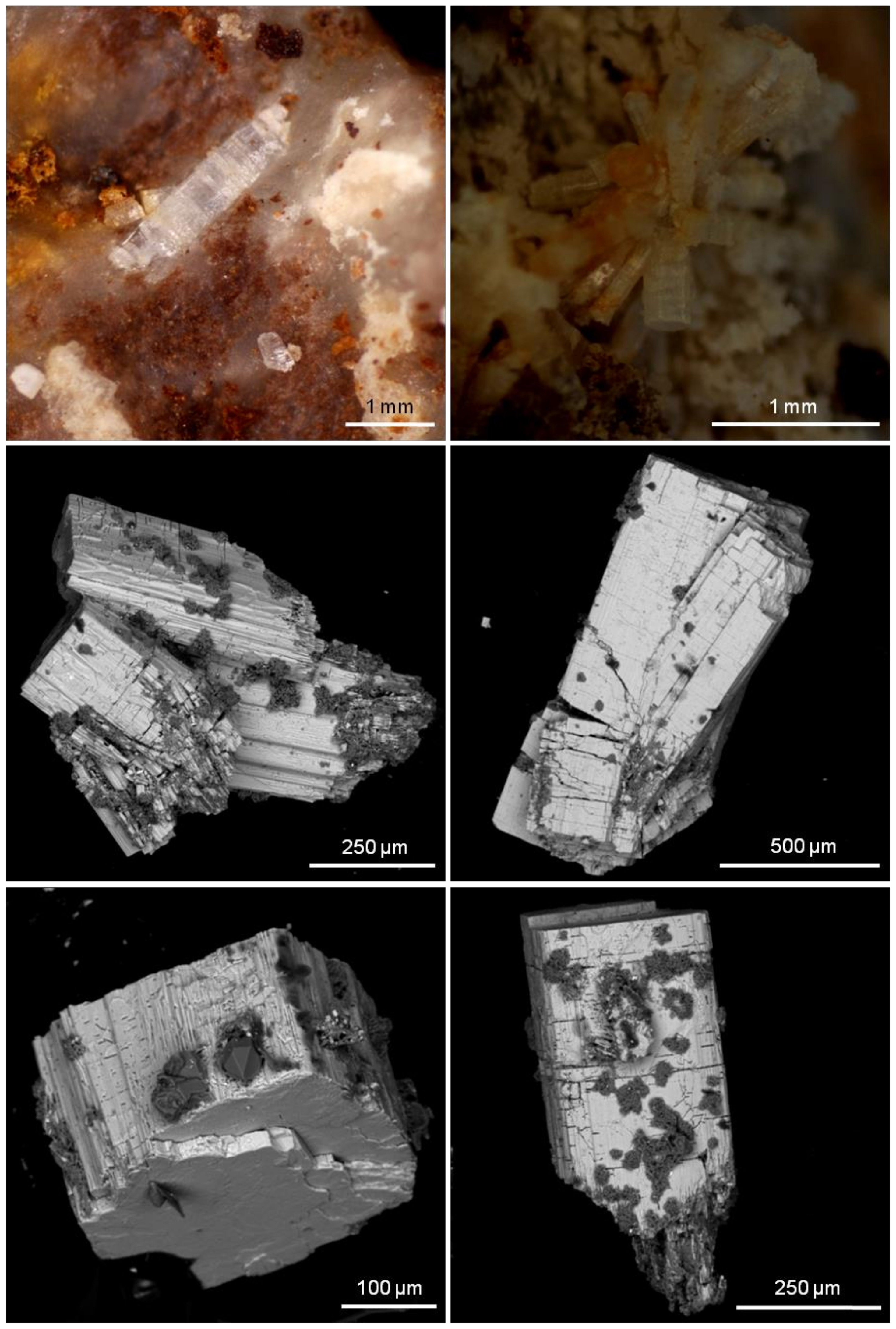
5. Morphology, Optical and Physical Properties of Rippite

Rippite commonly forms elongated prismatic crystals (up to 0.5–2 mm) and their intergrowths in carbonate matrix (Figure 2 and Figure 3), rarely in the “goethite” or “francolite” aggregates. The major forms are prism {100} and pinacoid {001}. Twinning is none observed. The mineral is colorless and it may be confused with fluorapatite due to elongated crystals.
The color of the powdered mineral is white. Rippite has vitreous luster and weak fluorescence. The second harmonic generation test indicated nonlinear optical properties of the mineral. Under laser the weak glow of rippite powder was steady and green that was comparable with colour characteristics of the reference sample (LiIO3). Cleavage is very perfect on (001) and perfect to distinct on (100); parting is none observed, fracture is stepped to uneven across cleavage (Figure 3). The Mohs’ hardness of rippite is 4–5, micro-indentation hardness is VHN50 = 210–487 kg/mm2, mean (n = 19)—307 kg/mm2. The density measured by flotation in the Clerici liquid (HCOOTl + Tl2[OOCCH2COO]) is 3.17(2) g/cm3. Density calculated from unit-cell dimensions and results of electron-microprobe analyses assumes 3.198 g/cm3. Rippite is optically uniaxial (+); ω = 1.737–1.739; ε = 1.747 (589 nm). The optical orientation is X = c. No dispersion and pleochroism were observed. The mineral is not soluble in concentrated HCl and H2SO4 that is comparable with the synthetic phase. The Gladstone–Dale’s compatibility factor for the holotype rippite is 0.005 (superior).
Fluorcalciopyrochlore octahedra and tainiolite blades occasionally occur as inclusions in rippite, and vice versa rippite (± quartz and other phases) sometimes fills the fissures in fluorcalciopyrochlore crystals (Figure 4, Figures S1 and S2). Such relationships indicate that rippite is later Nb-mineral than fluorcalciopyrochlore and crystallized together with tainiolite or later (Figure 4, Figures S1–S3). Most of rippite grains are partially resorpted by quartz especially along cleavage planes and even replaced by quartz in the outer parts of some crystals (Figure 3, Figure 4 and Figure 5, Figures S3–S5). “Goethite” impregnation is common of some grains occurring in altered carbonatite samples. In general, rippite is resistant to low-temperature alteration or weathering, but may be partially replaced by quartz (plus rutile and other phases) during carbonatite crystallization (Figure 5 and Figures S3–S5).

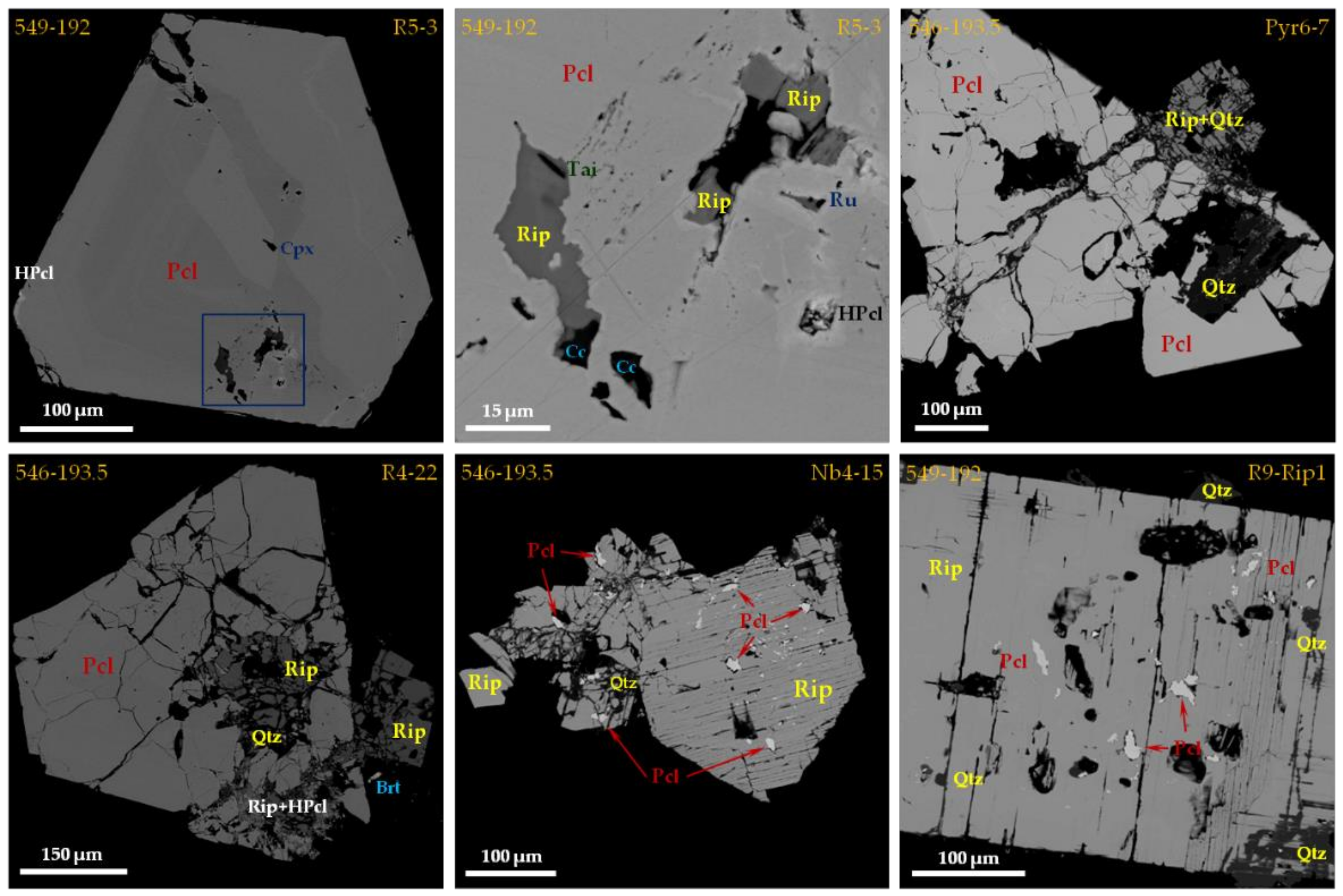
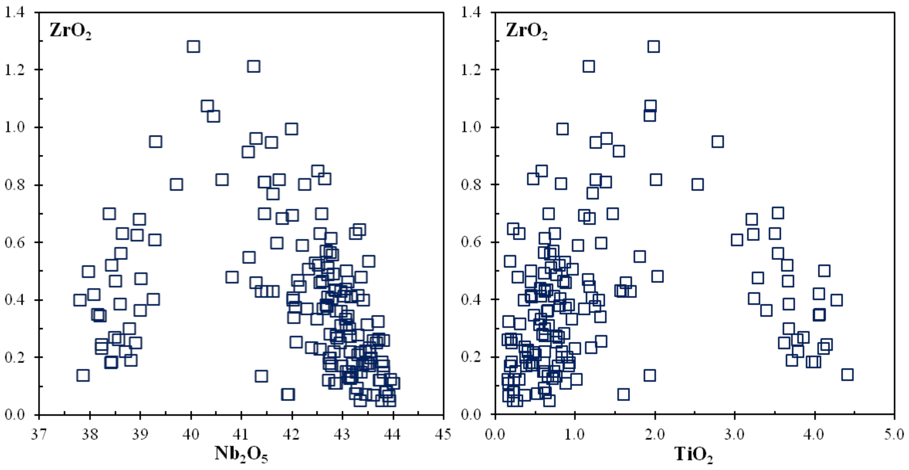
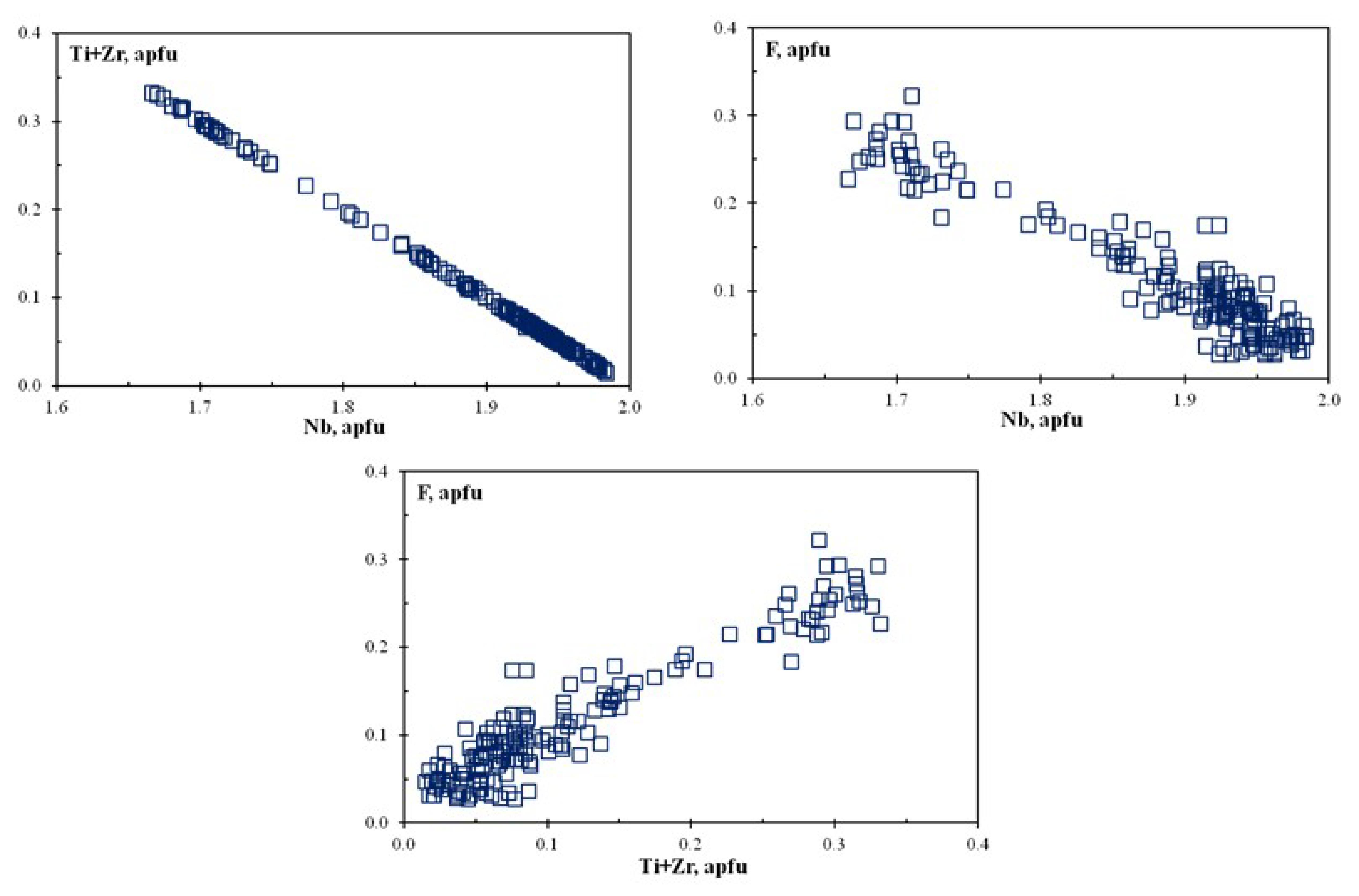
6. Chemical Composition of Rippite

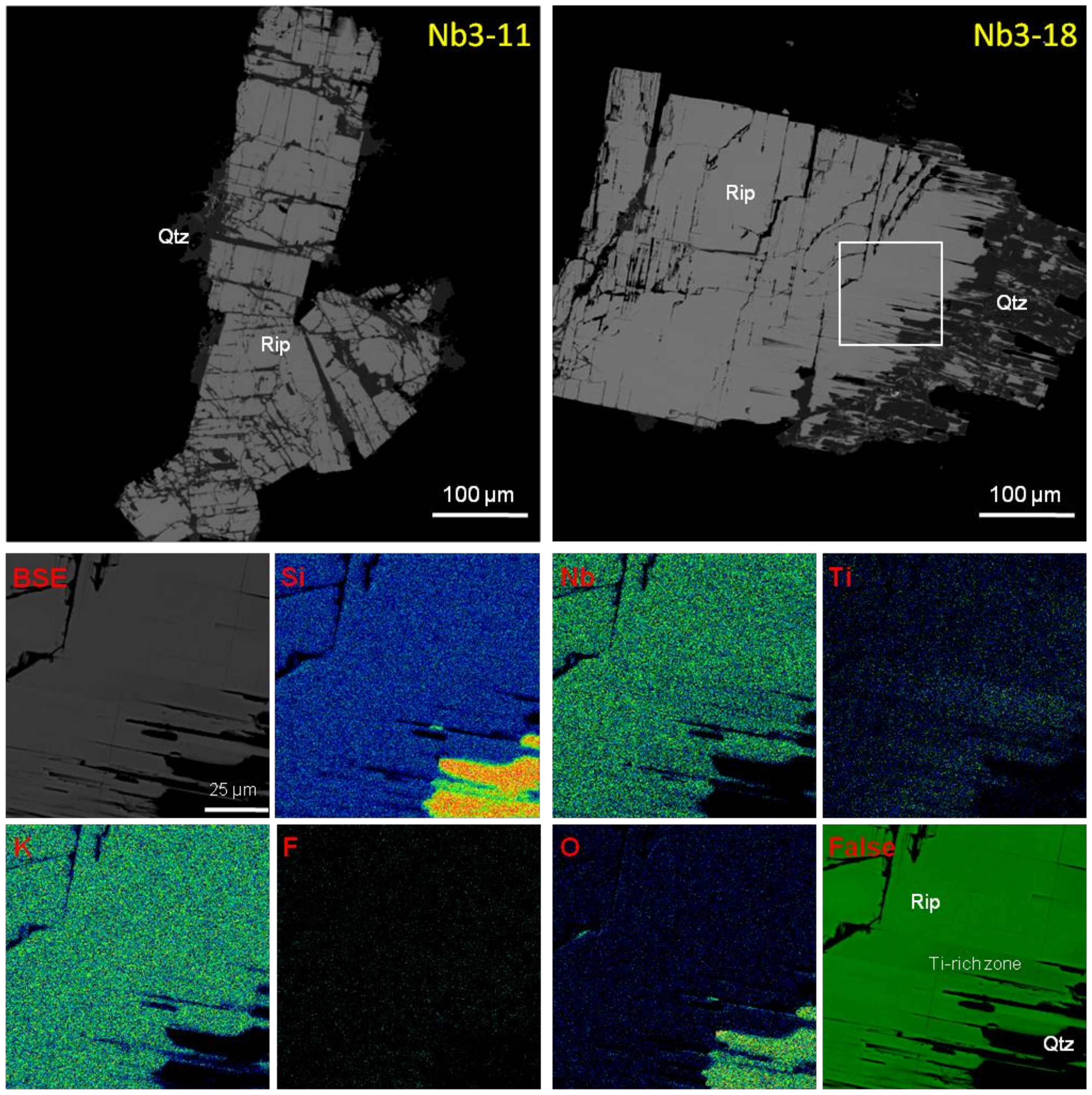
6.1. SEM and EMPA data

Majority of rippite prismatic crystals are homogeneous in composition and unzoned or weakly-zoned that is virtually invisible in the elemental SEM maps (Figures S3, S6 and S7). The contrast zonation is only fixed for rare grains with narrow Ti-rich outermost zone. Those core-to-rim variations are well indicated in the Nb and Ti elemental maps (Figure 5). In general the chemical composition of the mineral is close to the ideal structural formula K2Nb2(Si4O12)O2 (Table 2 and Table 3). The empirical formula of the holotype rippite (sample 546-193.5, mean of 110 analyses) is K2(Nb1.90Ti0.09Zr0.01)[Si4O12](O1.78OH0.12F0.10) (Table 2). Most of grains from holotype and other samples show compositions poor in TiO2 (<0.7–1.0 wt %) and F (<0.5 wt %), K2(Nb1.93Ti0.05Zr0.02) [Si4O12](O1.93F0.07) (Table 2 and Table 3). Raman, IR and SIMS data indicate very low content of H2O or (OH)-groups in such grains (see text below). High contents of TiO2 (up to 4.4 wt %) and F (up to 1.0 wt %) are common of the outermost zone in some grains, up to K2(Nb1.67Ti0.32Zr0.01)[Si4O12](O1.67F0.33) (Table 2, Figure 5). The ZrO2 amount is up to 1.3 wt %. However, there is no any clear correlation ZrO2 with TiO2 and Nb2O5 (Figure 6). All above-mentioned data strongly suggest the incorporation of (Ti,Zr) and F in the structure of rippite via the isomorphism Nb5+ + O2− → (Ti,Zr)4+ + F1− (Figure 7). The content of a hypothetical end-member K2Ti2[Si4O12]F2 may be up to 17 mol. % (Table 2). The database of chemistry for all rippite grains is given in Supplementary Table S1.

6.2. LA-ICP-MS and SIMS Data

Trace elements were measured by LA-ICP-MS for Ti-poor compositions of rippite from the Chuktukon calciocarbonatite. The REE and trace element concentrations are consistently low (<220 ppm in total) (Table 4). The substantial amounts (>2–5 ppm) are common of P (22–23 ppm), Ca (16 ppm), Ba (153–205 ppm), V (12–26 ppm), Sc (7.1–9.4 ppm), Hf (22–36 ppm) and Ta (7.3–11.3 ppm) (Table 4).
The contents of some light trace elements (Be, Li, B), F and H2O in low-Ti rippite were analyzed by SIMS (Table 4). The results indicate very low concentration of light elements (<5 ppm) and H2O (0.09–0.23 wt %). The content of F is consistent with EMPA-WDS data.

7. Optical Studies for Rippite

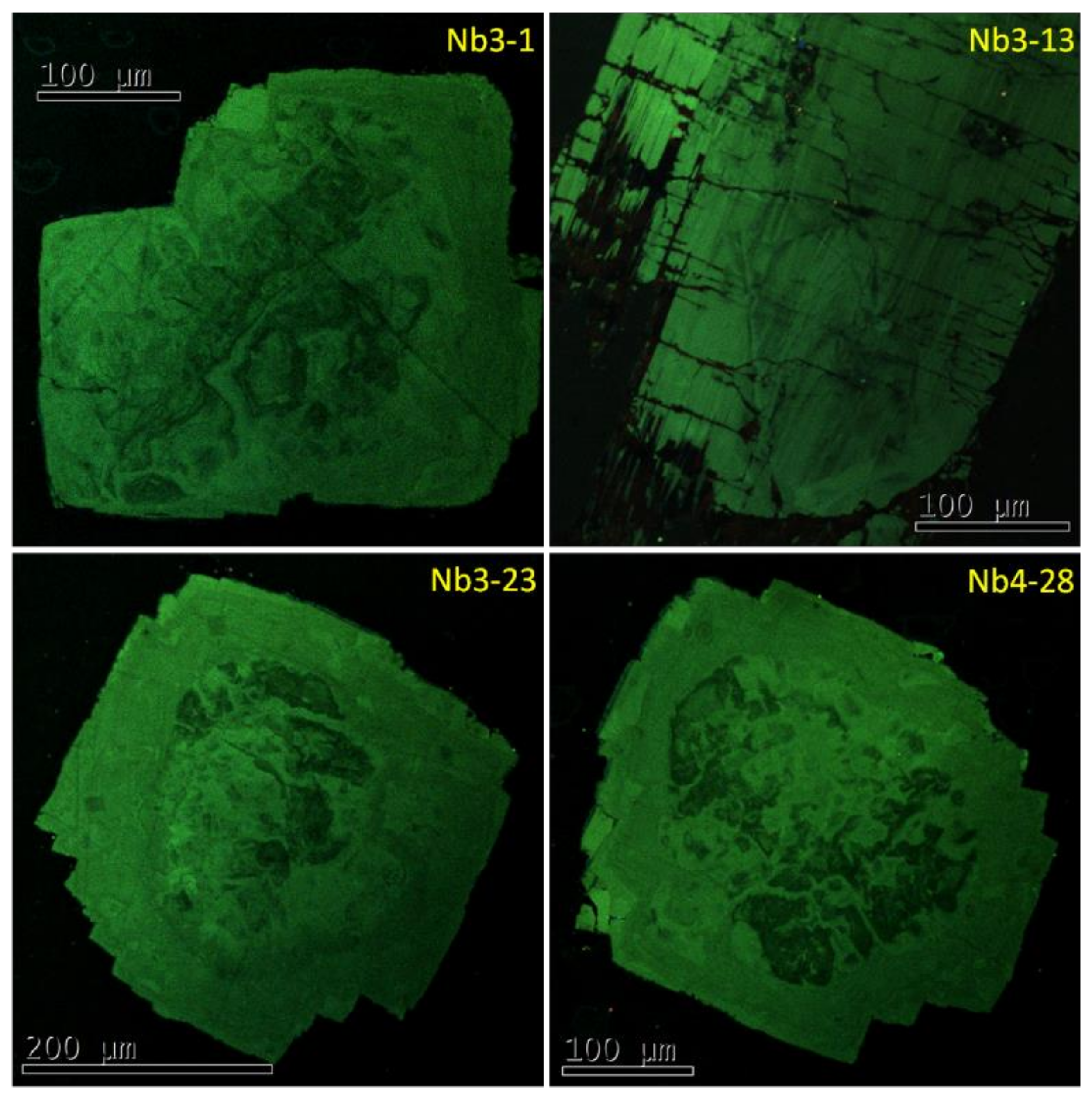
7.1. Cathodoluminescence

The CL images for single rippite crystals show green colour and very complex history during their growth (Figure 8). However, the individual zones and regions fixed in the CL images are weakly documented by the BSE images and elemental maps and do not reflect the variations in chemical composition (Figure 8, Figures S6 and S7). In general, the stages of nucleation, chaotic growth, dissolution and further growths may be observed in the CL images of individual rippite grains (Figure 8).

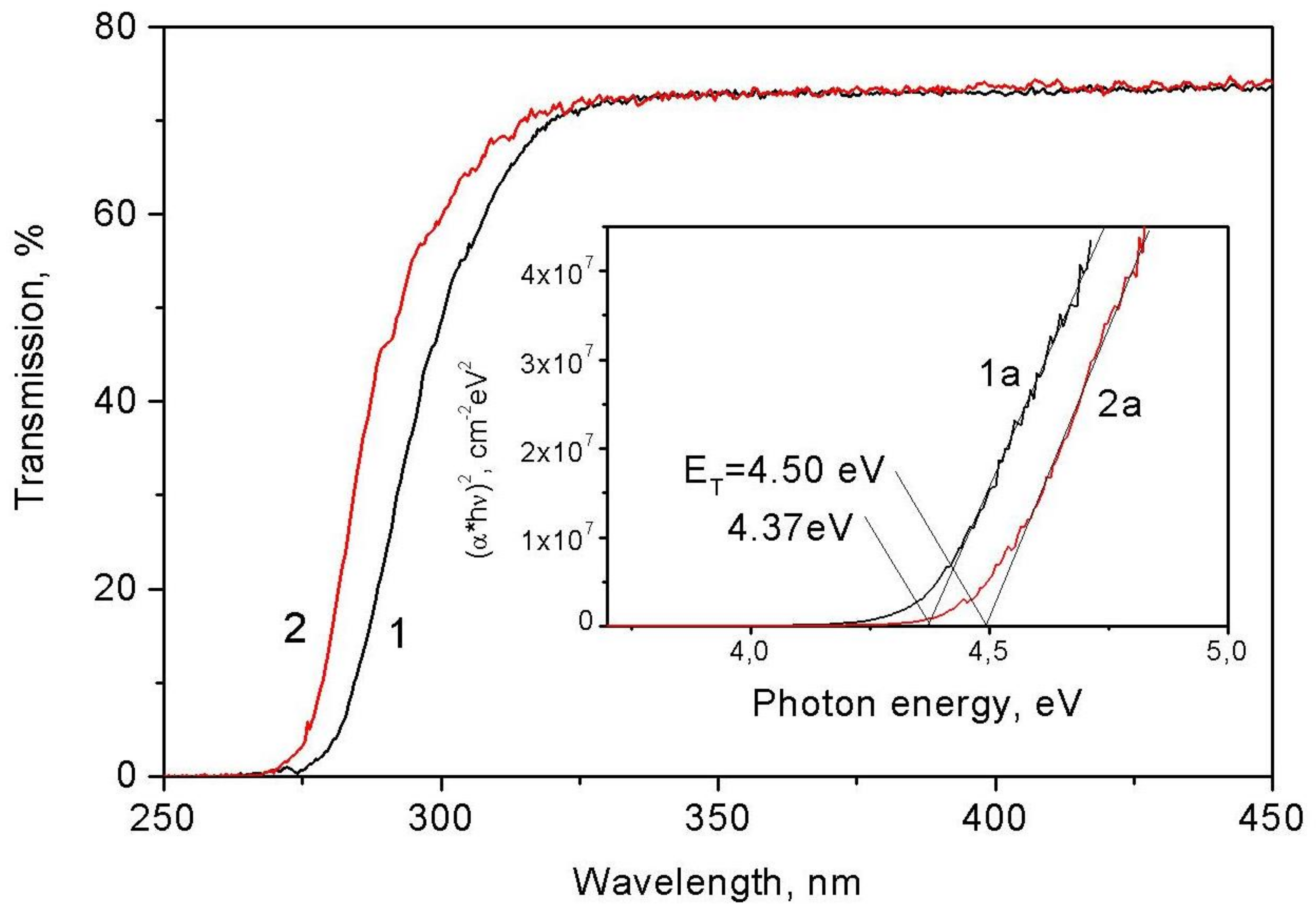
7.2. Transmission Spectra

The color of the powdered rippite is white and its crystals are almost colorless. Figure 9 shows the short-wave edge of the optical transmission spectra obtained for a crystal of about 150 × 150 mm2 and about 100 mm thick, at 300 and 80 K. It is seen that the crystal becomes transparent from a wavelength of about 275 nm in the UV region of the spectrum. The Tauc analysis [51] indicates that the spectra are straightened in coordinates (α*(hν))2 = f(hν), where α is the absorption coefficient and hν is the photon energy. This is evidence that the shape of the fundamental absorption edge is determined by the direct allowed band-to-band electronic transitions. The position of the intersection points of straight lines with the abscissa axis determines the values of the band gap at 300 and 80 K: Eg = 4.37 and 4.50 eV, respectively. Under the action of laser radiation with a wavelength of 1052 nm, second-harmonic radiation (526 nm) is generated in the rippite powder. The intensity of the second harmonic is comparable to that obtained from the powder of the reference sample, a well-known nonlinear optical crystal, lithium iodate (LiIO3), for which the second-order nonlinear susceptibility is d31 = 4.1 ± 0.4 pm/V [52].
Rippite has a weak fluorescence and, in addition, its samples show spontaneous luminescence in the range of 100–-400 K when heated or cooled at a rate of ~30 deg/min. This glow is typical for pyroelectrics [53] and indicates the absence of an inversion center in the structure. Indeed, rippite with a structure with a point group of 4mm belongs to one of the 10 crystallographic groups with a pyroelectric effect: 1, 2, 3, 4, 6, m, mm, mm2, 3m, 4mm and 6mm [54]. Such crystals have electric polarization even in the absence of an external electric field (spontaneous polarization). Pyroluminescence is the result of modifications in the atomic polarization in the crystal volume as temperature changes [53]. Flashes of light are the result of an electrical breakdown in pyroelectric fields, the intensity of which reaches hundreds of kV/cm.

7.3. Infrared and Raman Spectroscopy

Tetragonal rippite and its synthetic analog are related to space group P4bm (No. 100), its point group is C4v (4mm). Symmetry-representation analysis gives 105 modes of vibrations for rippite, which can be divided as 21A1 + 16A2 + 12B1 + 17B2+ 39E, where 20A1 + 38E vibrations are infrared active, 20A1 + 12B1+ 17B2 + 38E vibrations are Raman active, and 16A2 vibrations are inactive in both regions. Acoustic modes A1 + E are not included here. Among these, the silicate tetrahedrons will share the following internal vibrations 5A1 + 4B1 + 5B2 + 9E. External vibrations include translation and SiO4 libration modes. Their irreducible representations can be expressed as: 8A1 + 7B1 + 8B2 + 18E and 7A1 + B1 + 4B2 + 11E, respectively. The C4v point groups allow the simultaneous activity of some part of vibrations in both IR and Raman spectra [55]. The number of IR and Raman modes observed for rippite is fat fewer than predicted, because of the low intensity of some modes and orientation effects. Crystals of Ti-poor rippite were mainly used for the IR and Raman spectroscopic studies.

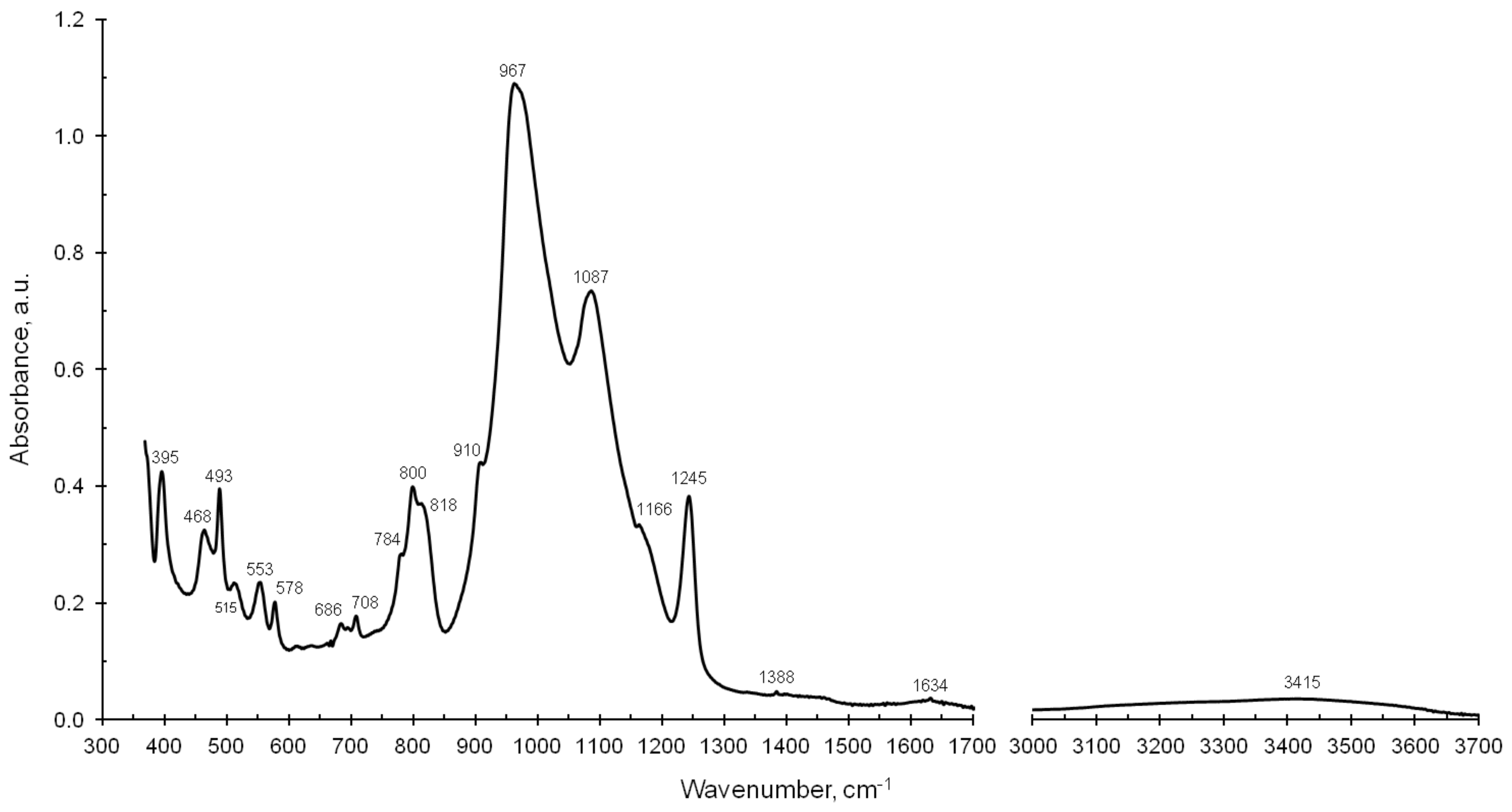
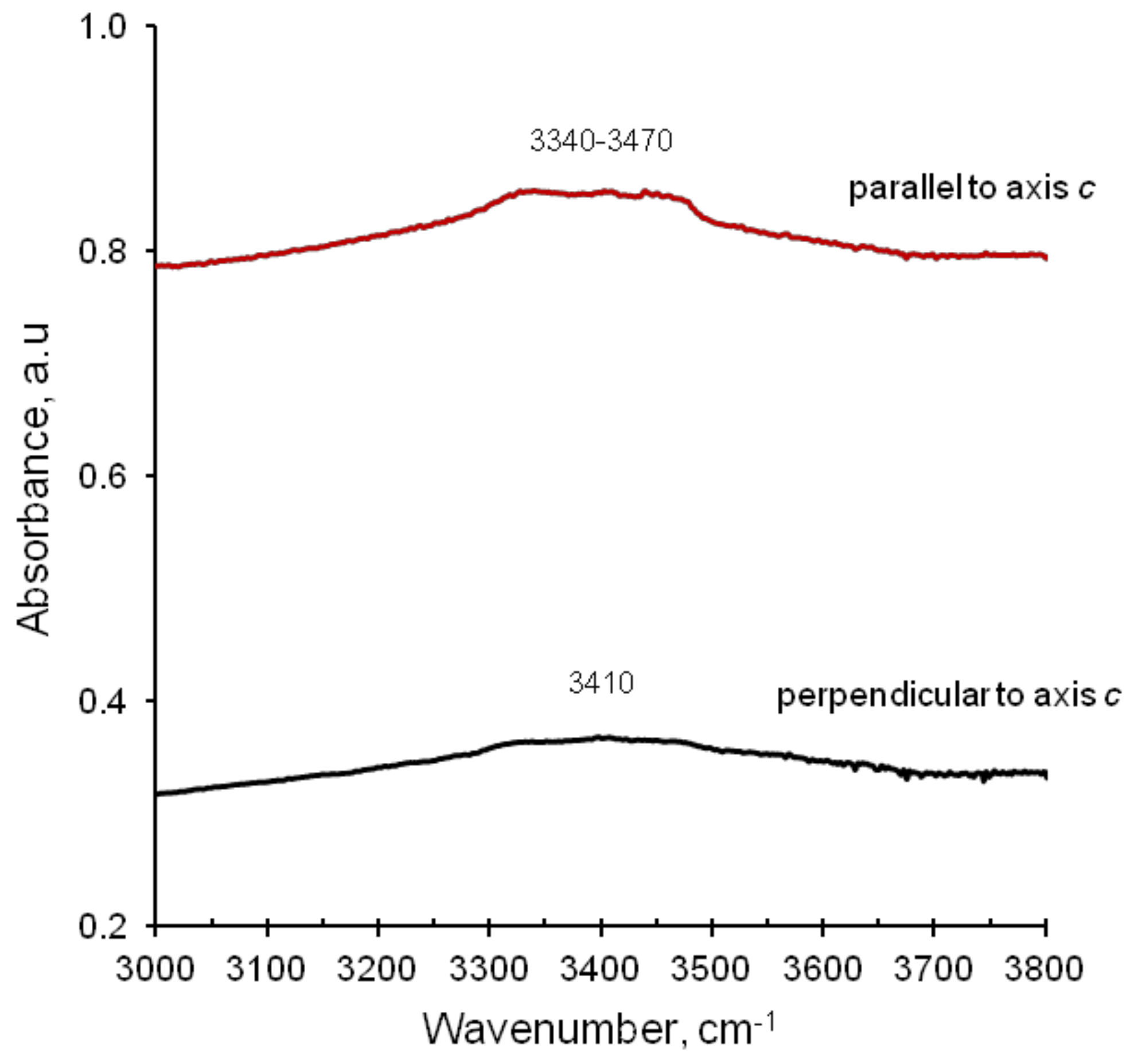
7.3.1. Infrared Spectra

The following absorption bands are more remarkable in the IR spectra of rippite (in cm−1): 395, 468, 493, 515, 553, 578, 686, 708, 784, 800, 818, 910, 967, 1087, 1166, 1245, 1388 and 1634 (powder KBr pellet, Figure 10). There are no clear bands in the 3000–4000 cm−1 region (Figure 10 and Figure 11).
The strongest absorption band of the IR spectrum of rippite is 967 cm−1 (Figure 10) and assigned to be the ν3 internal stretching mode of the SiO4 tetrahedra. The antisymmetric stretching modes of the Si-O-Si bonds and the stretching vibrations of Si-O of the SiO4 tetrahedra are active between 1300 and 960 cm−1; and symmetric stretching vibrational modes of the Si-O-Si bonds lying between 800 and 680 cm−1. The latest bands are associated with the presence of bridging oxides (Si-O-Si linkage) in the structure. The stretching modes of the Nb-O bonds in the NbO6 octahedra occur in the 600–800 cm−1 range as weak bands and shoulders, considering the stronger force constant of the Si-O bonds than that of the Nb-O ones. The bands below 580 cm−1 of the IR spectrum indicate silicon-oxygen bending vibrations and the stretching vibrations arising from K-O bonds. In addition, the IR and FTIR spectra of rippite show very weak and broad bands near 1634 and 3415 cm−1 (Figure 10 and Figure 11). However, heating of rippite in powder KBr pellet up to 380 °C and following IR measurements resulted to diminishing of these bands. Consequently, the presence of the substantial bands in the 1600–1700 and 3400–3500 cm−1 region in unheated rippite samples may be partially related to absorbed H2O. Nevertheless, we have to mention that our SIMS data indicate low H2O content (0.09–0.23 wt %) in the Ti-poor rippite. Thus, weak bands in the 3000–4000 cm−1 range, which may be related to the vibrations of (OH)-groups, unfortunately cannot outline the presence or absence of H2O and as result the true H2O concentration.

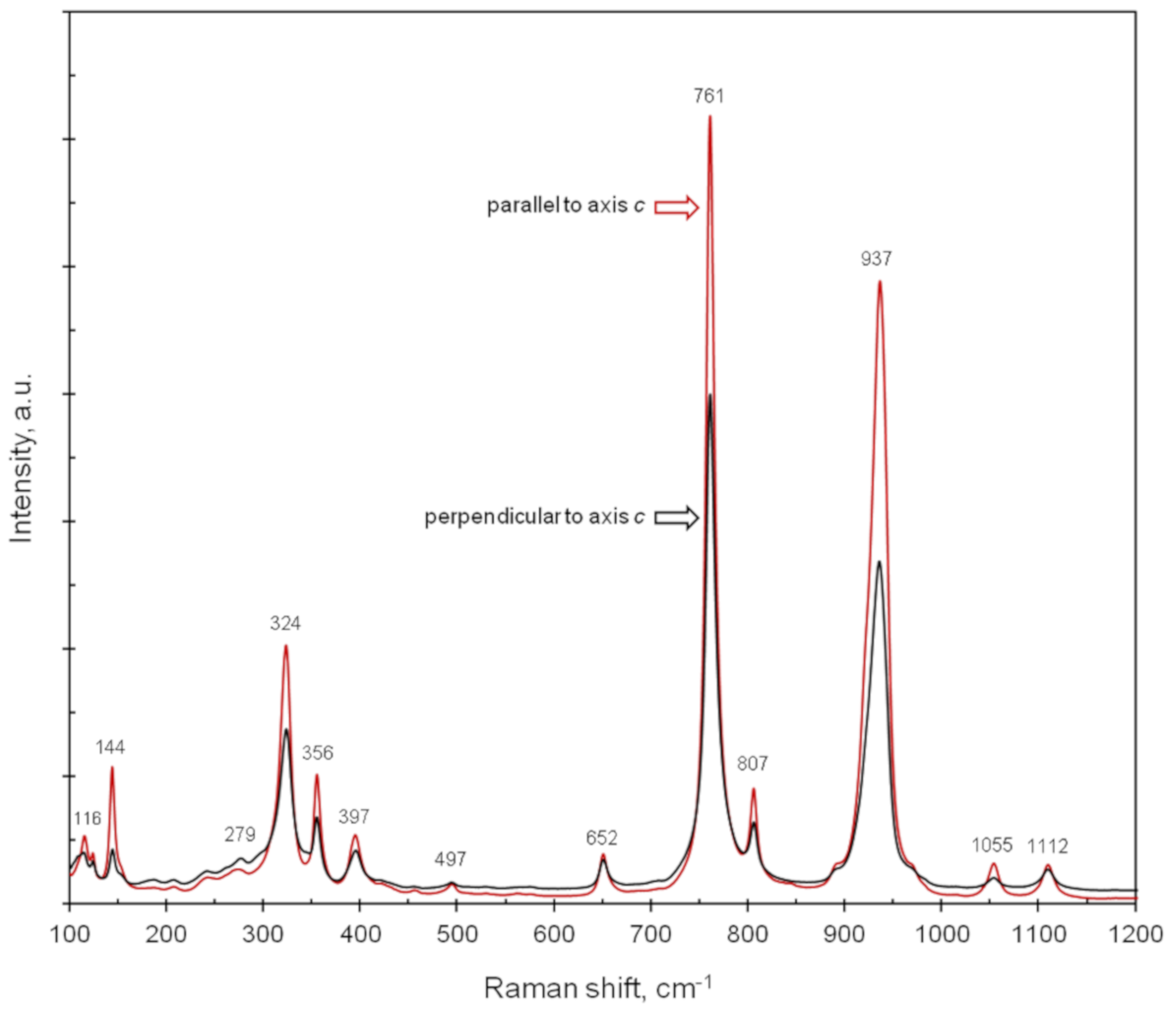
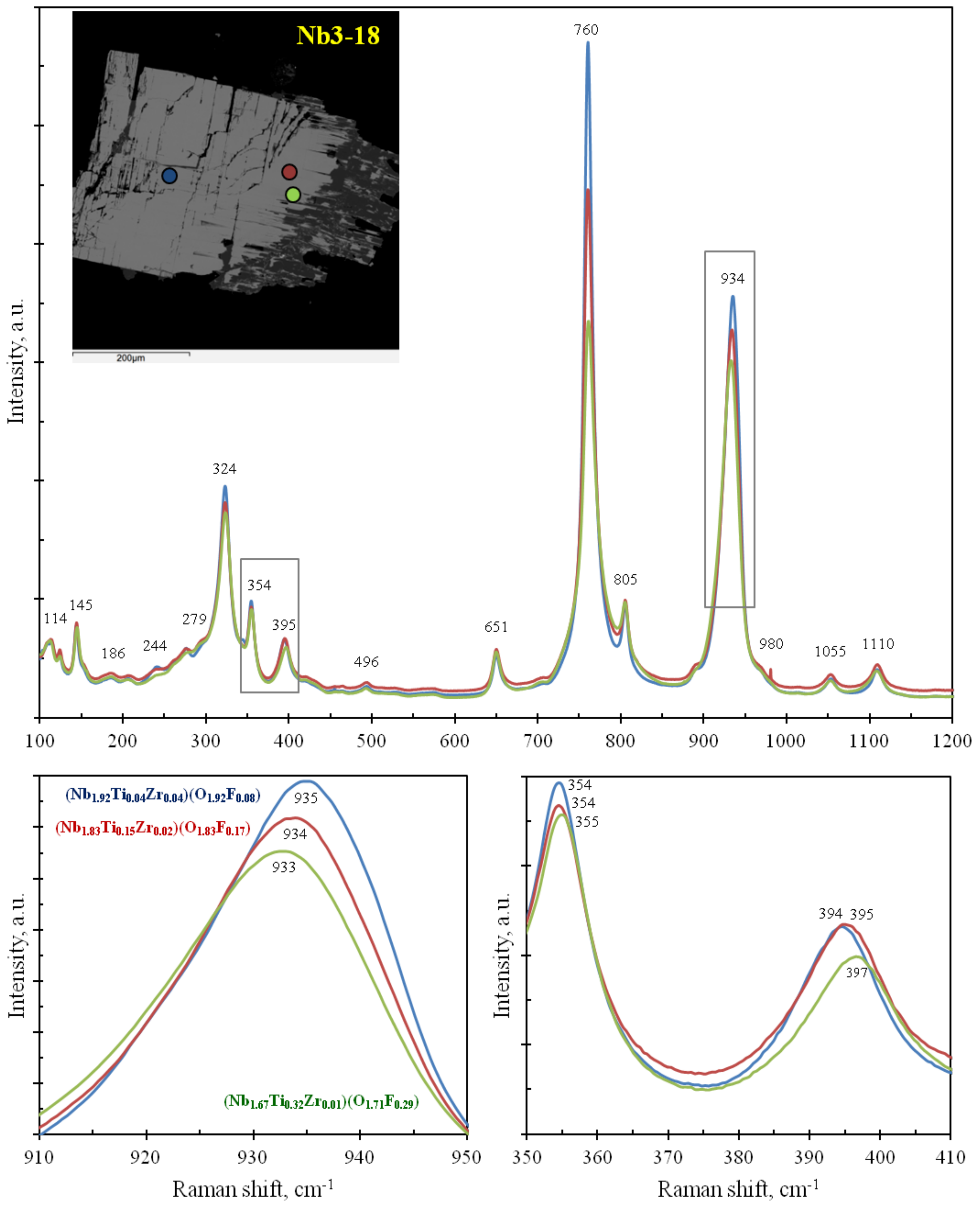
7.3.2. Raman Spectra

Main bands in oriented Raman spectra of rippite are (in cm−1): 116, 144, 279, 324, 356, 397, 497, 652, 761, 807, 937, 1055 and 1112 (Figure 12). There are no clear bands in the 3000–4000 cm−1 region.
In general, bands below 360 cm−1 are ascribed to vibrations in polyhedra and octahedra as well as librational vibrations of the Si4O12 group. Bands in the 390–700 cm−1 region are related to tetrahedral SiO4 bending vibrations (ν4 + ν2). The strongest Raman band at 761 cm−1 (Figure 12) is assigned to be the internal stretching mode ν1 of the SiO4 tetrahedra. The ν3 modes of the SiO4 tetrahedra seem to be bands in the 900–1200 cm−1 region.
In addition, we tried to obtain data for rippite concerning to the dependence between chemical composition and changing in Raman spectra. For this purpose, we used zoned crystal Nb3-18. All spectra were obtained during one analytical session and in same conditions. The most substantial differences are visible for the bands at 934, 395 and 354 cm−1 (Figure 13). The increasing of the Ti(+Zr) content in rippite lead to left shifting for the 934 cm−1 band and to right shifting for the 395 and 354 cm−1 bands in Raman spectra. Unfortunately, such insufficient deviations (in 1–2 cm−1) cannot be used for identification of chemical composition for rippite according to Raman spectra.

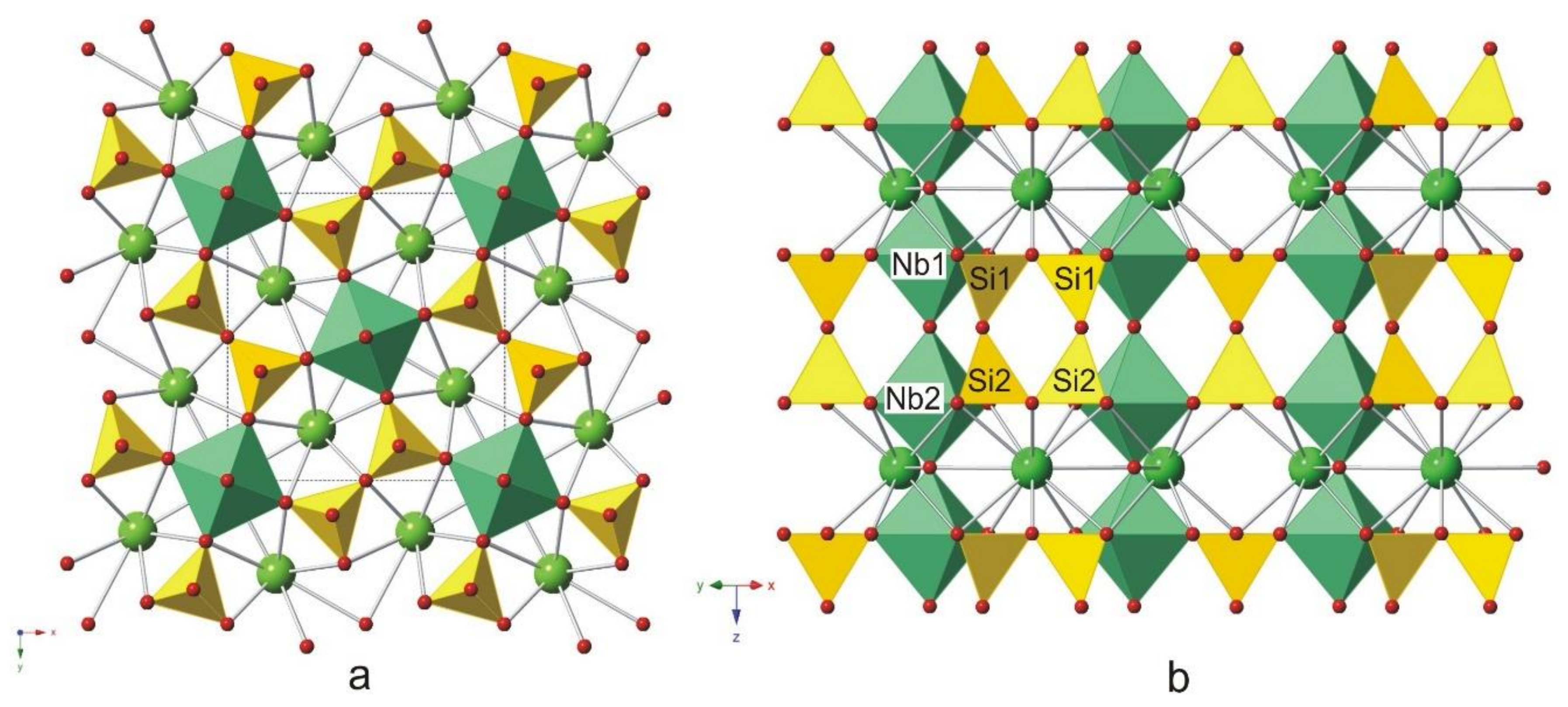
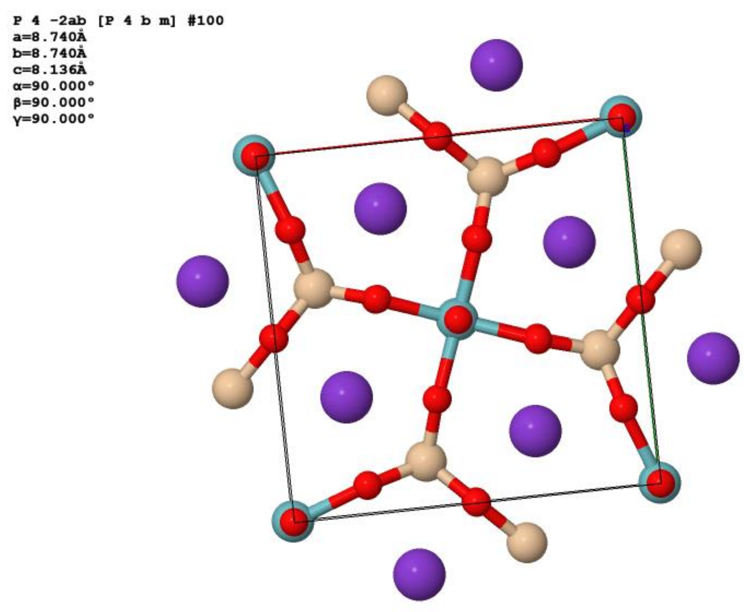
8. X-ray Crystallography and Crystal Structure of Rippite

The second harmonic generation test for rippite indicated the weak glow of powder, which was steady and green. The reference sample (LiIO3), which is used in nonlinear optics, showed the same colour characteristics. All this strongly assumes a noncentro-symmetrical structure for rippite.
X-ray powder diffraction data for rippite (in Å for MoKα) are given in Table 5. Unit cell parameters refined from the powder data using STOE Win XPOW 2.21 program package are as follows: tetragonal symmetry; space group P4bm; a = 8.735(3) Å; c = 8.127(5) Å; V = 620.1(4) Å3; Z = 2
Single-crystal X-ray studies were carried out on a fragment of low-Ti rippite grain and gave the following results: tetragonal symmetry; space group P4bm; a = 8.7388(2) Å; c = 8.1277(2) Å; V = 620.69(2) Å3; Z = 2. The crystal structure was studied for Ti-poor rippite K2(Nb1.93Ti0.05Zr0.02) [Si4O12]O(O0.93F0.07). It was solved by direct methods and refined on the basis of 949 independent reflections (836 reflections with I > 2σ(I)). The factor R1 for the crystal structure is 0.036. Crystallographic data for rippite are given in Table 6, Table 7, Table 8 and Table 9. The crystal structure is shown in Figure 14. Rippite is closely identical in the crystal structure to synthetic compound K2Nb2(Si4O12)O2 (or KNbSi2O7) [3,4,5,15] (Figure 15).
The Nb5+ cations occupy two sites, which also contain minor amounts of Ti4+ and Zr4+. The Nb sites are surrounded by six O atoms in octahedral coordination. The corner-shared NbO6 octahedra form chains running parallel to a four-fold symmetry axes. The chains are coupled together by four-membered Si4O12 rings so that each octahedron shares four corners with four SiO4 tetrahedra belonging to four different Si4O12 rings. Each Si4O12 ring shares its terminal O atoms with eight NbO6 octahedra of four neighboring chains linking them in the 3D heteropolyhedral framework. The four-membered rings are in the plane {110}; at that each ring is connected with two neighboring Nb-octahedra of one chain via shared O-atoms forming heteropolyhedral four-membered rings (Figure 14b). Positions of K+ are situated in channels which extend through the structure in the [001] direction. K+ cations are coordinated by twenty O atoms in a form of a two-capped pentagonal prism. Low content of F does not give possibility to determine its structural position.
It is should be noted that rippite is structurally related to cyclosilicates with [Si4O12]8− four- membered single rings. In structural and chemical aspects it seems to be in close with the labuntsovite-supergroup minerals, namely with vuoriyarvite-(K), K2(Nb,Ti)2(Si4O12)(O,OH)2∙4H2O (vuoriyarvite group, space group Cm, a = 14.692 Å, b = 14.164 Å, c = 7.859 Å, Z = 4; β = 117.87o, V = 1445.74 Å3) [56,57,58,59,60]. However, rippite is nominally free in H2O and (OH)-groups. In addition, the connection of the octahedral chains and [Si4O12]-rings is quite different in the crystal structure of rippite (this work) and the labuntsovite-supergroup minerals [56,57,58,59,60]. Unlike rippite the four-membered tetrahedral rings in the structure of vuoriyarvite-(K) are oriented perpendicularly to the NbO6-chains and two neighboring Nb-octahedra in one chain are connected by shared O-atoms with edge of the SiO4-tetrahedron. It led to considerable deformation of the octahedral chain. In general, we suggest that the rippite-related structure is a new structural type among cyclosilicates with the [Si4O12]8− single rings.
The compound K4(NbO)2[Si8O21] is somewhat close in element suite, chemistry and structure to rippite and synthetic K2(NbO)2Si4O12 [61,62]. It was synthesized in the hydrothermal system K2O-Nb2O5-SiO2-H2O (T = 450–500 °C; P = 1000–1200 bar) and its composition is (EMPA, in wt %): SiO2—51.10; Nb2O5—28.80; K2O—20.84 [61]. Unlike rippite and its synthetic analog this phase has complex modulated structure and triclinic symmetry: space group P-1; a = 33.92(2) Å, b = 10.950(6) Å, c = 7.737(4) Å, α = 76.42(4)o, β = 72.10(5)o, γ = 89.81(5)o, V = 2650.8 Å3 [62]. In addition, K4(NbO)2[Si8O21] is characterized by a relatively low nonlinear polarization in comparison with K2(NbO)2Si4O12 owing to the low concentration of the isolated NbO6 octahedra in its structure [63]. In contrast with this K-Nb-silicate, rippite and synthetic K2(NbO)2Si4O12 display a strong non-linear effect, as the NbO6 octahedra are linked in columns (Figure 14 and Figure 15).

9. Discussion and Concluding Remarks

In general the detailed studies for the Chuktukon calciocarbonatites (Chadobets upland, Krasnoyarsk Territory, Russia) gave the possibility of describing the chemical composition and crystal structure for a new mineral, rippite K2(Nb,Ti)2(Si4O12)(O,F)2, which belongs to a new structural type among the [Si4O12]-cyclosilicates and is close to the labuntsovite supergroup. This mineral is related to primary phases, which were crystallized after fluorcalciopyrochlore, the main carrier of Nb in the calciocarbonatites. The primary carbonatite paragenesis may be formed at temperatures near and below 837 °C according to the calcite-dolomite thermometer. The synthesis in the system SiO2-Nb2O5-K2O has indicated that the K2Nb2(Si4O12)O2 phase may be stable in the 510–735 °C temperature range [4,6,8,9,10,13,14].
Rippite is the second main mineral to concentrate Nb in the Chuktukon calciocarbonatites [1,2,29,32]. The presence of this mineral should be into account in processing schemes of carbonatitic Nb-ores, as in some rock species the modal contents of pyrochlore and rippite may be equal. The weathered carbonatites are the basis of the Chuktukon niobium-rare earth elements ore deposit, which is very promising for development in eastern Siberia in a future [40,41,42]. It is quite possible that unweathered calciocarbonatites will be also involved in processing schemes. In general, it is concerning not only to the Chuktukon calciocarbonatites, but and to other pyrochlore-rich carbonatites around the world [64]. At present rippite was found only in calciocarbonatites of the Chuktukon massif. However, we are not sure that this mineral is endemic for the Chuktukon rocks and may be also occur in other pyrochlore-containing carbonatites worldwide.
It should be noted that the mineral phase with ratio of the main components (K:Nb:Si) similar to rippite has been described as minute inclusions in late apatite aggregates from calciocarbonatites of the Araxá alkaline-carbonatite complex, Brazil [65]. According to chemistry (SiO2—37.9–39.3 wt %; Nb2O5—34.7–37.6 wt %; TiO2—0.3–1.6 wt %; K2O—14.2–14.4 wt %) and low total sums this mineral was identified as nenadkevichite (labuntsovite supergroup). Probably further studies will clearly outline whether the Araxá mineral is rippite or it is vuoriyarvite-K (labuntsovite supergroup).
The discovery of rippite also highlights some problems in the synthesis and growth of high-quality crystals suitable for industrial purposes. It is known that synthetic analog of rippite is a good material for nonlinear optics [3,4,5,6,7,8,9,10,11,12,13,14]. Ideal and large single crystals of KNbSi2O7 (near 1 cm) of high optical quality, which are valid for industrial purpose, are still rarity [14]. The mineral association of rippite-containing calciocarbonatites may be a key in understanding of favorable conditions for synthesis of the perfect KNbSi2O7 crystals (Ca-rich carbonate environment).
The compositional study of rippite strongly suggests the incorporation of (Ti,Zr) and F in the structure via the isomorphism Nb5+ + O2− → (Ti,Zr)4+ + F1−. As a result the content of a hypothetical end-member K2Ti2[Si4O12]F2 in rippite may be up to 17 mol. %. Unfortunately, there are no any data about synthesis of K2Ti2[Si4O12]F2 or K2Zr2[Si4O12]F2, which may be isostructural with K2Nb2(Si4O12)O2 and K2Ta2(Si4O12)O2 [3,4,5,16]. It is quite possible that Ti- and Zr-counterparts or their intermediate compositions with K2Nb2(Si4O12)O2 will have higher nonlinearity rather than pure K2Nb2(Si4O12)O2. From other side the minimal doping of the K2Nb2(Si4O12)O2 compound by Ti or Zr may lead to good conditions for single crystal fabrication.
It should be also mentioned that rippite has been successfully used as a geochronometer. The Ar-Ar age estimated for rippite from the Chuktukon calciocarbonatite (sample 546-193.5) is 231.1 ± 2.7 Ma [31].

Supplementary Materials

The following figures are available online at https://www.mdpi.com/2075-163X/10/12/1102/s1: Figure S1: Secondary polymineral inclusions with rippite in fluorcalciopyrochlore, Chuktukon calciocarbonatite, BSE images and elemental maps; Figure S2: Inclusions of fluorcalciopyrochlore in rippite, Chuktukon calciocarbonatite, BSE images and elemental maps; Figure S3: Relationship of rippite with tainiolite in the Chuktukon calciocarbonatite, BSE images and elemental maps; Figure S4: The outer zone in a rippite crystal and its replacement by quartz with formation of the TiO2 phase (rutile ?), Chuktukon calciocarbonatite, BSE image and elemental maps; Figure S5: Replacement of rippite by quartz with formation of the TiO2 phase (rutile ?), Chuktukon calciocarbonatite, BSE images and elemental maps; Figure S6: BSE and CL images and elemental maps for low-Ti rippite (grain Nb3-1), Chuktukon calciocarbonatite; Figure S7: BSE and CL images and elemental maps for low-Ti rippite (grain Nb3-23), Chuktukon calciocarbonatite. Supplementary materials also include Table S1: Chemical composition (WDS, wt %) of rippite from the Chuktukon calciocarbonatite, Chadobets upland, Siberia, Russia and CIF file for rippite.

Author Contributions

V.V.S. and A.G.D wrote the paper and performed the mineralogical description for carbonatites; the crystal structure and X-ray powder data for rippite, Y.V.S.; optical and physical properties for rippite, E.V.B.; Raman spectroscopy and SIMS, V.V.S.; SEM and CL, V.V.S., N.S.K. and A.G.D.; EMPA, V.V.S. and E.N.N.; LA ICP-MS—A.G.D.; IR spectroscopy, T.N.M. and I.N.K., second harmonic generation test, A.P.Y. and V.N.V. All authors have read and agreed to the published version of the manuscript.

Funding

Investigations of inclusions in minerals and physical and chemical properties of rippite were done on state assignment of IGM SB RAS (0330-2019-0002 and 0330-2016-0005) and GIN SB RAS (AAAA-A16-116122110027-2), and the Initiative Project of Ministry of Science and Higher Education of the Russian Federation, Act 211 of the Government of the Russian Federation (agreement no. 02.A03.21.0006). Geochemical, spectroscopic and chemical studies for rippite were supported by the Russian Science Foundation (grant 19-17-00019).

Acknowledgments

The author would like to thank M.V. Khlestov (IGM, Novosibirsk, Russia), S.G. Simakin and Ye.V. Potapov (YBPTI, Yaroslavl, Russia) and H. Bratz (Erlangen, Germany) for their technical assistance during SEM, SIMS and LA-ICP-MS studies. We are highly appreciative of the comments and suggestions of four anonymous reviewers.

Conflicts of Interest

The authors declare no conflict of interest.

References

  1. Doroshkevich, A.G.; Sharygin, V.V.; Seryotkin, Y.V.; Karmanov, N.S.; Belogub, E.V.; Moroz, T.N.; Nigmatulina, E.N.; Eliseev, A.P.; Vedenyapin, V.N.; Kupriyanov, I.N. Rippite, IMA 2016-025. CNMNC Newsletter no. 32, August 2016, page 919. Mineral. Mag. 2016, 80, 915–922. [Google Scholar]
  2. Sharygin, V.V.; Doroshkevich, A.G.; Seryotkin, Y.V.; Karmanov, N.S.; Belogub, E.V.; Moroz, T.N. A new K-Nb-cyclosilicate K2(Nb,Ti)2(Si4O12)O(O,F) from Chuktukon carbonatite massif, Chadobets upland, Russia. In Proceedings of the Abstract Volume of Second European Mineralogical Conference, Rimini, Italy, 11–15 September 2016; p. 421. [Google Scholar]
  3. Crosnier, M.P.; Guyomard, D.; Verbaere, A.; Piffard, Y.; Tournoux, M. K2(NbO)2Si4O12: A new material for non-linear optics. Ferroelectrics 1991, 124, 61–66. [Google Scholar] [CrossRef]
  4. Foster, M.C.; Arbogast, D.J.; Photinos, P.; Nielson, R.M.; Abrahams, S.C. K2(NbO)2Si4O12: A new ferroelectric. J. Appl. Crystallogr. 1999, 32, 421–425. [Google Scholar] [CrossRef]
  5. Crosnier, M.P.; Guyomard, D.; Verbaere, A.; Piffard, Y.; Tournoux, M. The potassium niobyl cyclotetrasilicate K2(NbO)2Si4O12. J. Solid State Chem. 1992, 98, 128–132. [Google Scholar] [CrossRef]
  6. Tanaka, H.; Yamamoto, M.; Takahashi, Y.; Benino, Y.; Fujiwara, T.; Komatsu, T. Crystalline phases and second harmonic intensities in potassium niobium silicate crystallized glasses. Opt. Mater. 2003, 22, 71–79. [Google Scholar] [CrossRef]
  7. Pernice, P.; Aronne, A.; Sigaev, V.N.; Sarkisov, P.D.; Molev, V.I.; Stefanovich, S.Y. Crystallization behavior of potassium niobium silicate glasses. J. Am. Ceram. Soc. 1999, 82, 3447–3452. [Google Scholar] [CrossRef]
  8. Sigaev, V.N.; Lotarev, S.V.; Ryzhenkov, V.S.; Golubev, N.V.; Stefanovich, S.Y.; Champagnon, B.; Vouagner, D.; Nardou, E.; Porcar, L.; Paleari, A.; et al. Nano-heterogeneous structure of (1-x)KNbO3–xSiO2 glasses in the low glass-forming oxide content range 0.05 ≤ x ≤ 0.3. J. Non-Cryst. Solids 2011, 357, 3136–3142. [Google Scholar] [CrossRef]
  9. Sigaev, V.N.; Ryzhenkov, V.S.; Lotarev, S.V.; Golubev, N.V.; Stefanovich, S.Y.; Okada, A. Glasses and their crystallization in the (1-x)KNbO3—xSiO2 system at low glass-forming oxide contents, 0 ≤ x ≤ 0.35. J. Non-Cryst. Solids 2010, 356, 958–965. [Google Scholar] [CrossRef]
  10. Sigaev, V.N.; Stefanovich, S.Y.; Champagnon, B.; Gregora, I.; Pernice, P.; Aronne, A.; LeParc, R.; Sarkisov, P.D.; Dewhurst, C. Amorphous nanostructuring in potassium niobium silicate glasses by SANS and SHG: A new mechanism for second-order optical non-linearity of glasses. J. Non-Cryst. Solids 2002, 306, 238–248. [Google Scholar] [CrossRef]
  11. Aronne, A.; Sigaev, V.N.; Pernice, P.; Fanelli, E.; Usmanova, L.Z. Non-isothermal crystallization and nanostructuring in potassium niobium silicate glasses. J. Non-Cryst. Solids 2004, 337, 121–129. [Google Scholar] [CrossRef]
  12. Santos, R.; Santos, L.F.; Almeida, R.M.; Deubener, J.; Wondraczek, L. Crystallization of niobium germanosilicate glasses. J. Solid State Chem. 2010, 183, 128–135. [Google Scholar] [CrossRef]
  13. Vernacotola, D.E.; Shelby, J.E. Potassium niobium silicate glasses. Phys. Chem. Glasses 1994, 35, 153–159. [Google Scholar]
  14. Sahashi, A.; Hoshina, T.; Takeda, H.; Tsurumi, T. Fabrication of ferroelectric silicate KNbSi2O7 single crystal. J. Ceram. Soc. Jpn. 2014, 122, 389–392. [Google Scholar] [CrossRef] [Green Version]
  15. Gopalakrishnan, J.; Ramesha, K.; Rangan, K.K.; Pandey, S. In Search of Inorganic Nonlinear Optical Materials for Second Harmonic Generation. J. Solid State Chem. 1999, 148, 75–80. [Google Scholar] [CrossRef]
  16. Lee, J.-G.; Höhn, P.; Greenblatt, M. A potassium tantalum (V) tetrasilicate KTaSi2O7. J. Solid State Chem. 1996, 123, 123–128. [Google Scholar] [CrossRef]
  17. Ripp, G.S.; Khodanovich, P.Y.; Smirnova, O.K. New data on the carbonatite nature of the Khalyuta deposit. Dokl. Earth Sci. 1999, 365, 351–353. [Google Scholar]
  18. Ripp, G.S.; Khodanovich, P.Y.; Smirnova, O.K. A new carbonatite province in West Transbaikalia. Geologiya i Geofizika 1999, 40, 73–81. [Google Scholar]
  19. Ripp, G.S.; Badmatsyrenov, M.V.; Skulyberdin, A.A. A new carbonatite occurrence in Northern Transbaikalia. Petrology 2002, 10, 391–394. [Google Scholar]
  20. Ripp, G.S.; Badmatsyrenov, M.V.; Doroshkevich, A.G.; Izbrodin, I.A. New carbonatite-bearing area in northern Transbaikalia. Petrology 2005, 13, 489–498. [Google Scholar]
  21. Ripp, G.S.; Doroshkevich, A.G.; Karmanov, N.S.; Kanakin, S.V. Micas from the Khaluta carbonatite deposit, western Transbaikal region. Geol. Ore Depos. 2009, 51, 812–821. [Google Scholar] [CrossRef]
  22. Ripp, G.S.; Doroshkevich, A.G.; Posokhov, V.F. Age of carbonatite magmatism in Transbaikalia. Petrology 2009, 17, 73–89. [Google Scholar] [CrossRef]
  23. Ripp, G.S.; Doroshkevich, A.G.; Posokhov, V.F.; Izbrodin, I.A.; Konopel’ko, D.L.; Sergeev, S.A. The age of carbonatites and mafic rocks (SHRIMP II and Rb-Sr dating) from the Oshurkovo apatite-bearing pluton (western Transbaikalia). Russ. Geol. Geophys. 2011, 52, 517–525. [Google Scholar] [CrossRef]
  24. Doroshkevich, A.G.; Ripp, G.S. Estimation of the conditions of formation of REE-carbonatites in western Transbaikalia. Geologiya i Geofizika 2004, 45, 492–500. [Google Scholar]
  25. Doroshkevich, A.G.; Wall, F.; Ripp, G.S. Calcite-bearing dolomite carbonatite dykes from Veseloe, North Transbaikalia, Russia and possible Cr-rich mantle xenoliths. Mineral. Petrol. 2007, 90, 19–49. [Google Scholar] [CrossRef]
  26. Doroshkevich, A.G.; Wall, F.; Ripp, G.S. Magmatic graphite in dolomite carbonatite at Pogranichnoe, North Transbaikalia, Russia. Contrib. Mineral. Petrol. 2007, 153, 339–353. [Google Scholar] [CrossRef]
  27. Doroshkevich, A.G.; Ripp, G.S.; Viladkar, S.G.; Vladykin, N.V. The Arshan REE carbonatites, southwestern Transbaikalia, Russia: Mineralogy, paragenesis and evolution. Can. Mineral. 2008, 46, 807–823. [Google Scholar] [CrossRef]
  28. Doroshkevich, A.G.; Ripp, G.S.; Moore, K.R. Genesis of the Khaluta alkaline-basic Ba-Sr carbonatite complex (West Transbaikalia, Russia). Mineral. Petrol. 2010, 98, 245–268. [Google Scholar] [CrossRef]
  29. Doroshkevich, A.G.; Chebotarev, D.A.; Sharygin, V.V.; Prokopyev, I.R.; Nikolenko, A.M. Petrology of alkaline silicate rocks and carbonatites of the Chuktukon massif, Chadobets upland, Russia: Sources, evolution and relation to the Triassic Siberian LIP. Lithos 2019, 332–333, 245–260. [Google Scholar] [CrossRef]
  30. Doroshkevich, A.G.; Sharygin, V.V.; Belousova, E.A.; Izbrodin, I.A.; Prokopyev, I.R. Zircon from the Chuktukon alkaline ultramafic carbonatite complex (Chadobets uplift, Siberian craton) as evidence of source heterogeneity. Lithos 2020. submitted. [Google Scholar]
  31. Chebotarev, D.A.; Doroshkevich, A.G.; Sharygin, V.V.; Yudin, D.S.; Ponomarchuk, A.V.; Sergeev, S.A. Geochronology of the Chuktukon carbonatite massif, Chadobets uplift (Krasnoyarsk Territory). Russ. Geol. Geophys. 2017, 58, 1222–1231. [Google Scholar] [CrossRef]
  32. Chebotarev, D.A.; Doroshkevich, A.G.; Klemd, R.; Karmanov, N.S. Evolution of Nb-mineralization in the Chuktukon carbonatite massif, Chadobets upland (Krasnoyarsk Territory, Russia). Periodico di Mineralogia 2017, 86, 99–118. [Google Scholar] [CrossRef]
  33. Sharygin, V.V. Tainiolite from Chuktukon Carbonatite Massif, Chadobets Upland, Russia. In Proceedings of the Abstract Volume of XXXIV International Conference “Magmatism of the Earth and Related Strategic Metal Deposits”, Miass, Russia, 4–9 August 2017; pp. 242–244. [Google Scholar]
  34. Sharygin, V.V.; Doroshkevich, A.G. Multiphase inclusions in zircons from Chuktukon carbonatite massif, Chadobets upland, Russia. In Proceedings of the Abstract Volume of XXXIV International Conference “Magmatism of the Earth and Related Strategic Metal Deposits”, Miass, Russia, 4–9 August 2017; pp. 244–247. [Google Scholar]
  35. Sharygin, V.V.; Doroshkevich, A.G.; Chebotarev, D.A. Na-Sr-Ba-REE-carbonates and phosphates in minerals of Chuktukon massif carbonatites, Chadobets upland, Krasnoyarsk territory. In Proceedings of the Abstract Volume of XVII Russian Fluid Inclusion Conference, Ulan-Ude, Russia, 12–17 September 2016; pp. 180–182. (In Russian). [Google Scholar]
  36. Kirichenko, V.T.; Zuev, V.K.; Perfilova, O.Y.; Sosnovskaya, O.V.; Smokotina, I.; Markovich, L.A.; Borodin, M.E.; Mironyuk, E. State Geological Map of Russian Federation, Scale 1:1000000 (Third Generation); Ser. Angaro-Yeniseysk. Sheet O-47 Bratsk. Explanatory Note; Cartografic Factory of VSEGEI: St. Petersburg, Russia, 2012; pp. 163–179. (In Russian) [Google Scholar]
  37. Dashkevich, N.N. Regional prediction of kimberlite magmatism in the southwestern Siberian Platform. In Geology and Mineral Resources of Krasnoyarsk Territory; Krasnoyarsk, Russia, 1999; pp. 31–42. (In Russian) [Google Scholar]
  38. Starosel’tsev, V.S. Identifying paleorifts as promising tectonic elements for active oil and gas generation. Russ. Geol. Geophys. 2009, 50, 358–364. [Google Scholar] [CrossRef]
  39. Prokopyev, I.; Starikova, A.; Doroshkevich, A.; Nugumanova, Y.; Potapov, V. Petrogenesis of ultramafic lamprophyres from the Terina complex (Chadobets upland, Russia): Mineralogy and melt inclusion composition. Minerals 2020, 10, 419. [Google Scholar] [CrossRef]
  40. Tsikyna, S.V. The Nb-REE Chuktukon Deposit: Modelling, Ore Typification and Assessment of Prospects. Ph.D. Thesis, The Krasnoyarsk Research Institute of Geology and Mineral Resources, Krasnoyarsk, Russia, 2004. (In Russian). [Google Scholar]
  41. Lomayev, V.G.; Serdyuk, S.S. The Chuktukon Nb-REE deposit—The priority object for modernization of the Russian rare-earth industry. J. Sib. Fed. Univ. Eng. Technol. 2011, 4, 132–154. (In Russian) [Google Scholar]
  42. Serdyuk, S.S.; Lomayev, V.G.; Kuzmin, V.I.; Flett, D.S.; Gudkova, N.V.; Kuzmin, D.V.; Mulagaleeva, M.A.; Kuzmina, V.N.; Kalyakin, S.N.; Logutenko, O.A. The Chuktukon niobium-rare earth metals deposit: Geology and investigation into the processing options of the ores. Miner. Eng. 2017, 113, 8–14. [Google Scholar] [CrossRef]
  43. Nosova, A.A.; Kargin, A.V.; Sazonova, L.V.; Dubinina, E.O.; Chugaev, A.V.; Lebedeva, N.M.; Yudin, D.S.; Larionova, Y.O.; Abersteiner, A.; Gareev, B.I.; et al. Sr-Nd-Pb isotopic systematic and geochronology of ultramafic alkaline magmatism of the southwestern margin of the Siberian Craton: Metasomatism of the sub-continental lithospheric mantle related to subduction and plume events. Lithos 2020, 364, 105509. [Google Scholar] [CrossRef]
  44. Kargin, A.V.; Nosova, A.A.; Postnikov, A.V.; Chugaev, A.V.; Postnikova, O.V.; Popova, L.P.; Poshibaev, V.V.; Sazonova, L.V.; Dokuchaev, A.Y.; Smirnova, M.D. Devonian ultramafic lamprophyre in the Irkineeva–Chadobets trough in the southwest of the Siberian Platform: Age, composition, and implications for diamond potential prediction. Geol. Ore Depos. 2016, 58, 383–403. [Google Scholar] [CrossRef]
  45. Nosova, A.A.; Sazonova, L.V.; Kargin, A.V.; Smirnova, M.D.; Lapin, A.V.; Shcherbakov, V.D. Olivine in ultramafic lamprophyres: Chemistry, crystallisation, and melt sources of Siberian pre- and post-trap aillikites. Contrib. Mineral. Petrol. 2018, 173, 55. [Google Scholar] [CrossRef]
  46. Lapin, A.V. About kimberlites of the Chadobets upland in connection with a problem of the formational-metallogeny analysis of the platform alkaline ultrabasic magmatic rocks. Otechestvennaya Geol. 2001, 4, 30–35. (In Russian) [Google Scholar]
  47. Lapin, A.V.; Lisitsyn, D.V. About mineralogical typomorphism of alkaline ultrabasic magmatic rocks of Chadobets upland. Otechestvennaya Geol. 2004, 6, 83–92. (In Russian) [Google Scholar]
  48. Jochum, K.P.; Dingwell, D.B.; Rocholl, A.; Stoll, B.; Hofmann, A.W.; Becker, S.; Besmehn, A.; Bessette, D.; Dietze, H.-J.; Dulski, P.; et al. The preparation and preliminary characterisation of eight geological MPI-DING reference glasses for in-situ microanalysis. Geostand. Newsl. J. Geostand. Geoanal. 2000, 24, 87–133. [Google Scholar] [CrossRef]
  49. Pearce, N.J.G.; Perkins, W.T.; Westgate, J.A.; Gorton, M.P.; Jackson, S.E.; Neal, C.R.; Chenery, S.P. A compilation of new and published major and trace element data for NIST SRM 610 and NIST SRM 612 glass reference materials. Geostand. Newsl. J. Geostand. Geoanal. 1997, 21, 115–144. [Google Scholar] [CrossRef]
  50. Van Achterbergh, E.; Ryan, C.G.; Jackson, S.E.; Griffin, W.L. Data reduction software for LA-ICP-MS: Appendix. In Laser Ablation-ICP-mass Spectrometry in the Earth Sciences: Principles and Applications; MAC Short Course Series; Sylvester, P.J., Ed.; Mineralog. Assoc. Canada (MAC): Ottawa, ON, Canada, 2001; Volume 29, pp. 239–243. [Google Scholar]
  51. Tauc, J. Optical properties and electronic structure of amorphous Ge and Si. Mater. Res. Bull. 1967, 3, 37–46. [Google Scholar] [CrossRef]
  52. Nikogosyan, D. Nonlinear optical crystals. In Complete Survey; Springer Science + Business Media Inc.: New York, NY, USA, 2005. [Google Scholar]
  53. Yelisseyev, A.P.; Isaenko, L.I.; Starikova, M.K. Optical study of defects in lithium iodate α-LiIO3. J. Opt. Soc. Am. B Opt. Phys. 2012, 29, 1430–1435. [Google Scholar] [CrossRef]
  54. Sirotin, Y.I.; Shaskolskaya, M.P. Fundamentals of Crystal Physics; Nauka: Moscow, Russia, 1975. (In Russian) [Google Scholar]
  55. Moroz, T.N.; Palchik, N.A. The uniqueness of determination of the space group symmetry by means of the vibrational spectroscopy methods. Crystallogr. Rep. 2009, 54, 734–737. [Google Scholar] [CrossRef]
  56. Rastsvetaeva, R.K.; Tamazyan, R.A.; Pushcharovsky, D.Y.; Nadezhina, T.N.; Voloshin, A.V. K-nenadkevichite, a new representative of the nenadkevichite-labuntsovite series. Crystallogr. Rep. 1994, 39, 908–914. [Google Scholar]
  57. Rastsvetaeva, R.K.; Tamazyan, R.A.; Pushcharovsky, D.Y.; Nadezhina, T.N. Crystal structure and microtwinning of K-rich nenadkevichite. Eur. J. Mineral. 1994, 6, 503–509. [Google Scholar] [CrossRef]
  58. Subbotin, V.V.; Voloshin, A.V.; Pakhomovskii, Y.A.; Bakhchisaraitsev, A.Y.; Pushcharovsky, D.Y.; Rastsvetaeva, R.K.; Nadezhina, T.N. Vuoriyarvite (K,Na)2(Nb,Ti)2Si4O12(O,OH)2·4H2O—A new mineral from carbonatites of the Vuoriyarvi massif, Kola Peninsula. Dokl. Akad. Nauk 1998, 358, 73–75. [Google Scholar]
  59. Chukanov, N.V.; Pekov, I.V.; Khomyakov, A.P. Recommended nomenclature for labuntsovite-group minerals. Eur. J. Mineral. 2002, 14, 165–173. [Google Scholar] [CrossRef]
  60. Chukanov, N.V.; Pekov, I.V.; Zadov, A.E.; Voloshin, A.V.; Subbotin, V.V.; Sorokhtina, N.V.; Rastsvetaeva, R.K.; Krivovichev, S.V. Minerals of the Labuntsovite Group; PH Nauka: Moscow, Russia, 2003; 323p. (In Russian) [Google Scholar]
  61. Rastsvetaeva, R.K.; Bolotina, N.B.; Pushcharovskii, D.Y.; Stefanovich, S.Y.; Nadezhina, T.N.; Dimitrova, O.V. K4(NbO)2[Si8O21]: Synthesis, structure, and properties. Crystallogr. Rep. 1994, 39, 915–922. [Google Scholar]
  62. Rastsvetaeva, R.K.; Bolotina, N.B.; Pushcharovskii, D.Y.; Nadezhina, T.N.; Dimitrova, O.V. Modulated crystal structure of K4(NbO)2[Si8O21]. J. Alloys Compd. 1994, 209, 145–150. [Google Scholar] [CrossRef]
  63. Pushcharovskii, D.Y. New approaches to the XRD studies of complicated crystal chemical phenomena in minerals. Croat. Chem. Acta 1999, 72, 135–145. [Google Scholar]
  64. Mitchell, R.H. Primary and secondary niobium mineral deposits associated with carbonatites. Ore Geol. Rev. 2015, 64, 626–641. [Google Scholar] [CrossRef]
  65. Traversa, G.; Gomes, C.B.; Brotzu, P.; Buraglini, N.; Morbidelli, L.; Principato, M.S.; Ronca, S.; Ruberti, E. Petrography and mineral chemistry of carbonatites and mica-rich rocks from the Araxá complex (Alto Paranaíba Province, Brazil). An. Acad. Bras. Ci. 2001, 73, 71–98. [Google Scholar] [CrossRef]
Figure 1. (a) Geological scheme of the Chadobets upland and (b) the Chuktukon massif within the upland and cross-section for the 546 drillhole ([36] and unpublished data from geological report of “Krasnoyarskgeols’emka”, 2010).
Minerals 10 01102 g001
Figure 2. General view and mineral relations for calciocarbonatite from the Chuktukon massif (holotype sample for rippite, 546–193.5), ordinary light and back-scattered electron (BSE) images. Symbols: “Goethite”, Gt—aggregate of goethite and other Fe-oxy/hydroxides; “Francolite”, Ap—Sr-rich carbonate-fluorapatite; “Psilomelane”, Psl—psilomelane aggregate (romanèchite + hollandite + others); Cc—calcite; Mnz—monazite-(Ce); Tai—tainiolite; Brt—barite; Rip—rippite; Pcl—fluorcalciopyrochlore; Dol—Fe–Mn–rich dolomite; Rt—rutile (Nb2O5—2.2–14.7 wt %; Fe2O3—0.3–3.9 wt %); Str—strontianite; Kfs—K-feldspar; Qtz—quartz; Zeol—K-Ca-zeolite (?).
Minerals 10 01102 g002
Figure 3. Discrete crystal fragments and intergrowths of rippite from the Chuktukon calciocarbonatite, ordinary light and BSE images (holotype sample 546–193.5). The rock fragments were partially or completely dissolved by diluted HCl acid. Some faces of rippite crystals are partially covered by quartz.
Minerals 10 01102 g003
Figure 4. Relations between fluorcalciopyrochlore and rippite in the Chuktukon calciocarbonatites, BSE images. Symbols: Rip—rippite; Pcl—fluorcalciopyrochlore; HPcl—Ba-Sr-Pb-rich hydro-/kenopyrochlore; Cpx—Ca–Na–rich clinopyroxene; Cc—calcite; Ru—rutile; Tai—tainiolite; Qtz—quartz; Brt—barite. For some details see Figures S1 and S2.
Minerals 10 01102 g004
Figure 5. Rippite crystals partially resorbed by quartz (BSE images) and elemental maps for area with Ti-rich composition (holotype sample 546-193.5). Rip—rippite; Qtz—quartz.
Minerals 10 01102 g005
Figure 6. Variations of ZrO2 versus Nb2O5 and TiO2 in rippite (in wt %).
Minerals 10 01102 g006
Figure 7. Compositional variations of rippite (in apfu).
Minerals 10 01102 g007
Figure 8. Cathodoluminescence images of rippite crystals.
Minerals 10 01102 g008
Figure 9. The short-wave edge of the rippite transmission spectra at 300 (curve 1) and 80 K (curve 2). In the insert: the Tauc plot for the case when the edge shape is determined by direct allowed electronic transitions. The band gap Eg for rippite is 4.37 and 4.50 eV at 300 and 80 K, respectively.
Minerals 10 01102 g009
Figure 10. IR spectrum of rippite in the 350–1700 and 3000–3700 cm−1 ranges (powder KBr pellet).
Minerals 10 01102 g010
Figure 11. FTIR oriented spectra of rippite in the 3000–3800 cm−1 range (single crystal on KBr plate).
Minerals 10 01102 g011
Figure 12. Oriented Raman spectra for single crystal of rippite.
Minerals 10 01102 g012
Figure 13. Raman spectra for a zoned rippite crystal showing dependence of some bands on chemical composition. For some details see Figure 5 and Figure 8 and Table 2 and Table S1.
Minerals 10 01102 g013
Figure 14. The crystal structure of rippite in (001) (a) and (110) (b) projections. Large green balls—K; small red balls—O.
Minerals 10 01102 g014
Figure 15. The crystal structure of the synthetic K2(NbO)2Si4O12 (KNbSi2O7) [15].
Minerals 10 01102 g015
Table 1. List of minerals found in the Chuktukon calciocarbonatites.
MineralFormulaMineralFormula
PyriteFeS2Ancylite-(Ce)(Ce,La,Nd)Sr(CO3)2(OH) H2O
FluoriteCaF2Daqingshanite-(Ce)(Sr,Ca,Ba)3(Ce,La)(CO3)3-x(PO4)(OH,F)2x
NeighboriteNaMgF3QuartzSiO2
ParascandolaiteKMgF3RutileTiO2
FluorapatiteCa5(PO4)3FBaddeleyiteZrO2
Carbonate-fluorapatite(Ca,Sr,Na)5(PO4,CO3)3FThorianiteThO2
Monazite-(Ce)(Ce,La,Nd)PO4HematiteFe2O3
FerrohagendorfiteNaCaFe2+Fe2+2(PO4)3LaachiteCa2Zr2Nb2TiFe2+O14
Florencite-(Ce)(Ce,La.Nd)Al3(PO4)2(OH)6“Nb-Fe3+-zirconolite”Ca2Zr2NbTi2Fe3+O14
“Sr-Ba-Ca-Na-REE-
phosphate”
(Na,REE)Ca2.5(Sr,Ba)3
(PO4)4(OH)
Fluorcalcio-
pyrochlore
(Ca,Na)2(Nb,Ti)2O6F
“Na-Ca-Fe-Mn-
phosphate”
Na4.8Ca0.6(Fe,Mn)3(PO4)4Hydroxykeno-
pyrochlore
(□,Ce,Ba)2(Nb,Ti)2O6(OH,F)
“Na-REE-Ca-Sr-Ba-
phosphate”
Na1.6REE0.6(Ba,Sr,Ca)0.8(PO4)OHydropyrochlore(H2O,Ba,Sr,□)2Nb2(O,OH)6(H2O)
“Ba-Sr-Ca-REE-H2O-
aluminophosphate”
(Ba,Sr,Ca,REE)Al(PO4) nH2OHydroxyplumbo-
pyrochlore
(Pb1.50.5)Nb2O6(OH)
Churchite-(Y)Y(PO4) 2H2OGoethiteα-FeOOH
AnhydriteCaSO4“Hydrogoethite”3Fe2O3·4H2O
BariteBaSO4BernaliteFe(OH)3 nH2O
CelestineSrSO4FerrihydriteFe3+10O14(OH)
CalciteCaCO3HollanditeBa(Mn4+6Mn3+2)O16
StrontianiteSrCO3Romanèchite(Ba,H2O)2(Mn4+,Mn3+)5O10
DolomiteCaMg(CO3)2K-feldsparKAlSi3O8
AnkeriteCa(Fe2+,Mg,Mn)(CO3)2DiopsideCaMgSi2O6
OlekminskiteSr(Sr,Ca,Ba)(CO3)2AegirineNaFe3+Si2O6
NyerereiteNa2Ca(CO3)2TainioliteKMg2Li(Si3O10)F2
ShortiteNa2Ca2(CO3)3RippiteK2(Nb,Ti)2(Si4O12)O(O,F)
Synchysite-(Ce)Ca(Ce,La,Nd)(CO3)2FZirconZrSiO4
Parisite-(Ce)Ca(Ce,La,Nd)2(CO3)3F2ThoriteThSiO4
Burbankite(Na,Ca)3(Sr,Ba,Ce)3(CO3)5KaoliniteAl2(Si2O5)(OH)4
Khanneshite(Na,Ca)3(Ba,Sr,Ce,Ca)3(CO3)5“K-Ca-zeolite”K3Ca5(Al3Si9O29)∙7–8H2O
New author’s data and data from [1,2,29,30,31,32,33,34,35] were used. The names in inverted commas mean poorly identified phases.
Table 2. Chemical composition (EMPA-WDS, wt %) of rippite from the Chuktukon calciocarbonatite (holotype sample 546-193.5), Chadobets upland, Siberia, Russia.
Component1 23 4 5 6 7 89
Grain Nb3-1 Nb3-18 Nb3-13 Nb3-23 Nb4-28
Position corcmrororcrcrcrincIdeal
n120sdRange2211111111111115
SiO240.280.2340.07–41.0240.1740.0740.9240.1740.2640.5440.6341.0240.2540.3440.0940.2140.1340.2340.2740.03
TiO21.111.060.15–4.410.520.154.050.581.182.013.024.410.451.390.260.740.570.820.910.00
ZrO20.370.260.05–1.280.170.260.350.850.680.820.610.140.420.960.260.630.330.800.26
Nb2O542.301.6138.23–44.0043.3743.8138.2042.5141.8140.6139.2837.8743.3041.2943.6142.5543.0642.2542.7244.28
Na2O0.010.020.00–0.060.010.000.000.000.000.000.040.000.030.000.000.000.020.020.03
K2O15.770.0915.68–16.0715.7215.7116.0315.7415.7715.8915.8616.0715.7415.8015.7115.7615.7115.7415.7415.69
F0.320.220.09–0.950.130.150.910.250.370.560.690.950.150.500.190.380.300.320.280.00
H2O0.180.050.12–0.260.18
Sum100.34 100.27100.15100.46100.10100.07100.43100.13100.46100.33100.28100.12100.27100.12100.18100.20100.00
O-F20.14 0.050.060.380.110.160.240.290.400.060.210.080.160.130.130.120.00
Sum100.20 100.22100.09100.0799.9999.92100.1999.84100.06100.27100.07100.04100.1199.9999.99100.08100.00
Formula based on 8 cations
Si4.000 4.0003.9993.9994.0014.0014.0004.0014.0014.0004.0004.0014.0004.0004.0004.0014.000
Ti0.083 0.0390.0110.2980.0430.0880.1490.2240.3240.0330.1040.0190.0550.0420.0610.0680.000
Zr0.018 0.0080.0130.0160.0410.0330.0390.0290.0070.0200.0470.0130.0310.0160.0390.012
Nb1.899 1.9531.9771.6881.9141.8781.8111.7481.6701.9451.8511.9671.9141.9401.8991.9192.000
K + Na2.000 2.0002.0002.0002.0001.9992.0002.0001.9992.0011.9992.0002.0002.0012.0002.0002.000
F0.101 0.0410.0490.2810.0790.1160.1750.2150.2930.0460.1570.0600.1200.0950.1010.0880.000
OH0.118 0.119
End–members, mol.%
K2Nb2(Si4O12)O20.95 0.980.990.840.960.940.910.870.830.970.930.980.960.970.950.961.00
K2Ti2(Si4O12)(F,OH)20.05 0.020.010.160.040.060.090.130.170.030.070.020.040.030.050.040.00
Notes: n—number of analyses; Ta2O5, BaO, SrO, FeO, Al2O3, MgO, MnO and CaO are below detection limits (<0.005 wt %). 1—average composition of holotype rippite; 2—average composition for crystals used for SIMS (determination of H2O and F); 3–7—individual crystals (see Figure 5, Figure 8 and Figure 12, Figures S6 and S7); 8 (inc)—from secondary inclusions in fluorcalciopyrochlore (Figure 4); 9—ideal composition K2Nb2(Si4O12)O2; c, m, r and or—core, middle, rim and outermost rim of crystals.
Table 3. Representative compositions (EMPA-WDS, wt %) of rippite from other Chuktukon calciocarbonatites, Chadobets upland, Siberia, Russia.
Sample514-231 514-231 549-192 549-192 549-192549-192
GrainR8-5 R8-4 R9-Rip2 R9-Rip3 R5-7R5-3
Positioncrcrcrcrorincinc
SiO240.2040.3040.3140.4340.1940.4440.1040.3541.0440.1740.75
TiO20.370.911.101.560.301.810.461.694.270.803.39
ZrO20.240.380.690.430.630.550.170.430.400.520.36
Nb2O543.5442.6842.0041.6243.2641.1543.3941.4037.8042.5239.00
Na2O0.070.000.000.040.050.050.040.030.030.030.06
K2O15.6415.8115.8015.7815.6715.7615.6715.7816.0415.7115.89
F0.110.120.500.290.230.510.340.570.740.550.84
Sum100.17100.20100.41100.15100.33100.27100.17100.25100.31100.31100.29
O-F20.050.050.210.120.100.220.140.240.310.230.35
Sum100.12100.15100.20100.03100.23100.05100.02100.01100.00100.0799.94
Formula based on 8 cations
Si4.0023.9984.0004.0014.0014.0004.0003.9994.0014.0004.000
Ti0.0270.0680.0820.1160.0220.1340.0340.1260.3130.0600.250
Zr0.0110.0190.0340.0210.0310.0270.0080.0210.0190.0250.017
Nb1.9591.9141.8841.8621.9471.8401.9571.8551.6661.9141.731
K + Na2.0002.0012.0002.0002.0001.9992.0012.0002.0002.0012.001
F0.0360.0360.1580.0900.0730.1600.1070.1790.2270.1740.261
End-members, mol.%
K2Nb2(Si4O12)O20.980.960.940.930.970.920.980.930.830.960.87
K2Ti2(Si4O12)(F,OH)20.020.040.060.070.030.080.020.070.170.040.13
Ta2O5, BaO, SrO, FeO, Al2O3, MgO, MnO and CaO are below detection limits (<0.005 wt %). c, m, r and or—core, middle, rim and outermost rim of crystals; inc—from secondary inclusions in fluorcalciopyrochlore. For samples: 514—number of drillhole; and 231—depths in drillhole. See Figure 4 and Figures S1–S4.
Table 4. Major and trace element composition of individual grains of rippite from the Chuktukon calciocarbonatite (holotype sample 546-193.5), Chadobets upland, Siberia, Russia.
Component12345678910
Grain5-1Rp5-2RpRp-1Rp-2Rp-3Rp-4Rp-5Rp-6-1Rp-6-2Rp-7
n2223333332
SiO2, wt %40.3140.3240.1640.1940.1540.1740.2040.1940.1540.11
TiO20.650.830.870.620.440.450.490.690.390.23
ZrO20.210.320.200.220.180.300.150.170.050.08
Nb2O542.8843.2242.7543.1743.4843.3743.4843.143.6943.87
Na2O 0.000.040.000.030.000.000.020.00
K2O15.5715.6715.7415.7015.7315.7015.7515.7515.7015.71
F 0.230.110.160.100.120.090.150.10
H2O 0.160.170.250.170.140.130.260.12
Sum99.61100.34100.10100.21100.39100.29100.33100.12100.40100.21
O-F2 0.100.050.070.040.050.040.060.04
Sum99.61100.34100.00100.16100.32100.25100.28100.08100.34100.17
Li, ppm0.360.370.220.310.110.050.110.131.230.03
Be 0.010.030.040.010.010.030.520.01
B 0.850.450.960.220.350.452.800.11
P2223
Ca1616
Ba201153
Sr1.310.70
Sum REE0.560.59
Y0.070.09
Sc7.19.4
Ga1.610.56
Cr0.951.00
V2612
Mn0.420.24
Co0.030.03
Ni0.250.26
Hf2236
Ta7.311.3
Th0.050.10
U0.610.35
Notes: n—number of analyses; Al2O3, MgO, MnO and CaO are below detection limits (<0.005 wt %). 1–2—SEM-EDS + LA-ICP-MS [32]; 3-10 – SEM-EDS + SIMS (with determination of H2O and F, author’s data).
Table 5. X-ray powder diffraction data for rippite (MoKα = 0.71073 Å, Gandolfi geometry, I > 1).
hkldcalc (Å)dobs (Å)Ihkldcalc (Å)dobs (Å)I
0018.1278.16141501.7131.71219
1106.1776.2051002241.697
1114.9174.94351341.6371.63914
0204.3674.383831521.5791.58018
0024.0634.082904401.5441.54613
1203.9063.914461251.5011.50211
0213.8473.850183501.498
1213.5213.530870441.4871.4917
1123.3953.405401441.4661.4695
2203.0883.096590601.4561.4605
0222.9752.985814421.4431.4468
1222.8162.822702441.4081.41011
1302.7622.768992601.3811.3836
2222.4592.46672351.3501.3513
2302.4232.42631541.3101.30916
2312.3222.32462621.308
0232.3022.29390451.304
1322.284 3601.3021.30111
0402.1842.18730261.294
1402.1192.120164441.2291.2333
0412.109 4601.2111.2115
1412.0502.036263541.206
0042.032 2701.2001.2065
2401.9531.955440641.1831.1843
1331.934 4621.1611.1612
1141.9301.930233701.1471.1486
0421.924 2641.142
0241.8421.841133721.1041.1043
3321.837 3551.102
2331.8061.81053641.0961.0982
2421.7601.762250821.0541.0583
The strongest diffraction lines are given in bold.
Table 6. Data collection and structure refinement details for rippite.
Crystal Data
Unit cell dimensions (Å)a = 8.73885(16), c = 8.1277(2)
Space groupP4bm (No. 100)
Volume (Å3)620.69(2)
Z2
Chemical formulaK2(Nb1.93Ti0.05Zr0.02)[Si4O12]O(O0.93F0.07)
Intensity Measurement
Crystal shapeFragment of prismatic crystal bounded by cleavage on (001)
Crystal size (mm)0.30 × 0.24 × 0.14
DiffractometerOxford Diffraction Xcalibur Gemini
X-ray radiationMo Kα
X-ray power50 kV 30 mA
MonochromatorGraphite
Temperature293 K
Detector to sample distance85 mm
Measurement methodOmega scan
Radiation width
Time per frame5 s
Max. θ°-range for Data collection32.04
Index ranges−6 ≤ h ≤ 12
−12 ≤ k ≤ 11
−11 ≤ l ≤ 9
No. of measured reflections3667
No. of unique reflections949
No. of observed reflections (I > 2σ(I))834
Refinement of the structure
No. of parameters used in refinement63
Rint0.0269
Flack x parameter0.15(16)
R1, I > 2σ(I)0.0360
R1 all Data0.0429
wR2 on (F2)0.0980
GooF1.109
Δρ min (e Å−3)−0.814 close to K1
Δρ max (e Å−3)0.855 close to Nb1
Table 7. Atom coordinates, Ueq2) values, and occupancies for rippite.
AtomOccupancyxyzUeq
Nb1Nb0.96Ti0.03Zr0.010.00.00.23217(13)0.0255(2)
Nb2Nb0.96Ti0.03Zr0.010.00.00.73373(12)0.0237(2)
K110.32254(8)0.82254(8)0.0003(3)0.0216(2)
Si110.12425(14)0.62425(14)0.3080(2)0.0139(3)
Si210.12401(13)0.62401(13)0.69906(18)0.0076(3)
O110.4240(4)0.2895(4)0.2361(4)0.0182(8)
O210.4229(5)0.2877(5)0.7680(3)0.0158(9)
O310.00.0−0.0020(14)0.0200(11)
O410.00.00.4943(17)0.0229(12)
O510.1204(3)0.6204(3)0.4974(8)0.0203(7)
O610.50.00.2356(8)0.0156(14)
O710.50.00.7679(7)0.0138(17)
Table 8. Anisotropic displacement parameters (Å2) for rippite.
AtomU11U22U33U12U13U23
Nb10.0105(2)0.0105(2)0.0555(5)0.00.00.0
Nb20.0114(2)0.0114(2)0.0483(5)0.00.00.0
K10.0237(3)0.0237(3)0.0176(4)−0.0041(3)0.0004(6)0.0004(6)
Si10.0141(5)0.0141(5)0.0133(7)−0.0007(6)−0.0001(4)−0.0001(4)
Si20.0098(4)0.0098(4)0.0032(6)0.0005(5)−0.0002(4)−0.0002(4)
O10.0189(16)0.0109(14)0.0247(15)−0.0009(13)−0.0029(12)−0.0022(12)
O20.0217(19)0.0147(18)0.0110(14)−0.0043(15)−0.0001(11)−0.0035(11)
O30.0161(12)0.0161(12)0.028(3)0.00.00.0
O40.0194(13)0.0194(13)0.030(3)0.00.00.0
O50.0264(9)0.0264(9)0.0080(14)−0.0090(12)−0.0054(18)−0.0054(18)
O60.0139(18)0.0139(18)0.019(3)0.005(3)0.00.0
O70.017(2)0.017(2)0.007(3)0.007(3)0.00.0
Table 9. Bond lengths (Å) for rippite.
Nb1–O31.903(12)Nb2–O41.946(13)
Nb1–O1(4×)1.956(4)Nb2–O2(4×)1.993(4)
Nb1–O42.131(13)Nb2–O32.148(12)
mean1.976mean2.011
K1–O2(2×)2.874(4)Si1–O51.541(7)
K1–O72.894(4)Si1–O1(2×)1.614(4)
K1–O1(2×)2.898(4)Si1–O61.644(3)
K1–O62.910(5)mean1.603
K1–O2(2×)3.073(4)
K1–O1(2×)3.088(4)Si2–O2(2×)1.590(4)
K1–O3(2×)3.2171(3)Si2–O71.632(2)
mean3.009Si2–O51.639(7)
mean1.623
Publisher’s Note: MDPI stays neutral with regard to jurisdictional claims in published maps and institutional affiliations.

