Communication

Determination of the Binding Epitope of an Anti-Mouse CCR9 Monoclonal Antibody (C9Mab-24) Using the 1× Alanine and 2× Alanine-Substitution Method

1
Department of Molecular Pharmacology, Tohoku University Graduate School of Medicine, 2-1 Seiryo-machi, Aoba-ku, Sendai 980-8575, Japan
2
Department of Antibody Drug Development, Tohoku University Graduate School of Medicine, 2-1 Seiryo-machi, Aoba-ku, Sendai 980-8575, Japan
*
Authors to whom correspondence should be addressed.
These authors contributed equally to this work.
Antibodies 2023, 12(1), 11; https://doi.org/10.3390/antib12010011
Submission received: 9 December 2022 / Revised: 7 January 2023 / Accepted: 25 January 2023 / Published: 31 January 2023
(This article belongs to the Special Issue Antibodies: 10th Anniversary)

Abstract

:
C-C chemokine receptor 9 (CCR9) is a receptor for C-C-chemokine ligand 25 (CCL25). CCR9 is crucial in the chemotaxis of immune cells and inflammatory responses. Moreover, CCR9 is highly expressed in tumors, including several solid tumors and T-cell acute lymphoblastic leukemia. Several preclinical studies have shown that anti-CCR9 monoclonal antibodies (mAbs) exert antitumor activity. Therefore, CCR9 is an attractive target for tumor therapy. In this study, we conducted the epitope mapping of an anti-mouse CCR9 (mCCR9) mAb, C9Mab-24 (rat IgG2a, kappa), using the 1× alanine (1× Ala)- and 2× alanine (2× Ala)-substitution methods via enzyme-linked immunosorbent assay. We first performed the 1× Ala-substitution method using one alanine-substituted peptides of the mCCR9 N-terminus (amino acids 1–19). C9Mab-24 did not recognize two peptides (F14A and F17A), indicating that Phe14 and Phe17 are critical for C9Mab-24-binding to mCCR9. Furthermore, we conducted the 2× Ala-substitution method using two consecutive alanine-substituted peptides of the mCCR9 N-terminus, and showed that C9Mab-24 did not react with four peptides (M13A–F14A, F14A–D15A, D16A–F17A, and F17A–S18A), indicating that 13-MFDDFS-18 is involved in C9Mab-24-binding to mCCR9. Overall, combining, the 1× Ala- or 2× Ala-scanning methods could be useful for understanding for target–antibody interaction.

1. Introduction

C-C chemokine receptor 9 (CCR9) is a member of the G-protein coupled receptors with seven transmembrane domains and four extracellular regions. Previous studies have shown that CCR9 is expressed on the surface of immature T lymphocytes and intestinal cells [1,2]. The C-C chemokine ligand 25 (CCL25), the only ligand for CCR9 [3,4], is mainly expressed in the thymus and intestinal epithelium [3,5,6,7]. CCL25 induces the chemotaxis of immature T cells into the thymus for their maturation [8]. Studies have demonstrated that CCR9 and CCL25 play important roles in inflammatory diseases, such as asthma [9], inflammatory bowel disease [10,11,12], and hepatitis [13,14]. Moreover, CCR9 is highly expressed in malignancies, such as lung cancer [15], breast cancer [16,17], ovarian cancer [18], melanoma [19,20], and T-cell acute lymphoblastic leukemia (T-ALL) [21]. CCR9 is expressed in more than 70% cases of T-ALL. However, it is expressed in a small subset of normal T cells [21]. The interaction of CCR9 and CCL25 activates the phosphatidylinositide 3-kinase (PI3K)/Akt signaling pathway, which is involved in the proliferation and survival of tumor cells [18,22,23].
Several anti-CCR9 mAbs have been developed for therapeutic uses. Anti-human CCR9 mAbs (clones 91R and 92R) exhibited cytotoxicity against CCR9-positive tumors via antibody-dependent cell-mediated cytotoxicity and complement-dependent cytotoxicity. These antibodies suppressed T-ALL proliferation in mouse xenograft models [24,25]. Moreover, Maciocia et al. developed a CCR9-specific mAb by gene-gun vaccination of rats with a plasmid encoding human CCR9. They further produced the chimeric antigen receptor (CAR)-T cells, and showed a potent antitumor effect in cell lines and patient-derived xenograft mouse models of T-ALL [21]. Therefore, CCR9 is considered as an attractive target for tumor therapy [5,26].
Using the N-terminal peptide immunization, we developed anti-mouse chemokine receptor mAbs against CCR2 [27], CCR3 [28], CCR4 [29], CCR6 [30], and CXCR6 [31]. Furthermore, we developed an anti-mouse CCR9 (mCCR9) mAb, C9Mab-24 (rat IgG2a, kappa), via peptide immunization of the mCCR9 N-terminus (amino acids 1–19) [32]. C9Mab-24 could be applied to flow cytometry in mCCR9-overexpressed Chinese hamster ovary-K1 cells and endogenously expressed RL2 cells [32]. In this study, we determined the binding epitope of C9Mab-24 using two different alanine scanning strategies via enzyme-linked immunosorbent assay (ELISA).

2. Materials and Methods

2.1. Development of C9Mab-24

Eurofins Genomics KK (Tokyo, Japan) synthesized a partial sequence of the N-terminal extracellular region of mCCR9 (Accession No.: NP_001160097) with cysteine at its C-terminus (mCCR9p1-19C; sequence: MMPTELTSLIPGMFDDFSYC). Subsequently, the keyhole limpet hemocyanin (KLH) was conjugated at the C-terminus of the peptide (mCCR9p1-19C-KLH). Chinese hamster ovary (CHO)-K1 cells were obtained from the American Type Culture Collection (Manassas, VA, USA). The expression plasmid of mCCR9 (pCMV6neo-mCCR9-Myc-DDK) was purchased from OriGene Technologies, Inc. (Rockville, MD, USA). The mCCR9 plasmid was transfected into CHO-K1 cells, using a neon transfection system (Thermo Fisher Scientific Inc., Waltham, MA, USA). Stable transfectants were established through cell sorting using a cell sorter (SH800; Sony Corp., Tokyo, Japan) using Roswell Park Memorial Institute (RPMI) 1640 medium (Nacalai Tesque, Inc.), containing 10% heat-inactivated fetal bovine serum (FBS, Thermo Fisher Scientific Inc.), 100 units/mL of penicillin, 100 μg/mL of streptomycin, and 0.25 μg/mL of amphotericin B (Nacalai Tesque, Inc.), and 0.5 mg/mL of G418 (Nacalai Tesque, Inc., Kyoto, Japan). CHO-K1, P3U1, mCCR9-overexpressed CHO-K1 (CHO/mCCR9), and RL2 were cultured in RPMI 1640 medium with 10% heat-inactivated FBS, 100 units/mL of penicillin, 100 μg/mL of streptomycin, and 0.25 μg/mL of amphotericin B. Cells were grown in a humidified incubator at 37 °C, at an atmosphere of 5% CO2 and 95% air.
A five-week old Sprague–Dawley rat was purchased from CLEA Japan (Tokyo, Japan). Animals were housed under specific pathogen-free conditions. All animal experiments were also conducted according to relevant guidelines and regulations to minimize animal suffering and distress in the laboratory. The Animal Care and Use Committee of Tohoku University (Permit number: 2019NiA-001) approved the animal experiments. The rat was monitored daily for health during the full four-week duration of the experiment. A reduction of more than 25% of the total body weight was defined as a humane endpoint. During sacrifice, the rat was euthanized through cervical dislocation, after which death was verified through respiratory and cardiac arrest. To develop mAbs against mCCR9, one rat was immunized intraperitoneally, using 100 µg mCCR9p1-19C-KLH peptide with Imject Alum (Thermo Fisher Scientific Inc.). The procedure included three additional immunizations, which were followed by a final booster intraperitoneal injection, two days before the harvest of spleen cells. Harvested spleen cells were subsequently fused with P3U1 cells, using PEG1500 (Roche Diagnostics, Indianapolis, IN, USA), after which hybridomas were grown in an RPMI medium supplemented with hypoxanthine, aminopterin, and thymidine for the selection (Thermo Fisher Scientific Inc.). Supernatants were subsequently screened with the mCCR9p1-19C peptide, using ELISA, following flow cytometry, using CHO/mCCR9, CHO-K1 and RL2 cells.

2.2. ELISA

The mCCR9 peptides, such as wild type (WT), 19 of 1× alanine (1× Ala)-substituted peptides (Table 1), and 18 of 2× alanine (2× Ala)-substituted peptides (Table 2), were synthesized using PEPscreen (Sigma-Aldrich Corp., St. Louis, MO, USA). Each peptide was immobilized on Nunc Maxisorp 96-well immuno plates (Thermo Fisher Scientific, Inc.) at a concentration of 1 μg/mL for 30 min at 37 °C. As a negative control, no peptide was immobilized on the immuno plates. After washing with phosphate-buffered saline containing 0.05% Tween20 (PBST), the wells were blocked with 1% bovine serum albumin containing PBST for 30 min at 37 °C. The plates were then incubated with C9Mab-24 (1 μg/mL), followed by a 1:20,000 dilution of peroxidase-conjugated anti-rat immunoglobulins (Sigma-Aldrich Corp.). Enzymatic reactions were performed using an ELISA POD Substrate TMB Kit (Nacalai Tesque, Inc.). Optical density was detected at 655 nm using an iMark microplate reader (Bio-Rad Laboratories, Inc., Berkeley, CA, USA).

3. Results

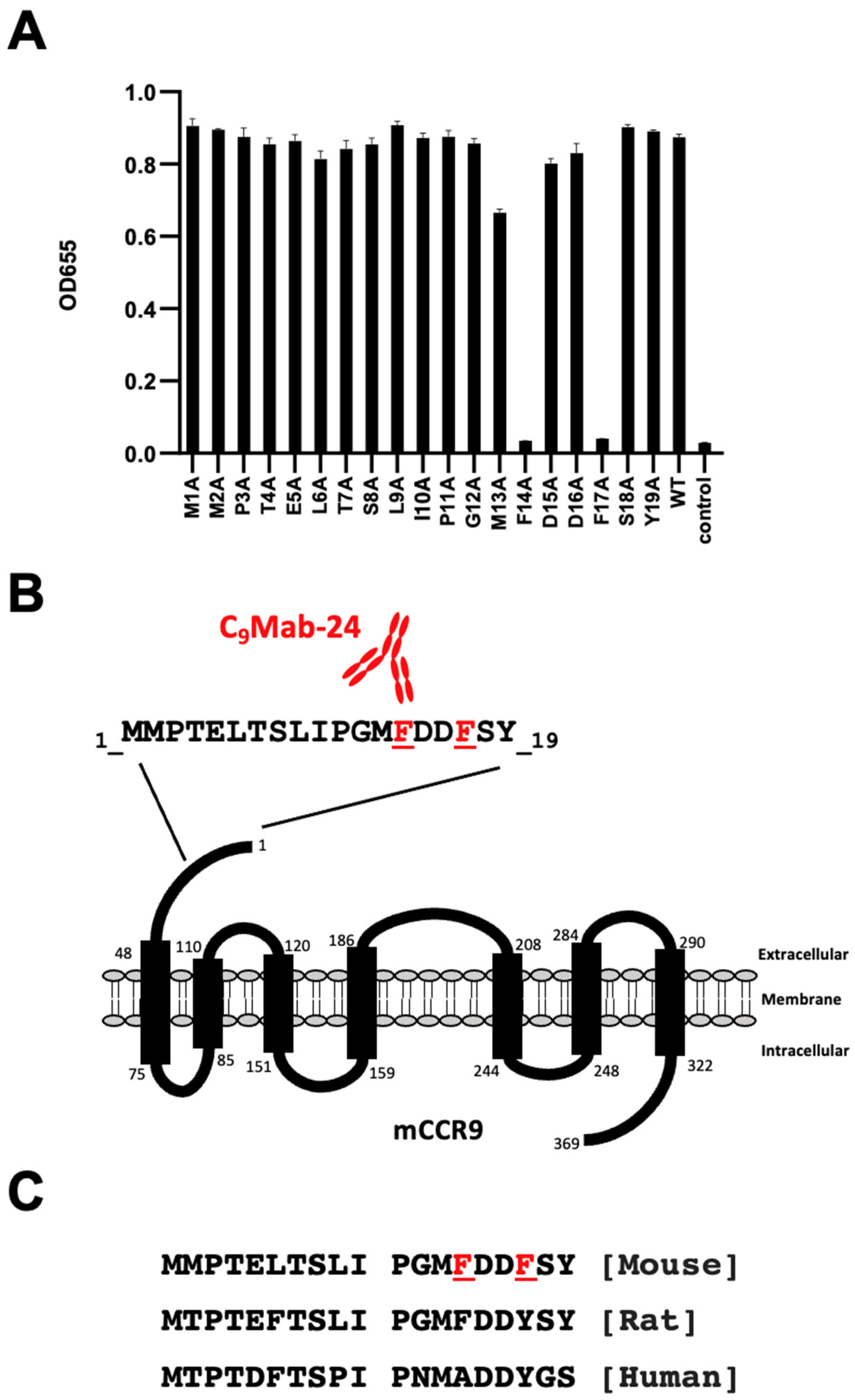
3.1. Epitope Determination Using 1× Ala-substituted mCCR9 Peptides

We previously developed an anti-mCCR9 mAb, C9Mab-24 (rat IgG2a, kappa) using peptide immunization of the mCCR9 N-terminus (amino acids 1–19) as shown in Materials and Methods [32]. C9Mab-24 is very useful for flow cytometry. Herein, we determined the binding epitope of C9Mab-24 using two different alanine scanning strategies via ELISA.
We synthesized 19 different 1× Ala-substituted mCCR9 peptides (Table 1). According to the ELISA results, C9Mab-24 reacted to 1× Ala-substituted peptides, such as M1A, M2A, P3A, T4A, E5A, L6A, T7A, S8A, L9A, I10A, P11A, G12A, M13A, D15A, D16A, S18A, and Y19A, as well as WT (positive control) (Figure 1A). In contrast, C9Mab-24 did not react with 1× Ala-substituted peptides, such as F14A and F17A as well as a negative control (Figure 1A). These results indicate that Phe14 and Phe17 of mCCR9 are the critical residues for the C9Mab-24 binding to mCCR9. Figure 1B summarizes the results. An alignment of mouse, rat, and human CCR9 sequences (residues, 1–19) is shown in Figure 1C.

3.2. Epitope Determination Using 2× Ala-substituted mCCR9 Peptides

The result of 1× Ala substitution showed that Phe14 and Phe17 of mCCR9 are the most critical for the C9Mab-24-mCCR9 interaction, but did not show that only Phe14 and Phe17 of mCCR9 are enough for C9Mab-24 binding to mCCR9. Therefore, we further investigated the C9Mab-24 epitope using 2× Ala-substituted mCCR9 peptides, as described previously [33].
We synthesized 18 different 2× Ala-substituted mCCR9 peptides (Table 2). According to the ELISA results, C9Mab-24 reacted to 2× Ala-substituted peptides, such as M1A–M2A, M2A–P3A, P3A–T4A, T4A–E5A, E5A–L6A, L6A–T7A, T7A–S8A, S8A–L9A, L9A–I10A, I10A–P11A, P11A–G12A, G12A–M13A, and S18A–Y19A as well as WT (positive control) (Figure 2A). In contrast, C9Mab-24 did not react with 2× Ala-substituted peptides, such as M13A–F14A, F14A–D15A, D16A–F17A, and F17A–S18A along with the negative control (Figure 2A). Moreover, the reactivity of C9Mab-24 with D15A–D16A was weaker than that with WT (Figure 2A). These results indicate that 13-MFDDFS-18 is involved in C9Mab-24-binding to mCCR9. Figure 2B summarizes the results.

4. Discussion

The alanine-scanning mutagenesis method was first applied to antibody–antigen interaction in the human growth hormone (hGH) and 21 different anti-hGH mAbs using ELISA [34]. Single alanine mutations were introduced at every residue within the side chains that have been suggested in mAbs recognition. Using the method, the high-resolution “functional epitopes” could be mapped for each mAb [34].
We have established various anti-chemokine receptor mAbs against mouse CCR3 [35,36], mouse CCR8 [37,38,39], and human CCR9 (hCCR9) (clone C9Mab-1) [40] using the Cell-Based Immunization and Screening (CBIS) method. We also identified the epitope of C9Mab-1 in the N-terminal region of hCCR9 [41]. Then, we immunized the peptide of the region, and established a clone, C9Mab-11, which possesses a binding affinity comparable to C9Mab-1 [42]. C9Mab-1 possesses a wider epitope (10-IPNMA-14 and 16-DY-17) [41] than that of C9Mab-11 (11-PNMA-14) (manuscript submitted). In this study, we identified the epitope of the anti-mCCR9 mAb, C9Mab-24, which was established by the N-terminal peptide immunization of mCCR9 [32]. Using the 2× Ala- and 1× Ala-substitution methods, we found that 13-MFDDFS-18 is involved in C9Mab-24-binding to mCCR9, and the phenylalanines at 14 and 17 are critical among the amino acids (Figure 1 and Figure 2). The combination of 2× Ala- and 1× Ala-substitution methods could contribute the determination of the region and center of the mAb epitope.
We previously applied the strategy to identify the epitope of an anti-mouse CXCR6 mAb, Cx6Mab-1 [33]. First, we could not identify the epitope of Cx6Mab-1 by 1× Ala-substitution methods. Next, we performed the 2× Ala-substitution method, and could determine the epitope of Cx6Mab-1. The 2× Ala substitution could be another option to determine the epitope of mAbs if the epitope was not determined by the 1× Ala-substitution method.
The N-terminal region of CCR9 is important for the CCL25 interaction. Anti-hCCR9 mAbs (91R and 92R) recognized the N-terminus of hCCR9, which was partially inhibited in the presence of CCL25 [3,25]. The epitopes of 91R and 92R are located within 11–16 amino acids of hCCR9 [24,25], which is almost identical to the epitopes of C9Mab-1 and C9Mab-11. C9Mab-24 also binds to the N-terminal region of mCCR9 (13-MFDDFS-18). Therefore, both the neutralizing and biological activities of C9Mab-24 should be investigated in the future.
The therapeutic success for refractory childhood leukemia relies on the development of CAR-T targeting B-cell-specific antigen CD19 for B-ALL [43]. Although the therapy targets the common B cell antigen, the background of the success is the ability to tolerate B-cell aplasia. Compared to B-cell aplasia, T-cell aplasia exhibits intolerable immunosuppression. Therefore, a major hurdle for the development of CAR-T cell therapy for T-ALL is the inability to find T-ALL antigens that are not expressed in normal T-cells [44]. To overcome this problem, CCR9 is expected for cell surface antigens unique to T-ALL cells. The CAR-CCR9, a CAR-T with a single-chain variable fragment (scFv) for hCCR9, demonstrated cytotoxicity against CCR9-positive but not CCR9-negative T-ALL cells [21]. Although the epitope of the scFv has not been reported, the investigation of a suitable epitope to exert the CAR-T-mediated cytotoxicity is thought to be important for the future therapeutic applications of our anti-CCR9 mAbs.

Author Contributions

H.K., T.T. and T.A. performed the experiments. M.K.K. and Y.K. designed the experiments. H.K., T.A., H.S. and Y.K. analyzed the data. H.K., T.A., H.S. and Y.K. wrote the manuscript. All authors read and approved the final manuscript and agreed to be accountable for all aspects of the research in ensuring that the accuracy or integrity of any part of the work is appropriately investigated and resolved. All authors have read and agreed to the published version of the manuscript.

Funding

This research was supported in part by the Japan Agency for Medical Research and Development (AMED) under Grant Numbers: JP22ama121008 (to Y.K.), JP22am0401013 (to Y.K.), JP22bm1004001 (to Y.K.), JP22ck0106730 (to Y.K.), and JP21am0101078 (to Y.K.), and by the Japan Society for the Promotion of Science (JSPS) Grants-in-Aid for Scientific Research (KAKENHI) grant nos. 21K20789 (to T.T.), 22K06995 (to H.S.), 21K15523 (to T.A.), 21K07168 (to M.K.K.), and 22K07224 (to Y.K.).

Institutional Review Board Statement

The animal study protocol was approved by the Animal Care and Use Committee of Tohoku University (Permit number: 2019NiA-001) for studies involving animals.

Data Availability Statement

All related data and methods are presented in this paper. Additional inquiries should be addressed to the corresponding authors.

Conflicts of Interest

The authors declare no conflict of interest involving this article.

References

  1. Wermers, J.D.; McNamee, E.N.; Wurbel, M.A.; Jedlicka, P.; Rivera-Nieves, J. The chemokine receptor CCR9 is required for the T-cell-mediated regulation of chronic ileitis in mice. Gastroenterology 2011, 140, 1526–1535. [Google Scholar] [CrossRef] [PubMed]
  2. Wu, W.; Doan, N.; Said, J.; Karunasiri, D.; Pullarkat, S.T. Strong expression of chemokine receptor CCR9 in diffuse large B-cell lymphoma and follicular lymphoma strongly correlates with gastrointestinal involvement. Hum. Pathol. 2014, 45, 1451–1458. [Google Scholar] [CrossRef] [PubMed]
  3. Zabel, B.A.; Agace, W.W.; Campbell, J.J.; Heath, H.M.; Parent, D.; Roberts, A.I.; Ebert, E.C.; Kassam, N.; Qin, S.; Zovko, M.; et al. Human G protein-coupled receptor GPR-9-6/CC chemokine receptor 9 is selectively expressed on intestinal homing T lymphocytes, mucosal lymphocytes, and thymocytes and is required for thymus-expressed chemokine-mediated chemotaxis. J. Exp. Med. 1999, 190, 1241–1256. [Google Scholar] [CrossRef] [PubMed]
  4. Allen, S.J.; Crown, S.E.; Handel, T.M. Chemokine: Receptor structure, interactions, and antagonism. Annu. Rev. Immunol. 2007, 25, 787–820. [Google Scholar] [CrossRef] [PubMed]
  5. Tu, Z.; Xiao, R.; Xiong, J.; Tembo, K.M.; Deng, X.; Xiong, M.; Liu, P.; Wang, M.; Zhang, Q. CCR9 in cancer: Oncogenic role and therapeutic targeting. J. Hematol. Oncol. 2016, 9, 10. [Google Scholar] [CrossRef]
  6. Kunkel, E.J.; Campbell, J.J.; Haraldsen, G.; Pan, J.; Boisvert, J.; Roberts, A.I.; Ebert, E.C.; Vierra, M.A.; Goodman, S.B.; Genovese, M.C.; et al. Lymphocyte CC chemokine receptor 9 and epithelial thymus-expressed chemokine (TECK) expression distinguish the small intestinal immune compartment: Epithelial expression of tissue-specific chemokines as an organizing principle in regional immunity. J. Exp. Med. 2000, 192, 761–768. [Google Scholar] [CrossRef]
  7. Papadakis, K.A.; Prehn, J.; Nelson, V.; Cheng, L.; Binder, S.W.; Ponath, P.D.; Andrew, D.P.; Targan, S.R. The role of thymus-expressed chemokine and its receptor CCR9 on lymphocytes in the regional specialization of the mucosal immune system. J. Immunol. 2000, 165, 5069–5076. [Google Scholar] [CrossRef]
  8. Igaki, K.; Komoike, Y.; Nakamura, Y.; Watanabe, T.; Yamasaki, M.; Fleming, P.; Yang, L.; Soler, D.; Fedyk, E.; Tsuchimori, N. MLN3126, an antagonist of the chemokine receptor CCR9, ameliorates inflammation in a T cell mediated mouse colitis model. Int. Immunopharmacol. 2018, 60, 160–169. [Google Scholar] [CrossRef]
  9. López-Pacheco, C.; Soldevila, G.; Du Pont, G.; Hernández-Pando, R.; García-Zepeda, E.A. CCR9 Is a Key Regulator of Early Phases of Allergic Airway Inflammation. Mediat. Inflamm. 2016, 2016, 3635809. [Google Scholar] [CrossRef]
  10. Wurbel, M.A.; McIntire, M.G.; Dwyer, P.; Fiebiger, E. CCL25/CCR9 interactions regulate large intestinal inflammation in a murine model of acute colitis. PLoS ONE 2011, 6, e16442. [Google Scholar] [CrossRef]
  11. Wurbel, M.A.; Le Bras, S.; Ibourk, M.; Pardo, M.; McIntire, M.G.; Coco, D.; Geha, R.S.; Fiebiger, E.; Snapper, S.B. CCL25/CCR9 interactions are not essential for colitis development but are required for innate immune cell protection from chronic experimental murine colitis. Inflamm. Bowel. Dis. 2014, 20, 1165–1176. [Google Scholar] [CrossRef]
  12. Wendt, E.; Keshav, S. CCR9 antagonism: Potential in the treatment of Inflammatory Bowel Disease. Clin. Exp. Gastroenterol. 2015, 8, 119–130. [Google Scholar] [CrossRef]
  13. Amiya, T.; Nakamoto, N.; Chu, P.S.; Teratani, T.; Nakajima, H.; Fukuchi, Y.; Taniki, N.; Yamaguchi, A.; Shiba, S.; Miyake, R.; et al. Bone marrow-derived macrophages distinct from tissue-resident macrophages play a pivotal role in Concanavalin A-induced murine liver injury via CCR9 axis. Sci. Rep. 2016, 6, 35146. [Google Scholar] [CrossRef]
  14. Nakamoto, N. [Role of inflammatory macrophages and CCR9/CCL25 chemokine axis in the pathogenesis of liver injury as a therapeutic target]. Nihon Rinsho Meneki Gakkai Kaishi 2016, 39, 460–467. [Google Scholar] [CrossRef]
  15. Lu, L.; Du, H.; Huang, H.; Wang, C.; Wang, P.; Zha, Z.; Wu, Y.; Liu, X.; Weng, C.; Fang, X.; et al. CCR9 Promotes Migration and Invasion of Lung Adenocarcinoma Cancer Stem Cells. Int. J. Med. Sci. 2020, 17, 912–920. [Google Scholar] [CrossRef]
  16. Johnson-Holiday, C.; Singh, R.; Johnson, E.; Singh, S.; Stockard, C.R.; Grizzle, W.E.; Lillard, J.W., Jr. CCL25 mediates migration, invasion and matrix metalloproteinase expression by breast cancer cells in a CCR9-dependent fashion. Int. J. Oncol. 2011, 38, 1279–1285. [Google Scholar] [CrossRef]
  17. Zhang, Z.; Sun, T.; Chen, Y.; Gong, S.; Sun, X.; Zou, F.; Peng, R. CCL25/CCR9 Signal Promotes Migration and Invasion in Hepatocellular and Breast Cancer Cell Lines. DNA Cell Biol. 2016, 35, 348–357. [Google Scholar] [CrossRef]
  18. Singh, R.; Stockard, C.R.; Grizzle, W.E.; Lillard, J.W., Jr.; Singh, S. Expression and histopathological correlation of CCR9 and CCL25 in ovarian cancer. Int. J. Oncol. 2011, 39, 373–381. [Google Scholar] [CrossRef]
  19. Fusi, A.; Liu, Z.; Kümmerlen, V.; Nonnemacher, A.; Jeske, J.; Keilholz, U. Expression of chemokine receptors on circulating tumor cells in patients with solid tumors. J. Transl. Med. 2012, 10, 52. [Google Scholar] [CrossRef]
  20. Kühnelt-Leddihn, L.; Müller, H.; Eisendle, K.; Zelger, B.; Weinlich, G. Overexpression of the chemokine receptors CXCR4, CCR7, CCR9, and CCR10 in human primary cutaneous melanoma: A potential prognostic value for CCR7 and CCR10? Arch. Dermatol. Res. 2012, 304, 185–193. [Google Scholar] [CrossRef]
  21. Maciocia, P.M.; Wawrzyniecka, P.A.; Maciocia, N.C.; Burley, A.; Karpanasamy, T.; Devereaux, S.; Hoekx, M.; O’Connor, D.; Leon, T.; Rapoz-D’Silva, T.; et al. Anti-CCR9 chimeric antigen receptor T cells for T-cell acute lymphoblastic leukemia. Blood 2022, 140, 25–37. [Google Scholar] [CrossRef] [PubMed]
  22. Li, B.; Wang, Z.; Zhong, Y.; Lan, J.; Li, X.; Lin, H. CCR9-CCL25 interaction suppresses apoptosis of lung cancer cells by activating the PI3K/Akt pathway. Med. Oncol. 2015, 32, 66. [Google Scholar] [CrossRef] [PubMed]
  23. Shen, X.; Mailey, B.; Ellenhorn, J.D.; Chu, P.G.; Lowy, A.M.; Kim, J. CC chemokine receptor 9 enhances proliferation in pancreatic intraepithelial neoplasia and pancreatic cancer cells. J. Gastrointest. Surg. 2009, 13, 1955–1962. [Google Scholar] [CrossRef] [PubMed]
  24. Somovilla-Crespo, B.; Martín Monzón, M.T.; Vela, M.; Corraliza-Gorjón, I.; Santamaria, S.; Garcia-Sanz, J.A.; Kremer, L. 92R Monoclonal Antibody Inhibits Human CCR9(+) Leukemia Cells Growth in NSG Mice Xenografts. Front. Immunol. 2018, 9, 77. [Google Scholar] [CrossRef]
  25. Chamorro, S.; Vela, M.; Franco-Villanueva, A.; Carramolino, L.; Gutiérrez, J.; Gómez, L.; Lozano, M.; Salvador, B.; García-Gallo, M.; Martínez, A.C.; et al. Antitumor effects of a monoclonal antibody to human CCR9 in leukemia cell xenografts. MAbs 2014, 6, 1000–1012. [Google Scholar] [CrossRef]
  26. Wu, X.; Sun, M.; Yang, Z.; Lu, C.; Wang, Q.; Wang, H.; Deng, C.; Liu, Y.; Yang, Y. The Roles of CCR9/CCL25 in Inflammation and Inflammation-Associated Diseases. Front. Cell Dev. Biol. 2021, 9, 686548. [Google Scholar] [CrossRef]
  27. Tanaka, T.; Li, G.; Asano, T.; Saito, M.; Kaneko, M.K.; Suzuki, H.; Kato, Y. Development of a Novel Anti-Mouse CCR2 Monoclonal Antibody (C(2)Mab-6) by N-Terminal Peptide Immunization. Monoclon. Antibodies Immunodiagn. Immunother. 2022, 41, 80–86. [Google Scholar] [CrossRef]
  28. Asano, T.; Suzuki, H.; Goto, N.; Tanaka, T.; Kaneko, M.K.; Kato, Y. Establishment of Novel Anti-Mouse CCR3 Monoclonal Antibodies (C(3)Mab-6 and C(3)Mab-7) by N-terminal Peptide Immunization. Monoclon. Antibodies Immunodiagn. Immunother. 2022, 41, 94–100. [Google Scholar] [CrossRef]
  29. Takei, J.; Suzuki, H.; Asano, T.; Tanaka, T.; Kaneko, M.K.; Kato, Y. Development of a Novel Anti-Mouse CCR4 Monoclonal Antibody (C(4)Mab-1) by N-Terminal Peptide Immunization. Monoclon. Antibodies Immunodiagn. Immunother. 2022, 41, 87–93. [Google Scholar] [CrossRef]
  30. Asano, T.; Tanaka, T.; Suzuki, H.; Li, G.; Nanamiya, R.; Tateyama, N.; Isoda, Y.; Okada, Y.; Kobayashi, H.; Yoshikawa, T.; et al. Development of a Novel Anti-Mouse CCR6 Monoclonal Antibody (C(6)Mab-13) by N-Terminal Peptide Immunization. Monoclon. Antibodies Immunodiagn. Immunother. 2022, 41, 343–349. [Google Scholar] [CrossRef]
  31. Kitamura, K.; Suzuki, H.; Kaneko, M.K.; Kato, Y. Cx(6)Mab-1: A Novel Anti-Mouse CXCR6 Monoclonal Antibody Established by N-Terminal Peptide Immunization. Monoclon. Antibodies Immunodiagn. Immunother. 2022, 41, 133–141. [Google Scholar] [CrossRef]
  32. Kobayashi, H.; Asano, T.; Suzuki, H.; Tanaka, T.; Yoshikawa, T.; Kaneko, M.K.; Kato, Y. Establishment of a Sensitive Monoclonal Antibody Against Mouse CCR9 (C9Mab-24) for Flow Cytometry. Monoclon. Antibodies Immunodiagn. Immunother. 2022, in press. [Google Scholar] [CrossRef] [PubMed]
  33. Isoda, Y.; Tanaka, T.; Suzuki, H.; Asano, T.; Nakamura, T.; Yanaka, M.; Handa, S.; Komatsu, Y.; Okuno, S.; Takahashi, N.; et al. Epitope Mapping of an Anti-Mouse CXCR6 Monoclonal Antibody (Cx(6)Mab-1) Using the 2 × Alanine Scanning Method. Monoclon. Antibodies Immunodiagn. Immunother. 2022, 41, 275–278. [Google Scholar] [CrossRef]
  34. Jin, L.; Fendly, B.M.; Wells, J.A. High resolution functional analysis of antibody-antigen interactions. J. Mol. Biol. 1992, 226, 851–865. [Google Scholar] [CrossRef]
  35. Asano, T.; Suzuki, H.; Tanaka, T.; Saito, M.; Li, G.; Goto, N.; Nanamiya, R.; Kaneko, M.K.; Kato, Y. C(3)Mab-3: A Monoclonal Antibody for Mouse CC Chemokine Receptor 3 for Flow Cytometry. Monoclon. Antibodies Immunodiagn. Immunother. 2022, 41, 74–79. [Google Scholar] [CrossRef]
  36. Asano, T.; Nanamiya, R.; Takei, J.; Nakamura, T.; Yanaka, M.; Hosono, H.; Tanaka, T.; Sano, M.; Kaneko, M.K.; Kato, Y. Development of Anti-Mouse CC Chemokine Receptor 3 Monoclonal Antibodies for Flow Cytometry. Monoclon. Antibodies Immunodiagn. Immunother. 2021, 40, 107–112. [Google Scholar] [CrossRef]
  37. Suzuki, H.; Saito, M.; Asano, T.; Tanaka, T.; Kitamura, K.; Kudo, Y.; Kaneko, M.K.; Kato, Y. C(8)Mab-3: An Anti-Mouse CCR8 Monoclonal Antibody for Immunocytochemistry. Monoclon. Antibodies Immunodiagn. Immunother. 2022, 41, 110–114. [Google Scholar] [CrossRef]
  38. Saito, M.; Tanaka, T.; Asano, T.; Nakamura, T.; Yanaka, M.; Handa, S.; Komatsu, Y.; Harigae, Y.; Tateyama, N.; Nanamiya, R.; et al. C(8)Mab-2: An Anti-Mouse C-C Motif Chemokine Receptor 8 Monoclonal Antibody for Immunocytochemistry. Monoclon. Antibodies Immunodiagn. Immunother. 2022, 41, 115–119. [Google Scholar] [CrossRef]
  39. Saito, M.; Suzuki, H.; Tanaka, T.; Asano, T.; Kaneko, M.K.; Kato, Y. Development of an Anti-Mouse CCR8 Monoclonal Antibody (C(8)Mab-1) for Flow Cytometry and Immunocytochemistry. Monoclon. Antibodies Immunodiagn. Immunother. 2022, 41, 333–338. [Google Scholar] [CrossRef]
  40. Nanamiya, R.; Takei, J.; Asano, T.; Tanaka, T.; Sano, M.; Nakamura, T.; Yanaka, M.; Hosono, H.; Kaneko, M.K.; Kato, Y. Development of Anti-Human CC Chemokine Receptor 9 Monoclonal Antibodies for Flow Cytometry. Monoclon. Antibodies Immunodiagn. Immunother. 2021, 40, 101–106. [Google Scholar] [CrossRef]
  41. Takei, J.; Asano, T.; Li, G.; Saito, M.; Suzuki, H.; Kaneko, M.K.; Kato, Y. Epitope Mapping of an Anti-Human CCR9 Monoclonal Antibody (C(9)Mab-1) Using Enzyme-Linked Immunosorbent Assay. Monoclon. Antibodies Immunodiagn. Immunother. 2021, 40, 239–242. [Google Scholar] [CrossRef] [PubMed]
  42. Tanaka, T.; Suzuki, H.; Isoda, Y.; Asano, T.; Nakamura, T.; Yanaka, M.; Handa, S.; Takahashi, N.; Okuno, S.; Yoshikawa, T.; et al. Development of a Sensitive Anti-Human CCR9 Monoclonal Antibody (C(9)Mab-11) by N-Terminal Peptide Immunization. Monoclon. Antibodies Immunodiagn. Immunother. 2022, 41, 303–310. [Google Scholar] [CrossRef] [PubMed]
  43. Maude, S.L.; Laetsch, T.W.; Buechner, J.; Rives, S.; Boyer, M.; Bittencourt, H.; Bader, P.; Verneris, M.R.; Stefanski, H.E.; Myers, G.D.; et al. Tisagenlecleucel in Children and Young Adults with B-Cell Lymphoblastic Leukemia. N. Engl. J. Med. 2018, 378, 439–448. [Google Scholar] [CrossRef] [PubMed]
  44. Gower, M.; Tikhonova, A.N. Avoiding fratricide: A T-ALL order. Blood 2022, 140, 3–4. [Google Scholar] [CrossRef]
Figure 1. Determination of the C9Mab-24 epitope for mCCR9 by ELISA using 1× Ala-substituted peptides. (A) Synthesized 1× Ala-substituted peptides of mCCR9 were immobilized on immuno plates. The plates were incubated with C9Mab-24 (1 μg/mL), followed by peroxidase-conjugated anti-rat immunoglobulins. (B) Schematic illustration of mCCR9 and the critical amino acids for C9Mab-24 epitope. (C) An alignment of mouse, rat, and human CCR9 sequences (residues, 1–19).
Antibodies 12 00011 g001
Figure 2. Determination of the C9Mab-24 epitope for mCCR9 by ELISA using 2× Ala-substituted peptides. (A) Synthesized 2× Ala-substituted peptides of mCCR9 were immobilized on immuno plates. The plates were incubated with C9Mab-24 (1 μg/mL), followed by peroxidase-conjugated anti-rat immunoglobulins. (B) Schematic illustration of mCCR9 and the C9Mab-24 epitope region.
Antibodies 12 00011 g002
Table 1. Identification of the C9Mab-24 epitope using 1× Ala-substituted mCCR9 peptides.
PeptidesSequencesC9Mab-24
WTMMPTELTSLIPGMFDDFSY++
M1AAMPTELTSLIPGMFDDFSY++
M2AMAPTELTSLIPGMFDDFSY++
P3AMMATELTSLIPGMFDDFSY++
T4AMMPAELTSLIPGMFDDFSY++
E5AMMPTALTSLIPGMFDDFSY++
L6AMMPTEATSLIPGMFDDFSY++
T7AMMPTELASLIPGMFDDFSY++
S8AMMPTELTALIPGMFDDFSY++
L9AMMPTELTSAIPGMFDDFSY++
I10AMMPTELTSLAPGMFDDFSY++
P11AMMPTELTSLIAGMFDDFSY++
G12AMMPTELTSLIPAMFDDFSY++
M13AMMPTELTSLIPGAFDDFSY++
F14AMMPTELTSLIPGMADDFSY
D15AMMPTELTSLIPGMFADFSY++
D16AMMPTELTSLIPGMFDAFSY++
F17AMMPTELTSLIPGMFDDASY
S18AMMPTELTSLIPGMFDDFAY++
Y19AMMPTELTSLIPGMFDDFSA++
++, OD655 ≥ 0.3; –, OD655 < 0.1.
Table 2. Identification of the C9Mab-24 epitope using 2× Ala-substituted mCCR9 peptides.
PeptidesSequencesC9Mab-24
WTMMPTELTSLIPGMFDDFSY++
M1A–M2AAAPTELTSLIPGMFDDFSY++
M2A–P3AMAATELTSLIPGMFDDFSY++
P3A–T4AMMAAELTSLIPGMFDDFSY++
T4A–E5AMMPAALTSLIPGMFDDFSY++
E5A–L6AMMPTAATSLIPGMFDDFSY++
L6A–T7AMMPTEAASLIPGMFDDFSY++
T7A–S8AMMPTELAALIPGMFDDFSY++
S8A–L9AMMPTELTAAIPGMFDDFSY++
L9A–I10AMMPTELTSAAPGMFDDFSY++
I10A–P11AMMPTELTSLAAGMFDDFSY++
P11A–G12AMMPTELTSLIAAMFDDFSY++
G12A–M13AMMPTELTSLIPAAFDDFSY++
M13A–F14AMMPTELTSLIPGAADDFSY
F14A–D15AMMPTELTSLIPGMAADFSY
D15A–D16AMMPTELTSLIPGMFAAFSY++
D16A–F17AMMPTELTSLIPGMFDAASY
F17A–S18AMMPTELTSLIPGMFDDAAY
S18A–Y19AMMPTELTSLIPGMFDDFAA++
++, OD655 ≥ 0.3; –, OD655 < 0.1.
Disclaimer/Publisher’s Note: The statements, opinions and data contained in all publications are solely those of the individual author(s) and contributor(s) and not of MDPI and/or the editor(s). MDPI and/or the editor(s) disclaim responsibility for any injury to people or property resulting from any ideas, methods, instructions or products referred to in the content.

