Article

Blowin’ in the Wind: Wind Dispersal Ability of Phytopathogenic Fusarium in a Wind Tunnel Experiment

Leibniz Centre for Agricultural Landscape Research (ZALF), 15374 Müncheberg, Germany
*
Author to whom correspondence should be addressed.
Atmosphere 2021, 12(12), 1653; https://doi.org/10.3390/atmos12121653
Submission received: 10 November 2021 / Revised: 6 December 2021 / Accepted: 7 December 2021 / Published: 9 December 2021
(This article belongs to the Special Issue Airborne Fungal and Pteridophyte Spores)

Abstract

:
Dispersal processes play an essential role in cereal diseases caused by phytopathogenic Fusarium. However, most empirical studies of Fusarium spore dispersal have focused on vertical transport by rain splash, while wind dispersal has been mostly neglected. Our objective was to determine the ability of Fusarium conidiospores to disperse via wind under controlled conditions in a wind tunnel study. Ten Fusarium species with diverse spore varieties were studied by placing them in the wind stream at wind velocities of 5 and 8 m s−1 and collecting them after 6 m and a period of 1 h using a newly developed air sampling box. Although spore concentrations were high in the releasing Petri Dishes, the tested isolates were recaptured in only 18 of 78 runs. F. equiseti and F. cerealis were the most frequently recovered species. Changing abiotic conditions, wind speed, and spore shapes had no significant effect on Fusarium spore recapture rates. Another experiment showed that conidiospores were rarely released from the grown mycelium. Therefore, the importance of wind alone as a dispersal medium for Fusarium conidiospores may have been overestimated so far. Further studies should investigate the importance of carrier media or mobile linkers combined with the wind dispersal of spores.

1. Introduction

Phytopathogenic fungi of the genus Fusarium infect wheat fields worldwide and cause one of the most prevalent diseases in cultivation, Fusarium head blight (FHB). Due to its ability to produce mycotoxins, FHB reduces yield and leads to economic losses even after harvest. In addition, Fusarium species overwinter on the crop residues in the field and in the soil, where they can linger for many years [1,2,3]. However, the nature of their dispersal, which is necessary for effective disease management, has not been thoroughly investigated.
Fusarium species are generally distributed across fields and often infest large regions when conditions are suitable [4,5,6,7]. Within large wheat fields, Fusarium species concentrate in areas with a more humid and, thus, usually cooler microclimate [8,9]. Therefore, higher Fusarium species abundances are observed mainly in spots with higher plant productivity and higher canopy cover [8,9].
The primary mechanism of dispersal of Fusarium spores is vertical transport via raindrop splashes [10,11]. Conidiospores are flung upward from the soil and debris onto the wheat plant. They are then carried further by splashes from the leaves to the wheat ears [12,13]. Experiments have shown that splashes can shoot spores up to 100 cm in an upward direction [10,14]. Rossi et al. (2002) conducted a spread of macrospores of Microdochium nivale, F. graminearum, F. avenaceum, F. culmorum, and F. poae in the field [15]. The dispersal was consistently associated with rain events. Therefore, they postulated that the spores were captured in tiny rain droplets and then transported via air turbulence [15].
An additional mechanism of spore dispersal is aerial transport. It has been shown that F. graminearum also utilizes wind dispersal through the production of ascospores [16,17]. Perithecia actively discharge ascospores up to a distance of more than 12 mm [17]. Weather conditions substantially impact the distribution of spores, as precipitation and periods of high humidity promote spore release [15,17,18]. Keller et al. (2011) released a clone of F. graminearum into wheat and barley fields via infected corn stalks [19]. Wheat and barley spikes were later collected at different distances from the inoculation source. The clone was mainly found within the first three meters and still located at over 18 m [20]. Nevertheless, in most Fusarium species, the sexual stage with ascospores is absent, which means they only produce micro and macroconidia [21].
Still, there are no comprehensive studies on exclusive wind dispersal of the conidia types in different Fusarium species. The studies that investigated the dispersal of Fusarium spores focused on spore dissemination at a short-range rather than the actual carriage of spores from a source A to a destination B over longer distances. Thus, the spread of Fusarium, except for F. graminearum, is generally assumed to be solely driven by splashes.
In our study on ten Fusarium species, we aim to determine the capacity of spores to disperse via the wind under controlled conditions. The measurements took place in a wind tunnel, which allowed for the precise setting of operating wind velocities. With the help of a custom-designed air sampler, spore flight was observed for one hour at wind velocities of 5 and 8 m s−1.
Our objective is to determine whether wind plays a relevant role in Fusarium spore dispersal. In addition, we attempted to reveal possible correlations between spore traits and wind dispersal. With the elucidation of wind impact on spore dispersal, we aim to contribute to a better understanding of the interaction in fungal movement processes and their infection behavior. We hope to provide valuable information for future effective crop protection strategies.

2. Materials and Methods

2.1. Cultivation of the Fungal Isolates

The Fusarium strains used in the experiment were obtained from the German Collection of Microorganisms and Cell Cultures (DSMZ, Braunschweig, Germany) and the culture collection of microorganisms at the Leibniz Centre for Agricultural Landscape Research (ZALF, Müncheberg, Germany). Ten different Fusarium strains were screened to cover the diversity of spore forms and sizes. Out of these, three strains came from the DSMZ: F. culmorum (DSM 62191), F. langsethiae (DSM113234), and F. poae (DSM 62376), and seven strains from the ZALF strain collection, all originating from wheat ears from Brandenburg (Germany): F. avenaceum (AH22), F. cerealis (AH28), F. equiseti (F40A), F. graminearum, F. oxysporum, F. sporotrichioides (AH83), and F. tricinctum (F643).
The fungi were grown on Petri Dishes (Ø 9 cm) in preparation for exposure in the wind tunnel. Mycelium and conidia from single-spore stock cultures were placed onto Synthetic Nutrient-Poor Agar (SNA) [22] and allowed to pre-cultivate for four days at 25 °C in the dark. This was followed by ten days of incubation at room temperature under artificial daylight and mixed black light (near UV, emission 310–360 nm) on a 12 h light/dark cycle to support spore formation.
F. graminearum was also used to produce perithecia and associated ascospores. Cultivation was performed on carrot agar according to the protocol of Cavinder et al. (2012) [23].

2.2. Construction of the Wind Tunnel Experiment

The dispersal experiment was conducted in March and April 2020 in the stationary wind tunnel of the ZALF Müncheberg, Germany. It was primarily constructed and used for wind erosion and particulate matter emission studies [24,25]. The wind tunnel can be adjusted to simulate boundary layer conditions of smooth or rough surfaces as common in agriculture, which can be changed in a controlled manner. Thus, in addition to the horizontal component, the airflow also has a turbulence-induced vertical component. The wind tunnel provides further a standardized and reproducible basis for conducting our investigation using unfiltered air taken at a height of 10 m from outside.
We constructed an Air Sampler Box (ASB, Figure 1a) to collect simultaneous airborne spores on three Petri Dishes over a certain period. The ASB works on the principles of an Andersen impactor, by directing the flow directly onto a collection medium, with only one stage but three parallel replications [26]. The air from the wind tunnel was aspirated through three tubes directly into a hermetically sealed box. In the box, the tubes ended above clean Petri Dishes (Figure 1b). A vacuum was set on the box with an airflow rate corresponding to the wind velocity in the tunnel for continuous sampling under isokinetic conditions. This was verified with a hot wire anemometer (Therm 642, LAMBRECHT meteo GmbH, Göttingen, Germany). Since we were interested in showing the possible dispersion and relative differences instead of an exact quantification, this experimental setup met the requirements. The ASB was installed at the end of the wind tunnel measuring section (6 m away from the Petri Dishes: Figure 1).
In order to measure spore release and dispersal, three Petri Dishes containing the same fungal species were placed at the beginning of the seven-meter-long measuring section of the wind tunnel. Spore release was initiated at wind velocities of 5 and 8 m s−1 and measured for each species for one hour and repeated three to four times, respectively. Windy (5 m s−1) and stormy (8 m s−1) conditions, corresponding to the year-round average weather data for Germany, were simulated with the selected wind speeds. The wind speeds were also chosen to produce sufficient friction on the Petri Dishes to release spores by the wind action. The Petri Dishes were placed at an angle of 45° to the air stream resulting in more shear force by direct wind exposure and avoiding wind shadows of the plate edges. Before setting up the Petri Dishes in the wind tunnel, the plates were checked for the presence of spores, and throughout the study period, random plates of the different Fusarium species were washed off and analyzed for spore titer per plate (see Section 2.3)
The wind tunnel chamber was aerated without samples at 8 m s−1 for 15 min between each run. This interval was also used to measure temperature and relative humidity with the Thermo-Hygrometer PCE-313A (PCE Instruments, Meschede, Germany) at the point where the air enters the tunnel. Once a day, a control run without fungi was measured with the ASB to record the background contamination.
The captured fungi, were grown on the inserted Petri Dishes with Potato Dextrose Agar (PDA; ROTH, Karlsruhe, Germany) with 0.4 mg mL−1 chloramphenicol (ROTH, Karlsruhe, Germany). Colony-forming units (cfu) were counted after 24, 48, and 72 h of incubation at room temperature. Fungal genera were determined via macroscopic and microscopic characteristics. In addition, to determine the Fusarium species, colonies were isolated onto Petri Dishes with PDA media to develop the characteristic macroscopic features. Furthermore, mycelium from the isolated fungi was transferred to Petri Dishes with SNA for the ideal spore production, further microscopically determined.

2.3. Comparison of the Spore Concentration on the Plates

For the species F. culmorum, F. graminearum, and F. sporotrichioides, the uniformity of spore distribution on a fully grown SNA plate were investigated. This served as a preliminary test for the idea of studying spore erosion before and after wind tunnel treatment (wtt). Five plates for each species were examined. The plate was divided so that the spore concentrations of the two halves could be compared. The surface mycelium, including the conidia, was removed, mixed with 3 mL of sterile 1/4 strength Ringer’s solution, and filtered through two-layered mull. The suspension concentration was determined using a Thoma counting chamber of 0.1 mm depth (Poly-Optik GmbH, Bad Blankenburg, Germany).

2.4. Comparison of Spore Concentration before and after the Wind Tunnel

In a subsequent test, the extent of spore removal in the wind tunnel was determined under the influence of different operating periods. This was intended to verify the accurate spore removal rate at high wind speed over different time intervals for two selected species. This test was performed for F. graminearum and F. sporotrichioides for a period of 15, 30, 60, and 120 min in replicates of three. Half of the mycelium was initially taken from three Petri Dishes with SNA media containing the same species to determine the spore concentration before the run (see: Section 2.3). The half-covered Petri Dishes were placed in the wind tunnel with a wind velocity of 5 m s−1. After 15, 30, 60, and 120 min, respectively, the plates were brought back to the laboratory to determine the spore concentration of the second half (see: Section 2.3).

2.5. Data Analysis

All data were statistically analyzed with OriginPro (Version 2019b; OriginLab Corporation, Northampton, MA, USA). Correlations between the collected data for temperature, relative humidity, time of measurement, wind speed, total cfu, and Fusarium cfu were evaluated via Pearson Correlation. Therefore, a two-tailed test of significance was used with a significance level of 0.05. A Kruskal-Wallis mean comparison with 0.05 and 0.1 significance levels was performed to detect changes between treatment duration in the wind tunnel.

3. Results

3.1. Spore Dispersal in the Wind Tunnel

3.1.1. Controls in the Wind Tunnel

The daily control runs recorded the airborne microbial load as the air was passed unfiltered into the wind tunnel. The average cfu over the experimental period was 97 cfu per plate, with values ranging from 26 cfu (2 April) to 337 cfu (13 March) per plate (Figure 2). On 13 March, by far the highest number of cfu was counted, but without any apparent explanation. The cfu differed only slightly within a measurement run, with one exception on 27 March.
Among the colonies grown on the trap plates, the following genera were found in varying abundance: Alternaria, Aspergillus, Aureobasidium, Botrytis, Cladosporium, Epicoccum, Mucor, Penicillium, Stachybotrys, Stemphylium, and Verticillium. Fusaria were also detected on the control plates. Based on the study’s research question, Fusarium colonies were counted and identified at the species level. These remained low in abundance compared to the total cfu. No fusaria were caught on numerous days. Single colonies of F. cerealis, F. culmorum, F. oxysporum, and F. solani were counted throughout the experiment. Only F. equiseti occurred slightly more frequently (1–6 cfu of F. equiseti on one plate).

3.1.2. Fusarium Strains in the Wind Tunnel

The sporulation of each species was revised at the beginning of the experiment. A high spore rate was found in all strains used, varying from 1 × 10⁵ conidiospores mL−1 to 5 × 10⁶ conidiospores mL−1. That means a total load of one Petri Dish contained at least 8 × 10⁵ conidiospores. The concentrations of one species always remained in a similar range.
Although the concentration of spores in the Petri Dishes set up was high, the tested Fusarium isolates were recaptured in 18 out of 78 runs. The determination was carried out only at the species level. F. equiseti were the most common species recaptured (14 times, Figure 3), followed by F. cerealis (nine times) and F. graminearum ascospores (5 times). However, considering the low cfu and the Fusarium cfu counted in the control runs, no reliable conclusion can be drawn for spore uptake of the specific isolates. In the majority of the runs, we did not recapture what we fed in. Even an increase in wind speed in the wind tunnel from 5 m s−1 to 8 m s−1 did not increase spores’ transport through the air. Only F. equiseti showed a slightly better spore transport via air at the higher wind speed. Five of the Fusarium species used produce only macroconidia that are longer and heavier than microconidia. Nevertheless, it is striking that the species forming both spore forms (F. langsethiae, F. oxysporum, F. poae, F. tricinctum, F. sporotrichioides) were extremely rarely recovered on the trap plates.
During the entire period, without distinguishing between wtt, control runs, and placed Fusarium isolates, a pattern can be discerned that makes certain species more prominent in wind dispersal. Among the Fusarium species detected, F. equiseti predominated, increasing abundance as the experimental period progressed. In contrast, F. poae, F. arthrosporioides, and F. tricinctum were seen only once or twice during the entire study (Figure 4).

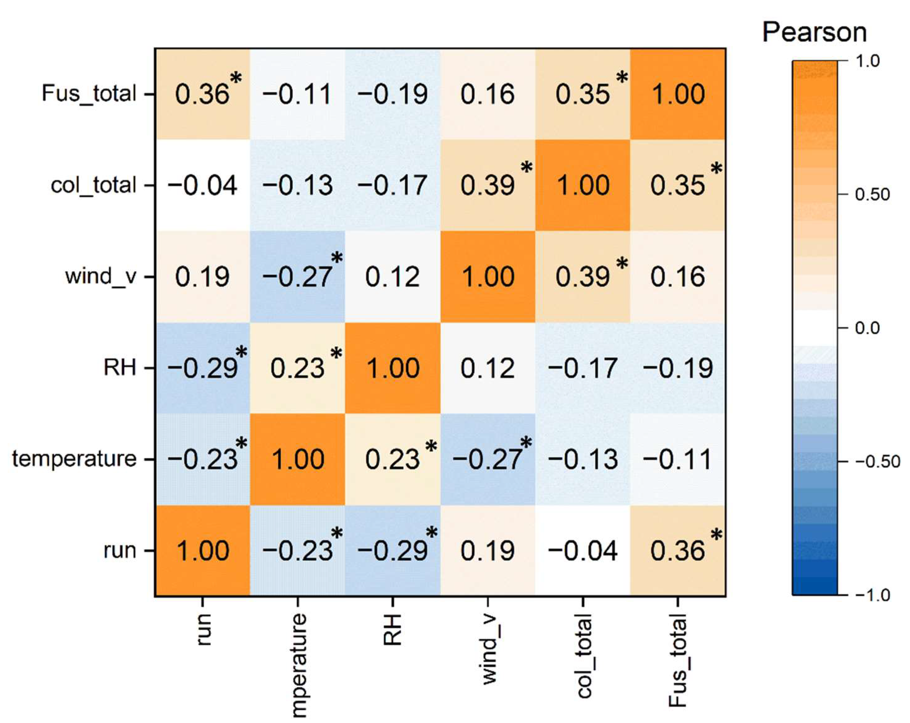
3.2. Correlation of Time, Abiotic Parameters, and cfu

Weather-related variations during the month-long experiment were recorded daily by measuring temperature and relative humidity. A Pearson correlation analysis investigated the extent to which these two factors, the measurement day itself and the wind speed, influenced the cfu caught (Figure 5). The extreme temperature values in the measured period were at a maximum of 15.7 °C and a minimum of 4.4 °C, with a median of 9.2 °C. Relative humidity ranged from 67.50% to 27.97%, with a median of 45.40%. Neither temperature nor relative humidity affected the total number of colonies nor Fusarium colonies. Significant correlations could be found between the progression over time, abiotic variables measured, and Fusarium colony numbers (Figure 5), and the temperature and relative humidity, whereas the number of counted Fusarium cfu rose with progressing time. In addition, a positive correlation between wind speed and total colony count was obtained (0.39, Figure 5).

3.3. Comparison of the Spore Concentration Distribution on the Plates

We tested whether the spore distribution on a Petri Dish is uniform in this experiment. The goal is to study the spore density before and after wind exposure to determine how many spores are released from the Petri Dishes. The sporulation of each species was revised at the beginning of the experiment. A high spore rate was found in all strains used. For three Fusarium species, the distribution on the plate was also examined. The concentrations of one species always remained in a similar range.
F. culmorum and F. sporotrichioides showed spore densities between 1 and 1.8 × 10⁶ conidiospores mL−1 after 14 days of incubation on SNA. F. graminearum, on the other hand, formed between 0.4 to 1.4 × 10⁵ conidiospores mL−1 and was lower by tenfold compared to the other two (Table 1). Spore concentration comparison of both halves of a fungal plate revealed a generally similar spore production with a high abundance of conidiospores on each half side of the plate. Nevertheless, there were also plates on which the distribution of spores was more uneven: The spore density detected varied from −34% to 106% across all three species, with the most variations in F. graminearum.

3.4. Comparison of Spore Concentration before and after the Wind Tunnel

Due to the low recapture rate of the Fusarium strains, an additional setup was used to study the spore removal rate. Despite the occasional uneven distribution of spores on the plates, the influence of the duration of the wtt on spore removal was investigated in this experiment. For this experiment, a Fusarium species that forms only macro-conidiospores (F. graminearum) and a species that forms both macro- and micro-conidiospores (F. sporotrichioides) were chosen.
The initial spore concentration of the first half with F. graminearum was consistent with the size order from the prior experiment (Table 2). It ranged from 0.04 to 3.56 × 10⁵ conidiospores mL−1 after growth on SNA media. F. sporotrichioides ranged from 4.78 to 12.84 × 10⁵ conidiospores mL−1 and confirmed the measured concentrations as well (Appendix A Table A1). There were no distinct concentration differences between the two halves despite the wind exposure of 15–120 min (Kruskal-Wallis, α = 0.05; Table 2). In some cases, the second half had a higher spore concentration. This result is consistent with the finding from Section 3.3 that the spores could be unevenly distributed on the plates.
Accordingly, the wind exposure did not lead to a significant decrease in conidiospore concentration, so we must assume that the airflow at the selected speed could not release spores from the mycelium of the Petri Dishes.

4. Discussion

The objective of the study was to determine the influence of wind on the spore dispersal of Fusaria. Our hypothesis that wind is an important dispersal medium was not supported by our experimental data, where only a small fraction of the Fusarium spores of different species was reliably detected with the established wind tunnel setup. Fusarium spores were detected on the trap plates during the experimental runs despite their poor dispersal. The species F. equiseti and F. cerealis stood out in the studies along with the ascospores of F. graminearum. Moreover, ascospores of F. graminearum were used as controls because of their already known ability to be transported by wind [27]. The total number of 662 Fusarium cfu were counted in the 90 different runs and, thus, about seven Fusarium cfu per run is very low.
Although a quantitative statement on the different spore flight ratios of Fusarium species would have been desirable, this was not the sole aim of this study. Our measurements allowed us to obtain qualitative information on the wind transport of some Fusarium conidiospores over a long distance (6 m). In particular, F. equiseti, F. cerealis, F. avenaceum, and F. culmorum. The ASB recaptured F. graminearum and F. oxysporum (in descending abundance). Given the technical capabilities of the ASB, these are not necessarily poor values. This is due to the efficiency with which it directs air to the trap plates; it is less than commercially available air samplers achieve. Still, it allowed us to perform measurements over a longer period (one hour) than the usual air samplers are designed to support. In addition, the regular use of air samplers only involves collection from the immediate environment but not transport over a defined distance from a source to a destination as in our experimental setup. However, our investigations of spore concentration after wtt showed that only a barely detectable fraction of spores detached from the plates.
To interpret the results appropriately, some physical–technical conditions must also be discussed. This concerns, in particular, the possible concentrations and dilution effects caused by the setup. At the wind velocity of 5 m s−1, an air volume of 8820 m3 (14,112 m3 at 8 m s−1) passes the measuring section of the tunnel within one hour of each run. This corresponds to wind paths of 18 km (5 m s−1) and 28.8 km (8 m s−1) and contrasts to the relatively small source distance of 0.09 m or area of 0.19 m2 of the three Petri dishes. Additionally, vertical dispersion is caused by the turbulent mixture inside the boundary layer, resulting in further dilution across the height of the airflow. Theoretically, the spores are mixed up in the wind tunnel to a height of 0.2 to 0.3 m if we regard them as small enough to equate their exchange behavior to that of momentum. Furthermore, it is not clear whether there was a continuous emission of the spores during the hour-long test runs or whether there was an abrupt emission immediately at the beginning or due to desiccation at an unspecified time.
The ASB is a further source of uncertainty for quantitative evaluations. It was designed according to the principles of an impactor but without prior calibrations of the separation efficiency. Since the deposition behavior can only be approximately described with commercially available impactors, the primary aim was a qualitative detection of the spores. In the case of the ASB design, the trapping efficiency results from a mixture of impaction and adhesion of the spores in the agar on the Petri Dishes. The Reynolds number (Re) of the spores is 6.5, which is exactly in the transition between Stokes’ and Newton’s laws of motion [28]. If all these limitations are considered, these first results from the ASB can be regarded as satisfactory and provide evidence for the airborne transport of spores.
Another limiting factor could be the entanglement of the spores in the mycelium during release. The spores may be released better at a later stage of development when the mycelium is more mature. This would lead to more spores in the mycelium overall, making them easier to expose at the surface. In addition, the mycelium might become drier, more brittle, and weaker overall with time, promoting the release of the spores. In addition, it should be taken into account that we worked with Petri Dishes densely covered with Fusarium. Such compact growth does not occur on plant surfaces or cereal crop residues remaining on the soil surface. However, it appears that a force greater than wind must act on the structures to release the spores.
In the literature, rain events are often described in this context since raindrops have enough force to loosen the spores and transport them vertically by splashing [5,11,29,30]. Different studies have shown that conidiospores are flung upward from the soil and debris onto the wheat plant and are further carried by splashes from the leaves to the wheat ears [12,13]. Jenkinson and Parry [29], as well as Hörberg [11], investigated the influence of raindrops on conidiospore dispersal in a laboratory study. Straw, infected with various Fusarium species (F. avenaceum [29], F. poae [11], and F. culmorum [11,29]), served as the inoculation source. Single water drops were dropped onto the straw from six meters. At different heights and distances, spore traps were installed. Conidiospores could be recaptured at altitudes from 45 cm to 60 cm, which is also reflected in other studies [10,11,14,29]. In addition, the conidiospores could reach a distance of up to 90 cm in the horizontal plane if no obstacles were in the way [11,29]. Although the conidiospores of all three species differed in their morphology, all showed similar distribution patterns.
Rainfall is an important driver in the development of FHB. Moreover, a positive correlation between increased precipitation and subsequent severe infection has been observed [30,31,32,33]. Weather conditions strongly influence spore release, with rainfall and periods of high humidity promoting spore release [15,17,18,34,35]. Nevertheless, our data showed no correlation between relative humidity and cfu counted. This would again indicate the importance of the raindrops as an extracting force. We measured relative humidity around 45% and never exceeded 67%. After a rain event, the relative humidity corresponds to over 80%, which lasts for several hours [2,11,15,18]. In further investigations, air humidification should be considered to create optimal release conditions.
The wind speeds used reflect windy (5 m s−1) to stormy (8 m s−1) conditions. By comparison, the average wind speed in Germany (10 m above the ground) is between 2.5 and 4 m s−1 (Deutscher Wetterdienst, https://www.dwd.de/DE/leistungen/windkarten/deutschland_und_bundeslaender.html, accessed on 8 December 2021). There were no significant differences in the release (see Section 3.4) and capture (see Section 3.1) of the spores as a function of wind velocity. Nevertheless, F. graminearum ascospores were shown to be released at 8 m s−1, which we were able to catch with the ASB. Since wind dispersal of ascospores of F. graminearum has already been demonstrated in several studies [17,35,36,37,38], this served as a positive control in our study. In the two runs at 8 m s−1, F. graminearum could be recaptured in five cfu.
Unlike conidiospores, ascospores are actively discharged from perithecia into the atmosphere [17,39,40] and subsequently passed on by the wind [16,35]. There are numerous studies on the dispersal of ascospores of F. graminearum, both over short distances within a field [18,19,34,35,41] and over long distances [37,42,43,44,45]. Within a field plot experiment from Manstretta et al. (2015), 93% of the total spores caught were ascospores [14]. The results showed a random distribution of ascospores within the wheat canopy, which were still traceable at the height of 90 cm. Markell and Francl (2003) detected twice as many airborne Fusarium ascospores than conidiospores in a fallowed border around a pre-inoculated wheat field [41]. This was also confirmed by a previous study conducted by Reis (1988), who reported that 98% of the spores of F. graminearum captured by spore traps were ascospores [46].
Nevertheless, in most Fusarium species, the sexual stage with ascospores is absent, which means that they only produce micro and macroconidia [21]. A striking aspect of our data analysis is that we barely caught any microspore formers. Microconidia are born in conidiogenous cells and are attached to the phialides in various arrangements. Depending on the species, they may be arranged singly, in chains, or false heads, while their shape and size differ [21]. The microconidia usually only reach a size of around 10 µm and smaller, while the macroconidia are usually between 20 and 60 µm in size [47].
It is also conceivable that mechanical tillage in the spring releases spores from the fungus-infested residues of previous crops that developed during the winter. The spores may bind to particles released by soil erosion and use them as a carrier medium over long distances [48,49,50]. In a collection of 1200 dust samples distributed across America, a total of about 112,000 bacteria and 57,000 fungal phylotypes were identified culture-independently [50]. Dietzel et al. likewise detected fungal plant disease pathogens in 1289 near-surface dust samples in the United States [51]. The most widespread fungal plant disease pathogens in deposited dust were Cladosporium sphaerospermum, Alternaria alternata, and Aureobasidium pullulans. F. oxysporum was also detected in elevated numbers [51]. In our results, F. equiseti and F. oxysporum appeared as the most frequent species. In this context, it is also interesting that both are often found in the soil samples [21,52]. Therefore, a possible link between soil and dust as a transport medium for conidiospores would be conceivable. However, further studies are required to prove this connection.
Our study conducted a unique experiment that tested different Fusarium species for their ability to travel a defined distance by wind. However, the importance of wind alone as a dispersal medium for Fusarium conidiospores has so far been overestimated. Additional forces such as wind turbulence, transport media, or mobile linkers appear to be more effective mechanisms for release and dispersal. Even if our results alone do not provide a clear conclusion, we see our study as a stimulus for further investigations. In particular, the remaining questions around the effect that the carrier medium has on spore dispersal are of great interest.

Author Contributions

Conceptualization, A.H. and M.E.H.M.; Data curation, A.H.; Formal analysis, A.H.; Funding acquisition, M.E.H.M.; Investigation, A.H.; Methodology, A.H. and R.F.; Project administration, M.E.H.M.; Resources, M.E.H.M.; Software, A.H.; Supervision, M.E.H.M.; Validation, A.H.; Visualization, A.H.; Writing—original draft, A.H., R.F., and M.E.H.M.; Writing—review and editing, A.H., R.F., and M.E.H.M. All authors have read and agreed to the published version of the manuscript.

Funding

This work was supported by Deutsche Forschungsgemeinschaft, in the framework of the BioMove Research Training Group (DFG-GRK 2118/1-2).

Institutional Review Board Statement

Not applicable.

Informed Consent Statement

Not applicable.

Data Availability Statement

The datasets generated during and/or analysed during the current study are available from the corresponding author on reasonable request.

Acknowledgments

Special thanks goes to Lina Hempel for her exceptional assistance in the implementation of the experiment. I also thank Steffen Münch for the technical training in the wind tunnel and Niklas Mahlau, Martina Peters and Thomas Müller for excellent technical assistance in the laboratory.

Conflicts of Interest

The authors declare no conflict of interest. The funders had no role in the design of the study; in the collection, analyses, or interpretation of data; in the writing of the manuscript, or in the decision to publish the results.

Appendix A

Table A1. Spore concentration on Fusarium-infected plates before and after wind tunnel treatment (wtt). This test was performed for F. graminearum and F. sporotrichioides for a period of 15, 30, 60, and 120 min in replicates of three. From three Petri Dishes containing the same species, half of the mycelium was taken initially to determine the spore concentration before the run using a Thoma counting chamber. Three half-covered Petri Dishes were placed in the wind tunnel (wt) with wind velocities of 5 m s−1. After 15, 30, 60, and 120 min, respectively, the plates were brought back to the laboratory to determine the spore concentration of the second half. Missing values were noted as “−”.
SpeciesTime in wt (min)Before wtt (×105
Conidiospores mL−1)
After wtt (×105
Conidiospores mL−1)
Percentage Variation (%)
F. graminearum150.310.13−60
0.940.80−15
0.200.50146
0.770.838
1.780.19−89
2.550.67−74
0.751.0439
0.460.29−37
2.680.29−89
302.003.2161
0.090.51467
0.562.20293
0.050.0620
1.330.28−79
0.800.44−45
1.640.30−82
0.020.09350
0.330.09−73
603.561.15−68
0.751.2567
1.000.71−29
0.050.14180
1.722.3335
-0.55
1.510.92−39
0.810.866
0.870.55−37
1200.430.39−9
0.040.17325
0.060.03−50
0.440.27−39
0.530.7541
1.030.88−15
0.110.110
0.630.36−43
0.500.08−84
F. sporotrichioides155.125.7512
4.437.0659
5.036.1522
4.816.3732
7.186.50−9
10.257.43−28
8.696.72−23
8.668.38−3
9.099.565
304.785.7119
5.784.75−18
9.036.50−28
7.417.886
6.169.1849
9.286.76−27
7.509.7830
9.598.81−8
8.036.84−15
606.626.56−1
7.43
10.313.34−68
6.217.5622
6.217.9127
7.596.34−16
7.345.63−23
6.226.342
7.445.69−24
1208.816.81−23
7.725.59−28
8.917.38−17
7.416.22−16
7.726.44−17
6.947.508
6.035.19−14
12.844.97−61
9.509.722

References

  1. Xu, X.; Nicholson, P. Community Ecology of Fungal Pathogens Causing Wheat Head Blight. Annu. Rev. Phytopathol. 2009, 47, 83–103. [Google Scholar] [CrossRef] [PubMed]
  2. Manstretta, V.; Rossi, V. Modelling the effect of weather on moisture fluctuations in maize stalk residues, an important inoculum source for plant diseases. Agric. For. Meteorol. 2015, 207, 83–93. [Google Scholar] [CrossRef]
  3. Maiorano, A.; Blandino, M.; Reyneri, A.; Vanara, F. Effects of maize residues on the Fusarium spp. infection and deoxynivalenol (DON) contamination of wheat grain. Crop Prot. 2008, 27, 182–188. [Google Scholar] [CrossRef]
  4. Pasquali, M.; Beyer, M.; Logrieco, A.; Audenaert, K.; Balmas, V.; Basler, R.; Boutigny, A.L.; Chrpová, J.; Czembor, E.; Gagkaeva, T.; et al. A European Database of Fusarium graminearum and F. culmorum Trichothecene Genotypes. Front. Microbiol. 2016, 7, 406. [Google Scholar] [CrossRef] [PubMed] [Green Version]
  5. Hofgaard, I.S.; Seehusen, T.; Aamot, H.U.; Riley, H.; Razzaghian, J.; Le, V.H.; Hjelkrem, A.-G.R.; Dill-Macky, R.; Brodal, G. Inoculum Potential of Fusarium spp. Relates to Tillage and Straw Management in Norwegian Fields of Spring Oats. Front. Microbiol. 2016, 7, 556. [Google Scholar] [CrossRef] [PubMed] [Green Version]
  6. Ioos, R.; Belhadj, A.; Menez, M. Occurrence and distribution of Microdochium nivale and Fusarium species isolated from barley, durum and soft wheat grains in France from 2000 to 2002. Mycopathologia 2004, 158, 351–362. [Google Scholar] [CrossRef]
  7. Talas, F.; Parzies, H.K.; Miedaner, T. Diversity in genetic structure and chemotype composition of Fusarium graminearum sensu stricto populations causing wheat head blight in individual fields in Germany. Eur. J. Plant Pathol. 2011, 131, 39–48. [Google Scholar] [CrossRef]
  8. Schiro, G.; Verch, G.; Grimm, V.; Müller, M. Alternaria and Fusarium Fungi: Differences in Distribution and Spore Deposition in a Topographically Heterogeneous Wheat Field. J. Fungi 2018, 4, 63. [Google Scholar] [CrossRef] [Green Version]
  9. Schiro, G.; Colangeli, P.; Müller, M.E.H. A Metabarcoding Analysis of the Mycobiome of Wheat Ears Across a Topographically Heterogeneous Field. Front. Microbiol. 2019, 10, 2095. [Google Scholar] [CrossRef]
  10. Paul, P.A.; El-Allaf, S.M.; Lipps, P.E.; Madden, L.V. Rain Splash Dispersal of Gibberella zeae Within Wheat Canopies in Ohio. Phytopathology 2004, 94, 1342–1349. [Google Scholar] [CrossRef] [Green Version]
  11. Hörberg, H.M. Patterns of splash dispersed conidia of Fusarium poae and Fusarium culmorum. Eur. J. Plant Pathol. 2002, 108, 73–80. [Google Scholar] [CrossRef]
  12. Parry, D.W.; Jenkinson, P.; Mcleod, L. Fusarium ear blight (scab) in small grain cereals—A review. Plant Pathol. 1995, 44, 207–238. [Google Scholar] [CrossRef]
  13. Sutton, J.C. Epidemiology of wheat head blight and maize ear rot caused by Fusarium graminearum. Can. J. Plant Pathol. 1982, 4, 195–209. [Google Scholar] [CrossRef]
  14. Manstretta, V.; Gourdain, E.; Rossi, V. Deposition patterns of Fusarium graminearum ascospores and conidia within a wheat canopy. Eur. J. Plant Pathol. 2015, 143, 873–880. [Google Scholar] [CrossRef]
  15. Rossi, V.; Languasco, L.; Pattori, E.; Giosuè, S. Dynamics of Airborne Fusarium Macroconidia in Wheat Fields Naturally Affected by Head Blight. J. Plant Pathol. 2002, 84, 53–64. [Google Scholar] [CrossRef]
  16. Keller, M.D.; Bergstrom, G.C.; Shields, E.J. The aerobiology of Fusarium graminearum. Aerobiologia 2013, 30, 123–136. [Google Scholar] [CrossRef]
  17. David, R.F.; Marr, L.C.; Schmale, D.G., III. Ascospore release and discharge distances of Fusarium graminearum under controlled temperature and relative humidity. Eur. J. Plant Pathol. 2016, 146, 59–69. [Google Scholar] [CrossRef]
  18. Paulitz, T.C. Diurnal Release of Ascospores by Gibberella zeae in Inoculated Wheat Plots. Plant Dis. 1996, 80, 674–678. [Google Scholar] [CrossRef]
  19. Keller, M.D.; Waxman, K.D.; Bergstrom, G.C.; Schmale, D.G., III. Local Distance of Wheat Spike Infection by Released Clones of Gibberella zeae Disseminated from Infested Corn Residue. Plant Dis. 2010, 94, 1151–1155. [Google Scholar] [CrossRef] [Green Version]
  20. Keller, M.D.; Thomason, W.E.; Schmale, D.G., III. The Spread of a Released Clone of Gibberella zeae from Different Amounts of Infested Corn Residue. Plant Dis. 2011, 95, 1458–1464. [Google Scholar] [CrossRef] [Green Version]
  21. Leslie, J.F.; Summerell, B.A. The Fusarium Laboratory Manual; Blackwell Publishing: Ames, IA, USA, 2007; ISBN 0813819199. [Google Scholar]
  22. Nierenberg, H. Untersuchungen über die morphologische und biologische Differenzierung in der Fusarium-Sektion Liseola. In Mitteilungen aus der Biologischen Bundesanstalt für Land-und Forstwirtschaft Berlin-Dahlem—169; Kommissionsverlag Paul Parey: Berlin, Germany; Hamburg, Germany, 1976; ISBN 3-489-16900-X. [Google Scholar]
  23. Cavinder, B.; Sikhakolli, U.; Fellows, K.M.; Trail, F. Sexual Development and Ascospore Discharge in Fusarium graminearum. J. Vis. Exp. 2012, 61, e3895. [Google Scholar] [CrossRef] [PubMed] [Green Version]
  24. Funk, R.; Reuter, H.I.; Hoffmann, C.; Engel, W.; Öttl, D. Effect of moisture on fine dust emission from tillage operations on agricultural soils. Earth Surf. Process. Landf. 2008, 33, 1851–1863. [Google Scholar] [CrossRef]
  25. Funk, R.; Papke, N.; Hör, B. Wind tunnel tests to estimate PM10 and PM2.5-emissions from complex substrates of open-cast strip mines in Germany. Aeolian Res. 2019, 39, 23–32. [Google Scholar] [CrossRef]
  26. Andersen, A.A. New sampler for the collection, sizing, and enumeration of viable airborne particles. J. Bacteriol. 1958, 76, 471–484. [Google Scholar] [CrossRef] [PubMed] [Green Version]
  27. Forrer, H.-R.; Pflugfelder, A.; Musa, T.; Vogelgsang, S. Low-Cost Spore Traps: An Efficient Tool to Manage Fusarium Head Blight through Improved Cropping Systems. Agronomy 2021, 11, 987. [Google Scholar] [CrossRef]
  28. Fischer, M.W.F.; Stolze-Rybczynski, J.L.; Davis, D.J.; Cui, Y.; Money, N.P. Solving the aerodynamics of fungal flight: How air viscosity slows spore motion. Fungal Biol. 2010, 114, 943–948. [Google Scholar] [CrossRef] [PubMed] [Green Version]
  29. Jenkinson, P.; Parry, D.W. Splash dispersal of conidia of Fusarium culmorum and Fusarium avenaceum. Mycol. Res. 1994, 98, 506–510. [Google Scholar] [CrossRef]
  30. Xu, X. Effects of environmental conditions on the development of Fusarium ear blight. Epidemiol. Mycotoxin Prod. Fungi 2003, 109, 683–689. [Google Scholar] [CrossRef]
  31. Birzele, B.; Meier, A.; Hindorf, H.; Krämer, J.; Dehne, H.-W. Epidemiology of Fusarium infection and deoxynivalenol content in winter wheat in the Rhineland, Germany. Mycotoxins Plant Dis. 2002, 108, 667–673. [Google Scholar] [CrossRef]
  32. Champeil, A.; Doré, T.; Fourbet, J.F. Fusarium head blight: Epidemiological origin of the effects of cultural practices on head blight attacks and the production of mycotoxins by Fusarium in wheat grains. Plant Sci. 2004, 166, 1389–1415. [Google Scholar] [CrossRef]
  33. Daamen, R.A.; Langerak, C.J.; Stol, W. Surveys of cereal diseases and pests in the Netherlands. 3. Monographella nivalis and Fusarium spp. in winter wheat fields and seed lots. Neth. J. Plant Pathol. 1991, 97, 105–114. [Google Scholar] [CrossRef]
  34. Fernando, W.G.D.; Miller, J.D.; Seaman, W.L.; Seifert, K.; Paulitz, T.C. Daily and seasonal dynamics of airborne spores of Fusarium graminearum and other Fusarium species sampled over wheat plots. Can. J. Bot. 2000, 78, 497–505. [Google Scholar]
  35. De Luna, L.; Paulitz, T.C.; Bujold, I.; Carisse, O. Ascospore gradients of Gibberella zeae from overwintered inoculum in wheat fields. Can. J. Plant Pathol. 2002, 24, 457–464. [Google Scholar] [CrossRef]
  36. Trail, F. Fungal cannons: Explosive spore discharge in the Ascomycota. FEMS Microbiol. Lett. 2007, 276, 12–18. [Google Scholar] [CrossRef] [Green Version]
  37. Prussin, A.J., II; Li, Q.; Malla, R.; Ross, S.D.; Schmale, D.G., III. Monitoring the Long-Distance Transport of Fusarium graminearum from Field-Scale Sources of Inoculum. Plant Dis. 2014, 98, 504–511. [Google Scholar] [CrossRef] [PubMed] [Green Version]
  38. Golan, J.J.; Pringle, A. Long-Distance Dispersal of Fungi. Microbiol. Spectr. 2017, 5, 1–24. [Google Scholar] [CrossRef] [PubMed] [Green Version]
  39. Elbert, W.; Taylor, P.E.; Andreae, M.O.; Pöschl, U. Contribution of fungi to primary biogenic aerosols in the atmosphere: Wet and dry discharged spores, carbohydrates, and inorganic ions. Eur. Geosci. Union 2007, 7, 4569–4588. [Google Scholar] [CrossRef] [Green Version]
  40. Trail, F.; Xu, H.; Loranger, R.; Gadoury, D. Physiological and environmental aspects of ascospore discharge in Gibberella zeae (anamorph Fusarium graminearum). Mycologia 2002, 94, 181–189. [Google Scholar] [CrossRef] [PubMed]
  41. Markell, S.G.; Francl, L.J. Fusarium Head Blight Inoculum: Species Prevalence and Gibberella zeae Spore Type. Plant Dis. 2003, 87, 814–820. [Google Scholar] [CrossRef] [PubMed] [Green Version]
  42. Del Ponte, E.M.; Shah, D.A.; Bergstrom, G.C. Spatial Patterns of Fusarium Head Blight in New York Wheat Fields Suggest Role of Airborne Inoculum. Plant Health Prog. 2003, 4, 6. [Google Scholar] [CrossRef] [Green Version]
  43. Maldonado-Ramirez, S.L.; Schmale, D.G.; Shields, E.J.; Bergstrom, G.C. The relative abundance of viable spores of Gibberella zeae in the planetary boundary layer suggests the role of long-distance transport in regional epidemics of Fusarium head blight. Agric. For. Meteorol. 2005, 132, 20–27. [Google Scholar] [CrossRef]
  44. Prussin, A.J., II; Marr, L.C.; Schmale, D.G., III; Stoll, R.; Ross, S.D. Experimental validation of a long-distance transport model for plant pathogens: Application to Fusarium graminearum. Agric. For. Meteorol. 2015, 203, 118–130. [Google Scholar] [CrossRef]
  45. Schmale, D.G., III; Ross, S.D.; Fetters, T.L.; Tallapragada, P.; Wood-Jones, A.K.; Dingus, B. Isolates of Fusarium graminearum collected 40–320 meters above ground level cause Fusarium head blight in wheat and produce trichothecene mycotoxins. Aerobiologia 2012, 28, 1–11. [Google Scholar] [CrossRef]
  46. Reis, E. Quantification of propagules of Gibberella zeae in the air by means of spore traps. Fitopatol. Bras. 1988, 13, 324–327. [Google Scholar]
  47. Gerlach, W.; Nirenberg, H. The Genus Fusarium—A Pictorial Atlas. In Mitteilungen aus der Biologischen Bundesanstalt fur Land-und Forstwirtschaft Berlin-Dahlem; Kommissionsverlag Paul Parey: Berlin, Germany; Hamburg, Germany, 1982; ISBN 3-489-20900-1. [Google Scholar]
  48. Acosta-Martínez, V.; Van Pelt, S.; Moore-Kucera, J.; Baddock, M.C.; Zobeck, T.M. Microbiology of wind-eroded sediments: Current knowledge and future research directions. Aeolian Res. 2015, 18, 99–113. [Google Scholar] [CrossRef] [Green Version]
  49. Favet, J.; Lapanje, A.; Giongo, A.; Kennedy, S.; Aung, Y.Y.; Cattaneo, A.; Davis-Richardson, A.G.; Brown, C.T.; Kort, R.; Brumsack, H.J.; et al. Microbial hitchhikers on intercontinental dust: Catching a lift in Chad. ISME J. 2013, 7, 850–867. [Google Scholar] [CrossRef] [PubMed]
  50. Barberán, A.; Ladau, J.; Leff, J.W.; Pollard, K.S.; Menninger, H.L.; Dunn, R.R.; Fierer, N. Continental-scale distributions of dust-associated bacteria and fungi. Proc. Natl. Acad. Sci. USA 2015, 112, 5756–5761. [Google Scholar] [CrossRef] [Green Version]
  51. Dietzel, K.; Valle, D.; Fierer, N.; U’ren, J.M.; Barberán, A. Geographical Distribution of Fungal Plant Pathogens in Dust Across the United States. Front. Ecol. Evol. 2019, 7, 304. [Google Scholar] [CrossRef] [Green Version]
  52. Munkvold, G.P. Fusarium Species and Their Associated Mycotoxins. In Methods in Molecular Biology; Springer: Berlin/Heidelberg, Germany, 2017; Volume 1542, ISBN 9781493967070. [Google Scholar]
Figure 1. Schematic of the wind tunnel setup. At the beginning of the working section, the three Petri Dishes containing the Fusarium species on Synthetic Nutrient-Poor Agar (SNA) to be examined are placed at an angle of 45°. The Air Sampler Box (ASB) is used to capture the spores that are in the air. The ASB is installed at the end of the wind tunnel working section (in 6 m distance to samples). Three tubes direct the air into the box (here elevated tubes for control measurements; during Fusarium wind tunnel treatment (wtt), tubes are at the same height as Petri Dishes); at the end, an adjustable vacuum cleaner is connected (a). Inside the box, the tubes end over sterile Petri Dishes with Potato Dextrose Agar media (PDA) to catch fungal spores (b). With this setup, a total of 10 Fusarium species were tested for wind dispersal at 5 and 8 m s−1 in three to four replicates.
Atmosphere 12 01653 g001
Figure 2. Total colony-forming units (cfu) counts per plate of all control runs held between the main runs once a day over the entire experimental period. Fungi were counted after 24, 48, and 72 h of incubation at room temperature on Potato Dextrose Agar (PDA). Each bar represents one of the three counted trap plates from the Air Sampler Box (ASB) with the corresponding total cfu. The mean cfu was 97 cfu per plate (dotted line) over the testing period.
Atmosphere 12 01653 g002
Figure 3. Recaptured Fusarium species after wind tunnel treatment (wtt) at 5 or 8 m s−1. Ten Fusarium isolates grown on Synthetic Nutrient-Poor Agar (SNA) were placed in the wind tunnel for an hour. Spores were captured from the air with the help of the Air Sampler box (ASB). Each bar represents the sum of cfu of the related Fusarium species caught over the experimental duration. Four replicate runs were performed for the wtt at 5 m s−1 and three replicate runs for 8 m s−1. F. graminearum ascospores wtt were only measured for 8 m s−1 in two repetitive runs.
Atmosphere 12 01653 g003
Figure 4. Identified Fusarium cfu on species level across all measurements (controls and actual dispersal measurements) regardless of wind tunnel treatment (wtt). The bars show the related Fusarium species counted on the three trap plates of the Air Sampler Box (ASB) across all runs. Data were collected over a month and reflected a total of 90 runs.
Atmosphere 12 01653 g004
Figure 5. Pearson correlation matrix between time, abiotic parameters such as temperature and relative humidity in the wind tunnel, the wind speed, and the number of cfu counted in total and caught Fusarium over the whole experimental period. The Pearson correlation coefficient is a value between +1 and −1, where 0 is no correlation at all. The data from 90 runs (controls and actual dispersal measurements) were processed in the analysis. A two-tailed test of significance was used. *: Correlation was significant at the 0.05 level.
Atmosphere 12 01653 g005
Table 1. Conidiospore concentration distribution on plates for three Fusarium species: F. culmorum, F. graminearum, and F. sporotrichioides. After 14 days of incubation on Synthetic Nutrient-Poor Agar (SNA) plates, spore concentrations were determined using a Thoma counting chamber. The plate was divided, and the two halves were counted separately. This experiment was repeated five times for each species. The percent variation was calculated to indicate the concentration differences between the plates.
SpeciesHalf 1 (×105 Conidiospores mL−1)Half 2 (×105 Conidiospores mL−1)Mean (×105 Conidiospores mL−1)Percentage Variation (%)
F. culmorum16.3810.8113.59−34
15.1616.0315.596
18.2518.2518.250
14.0912.8813.48−9
16.6610.3413.50−38
F. graminearum0.410.520.4627
0.561.160.86106
0.671.000.8449
0.920.630.77−32
1.361.361.360
F. sporotrichioides13.7515.9414.8516
15.8717.2516.569
14.8412.2513.55−17
11.8711.0311.45−7
15.4412.2513.85−21
Table 2. Summary of the statistical characteristic values of the tests from Section 3.3 (comparing the spore concentration on Petri Dishes) and Section 3.4 (comparison of spore concentrations before and after wind tunnel treatment). The average values of the respective Fusarium strains divided into their studied halves are shown. Along with the extreme values, the general mean, sample size, and the result of analysis of variance (ANOVA) with Kruskal-Wallis are indicated. Kruskal-Wallis ANOVA was performed at a significance level of 0.05. n.s.: not significant.
TestSpeciesHalf TotalMeanMinMaxKruskal-Wallis ANOVA
test 3.3
comparing spore
concentration on plate
F. culmorum1516.10814.0918.25n.s.
F. culmorum2513.66210.3418.25
F. graminearum150.7840.411.36n.s.
F. graminearum250.9320.521.36
F. sporotrichioides1514.35411.8714.84n.s.
F. sporotrichioides2513.74411.0312.25
test 3.4
spore concentration
after wtt
F. graminearum1350.8410.020.75n.s.
F. graminearum2360.6900.030.47
F. sporotrichioides1367.5584.437.46n.s.
F. sporotrichioides2356.8383.346.56
Publisher’s Note: MDPI stays neutral with regard to jurisdictional claims in published maps and institutional affiliations.

