Article

Insight into the Natural History of Pathogenic Variant c.919-2A>G in the SLC26A4 Gene Involved in Hearing Loss: The Evidence for Its Common Origin in Southern Siberia (Russia)

by
Valeriia Yu. Danilchenko
1,2,
Marina V. Zytsar
1,
Ekaterina A. Maslova
1,2,
Konstantin E. Orishchenko
1,2 and
Olga L. Posukh
1,2,*
1
Federal Research Center Institute of Cytology and Genetics, Siberian Branch of the Russian Academy of Sciences, 630090 Novosibirsk, Russia
2
Novosibirsk State University, 630090 Novosibirsk, Russia
*
Author to whom correspondence should be addressed.
Genes 2023, 14(4), 928; https://doi.org/10.3390/genes14040928
Submission received: 19 March 2023 / Revised: 10 April 2023 / Accepted: 12 April 2023 / Published: 17 April 2023
(This article belongs to the Section Molecular Genetics and Genomics)

Abstract

:
Pathogenic variants in the SLC26A4 gene leading to nonsyndromic recessive deafness (DFNB4), or Pendred syndrome, are some of the most common causes of hearing loss worldwide. Earlier, we found a high proportion of SLC26A4-related hearing loss with prevailing pathogenic variant c.919-2A>G (69.3% among all mutated SLC26A4 alleles that have been identified) in Tuvinian patients belonging to the indigenous Turkic-speaking Siberian people living in the Tyva Republic (Southern Siberia, Russia), which implies a founder effect in the accumulation of c.919-2A>G in Tuvinians. To evaluate a possible common origin of c.919-2A>G, we genotyped polymorphic STR and SNP markers, intragenic and flanking SLC26A4, in patients homozygous for c.919-2A>G and in healthy controls. The common STR and SNP haplotypes carrying c.919-2A>G were revealed, which convincingly indicates the origin of c.919-2A>G from a single ancestor, supporting a crucial role of the founder effect in the c.919-2A>G prevalence in Tuvinians. Comparison analysis with previously published data revealed the identity of the small SNP haplotype (~4.5 kb) in Tuvinian and Han Chinese carriers of c.919-2A>G, which suggests their common origin from founder chromosomes. We assume that c.919-2A>G could have originated in the geographically close territories of China or Tuva and subsequently spread to other regions of Asia. In addition, the time intervals of the c.919-2A>G occurrence in Tuvinians were roughly estimated.

1. Introduction

Pathogenic variants in the SLC26A4 gene (solute carrier family 26, member 4/pendrin, 7q22.3, OMIM 605646) are one of the most common causes of hearing loss worldwide. The SLC26A4 gene (21 exons) encodes the protein pendrin, which is involved in the transport of various anions [1]. High levels of SLC26A4 expression are observed in the inner ear, thyroid, and kidneys [2]. Several hundred pathogenic SLC26A4 variants (the Deafness Variation Database: https://deafnessvariationdatabase.org/gene/SLC26A4, accessed on 13 February 2023) are currently known to be associated with varying phenotypes. They can lead to nonsyndromic recessive deafness (DFNB4) or Pendred syndrome. The DFNB4 (OMIM 600791) is characterized by the prelingual or perilingual onset of sensorineural or mixed hearing loss, which may be fluctuating or progressive. Pendred syndrome (PDS, OMIM 274600) is an autosomal recessive disorder associated with sensorineural hearing loss and goiter. In the inner ear, deficiency or dysfunction of pendrin presumably leads to the development of endolymphatic hydrops due to defects in anion and fluid transport. As a result, SLC26A4-related hearing loss is most commonly accompanied by the enlarged vestibular aqueduct (EVA) and/or other malformations of the inner ear structures [3].
At present, numerous studies aimed to investigate the prevalence of SLC26A4-related hearing loss, as well as the distribution of pathogenic SLC26A4 variants in various regions of the world. The spectrum of SLC26A4 pathogenic variants found in Asian populations appears to differ from that in populations of Caucasian origin: the variants c.919-2A>G and c.2168A>G (p.His723Arg) are the most common in East Asia, while they are very rare or absent in Europe; variants c.1001+1G>A, c.412G>T (p.Val138Phe), c.1246A>C (p.Thr416Pro), c.707T>C (p.Leu236Pro), and c.626G>T (p.Gly209Val) are prevalent in many Caucasian populations [4,5,6,7,8,9,10]. Only several SLC26A4 pathogenic variants (c.1226G>A (p.Arg409His), c.1229C>T (p.Thr410Met), c.1334T>G (p.Leu445Trp), and c.1790T>C (p.Leu597Ser)) are ubiquitously found (with varying frequencies) in all regions of the world. The accumulation of a number of specific pathogenic SLC26A4 variants in certain populations was suggested to be a result of the founder effect, as evidenced by conservation of haplotypes formed by closely linked genetic markers: STRs (short tandem repeats) or SNPs (single nucleotide polymorphisms). Common specific haplotypes were found for several recurrent pathogenic SLC26A4 variants: c.2168A>G (p.His723Arg) in Japanese and Koreans; c.919-2A>G in Chinese [8,11,12]; c.412G>T (p.Val138Phe) in German patients [13]; c.1541A>G (p.Gln514Arg) in Spanish patients [14]; c.965dup (p.Asn322LysfsTer8) in Iranian patients [15]; and c.269C>T (p.Ser90Leu), c.716T>A (p.Val239Asp), and c.1337A>G (p.Gln446Arg) in families from Pakistan [10,16].
The c.919-2A>G variant (rs111033313) was shown to be recurrent in multiple East Asian populations [7,8,11]. This variant (originally named 1143-2A>G, later IVS7-2A>G) was firstly found in a Turkish family with Pendred syndrome [17]. The c.919-2A>G is located at the canonical acceptor splice site -2 in the intron region between exons 7 and 8 and leads to a skipping of exon 8. The deletion of exon 8 generates a new stop codon at position 311, which results in a premature truncated protein of only 310 amino acids [18].
Subsequently, in numerous studies, c.919-2A>G has been frequently identified in patients from Asian countries (mainland China and Taiwan, Mongolia, Korea, and Japan) and observed with the highest frequency in China, while c.919-2A>G is very rare or absent in other countries [4,5,8,12,19,20,21,22,23,24,25,26]. The observed frequencies of c.919-2A>G in global populations, according to the Genome Aggregation Database (gnomAD, https://gnomad.broadinstitute.org/, accessed on 13 February 2023), are as follows: 0.005378 in East Asian; 0.00001470 in European (non-Finnish); and 0.0 in South Asian, European (Finnish), Ashkenazi Jewish, Middle Eastern, Amish, African/African American, Latino/Admixed American, and Other.
In our recent study [27], we performed a thorough analysis of the SLC26A4 gene by Sanger sequencing in the large cohorts of patients with hearing loss belonging to two neighboring indigenous Turkic-speaking Siberian peoples (Tuvinians and Altaians) (in the Tyva Republic and the Altai Republic, Southern Siberia, Russia). We found that 28.2% (62/220) of enrolled Tuvinian patients from the Tyva Republic (Tuva) had biallelic pathogenic SLC26A4 variants. This rate of the SLC26A4-related hearing loss in Tuvinian patients appeared to be one of the highest among populations worldwide. The majority of Tuvinian patients were homozygous or compound heterozygous for c.919-2A>G. The proportion of this variant was 69.3% (95/137) among all SLC26A4 mutant alleles identified in Tuvinian patients, and its carrier frequency in the Tuvinian population was 5.1% (8/157) [27].
A high rate of c.919-2A>G in Tuvinians implies a crucial role of the founder effect in its prevalence in this indigenous Siberian people. In this regard, we aimed to test a presumable common origin of c.919-2A>G in Tuvinians by analyzing the genetic background (haplotypes) of c.919-2A>G in the carriers of this SLC26A4 pathogenic variant.

2. Materials and Methods

2.1. Subjects

Genotyping of genetic markers (STRs and SNPs) for the haplotype analysis was carried out in the sample of unrelated Tuvinian patients with hearing loss who were homozygous for variant c.919-2A>G (n = 23) and in the ethnically matched control sample (Tuvinians), which was represented by unrelated healthy individuals without c.919-2A>G (n = 63). Both samples were formed after our recent analysis of the SLC26A4 gene in Tuvinians belonging to indigenous population of the Tyva Republic (Southern Siberia, Russia) [27].

2.2. Ethics Statement

Written informed consent was obtained from all individuals or their legal guardians before they participated in the study. The study was conducted in accordance with the Declaration of Helsinki, and the protocol was approved by the Bioethics Commission at the Institute of Cytology and Genetics SB RAS, Novosibirsk, Russia (Protocol No. 9, 24 April 2012).

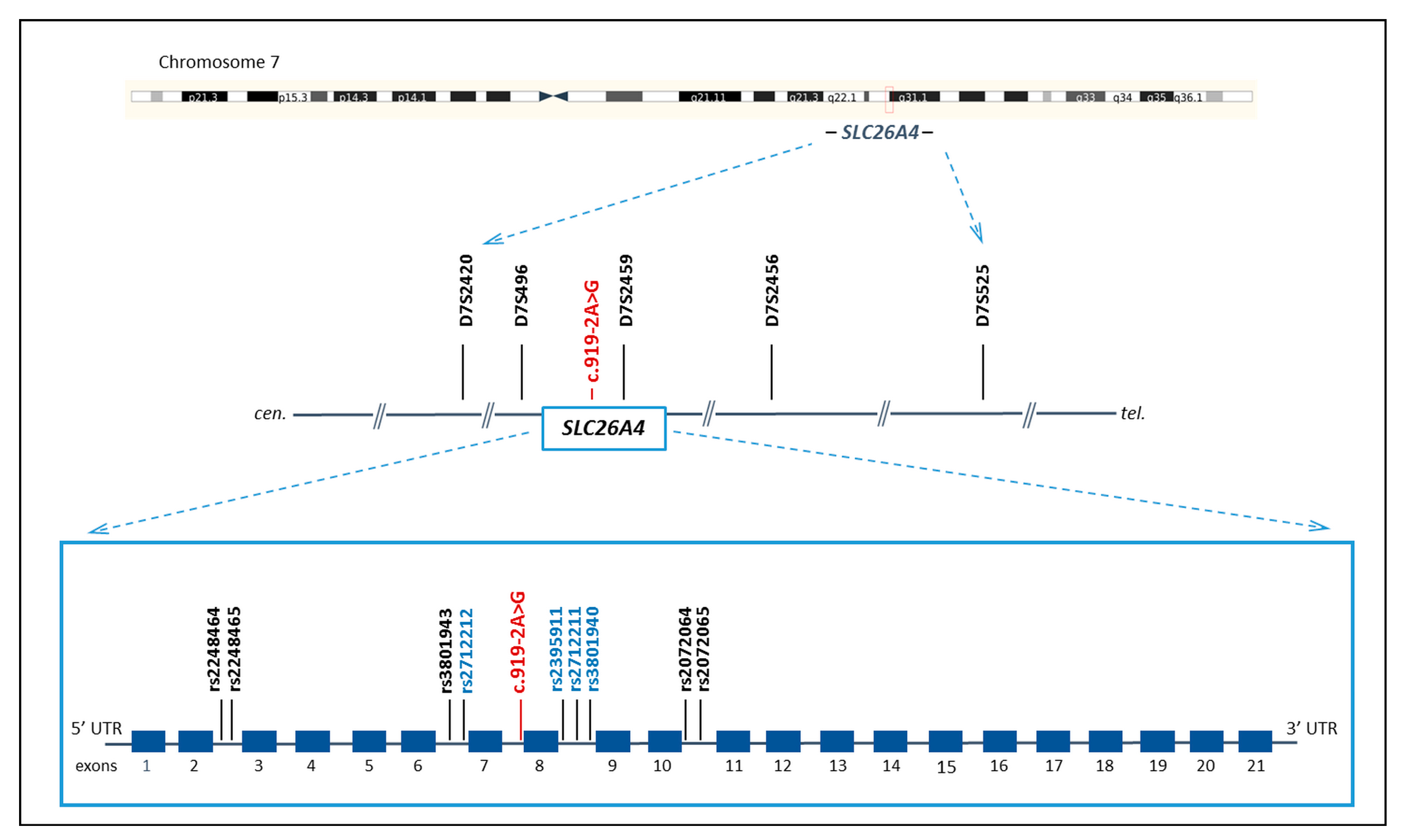
2.3. STRs and SNPs Genotyping

To analyze the c.919-2A>G genetic background, we genotyped five STRs in the region of chromosome 7, including four STRs flanking the SLC26A4 gene at different physical distances: centromeric D7S2420 (~0.43 Mb) and D7S496 (~0.17 Mb); telomeric D7S2456 (~0.36 Mb) and D7S525 (~2.32 Mb); and intragenic D7S2459, located approximately 7.6 kb away from c.919-2A>G. These STRs have been previously used for linkage analysis to define the genetic interval linked to Pendred syndrome or DFNB4 and haplotype analysis in analyzed pedigrees [15,28,29,30,31,32,33]. The total length of the region flanked by distal markers D7S2420 and D7S525 was approximately 2.8 Mb.
To study the fine structure of haplotypes including c.919-2A>G, nine intragenic SNPs (rs2248464, rs2248465, rs3801943, rs2712212, rs2395911, rs2712211, rs3801940, rs2072064, and rs2072065) that closely flanked c.919-2A>G were also genotyped. The choice of analyzed SNPs was based on their physical distances to c.919-2A>G and the variability (a minor allele frequency greater than 0.1) in global populations according to the Genome Aggregation Database (gnomAD, https://gnomad.broadinstitute.org/, accessed on 13 February 2023) (Table S1). Four of them, rs2712212, rs2395911, rs2712211, and rs3801940, were included for comparative analysis with the data from the study by Wu et al. [12], where these SNPs (designated JST160568, JST089508, JST160566, and JST160565, respectively) were used to detect evidence of the founder effect for the c.919-2A>G variant in Taiwanese hearing-impaired patients [12]. The locations and physical distances of analyzed SNPs to c.919-2A>G were as follows: centromeric rs2248464 (intron 2, 20.28 kb), rs2248465 (intron 2, 20.27 kb), rs3801943 (intron 6, 2.23 kb), and rs2712212 (intron 6, 2.18 kb) and telomeric rs2395911 (intron 8, 0.22 kb), rs2712211 (intron 8, 2.02 kb), rs3801940 (intron 8, 2.32 kb), rs2072064 (intron 10, 10.62 kb), and rs2072065 (intron 10, 10.76 kb). The total length of the region flanked by the distal markers rs2248464 and rs2072065 was 31.039 kb.
The location of all analyzed genetic markers (STRs and SNPs) on chromosome 7 is presented in Figure 1.
The STRs genotyping was performed by fragment analysis. To amplify fragments containing STRs, the primer sequences were taken from the Ensembl genome browser (http://www.ensembl.org, accessed on 15 September 2022). One (forward) from each primer pair was marked on the 5′ end by different fluorescent dyes (Applied Biosystems 5´ Labeled/Unlabeled Primer Pairs, Thermo Fisher Scientific, Waltham, MA, USA). The SNP genotyping was performed by Sanger sequencing. To amplify fragments containing SNPs, the primer pairs were designed using Primer Premier 5 tools (https://www.bioprocessonline.com/doc/primer-premier-5-design-program-0001). All used primers are summarized in Table S2. Fragment analysis and Sanger sequencing were performed in the SB RAS Genomics Core Facility (Institute of Chemical Biology and Fundamental Medicine SB RAS, Novosibirsk, Russia).

2.4. Reconstruction of STR and SNP Haplotypes

The STR and SNP genotyping data were used for the reconstruction and calculation of haplotype frequencies performed by the Expectation–Maximization (EM) algorithm of the Arlequin 3.5.1.2 software [34]. Linkage disequilibrium between the STR and SNP alleles and the c.919-2A>G variant was calculated using δ = (Pd − Pn)/(1 − Pn), where δ is the measure of linkage disequilibrium; Pd is the marker allele frequency among mutant chromosomes carrying c.919-2A>G (the sample of patients homozygous for c.919-2A>G); and Pn is the frequency of the same allele among normal chromosomes (control sample) [35].

2.5. Estimation of c.919-2A>G Age

The estimation of the c.919-2A>G age was performed by the DMLE+ v2.3 software method (Disequilibrium Mapping and Likelihood Estimation, http://dmle.org/) [36] and by the single-marker method using the algorithm [37] g = log [1 − Q/(1 − Pn)]/log(1 − Ѳ), where g is the number of generations passed from the moment of the mutation appearance to the present; Q is the share of mutant chromosomes unlinked with the founder haplotype; Pn is the population frequency of allele included in the founder haplotype; and Ѳ is the recombinant fraction calculated from physical distance between marker and mutation (under the assumption that 1 cM = 1000 kb). (See details in Supplementary Materials File S1).

2.6. Statistical Analysis

Fisher’s exact test with a significance level of p < 0.05 was applied to compare the allele and haplotype frequencies between the examined samples of patients and controls.

3. Results

We assumed that the high prevalence of c.919-2A>G in the SLC26A4 gene in Tuvinians is a consequence of the founder effect. To test whether c.919-2A>G shares a common haplotype, we performed genotyping of polymorphic genetic markers: five STRs (four of them are flanking the SLC26A4 gene and one is intragenic) and nine intragenic SNPs closely linked to c.919-2A>G in 23 unrelated Tuvinian patients homozygous for c.919-2A>G. We also genotyped the same genetic markers in 63 healthy unrelated Tuvinians without c.919-2A>G. The results of the STR and SNP genotyping in the sample of the c.919-2A>G homozygotes and in the control sample are summarized in Tables S3 and S4.

3.1. STR and SNP Haplotypes

STR haplotypes. Data on genotyping of five STRs (D7S2420, D7S496, D7S2459, D7S2456, and D7S525) were used to reconstruct STR haplotypes by the Expectation–Maximization (EM) algorithm of the Arlequin 3.5.1.2 software [34] both in Tuvinian deaf patients homozygous for c.919-2A>G and in the ethnically matched control sample. The boundaries of the shared STR haplotypes were determined by observed linkage disequilibrium between certain alleles of distal markers (D7S2420 and D7S525) and c.919-2A>G (Table S3). The total length of the region flanked by D7S2420 and D7S525 is ~2.8 Mb. Four different haplotypes formed by specific alleles of all five STRs were found to be associated with c.919-2A>G in homozygotes for c.919-2A>G, while none of these haplotypes were detected in the control sample (Table 1). Among these haplotypes, the 278-120-147-244-227 haplotype was the most common (91.3%) among mutant chromosomes bearing c.919-2A>G.
SNP haplotypes. The SNP haplotypes were reconstructed based on the results of the genotyping of nine intragenic SNPs closely flanking c.919-2A>G (rs2248464, rs2248465, rs3801943, rs2712212, rs2395911, rs2712211, rs3801940, rs2072064, and rs2072065) (Table 1). Certain alleles of all analyzed SNPs showed strong linkage to c.919-2A>G (Table S4), thereby forming the only specific haplotype A-C-T-A-G-G-C-A-C for all c.919-2A>G carriers (100%), while the frequency of this haplotype reached only 2.8% in the control sample (Table 1).
Four of the SNPs (rs2712212, rs2395911, rs2712211, and rs3801940) were early analyzed in the c.919-2A>G carriers from Taiwan in the study by Wu et al. [12], where the core haplotype T-C-C-G composed of certain alleles of these SNPs (designated in their study as JST160568, JST089508, JST160566, and JST160565, respectively) was revealed in a majority of chromosomes of the c.919-2A>G homozygotes, favoring the origin of c.919-2A>G from a common ancestor. In our study, when considering a haplotype constituted by these SNPs, a single haplotype (corresponding to T-C-C-G in the study by Wu et al. [12]) was found in all homozygotes for c.919-2A>G (100%) (Table 1), while this haplotype was the second by frequency (25.9%) among eight different SNP haplotypes found in the control sample.

3.2. Estimation of c.919-2A>G Age

Common STR and SNP haplotypes found for pathogenic SLC26A4 variant c.919-2A>G, which is predominant in Tuvinians, suggest that c.919-2A>G originated from a single ancestor. We tried to evaluate the approximate “age” of c.919-2A>G by estimation of the numbers of generations (g) and years (with the assumption that g = 25 years) passed from the ancestral mutation event by the single-marker method (when appropriate) and by the DMLE+ v.2.3 program (Supplementary Materials File S1). Allele 227 of the distal STR marker D7S525 (~2.32 Mb from c.919-2A>G), which was found in strong linkage disequilibrium with c.919-2A>G (Table S3), was used when applying the single-marker method. We were not able to apply the single-marker method for SNP markers due to the lack of recombination in all SNPs analyzed (Table S4). The results of the c.919-2A>G age evaluation are summarized in Table 2.
The c.919-2A>G age estimations gave three time intervals depending on different population growth rates (d = 0.05, 0.1, and 0.2) that we applied for calculations (Table 2), thus demonstrating the sensitivity of the methods used from demographic parameters (Supplementary Materials File S1). In addition, we also calculated (using the DMLE+ v.2.3 program) the age of c.919-2A>G based on the SNP internal haplotype A-G-G-C (rs2712212-rs2395911-rs2712211-rs3801940) (Table 1). The variations in the c.919-2A>G age in that case, being 106–182 generations (2650–4550 years), 112–192 (2800–4800 years), and 105–188 generations (2625–4700 years) with d = 0.05, 0.1, and 0.2, respectively, indirectly indicate a more ancient age of this haplotype (Table 2).

4. Discussion

Understanding the regional or ethnospecific landscape of different pathogenic SLC26A4 variants is still far from clear due to the heterogeneity in size and phenotypic characteristics of the examined cohorts of patients and the variable sensitivity of the SLC26A4 molecular diagnostics in different studies. In particular, the proportion of c.919-2A>G, a well-known pathogenic variant, among other mutant alleles of the SLC26A4 gene found in different cohorts of patients with SLC26A4-related hearing loss, remains often uncertain. To assess such data worldwide, we reviewed the literature and selected relevant papers according to the following main criteria: the methodology of the SLC26A4 gene analysis, implying sequencing of all coding exons of SLC26A4 with flanking regions, which allowed us to conclude the presence or absence of c.919-2A>G in the studied patients (at that, more than two unrelated families were studied) and a mandatory indication of the territorial affiliation and/or the ethnicity of patients. In addition, if the required information was not available, we calculated ourselves the proportion of alleles carrying c.919-2A>G among all mutant SLC26A4 alleles identified in patients. Based on the obtained data, we came to the conclusion that the spatial distribution of c.919-2A>G can be limited to the territory of Eurasia, since c.919-2A>G was not found on other world continents, as follows from the relevant studies [38,39,40,41,42,43,44,45,46]. Figure 2 represents a hot map demonstrating the c.919-2A>G prevalence in patients with SLC26A4-related hearing loss in Eurasia.
The highest frequency of c.919-2A>G in patients with hearing loss is observed in China and Mongolia. The SLC26A4 pathogenic variants are the second-most common cause of deafness in China. The data on the c.919-2A>G prevalence were obtained for patients of Han Chinese ethnicity from various regions of China as well as for some minor ethnic groups (Hui, Uighur, Tibetan, Tu, Mongolian, etc.) living in China. Numerous studies revealed that the frequency of c.919-2A>G detected in patients sufficiently exceeds 40%, reaching 60–70% in some regions of China [4,12,47,48,49,50,51,52,53,54,55]. The c.919-2A>G variant is observed with frequency in the range of 60–70% in Mongolian patients from Mongolia and Mongolians living in the northwest of China [22,25]. In Korea and Japan, the c.919-2A>G appears to be the second-most common, by frequency, pathogenic SLC26A4 variant (after c.2168A>G (p.His723Arg)) in patients with hearing loss, and its frequency falls within 20–40% in Korea and 5–10% in Japan, respectively [5,11,19,56,57]. In Thailand, c.919-2A>G was found in one third of all mutated SLC26A4 alleles in a small sample of patients with Pendred syndrome [58]. The c.919-2A>G has also been found in several Iranian families [9], as well as in Turkish families; thus, the “area” of c.919-2A>G apparently extends to Turkey as a result of historical migration of Turks from Central Asia to Anatolia [59].
The detection of c.919-2A>G in multiple patients from different Asian populations suggests that it might have arisen on a common ancestral founder chromosome. To our knowledge, there are only a few studies aimed at confirming this hypothesis by analyzing the genetic background (haplotypes) of c.919-2A>G [11,12,18,60]. The study by Park et al. [11] was the first study where haplotypes bearing c.919-2A>G were analyzed: three STRs (D7S496, D7S2459, and D7S2456) were used for haplotype analysis in several probands of different ethnicities (Korean, Chinese, and Japanese) who were homozygous or heterozygous for c.919-2A>G. The authors did not reveal a strong association of certain STR alleles with c.919-2A>G on different chromosomes and suggested that c.919-2A>G may be an older founder mutation that has undergone ancestral recombination events with the flanking STR markers. Nevertheless, they did not rule out that c.919-2A>G is a hot spot for recurrent mutational events, despite this allele not being observed in western populations [11]. Subsequently, Yang et al. (2005) analyzed the c.919-2A>G associated haplotypes by the genotyping of five STRs (D7S2549, D7S2420, D7S496, D7S2459, and D7S2456) in four Taiwanese families [18]. Haplotype analysis showed a significant haplotype between markers D7S2420 and D7S2456 common to the family members carrying c.919-2A>G, suggesting that they may be derived from a common ancestor [18]. In the study by Reiisi et al. (2014), different STR haplotypes (defined by the specific alleles of D7S2420, D7S496, D7S2459, and D7S2456) were revealed in two Iranian families carrying SLC26A4 variants c.919-2A>G or c.416G>T (p.Gly139Val) in each of them: 2-2-1-2 for the c.919-2A>G-associated haplotype in one family and 1-1-1-1 for the c.416G>T (p.Gly139Val)-associated haplotype in another family [60]. In the study by Wu et al. (2005), the evidence of a common ancestral origin for c.919-2A>G was also obtained, since on the majority of chromosomes with c.919-2A>G in patients homozygous or heterozygous for c.919-2A>G from Taiwan (Han Chinese), the core haplotype consisting of four SNPs closely flanking c.919-2A>G (JST160568, JST089508, JST160566, and JST160565) was revealed [12].
In our recent study [27], we revealed a high rate of the SLC26A4-related hearing loss in Tuvinian patients belonging to indigenous Siberian people living in Southern Siberia (Russia). At that, we found that the frequency of c.919-2A>G reaches 69.3% among all SLC26A4 mutant alleles identified in Tuvinian patients, which allowed us to suggest a role of the founder effect in the accumulation of c.919-2A>G in these indigenous Siberian people.
To evaluate a presumable common origin of c.919-2A>G in Tuvinians, we performed haplotype analysis by the genotyping of polymorphic genetic markers (STRs and SNPs) both within and flanking the SLC26A4 gene in homozygous carriers of this SLC26A4 pathogenic variant. Our choice of analyzed five STRs (D7S2420, D7S496, D7S2459, D7S2456, and D7S525), surrounding c.919-2A>G, was based on their use in previous studies in the haplotype analysis for several recurrent pathogenic SLC26A4 variants: c.707T>C (p.Leu236Pro) and c.1246A>C (p.Thr416Pro) in families originating from Western Europe and the USA [61]; c.2168A>G (p.His723Arg) in Korean and Japanese families, c.2027T>A (p.Leu676Gln) in Mongolian patients, and c.269C>T (p.Ser90Leu) in Pakistani patients [11]; c.412G>T (p.Val138Phe) and c.85G>C (p.Glu29Gln) in German and Danish patients [13,62]; c.919-2A>G in patients of Asian origin [11,18] and in Iranian families [60]; c.1541A>G (p.Gln514Arg) in Spanish patients [14]; c.416G>T (p.Gly139Val) in Iranian families [60]; c.716T>A (p.Val239Asp) in Pakistani and Iranian patients [16,63]; and c.1003T>C (p.Phe335Leu), c.1554G>A (p.Trp518Ter), c.84C>A (p.Ser28Arg), and c.2235+2T>C in Brazilian patients [38]. In addition, in the study by Mojtabavi Naeini et al. [64], the characteristics and the allelic and haplotype frequencies of D7S2420, D7S496, and D7S2459 were examined in five ethnic groups (Fars, Azari, Turkmen, Gilaki, and Arab) of the Iranian population. We revealed the 278-120-147-244-227 haplotype (D7S2420-D7S496-/c.919-2A>G/-D7S2459-D7S2456-D7S525), encompassing about 2.8 Mb, in the majority of mutant chromosomes bearing c.919-2A>G (91.3%) (Table 1). This haplotype, as well as the other three STR haplotypes found in homozygotes for c.919-2A>G, was absent in the control sample, which emphasizes the specificity of the genetic background for c.919-2A>G in Tuvinians.
In addition, we genotyped nine intragenic SNPs flanking c.919-2A>G and found the only haplotype A-C-T-A-G-G-C-A-C constituted by the specific allelic combination of all SNPs (rs2248464-rs2248465-rs3801943-rs2712212-/c.919-2A>G/-rs2395911-rs2712211-rs3801940-rs2072064-rs2072065), encompassing 31.039 kb, in all homozygotes for c.919-2A>G, while the frequency of this haplotype reached only 2.8% in the control sample (Table 1).
Thus, based on the common STR and SNP haplotypes bearing c.919-2A>G found in Tuvinians, we obtained convincing evidence supporting the origin of c.919-2A>G from a single ancestor, and the observed accumulation of c.919-2A>G in this indigenous Siberian people may be explained by the founder effect.
In addition, we roughly estimated the potential time intervals of the c.919-2A>G occurrence in the Tuva. As far as we know, there are no age estimations for any pathogenic variants of the SLC26A4 gene yet, and the age of c.919-2A>G was evaluated by us for the first time. It is worth noting that the methods applied for the estimation of the age of mutation are sensitive to demographic parameters (Supplementary Materials File S1) [36,37,65,66,67]. In view of the lack of reliable data on the variation of the population size of Tuvinians throughout their history, the time of the c.919-2A>G occurrence in the Tuva territory should be considered only as an approximate value. Nevertheless, the partly overlapping time intervals obtained at different population growth rates (d = 0.05, d = 0.1, and d = 0.5) are almost coincided for STR markers (2575–4950 years, 1575–2675 years, and 875–1475 years) and SNP markers (2275–4775 years, 1325–2575 years, and 725–1350 years) (Table 2).
Now, Tuvinians live mainly in the Tyva Republic (Tuva) located in Southern Siberia (Russia), which is bordered by Mongolia in the south and the east. Besides the Tyva Republic, relatively small groups of Tuvinians also live in the northern part of Mongolia and in the Xinjiang Uygur Autonomous Region of China [68,69]. Tuva is located in the geographical center of the Asian continent, and the ancient population of Tuva experienced different gene flows from neighboring regions. At different times, Tuva was at the periphery of a powerful state of Huns (the 2nd century BC—the 1st century AD) or was incorporated in the Ancient Turkic Khaganate (the 6th–8th centuries), the Uyghur Khaganate (the 8th–9th centuries), the Yenisei Kyrgyz Khaganate (the 9th–12th centuries), and also in the Mongol Empire (the 13th–14th centuries). These historical events had a certain impact on the formation of the Tuvinian ethnic group [70,71]. We believe that c.919-2A>G could have appeared in the ancestors of the modern Tuvinian population as a result of different gene flows before the final formation of the Tuvinian ethnos, which was completed by the end of the 13th–14th centuries [70,71].
A very interesting finding of our study was the identity of the “internal” haplotype A-G-G-C (rs2712212-/c.919-2A>G/-rs2395911-rs2712211-rs3801940), encompassing ~4.5 kb, found in the c.919-2A>G homozygotes from Tuva (Tuvinians) and the core haplotype (formed by the same SNPs) in the c.919-2A>G carriers from Taiwan (Han Chinese) [12]. This finding indicates the common ancestor for “Tuvinian” and “Chinese” founder chromosomes with c.919-2A>G. Thus, we speculate that c.919-2A>G could have arisen in the geographically close territories of China or Tuva and subsequently spread to other regions of Asia.

5. Conclusions

The common STR and SNP haplotypes carrying c.919-2A>G, found in Tuvinian patients, convincingly indicate the origin of this SLC26A4 pathogenic variant from a common ancestor that supports a crucial role of the founder effect in the accumulation of c.919-2A>G in the indigenous Siberian people living in Southern Siberia. The identity of small haplotype (~4.5 kb) bearing c.919-2A>G found in Tuvinian and Han Chinese carriers of c.919-2A>G indicates their common founder chromosomes with c.919-2A>G. The SLC26A4 pathogenic variant c.919-2A>G could have arisen in the geographically close territories of China or Tuva and subsequently spread to other regions of Asia.

Supplementary Materials

The following supporting information can be downloaded at https://www.mdpi.com/article/10.3390/genes14040928/s1: Table S1: Allelic frequencies of analyzed SNPs according to data from the Genome Aggregation Database (gnomAD v3.1.2, https://gnomad.broadinstitute.org/); Table S2: Primer sequences for STR and SNP genotyping; Table S3: The allelic frequencies of STRs (D7S2420, D7S496, D7S2459, D7S2456, and D7S525) in patients homozygous for c.919-2A>G and in the control sample; Table S4: The genotypes and the allelic frequencies of analyzed SNPs in patients homozygous for c.919-2A>G and in the control sample; Supplementary Materials File S1: Estimation of the c.919-2A>G age.

Author Contributions

Conceptualization, O.L.P.; Methodology, V.Y.D., O.L.P. and M.V.Z.; Formal analysis, V.Y.D., M.V.Z. and E.A.M.; Investigation, V.Y.D., M.V.Z. and E.A.M.; Resources, O.L.P., M.V.Z. and K.E.O.; Data curation, O.L.P., V.Y.D. and M.V.Z.; Writing—original draft preparation, V.Y.D., M.V.Z. and O.L.P.; Writing—review and editing, V.Y.D., M.V.Z., O.L.P., E.A.M. and K.E.O.; Supervision, O.L.P. All authors have read and agreed to the published version of the manuscript.

Funding

This work was supported by the Russian Science Foundation (grant No. 21-75-00030, https://rscf.ru/en/project/21-75-00030/, to M.V.Z. and E.A.M.); by the projects of the Institute of Cytology and Genetics SB RAS (No. FWNR-2022-0003 to V.Y.D. and No. FWNR-2022-0021 to O.L.P.); and by the Ministry of Education and Science of Russian Federation (grant No. FSUS-2020-0040 to O.L.P. and K.E.O.).

Institutional Review Board Statement

This study was conducted in accordance with the Declaration of Helsinki, and the protocol was approved by the Bioethics Commission at the Institute of Cytology and Genetics SB RAS, Novosibirsk, Russia (Protocol No. 9, 24 April 2012).

Informed Consent Statement

Written informed consent was obtained from all individuals or their legal guardians before they participated in the study.

Data Availability Statement

The data presented in this study are available in this article and Supplementary Materials.

Acknowledgments

The authors are sincerely grateful to all participants of the study. We also thank Bady-Khoo M.S. for help during the recruitment of participants.

Conflicts of Interest

The authors declare no conflict of interest.

References

  1. Everett, L.A.; Glaser, B.; Beck, J.C.; Idol, J.R.; Buchs, A.; Heyman, M.; Adawi, F.; Hazani, E.; Nassir, E.; Baxevanis, A.D.; et al. Pendred syndrome is caused by mutations in a putative sulphate transporter gene (PDS). Nat. Genet. 1997, 17, 411–422. [Google Scholar] [CrossRef]
  2. Everett, L.A.; Morsli, H.; Wu, D.K.; Green, E.D. Expression pattern of the mouse ortholog of the Pendred’s syndrome gene (Pds) suggests a key role for pendrin in the inner ear. Proc. Natl. Acad. Sci. USA 1999, 96, 9727–9732. [Google Scholar] [CrossRef]
  3. Honda, K.; Griffith, A.J. Genetic architecture and phenotypic landscape of SLC26A4-related hearing loss. Hum. Genet. 2022, 141, 455–464. [Google Scholar] [CrossRef]
  4. Albert, S.; Blons, H.; Jonard, L.; Feldmann, D.; Chauvin, P.; Loundon, N.; Sergent-Allaoui, A.; Houang, M.; Joannard, A.; Schmerber, S.; et al. SLC26A4 gene is frequently involved in nonsyndromic hearing impairment with enlarged vestibular aqueduct in Caucasian populations. Eur. J. Hum. Genet. 2006, 14, 773–779. [Google Scholar] [CrossRef] [PubMed]
  5. Du, W.; Guo, Y.; Wang, C.; Wang, Y.; Liu, X. A systematic review and meta-analysis of common mutations of SLC26A4 gene in Asian populations. Int. J. Pediatr. Otorhinolaryngol. 2013, 77, 1670–1676. [Google Scholar] [CrossRef] [PubMed]
  6. Miyagawa, M.; Nishio, S.Y.; Usami, S.; Deafness Gene Study Consortium. Mutation spectrum and genotype-phenotype correlation of hearing loss patients caused by SLC26A4 mutations in the Japanese: A large cohort study. J. Hum. Genet. 2014, 59, 262–268. [Google Scholar] [CrossRef]
  7. Lu, Y.J.; Yao, J.; Wei, Q.J.; Xing, G.Q.; Cao, X. Diagnostic Value of SLC26A4 Mutation Status in Hereditary Hearing Loss With EVA: A PRISMA-Compliant Meta-Analysis. Medicine 2015, 94, e2248. [Google Scholar] [CrossRef]
  8. Tsukada, K.; Nishio, S.Y.; Hattori, M.; Usami, S. Ethnic-specific spectrum of GJB2 and SLC26A4 mutations: Their origin and a literature review. Ann. Otol. Rhinol. Laryngol. 2015, 124 (Suppl. S1), 61S–76S. [Google Scholar] [CrossRef]
  9. Koohiyan, M. A systematic review of SLC26A4 mutations causing hearing loss in the Iranian population. Int. J. Pediatr. Otorhinolaryngol. 2019, 125, 1–5. [Google Scholar] [CrossRef] [PubMed]
  10. Naz, S. Molecular genetic landscape of hereditary hearing loss in Pakistan. Hum. Genet. 2022, 141, 633–648. [Google Scholar] [CrossRef]
  11. Park, H.J.; Shaukat, S.; Liu, X.Z.; Hahn, S.H.; Naz, S.; Ghosh, M.; Kim, H.N.; Moon, S.K.; Abe, S.; Tukamoto, K.; et al. Origins and frequencies of SLC26A4 (PDS) mutations in east and south Asians: Global implications for the epidemiology of deafness. J. Med. Genet. 2003, 40, 242–248. [Google Scholar] [CrossRef] [PubMed]
  12. Wu, C.C.; Yeh, T.H.; Chen, P.J.; Hsu, C.J. Prevalent SLC26A4 mutations in patients with enlarged vestibular aqueduct and/or Mondini dysplasia: A unique spectrum of mutations in Taiwan, including a frequent founder mutation. Laryngoscope 2005, 115, 1060–1064. [Google Scholar] [CrossRef]
  13. Borck, G.; Roth, C.; Martiné, U.; Wildhardt, G.; Pohlenz, J. Mutations in the PDS gene in German families with Pendred’s syndrome: V138F is a founder mutation. J. Clin. Endocrinol. Metab. 2003, 88, 2916–2921. [Google Scholar] [CrossRef]
  14. Pera, A.; Dossena, S.; Rodighiero, S.; Gandía, M.; Bottà, G.; Meyer, G.; Moreno, F.; Nofziger, C.; Hernández-Chico, C.; Paulmichl, M. Functional assessment of allelic variants in the SLC26A4 gene involved in Pendred syndrome and nonsyndromic EVA. Proc. Natl. Acad. Sci. USA 2008, 105, 18608–18613. [Google Scholar] [CrossRef] [PubMed]
  15. Mohseni, M.; Honarpour, A.; Mozafari, R.; Davarnia, B.; Najmabadi, H.; Kahrizi, K. Identification of a founder mutation for Pendred syndrome in families from northwest Iran. Int. J. Pediatr. Otorhinolaryngol. 2014, 78, 1828–1832. [Google Scholar] [CrossRef]
  16. Anwar, S.; Riazuddin, S.; Ahmed, Z.M.; Tasneem, S.; Ateeq-ul-Jaleel; Khan, S.Y.; Griffith, A.J.; Friedman, T.B.; Riazuddin, S. SLC26A4 mutation spectrum associated with DFNB4 deafness and Pendred’s syndrome in Pakistanis. J. Hum. Genet. 2009, 54, 266–270. [Google Scholar] [CrossRef] [PubMed]
  17. Coucke, P.J.; van Hauwe, P.; Everett, L.A.; Demirhan, O.; Kabakkaya, Y.; Dietrich, N.L.; Smith, R.J.; Coyle, E.; Reardon, W.; Trembath, R.; et al. Identification of two different mutations in the PDS gene in an inbred family with Pendred syndrome. J. Med. Genet. 1999, 36, 475–477. [Google Scholar] [PubMed]
  18. Yang, J.J.; Tsai, C.C.; Hsu, H.M.; Shiao, J.Y.; Su, C.C.; Li, S.Y. Hearing loss associated with enlarged vestibular aqueduct and Mondini dysplasia is caused by splice-site mutation in the PDS gene. Hear Res. 2005, 199, 22–30. [Google Scholar] [CrossRef]
  19. Tsukamoto, K.; Suzuki, H.; Harada, D.; Namba, A.; Abe, S.; Usami, S. Distribution and frequencies of PDS (SLC26A4) mutations in Pendred syndrome and nonsyndromic hearing loss associated with enlarged vestibular aqueduct: A unique spectrum of mutations in Japanese. Eur. J. Hum. Genet. 2003, 11, 916–922. [Google Scholar] [CrossRef]
  20. Wang, Q.J.; Zhao, Y.L.; Rao, S.Q.; Guo, Y.F.; Yuan, H.; Zong, L.; Guan, J.; Xu, B.C.; Wang, D.Y.; Han, M.K.; et al. A distinct spectrum of SLC26A4 mutations in patients with enlarged vestibular aqueduct in China. Clin. Genet. 2007, 72, 245–254. [Google Scholar] [CrossRef]
  21. Dai, P.; Li, Q.; Huang, D.; Yuan, Y.; Kang, D.; Miller, D.T.; Shao, H.; Zhu, Q.; He, J.; Yu, F.; et al. SLC26A4 c.919-2A>G varies among Chinese ethnic groups as a cause of hearing loss. Genet. Med. 2008, 10, 586–592. [Google Scholar] [CrossRef] [PubMed]
  22. Yang, X.L.; Bai-Cheng, X.; Chen, X.J.; Pan-Pan, B.; Jian-Li, M.; Xiao-Wen, L.; Zhang, Z.W.; Wan, D.; Zhu, Y.M.; Guo, Y.F. Common molecular etiology of patients with nonsyndromic hearing loss in Tibetan, Tu nationality, and Mongolian patients in the northwest of China. Acta Otolaryngol. 2013, 133, 930–934. [Google Scholar] [CrossRef]
  23. Lee, H.J.; Jung, J.; Shin, J.W.; Song, M.H.; Kim, S.H.; Lee, J.H.; Lee, K.A.; Shin, S.; Kim, U.K.; Bok, J.; et al. Correlation between genotype and phenotype in patients with bi-allelic SLC26A4 mutations. Clin. Genet. 2014, 86, 270–275. [Google Scholar] [CrossRef] [PubMed]
  24. Liu, Y.; Ao, L.; Ding, H.; Zhang, D. Genetic frequencies related to severe or profound sensorineural hearing loss in Inner Mongolia Autonomous Region. Genet. Mol. Biol. 2016, 39, 567–572. [Google Scholar] [CrossRef] [PubMed]
  25. Erdenechuluun, J.; Lin, Y.-H.; Ganbat, K.; Bataakhuu, D.; Makhbal, Z.; Tsai, C.-Y.; Lin, Y.-H.; Chan, Y.-H.; Hsu, C.-J.; Hsu, W.-C.; et al. Unique spectra of deafness-associated mutations in Mongolians provide insights into the genetic relationships among Eurasian populations. PLoS ONE 2018, 13, e0209797. [Google Scholar] [CrossRef]
  26. Wu, C.C.; Tsai, C.Y.; Lin, Y.H.; Chen, P.Y.; Lin, P.H.; Cheng, Y.F.; Wu, C.M.; Lin, Y.H.; Lee, C.Y.; Erdenechuluun, J.; et al. Genetic Epidemiology and Clinical Features of Hereditary Hearing Impairment in the Taiwanese Population. Genes 2019, 10, 772. [Google Scholar] [CrossRef]
  27. Danilchenko, V.Y.; Zytsar, M.V.; Maslova, E.A.; Bady-Khoo, M.S.; Barashkov, N.A.; Morozov, I.V.; Bondar, A.A.; Posukh, O.L. Different Rates of the SLC26A4-Related Hearing Loss in Two Indigenous Peoples of Southern Siberia (Russia). Diagnostics 2021, 11, 2378. [Google Scholar] [CrossRef]
  28. Baldwin, C.T.; Weiss, S.; Farrer, L.A.; De Stefano, A.L.; Adair, R.; Franklyn, B.; Kidd, K.K.; Korostishevsky, M.; Bonné-Tamir, B. Linkage of congenital, recessive deafness (DFNB4) to chromosome 7q31 and evidence for genetic heterogeneity in the Middle Eastern Druze population. Hum. Mol. Genet. 1995, 4, 1637–1642. [Google Scholar] [CrossRef]
  29. Coucke, P.; Van Camp, G.; Demirhan, O.; Kabakkaya, Y.; Balemans, W.; Van Hauwe, P.; Van Agtmael, T.; Smith, R.J.; Parving, A.; Bolder, C.H.; et al. The gene for Pendred syndrome is located between D7S501 and D7S692 in a 1.7-cM region on chromosome 7q. Genomics 1997, 40, 48–54. [Google Scholar] [CrossRef]
  30. Gausden, E.; Coyle, B.; Armour, J.A.; Coffey, R.; Grossman, A.; Fraser, G.R.; Winter, R.M.; Pembrey, M.E.; Kendall-Taylor, P.; Stephens, D.; et al. Pendred syndrome: Evidence for genetic homogeneity and further refinement of linkage. J. Med. Genet. 1997, 34, 126–129. [Google Scholar] [CrossRef]
  31. López-Bigas, N.; Rabionet, R.; de Cid, R.; Govea, N.; Gasparini, P.; Zelante, L.; Arbonés, M.L.; Estivill, X. Splice-site mutation in the PDS gene may result in intrafamilial variability for deafness in Pendred syndrome. Hum. Mutat. 1999, 14, 520–526. [Google Scholar] [CrossRef]
  32. Gonzalez Trevino, O.; Karamanoglu Arseven, O.; Ceballos, C.J.; Vives, V.I.; Ramirez, R.C.; Gomez, V.V.; Medeiros-Neto, G.; Kopp, P. Clinical and molecular analysis of three Mexican families with Pendred’s syndrome. Eur. J. Endocrinol. 2001, 144, 585–593. [Google Scholar] [CrossRef] [PubMed]
  33. Yazdanpanahi, N.; Tabatabaiefar, M.A.; Bagheri, N.; Azadegan Dehkordi, F.; Farrokhi, E.; Hashemzadeh Chaleshtori, M. The role and spectrum of SLC26A4 mutations in Iranian patients with autosomal recessive hereditary deafness. Int. J. Audiol. 2015, 54, 124–130. [Google Scholar] [CrossRef] [PubMed]
  34. Excoffier, L.; Lischer, H.E. Arlequin suite ver 3.5: A new series of programs to perform population genetics analyses under Linux and Windows. Mol. Ecol. Resour. 2010, 10, 564–567. [Google Scholar] [CrossRef]
  35. Bengtsson, B.O.; Thomson, G. Measuring the strength of associations between HLA antigens and diseases. Tissue Antigens 1981, 18, 356–363. [Google Scholar] [CrossRef]
  36. Rannala, B.; Bertorelle, G. Using linked markers to infer the age of a mutation. Hum. Mutat. 2001, 18, 87–100. [Google Scholar] [CrossRef] [PubMed]
  37. Risch, N.; de Leon, D.; Ozelius, L.; Kramer, P.; Almasy, L.; Singer, B.; Fahn, S.; Breakefield, X.; Bressman, S. Genetic analysis of idiopathic torsion dystonia in Ashkenazi Jews and their recent descent from a small founder population. Nat. Genet. 1995, 9, 152–159. [Google Scholar] [CrossRef]
  38. Nonose, R.W.; Lezirovitz, K.; de Mello Auricchio, M.T.B.; Batissoco, A.C.; Yamamoto, G.L.; Mingroni-Netto, R.C. Mutation analysis of SLC26A4 (Pendrin) gene in a Brazilian sample of hearing-impaired subjects. BMC Med. Genet. 2018, 19, 73. [Google Scholar] [CrossRef]
  39. Pryor, S.P.; Madeo, A.C.; Reynolds, J.C.; Sarlis, N.J.; Arnos, K.S.; Nance, W.E.; Yang, Y.; Zalewski, C.K.; Brewer, C.C.; Butman, J.A.; et al. SLC26A4/PDS genotype-phenotype correlation in hearing loss with enlargement of the vestibular aqueduct (EVA): Evidence that Pendred syndrome and non-syndromic EVA are distinct clinical and genetic entities. J. Med. Genet. 2005, 42, 159–165. [Google Scholar] [CrossRef]
  40. Dai, P.; Stewart, A.K.; Chebib, F.; Hsu, A.; Rozenfeld, J.; Huang, D.; Kang, D.; Lip, V.; Fang, H.; Shao, H.; et al. Distinct and novel SLC26A4/Pendrin mutations in Chinese and U.S. patients with nonsyndromic hearing loss. Physiol. Genom. 2009, 38, 281–290. [Google Scholar] [CrossRef]
  41. Dahl, H.H.; Ching, T.Y.; Hutchison, W.; Hou, S.; Seeto, M.; Sjahalam-King, J. Etiology and audiological outcomes at 3 years for 364 children in Australia. PLoS ONE 2013, 8, e59624. [Google Scholar] [CrossRef] [PubMed]
  42. de Moraes, V.C.; dos Santos, N.Z.; Ramos, P.Z.; Svidnicki, M.C.; Castilho, A.M.; Sartorato, E.L. Molecular analysis of SLC26A4 gene in patients with nonsyndromic hearing loss and EVA: Identification of two novel mutations in Brazilian patients. Int. J. Pediatr. Otorhinolaryngol. 2013, 77, 410–413. [Google Scholar] [CrossRef] [PubMed]
  43. Carvalho, S.D.C.E.S.; Grangeiro, C.H.P.; Picanço-Albuquerque, C.G.; Dos Anjos, T.O.; De Molfetta, G.A.; Silva, W.A., Jr.; Ferraz, V.E.F. Contribution of SLC26A4 to the molecular diagnosis of nonsyndromic prelingual sensorineural hearing loss in a Brazilian cohort. BMC Res. Notes 2018, 11, 546. [Google Scholar] [CrossRef] [PubMed]
  44. Talbi, S.; Bonnet, C.; Riahi, Z.; Boudjenah, F.; Dahmani, M.; Hardelin, J.P.; Wong Jun Tai, F.; Louha, M.; Ammar-Khodja, F.; Petit, C. Genetic heterogeneity of congenital hearing impairment in Algerians from the Ghardaïa province. Int. J. Pediatr. Otorhinolaryngol. 2018, 112, 1–5. [Google Scholar] [CrossRef] [PubMed]
  45. Torre-González, C.; Villanueva-García, D.; García-Delgado, C.; Castillo-Castillo, S.; Huante-Guido, M.; Chichitz-Madrigal, J.; Juárez-Torres, M.E.; Sánchez-Sandoval, A.L.; Barrón-Palma, E.V.; Morán-Barroso, V.F. Congenital hearing loss: A literature review of the genetic etiology in a Mexican population. Bol. Med. Hosp. Infant. Mex. 2022, 79, 206–214. [Google Scholar] [CrossRef]
  46. Wonkam, A.; Adadey, S.M.; Schrauwen, I.; Aboagye, E.T.; Wonkam-Tingang, E.; Esoh, K.; Popel, K.; Manyisa, N.; Jonas, M.; deKock, C.; et al. Exome sequencing of families from Ghana reveals known and candidate hearing impairment genes. Commun. Biol. 2022, 5, 369. [Google Scholar] [CrossRef] [PubMed]
  47. Yuan, Y.; Guo, W.; Tang, J.; Zhang, G.; Wang, G.; Han, M.; Zhang, X.; Yang, S.; He, D.Z.; Dai, P. Molecular epidemiology and functional assessment of novel allelic variants of SLC26A4 in non-syndromic hearing loss patients with enlarged vestibular aqueduct in China. PLoS ONE 2012, 7, e49984. [Google Scholar] [CrossRef] [PubMed]
  48. Dai, P.; Yuan, Y.; Huang, D.; Zhu, X.; Yu, F.; Kang, D.; Yuan, H.; Wu, B.; Han, D.; Wong, L.J. Molecular etiology of hearing impairment in Inner Mongolia: Mutations in SLC26A4 gene and relevant phenotype analysis. J. Transl. Med. 2008, 6, 74. [Google Scholar] [CrossRef]
  49. Chai, Y.; Huang, Z.; Tao, Z.; Li, X.; Li, L.; Li, Y.; Wu, H.; Yang, T. Molecular etiology of hearing impairment associated with nonsyndromic enlarged vestibular aqueduct in East China. Am. J. Med. Genet. A 2013, 161A, 2226–2233. [Google Scholar] [CrossRef]
  50. Xin, F.; Yuan, Y.; Deng, X.; Han, M.; Wang, G.; Zhao, J.; Gao, X.; Liu, J.; Yu, F.; Han, D.; et al. Genetic mutations in nonsyndromic deafness patients of Chinese minority and Han ethnicities in Yunnan, China. J. Transl. Med. 2013, 11, 312. [Google Scholar] [CrossRef]
  51. Chen, K.; Zong, L.; Liu, M.; Wang, X.; Zhou, W.; Zhan, Y.; Cao, H.; Dong, C.; Tang, H.; Jiang, H. Developing regional genetic counseling for southern Chinese with nonsyndromic hearing impairment: A unique mutational spectrum. J. Transl. Med. 2014, 12, 64. [Google Scholar] [CrossRef] [PubMed]
  52. Du, W.; Wang, Q.; Zhu, Y.; Wang, Y.; Guo, Y. Associations between GJB2, mitochondrial 12S rRNA, SLC26A4 mutations, and hearing loss among three ethnicities. Biomed. Res. Int. 2014, 2014, 746838. [Google Scholar] [CrossRef] [PubMed]
  53. Duan, S.H.; Zhu, Y.M.; Wang, Y.L.; Guo, Y.F. Common molecular etiology of nonsyndromic hearing loss in 484 patients of 3 ethnicities in northwest China. Acta Otolaryngol. 2015, 135, 586–591. [Google Scholar] [CrossRef] [PubMed]
  54. Huang, B.; Han, M.; Wang, G.; Huang, S.; Zeng, J.; Yuan, Y.; Dai, P. Genetic mutations in non-syndromic deafness patients in Hainan Province have a different mutational spectrum compared to patients from Mainland China. Int. J. Pediatr. Otorhinolaryngol. 2018, 108, 49–54. [Google Scholar] [CrossRef] [PubMed]
  55. Wu, L.; Liu, Y.; Wu, J.; Chen, S.; Tang, S.; Jiang, Y.; Dai, P. Study on the relationship between the pathogenic mutations of SLC26A4 and CT phenotypes of inner ear in patient with sensorineural hearing loss. Biosci. Rep. 2019, 39, BSR20182241. [Google Scholar] [CrossRef]
  56. Park, H.J.; Lee, S.J.; Jin, H.S.; Lee, J.O.; Go, S.H.; Jang, H.S.; Moon, S.K.; Lee, S.C.; Chun, Y.M.; Lee, H.K.; et al. Genetic basis of hearing loss associated with enlarged vestibular aqueducts in Koreans. Clin. Genet. 2005, 67, 160–165. [Google Scholar] [CrossRef]
  57. Rah, Y.C.; Kim, A.R.; Koo, J.W.; Lee, J.H.; Oh, S.H.; Choi, B.Y. Audiologic presentation of enlargement of the vestibular aqueduct according to the SLC26A4 genotypes. Laryngoscope 2015, 125, E216–E222. [Google Scholar] [CrossRef]
  58. Snabboon, T.; Plengpanich, W.; Saengpanich, S.; Sirisalipoch, S.; Keelawat, S.; Sunthornyothin, S.; Khovidhunkit, W.; Suwanwalaikorn, S.; Sridama, V.; Shotelersuk, V. Two common and three novel PDS mutations in Thai patients with Pendred syndrome. J. Endocrinol. Investig. 2007, 30, 907–913. [Google Scholar] [CrossRef]
  59. Cengiz, F.B.; Yilmazer, R.; Olgun, L.; Sennaroglu, L.; Kirazli, T.; Alper, H.; Olgun, Y.; Incesulu, A.; Atik, T.; Huesca-Hernandez, F.; et al. Novel pathogenic variants underlie SLC26A4-related hearing loss in a multiethnic cohort. Int. J. Pediatr. Otorhinolaryngol. 2017, 101, 167–171. [Google Scholar] [CrossRef]
  60. Reiisi, S.; Sanati, M.H.; Tabatabaiefar, M.A.; Ahmadian, S.; Reiisi, S.; Parchami, S.; Porjafari, H.; Shahi, H.; Shavarzi, A.; Hashemzade Chaleshtori, M. The Study of SLC26A4 Gene Causing Autosomal Recessive Hearing Loss by Linkage Analysis in a Cohort of Iranian Populations. Int. J. Mol. Cell. Med. 2014, 3, 176–182. [Google Scholar]
  61. van Hauwe, P.; Everett, L.A.; Coucke, P.; Scott, D.A.; Kraft, M.L.; Ris-Stalpers, C.; Bolder, C.; Otten, B.; de Vijlder, J.J.; Dietrich, N.L.; et al. Two frequent missense mutations in Pendred syndrome. Hum. Mol. Genet. 1998, 7, 1099–1104. [Google Scholar] [CrossRef] [PubMed]
  62. Rendtorff, N.D.; Schrijver, I.; Lodahl, M.; Rodriguez-Paris, J.; Johnsen, T.; Hansén, E.C.; Nickelsen, L.A.; Tümer, Z.; Fagerheim, T.; Wetke, R.; et al. SLC26A4 mutation frequency and spectrum in 109 Danish Pendred syndrome/DFNB4 probands and a report of nine novel mutations. Clin. Genet. 2013, 84, 388–391. [Google Scholar] [CrossRef] [PubMed]
  63. Azadegan-Dehkordi, F.; Ahmadi, R.; Bahrami, T.; Yazdanpanahi, N.; Farrokhi, E.; Tabatabaiefar, M.A.; Hashemzadeh-Chaleshtori, M. A novel variant of SLC26A4 and first report of the c.716T>A variant in Iranian pedigrees with non-syndromic sensorineural hearing loss. Am. J. Otolaryngol. 2018, 39, 719–725. [Google Scholar] [CrossRef] [PubMed]
  64. Mojtabavi Naeini, M.; Mesrian Tanha, H.; Hashemzadeh Chaleshtori, M.; Vallian, S. Genotyping data and novel haplotype diversity of STR markers in the SLC26A4 gene region in five ethnic groups of the Iranian population. Genet. Test. Mol. Biomarkers 2014, 18, 820–825. [Google Scholar] [CrossRef]
  65. Slatkin, M.; Rannala, B. Estimating allele age. Annu. Rev. Genom. Hum. Genet. 2000, 1, 225–249. [Google Scholar] [CrossRef]
  66. Labuda, D.; Zietkiewicz, E.; Labuda, M. The genetic clock and the age of the founder effect in growing populations: A lesson from French Canadians and Ashkenazim. Am. J. Hum. Genet. 1997, 61, 768–771. [Google Scholar] [CrossRef]
  67. Colombo, R. Age estimate of the N370S mutation causing Gaucher disease in Ashkenazi Jews and European populations: A reappraisal of haplotype data. Am. J. Hum. Genet. 2000, 66, 692–697. [Google Scholar] [CrossRef]
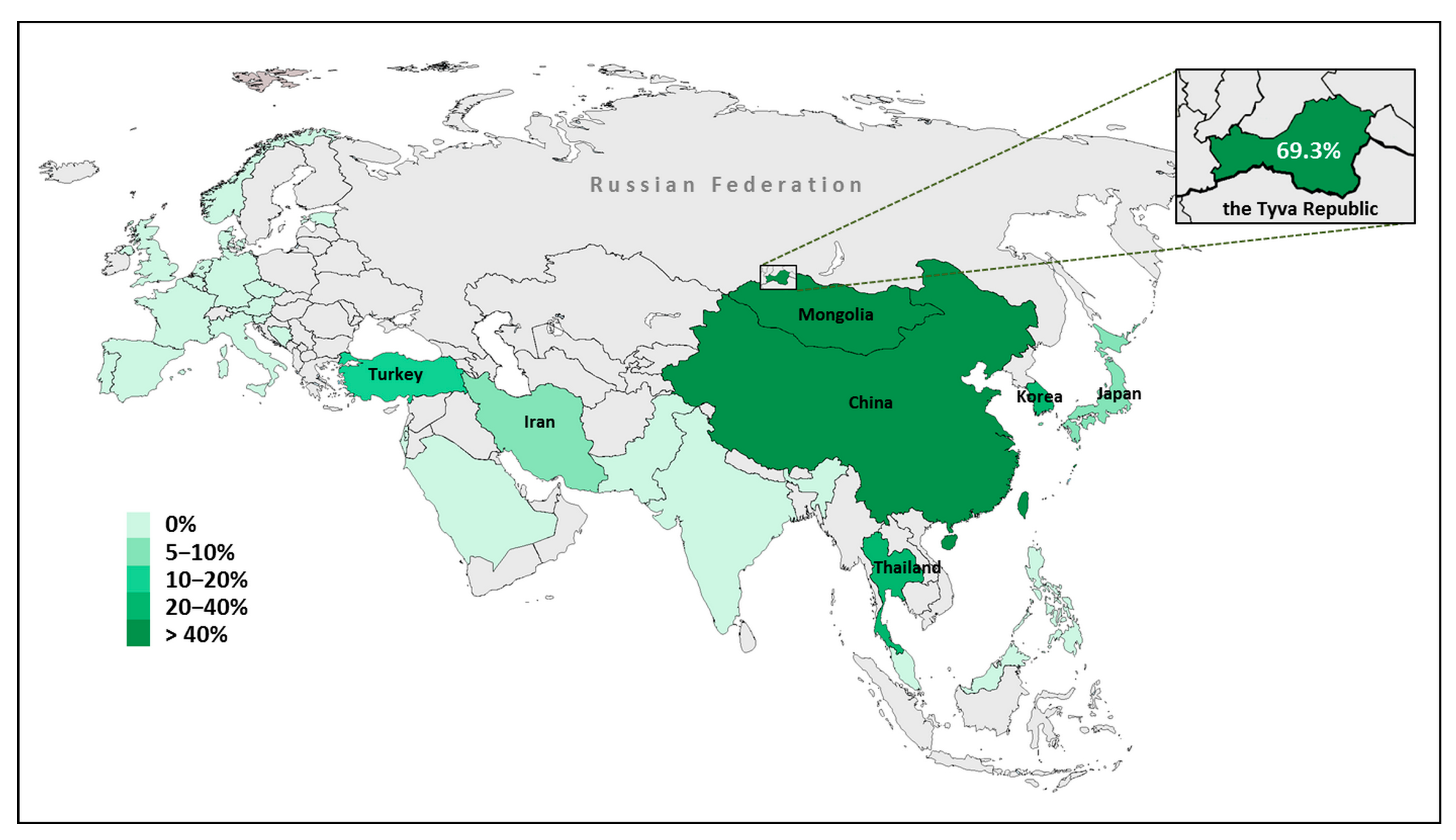
  68. Mongush, M.V. Tuvans of Mongolia and China. Int. J. Cent. Asian Stud. 1996, 1, 225–243. [Google Scholar]
  69. Chen, Z.; Zhang, Y.; Fan, A.; Zhang, Y.; Wu, Y.; Zhao, Q.; Zhou, Y.; Zhou, C.; Bawudong, M.; Mao, X.; et al. Brief communication: Y-chromosome haplogroup analysis indicates that Chinese Tuvans share distinctive affinity with Siberian Tuvans. Am. J. Phys. Anthropol. 2011, 144, 492–497. [Google Scholar] [CrossRef]
  70. Vainshtein, S.I.; Mannay-Ool, M.H. History of Tyva, 2nd ed.; Science: Novosibirsk, Russia, 2001. (In Russian) [Google Scholar]
  71. Mannai-ool, M.K. Tuvan People. The Origin and Formation of the Ethnos; Nauka Publ.: Novosibirsk, Russia, 2004; pp. 99–166. (In Russian) [Google Scholar]
Figure 1. Schematic structure of the SLC26A4 gene and the location of genetic markers (five STRs and nine SNPs) that were used to reconstruct the c.919-2A>G haplotypes. Location of SLC26A4 gene is shown by red square. The c.919-2A>G variant is marked by red color. Four of SNP markers from the study by Wu et al. [12] are marked by blue color.
Genes 14 00928 g001
Figure 2. The hot map demonstrating the proportion of c.919-2A>G among all mutated SLC26A4 alleles revealed in patients with SLC26A4-related hearing loss in the territory of Eurasia. The geographic regions for which no data are available are marked by gray color.
Genes 14 00928 g002
Table 1. The frequencies of STR and SNP haplotypes found among the chromosomes bearing c.919-2A>G, in comparison with the normal chromosomes.
STR Haplotypes
D7S2420-D7S496-/c.919-2A>G/-D7S2459-D7S2456-D7S525
(~2.8 Mb)
Frequency of HaplotypesX2p
Mutant
Chromosomes
Normal
Chromosomes
278-120-147-244-2270.91300.0150<10−35
278-120-147-244-2290.04350.02.40.0704
278-120-147-244-2210.02170.00.280.2674
278-120-147-244-2250.02170.00.280.2674
Other haplotypes0.01.0--
SNP Haplotypes
rs2248464-rs2248465-rs3801943-rs2712212*-/c.919-2A>G/-rs2395911*-rs2712211*-rs3801940*-rs2072064-rs2072065
(31.039 kb)
Frequency of HaplotypesX2p
Mutant
Chromosomes
Normal
Chromosomes
A-C-T-A-G-G-C-A-C1.00.0280150<10−36
Other haplotypes0.00.9720--
Designations of the STR alleles included in haplotypes correspond to the size of the PCR products (in nucleotides). The most common haplotypes are shown in bold. *—rs2712212, rs2395911, rs2712211, and rs3801940 correspond to SNPs analyzed in the study by Wu et al. [12]. The haplotype A-G-G-C (rs2712212*-rs2395911*-rs2712211*-rs3801940*) is underlined. Its allelic composition corresponds to the core haplotype T-C-C-G in the study by Wu et al. [12]. Statistically significant (p < 0.05) differences in haplotype frequencies are in bold.
Table 2. The results of the c.919-2A>G age dating.
Genetic Markers Used for CalculationsdThe Single-Marker MethodThe DMLE + Calculation
gAgeg (95% CI)Age (95% CI)
STR markers *0.0522550 years103–1982575–4950 years
0.121525 years63–1071575–2675 years
0.217425 years35–59875–1475 years
SNP markers0.05--91–1912275–4775 years
0.153–1031325–2575 years
0.229–54725–1350 years
*—The distal STR marker D7S525 was used for the c.919-2A>G age estimation by the single-marker method, and the STR haplotypes were used for c.919-2A>G age estimation by the DMLE+ v.2.3 program. d—population growth rate. g—the number of generations; the age of mutation was calculated as g × 25 years. CI—confidence interval.
Disclaimer/Publisher’s Note: The statements, opinions and data contained in all publications are solely those of the individual author(s) and contributor(s) and not of MDPI and/or the editor(s). MDPI and/or the editor(s) disclaim responsibility for any injury to people or property resulting from any ideas, methods, instructions or products referred to in the content.

