Article

Genome-Wide Associations and Confirmatory Meta-Analyses in Diabetic Retinopathy

1
Department of Medicine, Brigham and Women’s Hospital, Mass General Brigham, Harvard Medical School, Boston, MA 02445, USA
2
Department of Ophthalmology, Massachusetts Eye and Ear, Mass General Brigham, Harvard Medical School, Boston, MA 02445, USA
*
Author to whom correspondence should be addressed.
These authors contributed equally to this work.
Genes 2023, 14(3), 653; https://doi.org/10.3390/genes14030653
Submission received: 31 January 2023 / Revised: 21 February 2023 / Accepted: 3 March 2023 / Published: 5 March 2023
(This article belongs to the Special Issue Feature Papers in Human Genomics and Genetic Diseases)

Abstract

:
The present study aimed to summarize and validate the genomic association signals for diabetic retinopathy (DR), proliferative DR, and diabetic macular edema/diabetic maculopathy. A systematic search of the genome-wide association study (GWAS) catalog and PubMed/MELINE databases was conducted to curate a comprehensive list of significant GWAS discoveries. The top signals were then subjected to meta-analysis using established protocols. The results indicate the need for improved consensus among DR GWASs, highlighting the importance of validation efforts. A subsequent meta-analysis confirmed the association of two SNPs, rs4462262 (ZWINT-MRPS35P3) (odds ratio = 1.38, p = 0.001) and rs7903146 (TCF7L2) (odd ratio = 1.30, p < 0.001), with DR in independent populations, strengthening the evidence of their true association. We also compiled a list of candidate SNPs for further validation. This study highlights the importance of consistent validation and replication efforts in the field of DR genetics. The two identified gene loci warrant further functional investigation to understand their role in DR pathogenesis.

1. Introduction

Diabetic retinopathy (DR) is a leading cause of visual impairment. It leads to blinding complications and affects more than one-third of global diabetic patients, posing blindness to one in three DR patients [1]. DR is a progressive retinal microvascular complication of diabetes mellitus. Its pathology involves microaneurysms, hemostatic changes, and capillary occlusion in the retina. The vessel permeability increases, resulting in retinal nonperfusion, neovascularization, and vessel wall thickening. Further progress into macular edema, vitreous hemorrhage, or retina distortion can lead to severe visual loss and blindness [2]. Effective interventions, such as tight glycemic and blood pressure control, lipid-lowering, and laser therapies, could delay its progression and preserve vision. Early diagnosis and appropriate treatment can help prevent visual damage and the blinding consequences [3,4,5,6,7,8].
Genetic studies play an important role in uncovering DR-related genes and shedding light on the underlying pathophysiology and potential diagnostic and therapeutic markers. Over 300 DR candidate gene association studies have been published [9]. Common variants within genes involved in various processes, such as angiogenesis, glucose metabolism, lipid metabolism, inflammation, vascular regulation, cell communication, and extracellular matrix, have been evaluated for their associations with DR and its subtypes, with noticeable inconsistent findings [10,11]. In an effort to address inconsistencies and uncover a broader range of genetic factors, the field has shifted towards genome-wide association studies (GWASs) for investigating DR.
Genome-wide association studies exam the entire genome in populations to identify single nucleotide polymorphisms (SNPs) that are associated with a particular trait or disease. GWASs have been conducted in different populations to investigate the genetic architecture of DR [12,13,14,15,16,17,18,19,20] and its subtypes [21,22,23,24,25,26,27,28], such as proliferative diabetic retinopathy (PDR) and diabetic macular edema (DME)/diabetic maculopathy. These GWASs have reported more than 100 gene loci and SNPs that significantly or suggestively alter the risk of DR in European, Asian, and African populations. However, the reported SNPs varied among the DR GWASs [10,11,12,13,29]. Therefore, a systematic summary and evaluation of existing DR GWAS results is needed to prioritize the efforts in replicating these GWAS signals in independent populations, ultimately advancing our understanding about the DR genetics.
In this study, we first summarized all reported SNPs from published GWASs on DR and its subtypes. Second, we sifted through SNPs of genome-wide or suggestive significance to identify overlapping signals between independent GWASs. Finally, we calculated the summary outcomes for the top GWAS SNPs using a meta-analysis and all available genetic data from independent replication studies. Our study provides a comprehensive summary and update, emphasizing the urgent need for improved consensus among DR GWASs. Through a meta-analysis of the reported GWAS SNPs, we confirmed the association of two SNPs, rs4462262 (ZWINT-MRPS35P3) and rs7903146 (TCF7L2), with DR. However, to ensure the robustness and generalizability of GWAS findings, further replication of the candidate SNPs summarized in our study is necessary.

2. Materials and Methods

2.1. Identification of DR GWASs and Data Extraction

Genome-wide association studies on DR were identified via searching the GWAS catalog [30], literature databases, and citation lists of relevant publications. The search strategies used for the identification of DR GWASs in PubMed/MEDLINE are detailed in Table S1. The search terms used in the GWAS catalog were DR (trait ID in the Experimental Factor Ontology (EFO): EFO_0003770), PDR (EFO_0009322), DME (EFO_0009321), and diabetic maculopathy (EFO_0010133). We summarized all records that met the following criteria: (1) studies tested the associations of genetic variants with DR and its subtypes (i.e., nonproliferative DR (also known as NPDR), PDR, sight-threatening DR, DME, or diabetic maculopathy) at a genomic scale; (2) study population was clearly defined; (3) background disease was type 1 diabetes (T1D), type 2 diabetes (T2D), or both T1D and T2D; (4) diagnosis of DR was based on clinical data; and (5) publicly reported genome-wide significant variants.
The data extraction process for the SNPs associated with DR involved screening and extracting data from both GWAS catalog datasets and identified GWASs from a literature search. Two authors, X.Y.T. and S.S.R., reviewed the records, performed the data extraction, and resolved any discrepancies through consensus. The extracted data were then organized into summary tables for further analysis.

2.2. Identification of Overlapping Signals between GWASs

To identify overlapping SNPs between documented DR GWASs, we took a multistep approach. Three DR phenotypes were analyzed separately, including DR, PDR/sight-threatening DR, and DME/diabetic maculopathy. Within each phenotype group, the first step was to search for identical SNPs present in two or more independent DR GWASs. Secondly, for each DR phenotype, we searched for SNPs that were near each other within a ±100 K base pair window. This step aimed to identify SNPs that may be in linkage disequilibrium and may have a similar effect on the DR phenotypes. This method of searching for identical and nearby SNPs between different DR GWASs allowed for the identification of SNPs with the best available genetic evidence supporting their effects on the risk of DR and provided a list of SNPs for the subsequent meta-analysis.

2.3. Genetic Meta-Analysis

A genetic meta-analysis was conducted for any SNPs or gene loci repeatedly identified by independent DR GWASs and SNPs with a genome-wide significant p-value (<1 × 10⁻⁷) following previously published protocols [31,32,33,34,35]. Briefly, we performed the literature search using Boolean logic and search terms with controlled vocabularies (i.e., Medical Subject Heading terms) in the PubMed/MEDLINE databases. The search terms were constructed as follows: (Identified genes and genetic loci OR SNP IDs) AND (“diabetic retinopathy” (MeSH Terms) OR (“diabetic” (All Fields) AND “retinopathy” (All Fields)) OR “diabetic retinopathy” (All Fields)) (Table S2). Additionally, we scanned the reference lists of relevant research articles, reviews, and meta-analyses identified during the screening process to include all relevant genetic data. The last search was conducted on 28 January 2023.
We included SNPs and studies that met the following criteria in the genetic meta-analysis: (1) original genetic case-control studies that enrolled unrelated individuals from explicitly defined populations; (2) studies that used T1D, T2D, or both as the background conditions; (3) odds ratio and 95% confidence intervals (CIs)/standard error (SE) were reported or calculable based on the reported data; and (4) SNPs with genome-wide significant p-values (<1 × 10⁻⁷) in the documented GWAS discovery cohorts, or the gene loci contained genome-wide significant SNPs. We excluded animal studies, case reports, reviews, abstracts, conference proceedings, and editorials. Two investigators (X.T.Y. and S.S.R.) screened, reviewed, and extracted all the data independently. Disagreements were resolved through consensus. If the allele counts were not provided, we calculated them from the genotype data. If only the OR and 95% CI were reported, we estimated the SE using the equation SE = (β−ln(lower limit of 95% CI))/1.96, where β = ln (OR) [36]. Fully adjusted outcomes were used in the meta-analysis when available. If duplicated cohorts were identified, we used the larger and more recent cohort in the meta-analysis. We also adopted the Newcastle Ottawa Scale (NOS) (accessed via http://www.ohri.ca/programs/clinical_epidemiology/oxford.asp, accessed on 1 January 2023) to evaluate the overall quality of the case-control studies (Appendix A) [37,38,39]. A study with ≤6 stars was considered high risk for introducing biases; therefore, it was subject to removal in the sensitivity analysis [40].
We performed a meta-analysis for each SNP if it was reported in two or more independent cohorts. The genetic association was evaluated using an allelic model, i.e., effect allele frequency vs. reference allele frequency. We calculated the summary OR and 95% CI for each polymorphism using the random-effects model. The heterogeneity was tested using the I2 statistics [41]. An I2 value lower than 50% indicated low heterogeneity. We plotted the funnel plots to assess the publication bias [42,43,44]. We also conducted a sensitivity analysis to confirm the associations by sequentially omitting each of the studies one at a time if the studies deviated from the HWE or were of suboptimal quality [31,45]. We performed the meta-analysis using RevMan 5 (https://training.cochrane.org/online-learning/core-software/revman/, accessed on 1 January 2023). Summary p-values of <0.05 were considered statistically significant.

2.4. Functional Annotation of SNPs and Gene Loci

To understand the functional relevance of the identified SNPs related to DR, we employed in silico functional prediction scores, such as SIFT [46], PolyPhen [47], CADD [48], and RegulomeDB [49]. Moreover, we also assessed the SNPs in high (D-prime ≥ 0.8 and r2 ≥ 0.8) linkage disequilibrium (LD) with the identified SNPs. The LD data for European populations from the 1000 Genome project were used [50]. Additionally, we analyzed the expression quantitative trait loci (eQTL) databases through the Genotype-Tissue Expression (GTEx) portal to gain direct knowledge of the effects of risk alleles on nearby gene expression [51].

3. Results

We identified 14 GWASs conducted in diverse populations, including European [12,21,23,24,25,27], Asian [12,21,23,24,25,27], African [26,28], and Arabian [15], through searching the GWAS catalog and literature databases (Figure S1 and Table 1). With the exception of one GWAS that focused on T1D [21] and another GWAS that included both T1D and T2D [15], the majority of the GWASs enrolled individuals with DR in a T2D context. Only half of the GWASs tested significant SNPs discovered in the initial stage in at least one replication cohort [14,17,22,23,25,26,28]. Although the majority of GWASs had small sample sizes, with less than 1000 participants, no power analysis results were reported (Table 1). Additionally, we found three pertinent GWASs that used samples from the UK Biobank [18,19,20]. However, we excluded them from the summary table, as they primarily used nondiabetic population controls.
It is worth noting that there have been exciting developments in the field of DR genetics in recent years. These advancements include exome sequencing for rare and functional variants [15], and a more in-depth analysis of DR GWAS datasets has provided further understanding of the underlying pathological pathways [52], causal factors for DR [53,54,55], ability to predict DR using polygenic risk scores [56], and pharmacogenetic responses to treatment [57].

3.1. Genome-Wide Associations of DR

A total of six GWASs identified 76 SNPs located in 61 gene loci, with p-values that suggest a potential or confirmed genome-wide significance for DR (p < 5 × 10⁻⁵) (Table S3) [13,14,16,17,21,26]. Our subsequent analysis revealed a lack of consensus among the existing DR GWASs, as we found no overlap in SNPs from different studies within a ±100 K base pair window.
Seven out of the seventy-six SNPs exhibited genome-wide significance (p < 1.0 × 10⁻⁷), with the top seven being rs17376456 (KIAA0825/C5orf36, p = 3.0 × 10⁻¹⁵), rs2038823 (HS6ST3, p = 5.0 × 10⁻¹¹), rs12630354 (THRAP3P1-STT3B, p = 7.0 × 10⁻¹⁰), rs4838605 (ARHGAP22, p = 2.0 × 10⁻⁹), rs12219125 (AMD1P1-PLXDC2, p = 9.0 × 10⁻⁹), rs202069793 (OR13D3P-OR13D1, p = 6.0 × 10⁻⁸), and rs4462262 (ZWINT-MRPS35P3, p = 9.0 × 10⁻⁸) (Table S3). Notably, two of these seven SNPs, rs12630354 and rs202069793, were validated in separate populations in the original GWASs [17,26]. All seven SNPs were included in subsequent meta-analysis.

3.2. Genome-Wide Associations of PDR and DME

In our investigation of PDR, we identified six GWASs that tested PDR against a T2D background (Table S4) [22,23,24,25,26,28], with all but one study including a validation cohort [24] (Table 1). A total of 29 SNPs located near 27 gene loci demonstrated suggestive or lower p values. Among them, eight SNPs showed genomic significance, including rs3081219 (WDR72, p = 1.0×10⁻⁹), rs3913535 (NOX4, p = 4.0 × 10⁻⁹), rs11018670 (FOLH1B, p = 1.0 × 10⁻⁸), rs72740408 (HNRNPA1P46, p = 2.0 × 10⁻⁸), rs184340784 (LINC01646, p = 4.0 × 10⁻⁸), rs1065386 (HLA-B, p = 5.0 × 10⁻⁸), rs4726066 (PRKAG2, p = 5.0 × 10⁻⁸), and rs200295620 (GOLIM4-EGFEM1P, p = 7.0 × 10⁻⁸).
Two GWASs were identified for DME, which collectively found 12 SNPs located near 10 gene loci (Table S5) [24,27]. Notably, only one of the SNPs, rs9966620, was genome-wide significant (p = 7.0 × 10⁻⁸). Neither of the two GWASs included a validation cohort (Table 1).
Furthermore, our analysis revealed a lack of overlapping SNPs or gene loci among different GWASs, and neither PDR nor DME had overlapping SNPs or gene loci among the different GWASs within a ±100 K base pair window, which was comparable to the results obtained from the DR GWASs.

3.3. Genetic Meta-Analysis of Top GWAS Signals

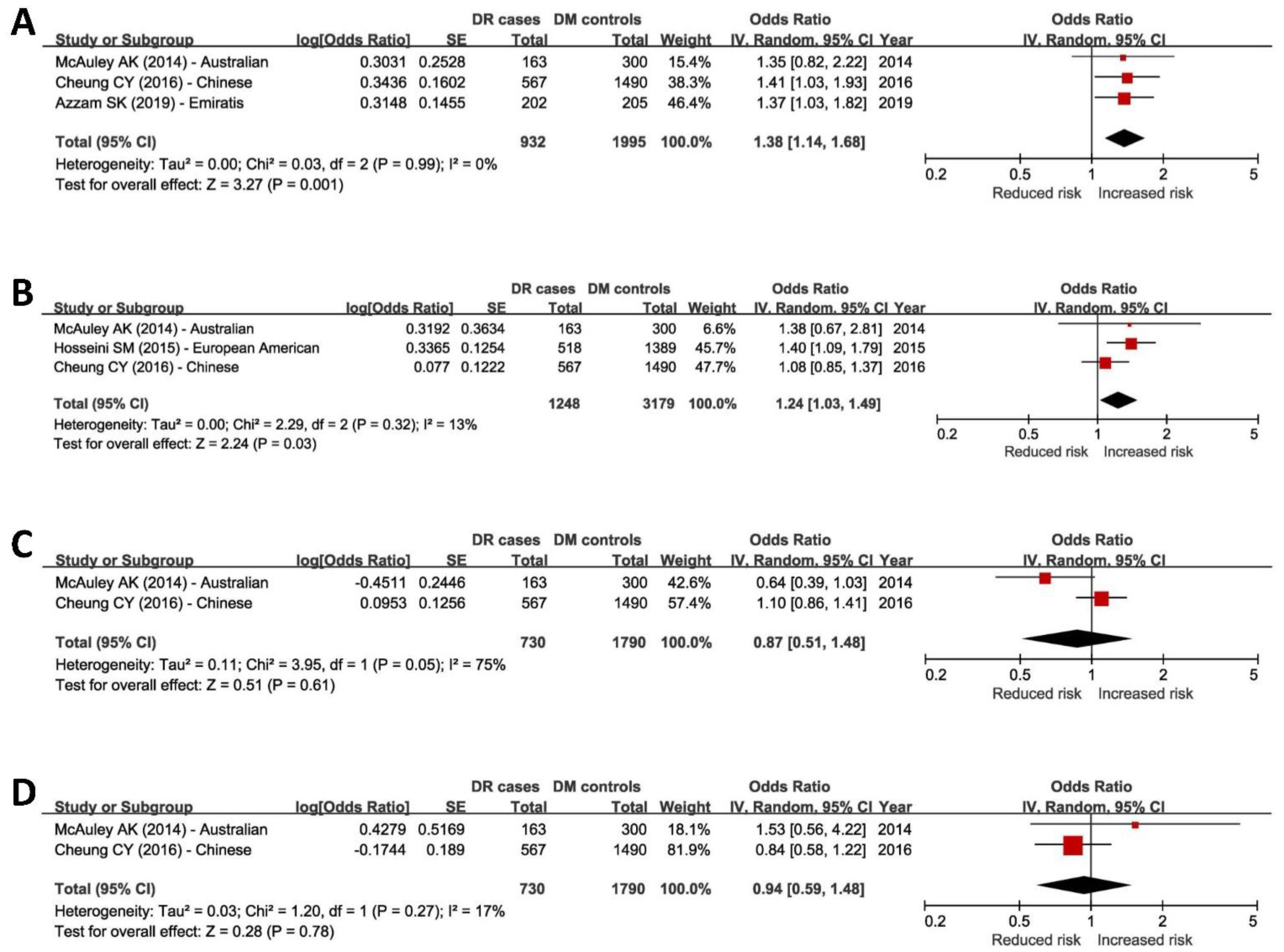
3.3.1. Meta-Analysis of Top DR-Associated SNPs

Replication studies and meta-analyses are crucial for validating findings and strengthening evidence for a true association, especially given the lack of consensus among existing GWASs. In our meta-analysis, we identified 37 records of published studies from the literature search and extracted data from 20 replication studies (Figure S2). Four of the seven top DR GWAS SNPs (Table S3) were replicated in two or more independent studies, including rs4462262 (ZWINT-MRPS35P3) [58,59,60], rs12219125 (PLXDC2-NEBL) [59,60,61], rs4838605 (ARHGAP22) [58,59,60], and rs17376456 (C5orf36) [59,60]. Notably, all four of these SNPs were reported without an internal validation cohort in the initial GWAS [13]. The remaining three of the seven GWAS SNPs, rs2038823 [13], rs12630354 [17], and rs202069793 [26], have not been replicated in independent studies. Additionally, we found five replication studies [62,63,64,65,66] that tested the DR-association of rs7903146 in the TCF7L2 gene locus, which was identified by rs34872471 (p = 4 × 10⁻¹⁵) in a UK Biobank GWAS using population controls [20]. These two SNPs were in high linkage disequilibrium in European populations (r2 ≥ 0.99) [50]. Therefore, we conducted a meta-analysis for rs7903146.
Our meta-analysis confirmed the significant association of rs4462262 (OR = 1.38, p = 0.001), rs12219125 (OR = 1.24, p = 0.03), and rs7903146 (OR = 1.30, p < 0.001) with DR in replication cohorts (Figure 1 and Figure S4). The heterogeneity measurements (I2) were low for all of these combined outcomes. However, we found no evidence of association between rs4838605 and rs17376456 with DR in our analysis (p > 0.05) (Figure 1).
All studies included in the meta-analysis scored six or higher on the NOS quality assessments (Table S6). Funnel plots showed no significant deviations (Figures S3 and S4), indicating a lower risk of introducing potential biases by pooling results from these studies. The sensitivity analysis confirmed the stability of most of the meta-analytical results by omitting one study at a time and recalculating the combined outcomes. Only rs12219125 became insignificant after removing Hosseini S. M.’s study [61], highlighting the need for further replication efforts.

3.3.2. Meta-Analysis of Top PDR- and DME-Associated SNPs

Our literature search did not identify replication efforts for the top GWAS hits of PDR and DME, preventing us from performing meta-analyses for these DR phenotypes.

3.4. Biological Relevance of rs4462262 and rs7903146 to DR

The SNP rs4462262 is an intergenic variant, located approximately 750 kbp upstream to the nearest gene, IPMK. A microRNA, MIR3924, was situated approximately 110 kbp upstream of the SNP. However, the functional annotations for rs4462262 and 54 other SNPs in high LD with rs4462262 were unremarkable (Table 2), providing limited evidence for the biological relevance of these variants.
In contrast, the SNP rs7903146 is located in intron 3 of TCF7L2 and is significantly associated with the expression of TCF7L2 (p = 2.9 × 10⁻⁷) (Table 2). Another SNP, rs7074440, in high LD with rs7903146 (D’ = 0.95 and r2 = 0.91) has a CADD score of 10.1 and is also significantly associated with the expression of TCF7L2 (p = 2.1 × 10⁻⁶) (Table 2), indicating a potentially functional role in TCF7L2 regulation.

4. Discussion

In this study, we comprehensively summarized the genomic association signals of DR, PDR, and DME/diabetic maculopathy from published GWASs. Our results emphasized the urgent need for improved consensus among DR GWASs, underlining the importance of validation and replication efforts. We confirmed the association of two SNPs, rs4462262 (ZWINT-MRPS35P3) and rs7903146 (TCF7L2), with DR through meta-analysis in independent populations, strengthening the evidence of their true association. Moreover, we provided a list of candidate SNPs for further validation studies.
The lack of consensus among DR GWASs is a complex issue that arises from several potential factors. One such factor is the clinical heterogeneity of DR populations, which can lead to conflicting results between studies. A possible solution is to carefully stratify patients based on specific clinical characteristics and to conduct studies in homogeneous patient populations. Second, ethnic differences in SNP allele frequencies can impact the strength and direction of genetic associations. To address this issue, large-scale studies with diverse populations are needed to increase the generalizability of results. Additionally, the standardization of SNP definitions and frequency databases can improve the accuracy of genetic association analyses [67]. Third, the study definition of DR and its subtypes could be another factor contributing to the lack of consensus. To address this, a standardized definition and diagnostic criteria, such as the ETDRS grading system [68,69], should be universally adopted by the DR genetic research community to ensure consistency between studies. Fourth, whether to include a validation design within a GWAS is another potential factor. Including a validation design within a GWAS can increase the confidence in the findings and help to reduce false positive results. This is particularly important in a complex disease such as DR, where multiple genetic and environmental factors are likely to be involved [11,70]. Finally, differences in the statistical power between studies can also contribute to the lack of consensus among DR GWASs. To overcome this issue, larger sample sizes and more powerful study designs are needed to increase the chances of detecting true associations, especially in rare variant studies. Addressing these potential factors through careful study design and increased collaboration among DR researchers can help to improve the consistency of DR GWAS results and eventually lead to a better understanding of the underlying mechanisms of DR.
In our meta-analysis, we noticed a lack of replication studies for DR GWASs findings. Replication is a critical step to confirm the validity of a discovery and strengthens the evidence for a true association. Without replication, the results of a study may be considered unreliable or of limited generalizability, especially when the study is based on a small sample size or has limited statistical power. However, replication studies are often challenging and resource intensive, which can limit the number of independent validation studies performed for DR GAWS discoveries. This is particularly true in the case of DR genetics, which are often complex due to the heterogeneity of DR populations, differences in SNP allele frequencies across ethnic groups, and variations in the definition of DR and its subtypes. Therefore, it is essential to prioritize replication efforts in DR genetics research, which directly pointed us towards conducting this study.
The association between rs7903146 and DR is significant, and the regulatory effects of this SNP and other SNPs in LD provide evidence supporting the functional role of TCF7L2 in DR pathogenesis. As a transcriptional regulator of the canonical Wnt signaling pathway, TCF7L2 plays a vital role in cell proliferation and fate specification. The involvement of TCF7L2 in DR pathogenesis can be two-fold. First, TCF7L2 may promote pathological retinal neovascularization via the ER stress-dependent upregulation of VEGFA, making retinal cells from rs7903146-TT patients more sensitive to VEGF upregulation and at greater risk of developing PDR [63]. Second, TCF7L2 may also increase the risk of DR development and progression by influencing the responses to glycemic control drugs, such as glipizide and metformin, which are two commonly used glycemic control drugs [71]. In addition, good glycemic control is closely associated with short- and long-term beneficial effects on DR prevention and intervention [72,73]. However, the functional relevance of rare mutations in the TCF7L2 gene and their impact on DR pathophysiology in diverse ethnic groups remain unknown, and further sequencing studies of large cohorts are needed to understand their role. In addition, understanding the functional regions of the TCF7L2 gene and their impact on specific phenotypes will also be important for comprehending their role in diverse human diseases, including DR [74,75].
On the other hand, the lack of functional annotations for rs4462262 and 54 other SNPs in high LD with rs4462262 limits our understanding of their functional relevance in DR pathogenesis. To reveal their role in DR, the identification of rare and common variants that truly alter the risk of DR and functional investigations are crucial. The confirmation and functional annotation of these variants could be a critical step in discovering new genetic markers for DR, facilitating better prediction of disease susceptibility and tailoring individualized treatments for patients.
This study has several potential limitations, including the limited number of independent replication cohorts and subphenotypes, limited diversity of population ancestries, and the use of population controls instead of DR-free diabetic controls in the DR GWASs using the UK Biobank datasets. These limitations affected our ability to confirm more GWAS discoveries.

5. Conclusions

Our study underlines the urgent need for consistent replication efforts in the field of DR genetics. Two SNPs, rs4462262 (ZWINT-MRPS35P3) and rs7903146 (TCF7L2), showed consistent association with DR. Confirmation and functional investigations of reported and new variants are needed for a better understanding of the underlying mechanisms of DR development.

Supplementary Materials

The following are available online at https://www.mdpi.com/article/10.3390/genes14030653/s1. Table S1. Search strategies for the identification of genome-wide association studies of diabetic retinopathy in PubMed/MEDLINE; Table S2. Search strategies for genetic association studies of top genome-wide association signals of diabetic retinopathy. Table S3. Summary of genome-wide significant gene loci in diabetic retinopathy; Table S4. Summary of genome-wide significant loci in proliferative diabetic retinopathy, severe diabetic retinopathy, and vision-threatening diabetic retinopathy. Table S5. Summary of genome-wide significant loci in diabetic macular edema/diabetic maculopathy. Table S6. Quality assessments of included case-control studies. Figure S1. GWAS catalog and literature search and results of the literature review. Figure S2. Literature search for replication studies of top GWAS signals. Figure S3. Funnel plots of the study outcomes used in the meta-analysis. Figure S4. Meta-analysis of the genetic association of rs7903146 (T, TCF7L2) with diabetic retinopathy.

Author Contributions

Study conceptualization: S.R. and X.Y.; Acquisition of data: X.Y. and S.R.; Assessment of quality of studies: X.Y.; Data analysis: S.R.; Drafting of the manuscript: X.Y. and S.R.; Critical revision of the manuscript for important intellectual content: S.R. All authors have read and agreed to the published version of the manuscript.

Funding

This research received no external funding.

Institutional Review Board Statement

Ethical review and approval were waived for this study due to the usage of published GWAS datasets and genetic candidate gene association study data.

Informed Consent Statement

Written informed consent was obtained in the original publications.

Data Availability Statement

We used publicly available software for the analyses and provided a list of the specific programs used in Section 2.

Acknowledgments

We would like to express our sincere gratitude to all the authors of the genome-wide association studies for generously sharing their summary datasets. Your contributions have been invaluable to our research.

Conflicts of Interest

The authors declare no conflict of interest.

Appendix A

Newcastle—Ottawa Quality Assessment Scale
Case Control Studies
(Accessed via http://www.ohri.ca/programs/clinical_epidemiology/oxford.asp) (accessed on 20 January 2023)
Note: A study can be awarded a maximum of one star for each numbered item within the Selection and Exposure categories. A maximum of two stars can be given for Comparability.
Selection
1. Is the case definition adequate?
 a. yes, with independent validation
 b. yes, e.g., record linkage or based on self reports
 c. no description
2. Representativeness of the cases
 a. consecutive or obviously representative series of cases
 b. potential for selection biases or not stated
3. Selection of Controls
 a. community controls
 b. hospital controls
 c. no description
4. Definition of Controls
 a. no history of disease (endpoint.)
 b. no description of source
Comparability
1. Comparability of cases and controls on the basis of the design or analysis
 a. study controls for _______________ (Select the most important factor: ethnicity)
 b. study controls for any additional factor (This criteria could be modified to indicate specific control for a second important factor.)
Exposure
1. Ascertainment of exposure
 a. secure record (e.g., surgical records)
 b. structured interview where blind to case/control status
 c. interview not blinded to case/control status
 d. written self report or medical record only
 e. no description
2. Same method of ascertainment for cases and controls
 a. yes
 b. no
3. Non-Response rate
 a. same rate for both groups
 b. non respondents described
 c. rate different and no designation

References

  1. Yau, J.W.; Rogers, S.L.; Kawasaki, R.; Lamoureux, E.L.; Kowalski, J.W.; Bek, T.; Chen, S.J.; Dekker, J.M.; Fletcher, A.; Grauslund, J.; et al. Global prevalence and major risk factors of diabetic retinopathy. Diabetes Care 2012, 35, 556–564. [Google Scholar] [CrossRef] [PubMed] [Green Version]
  2. Scott, I.U.; Flynn, H.W.; Smiddy, W.E.; Ophthalmology, A.A.O. Diabetes and Ocular Disease: Past, Present, and Future Therapies, 2nd ed.; Scott, I.U., Harry, W., Flynn, J., Smiddy, W.E., Eds.; Oxford University Press in cooperation with the American Academy of Ophthalmology: New York, NY, USA, 2010; p. 496. [Google Scholar]
  3. UK Prospective Diabetes Study Group. Tight blood pressure control and risk of macrovascular and microvascular complications in type 2 diabetes: UKPDS 38. Bmj 1998, 317, 703–713. [Google Scholar] [CrossRef] [Green Version]
  4. Diabetes, C.; Complications Trial/Epidemiology of Diabetes, I.; Complications Research, G.; Lachin, J.M.; Genuth, S.; Cleary, P.; Davis, M.D.; Nathan, D.M. Retinopathy and nephropathy in patients with type 1 diabetes four years after a trial of intensive therapy. N. Engl. J. Med. 2000, 342, 381–389. [Google Scholar] [CrossRef] [Green Version]
  5. Group, A.S.; Group, A.E.S.; Chew, E.Y.; Ambrosius, W.T.; Davis, M.D.; Danis, R.P.; Gangaputra, S.; Greven, C.M.; Hubbard, L.; Esser, B.A.; et al. Effects of medical therapies on retinopathy progression in type 2 diabetes. N. Engl. J. Med. 2010, 363, 233–244. [Google Scholar] [CrossRef] [Green Version]
  6. Keech, A.C.; Mitchell, P.; Summanen, P.A.; O’Day, J.; Davis, T.M.; Moffitt, M.S.; Taskinen, M.R.; Simes, R.J.; Tse, D.; Williamson, E.; et al. Effect of fenofibrate on the need for laser treatment for diabetic retinopathy (FIELD study): A randomised controlled trial. Lancet 2007, 370, 1687–1697. [Google Scholar] [CrossRef] [PubMed]
  7. The Diabetic Retinopathy Study Research Group. Photocoagulation treatment of proliferative diabetic retinopathy. Clinical application of Diabetic Retinopathy Study (DRS) findings, DRS Report Number 8. Ophthalmology 1981, 88, 583–600. [Google Scholar]
  8. Hovind, P.; Tarnow, L.; Rossing, K.; Rossing, P.; Eising, S.; Larsen, N.; Binder, C.; Parving, H.H. Decreasing incidence of severe diabetic microangiopathy in type 1 diabetes. Diabetes Care 2003, 26, 1258–1264. [Google Scholar] [CrossRef] [Green Version]
  9. Rong, S.S.; Tam, P.O.S.; Pang, C.P.; Chen, L.J. Global Review and Meta-analysis of Diabetic Retinopathy Genetic Studies Highlight Gaps in the Pathogenesis between Various Populations. Investig. Ophthalmol. Vis. Sci. 2013, 54, 6197. [Google Scholar]
  10. Bhatwadekar, A.D.; Shughoury, A.; Belamkar, A.; Ciulla, T.A. Genetics of Diabetic Retinopathy, a Leading Cause of Irreversible Blindness in the Industrialized World. Genes 2021, 12, 1200. [Google Scholar] [CrossRef]
  11. Cabrera, A.P.; Mankad, R.N.; Marek, L.; Das, R.; Rangasamy, S.; Monickaraj, F.; Das, A. Genotypes and Phenotypes: A Search for Influential Genes in Diabetic Retinopathy. Int. J. Mol. Sci. 2020, 21, 2712. [Google Scholar] [CrossRef] [Green Version]
  12. Fu, Y.P.; Hallman, D.M.; Gonzalez, V.H.; Klein, B.E.; Klein, R.; Hayes, M.G.; Cox, N.J.; Bell, G.I.; Hanis, C.L. Identification of Diabetic Retinopathy Genes through a Genome-Wide Association Study among Mexican-Americans from Starr County, Texas. J. Ophthalmol. 2010, 2010, 861291. [Google Scholar] [CrossRef] [PubMed]
  13. Huang, Y.C.; Lin, J.M.; Lin, H.J.; Chen, C.C.; Chen, S.Y.; Tsai, C.H.; Tsai, F.J. Genome-wide association study of diabetic retinopathy in a Taiwanese population. Ophthalmology 2011, 118, 642–648. [Google Scholar] [CrossRef] [PubMed]
  14. Awata, T.; Yamashita, H.; Kurihara, S.; Morita-Ohkubo, T.; Miyashita, Y.; Katayama, S.; Mori, K.; Yoneya, S.; Kohda, M.; Okazaki, Y.; et al. A genome-wide association study for diabetic retinopathy in a Japanese population: Potential association with a long intergenic non-coding RNA. PLoS ONE 2014, 9, e111715. [Google Scholar] [CrossRef] [Green Version]
  15. Shtir, C.; Aldahmesh, M.A.; Al-Dahmash, S.; Abboud, E.; Alkuraya, H.; Abouammoh, M.A.; Nowailaty, S.R.; Al-Thubaiti, G.; Naim, E.A.; Alyounes, B.; et al. Exome-based case-control association study using extreme phenotype design reveals novel candidates with protective effect in diabetic retinopathy. Hum. Genet. 2016, 135, 193–200. [Google Scholar] [CrossRef]
  16. Hsieh, A.R.; Huang, Y.C.; Yang, Y.F.; Lin, H.J.; Lin, J.M.; Chang, Y.W.; Wu, C.M.; Liao, W.L.; Tsai, F.J. Lack of association of genetic variants for diabetic retinopathy in Taiwanese patients with diabetic nephropathy. BMJ Open Diabetes Res. Care 2020, 8, e000727. [Google Scholar] [CrossRef] [PubMed] [Green Version]
  17. Imamura, M.; Takahashi, A.; Matsunami, M.; Horikoshi, M.; Iwata, M.; Araki, S.I.; Toyoda, M.; Susarla, G.; Ahn, J.; Park, K.H.; et al. Genome-wide association studies identify two novel loci conferring susceptibility to diabetic retinopathy in Japanese patients with type 2 diabetes. Hum. Mol. Genet. 2021, 30, 716–726. [Google Scholar] [CrossRef]
  18. Backman, J.D.; Li, A.H.; Marcketta, A.; Sun, D.; Mbatchou, J.; Kessler, M.D.; Benner, C.; Liu, D.; Locke, A.E.; Balasubramanian, S.; et al. Exome sequencing and analysis of 454,787 UK Biobank participants. Nature 2021, 599, 628–634. [Google Scholar] [CrossRef] [PubMed]
  19. Jiang, L.; Zheng, Z.; Fang, H.; Yang, J. A generalized linear mixed model association tool for biobank-scale data. Nat. Genet. 2021, 53, 1616–1621. [Google Scholar] [CrossRef]
  20. Xue, Z.; Yuan, J.; Chen, F.; Yao, Y.; Xing, S.; Yu, X.; Li, K.; Wang, C.; Bao, J.; Qu, J.; et al. Genome-wide association meta-analysis of 88,250 individuals highlights pleiotropic mechanisms of five ocular diseases in UK Biobank. EBioMedicine 2022, 82, 104161. [Google Scholar] [CrossRef]
  21. Grassi, M.A.; Tikhomirov, A.; Ramalingam, S.; Below, J.E.; Cox, N.J.; Nicolae, D.L. Genome-wide meta-analysis for severe diabetic retinopathy. Hum. Mol. Genet. 2011, 20, 2472–2481. [Google Scholar] [CrossRef] [Green Version]
  22. Sheu, W.H.; Kuo, J.Z.; Lee, I.T.; Hung, Y.J.; Lee, W.J.; Tsai, H.Y.; Wang, J.S.; Goodarzi, M.O.; Klein, R.; Klein, B.E.; et al. Genome-wide association study in a Chinese population with diabetic retinopathy. Hum. Mol. Genet. 2013, 22, 3165–3173. [Google Scholar] [CrossRef] [Green Version]
  23. Burdon, K.P.; Fogarty, R.D.; Shen, W.; Abhary, S.; Kaidonis, G.; Appukuttan, B.; Hewitt, A.W.; Sharma, S.; Daniell, M.; Essex, R.W.; et al. Genome-wide association study for sight-threatening diabetic retinopathy reveals association with genetic variation near the GRB2 gene. Diabetologia 2015, 58, 2288–2297. [Google Scholar] [CrossRef] [PubMed]
  24. Graham, P.S.; Kaidonis, G.; Abhary, S.; Gillies, M.C.; Daniell, M.; Essex, R.W.; Chang, J.H.; Lake, S.R.; Pal, B.; Jenkins, A.J.; et al. Genome-wide association studies for diabetic macular edema and proliferative diabetic retinopathy. BMC Med. Genet. 2018, 19, 71. [Google Scholar] [CrossRef] [Green Version]
  25. Meng, W.; Shah, K.P.; Pollack, S.; Toppila, I.; Hebert, H.L.; McCarthy, M.I.; Groop, L.; Ahlqvist, E.; Lyssenko, V.; Agardh, E.; et al. A genome-wide association study suggests new evidence for an association of the NADPH Oxidase 4 (NOX4) gene with severe diabetic retinopathy in type 2 diabetes. Acta Ophthalmol. 2018, 96, e811–e819. [Google Scholar] [CrossRef] [Green Version]
  26. Pollack, S.; Igo, R.P., Jr.; Jensen, R.A.; Christiansen, M.; Li, X.; Cheng, C.Y.; Ng, M.C.Y.; Smith, A.V.; Rossin, E.J.; Segrè, A.V.; et al. Multiethnic Genome-Wide Association Study of Diabetic Retinopathy Using Liability Threshold Modeling of Duration of Diabetes and Glycemic Control. Diabetes 2019, 68, 441–456. [Google Scholar] [CrossRef] [PubMed] [Green Version]
  27. Meng, W.; Chan, B.W.; Ezeonwumelu, C.; Hébert, H.L.; Campbell, A.; Soler, V.; Palmer, C.N. A genome-wide association study implicates that the TTC39C gene is associated with diabetic maculopathy with decreased visual acuity. Ophthalmic Genet. 2019, 40, 252–258. [Google Scholar] [CrossRef]
  28. Liu, C.; Chen, G.; Bentley, A.R.; Doumatey, A.; Zhou, J.; Adeyemo, A.; Yang, J.; Rotimi, C. Genome-wide association study for proliferative diabetic retinopathy in Africans. NPJ Genom. Med. 2019, 4, 20. [Google Scholar] [CrossRef] [PubMed] [Green Version]
  29. Sharma, A.; Valle, M.L.; Beveridge, C.; Liu, Y.; Sharma, S. Unraveling the role of genetics in the pathogenesis of diabetic retinopathy. Eye 2019, 33, 534–541. [Google Scholar] [CrossRef] [Green Version]
  30. Buniello, A.; MacArthur, J.A.L.; Cerezo, M.; Harris, L.W.; Hayhurst, J.; Malangone, C.; McMahon, A.; Morales, J.; Mountjoy, E.; Sollis, E.; et al. The NHGRI-EBI GWAS Catalog of published genome-wide association studies, targeted arrays and summary statistics 2019. Nucleic Acids Res. 2019, 47, D1005–D1012. [Google Scholar] [CrossRef] [Green Version]
  31. Ma, L.; Li, Z.; Liu, K.; Rong, S.S.; Brelen, M.E.; Young, A.L.; Kumaramanickavel, G.; Pang, C.P.; Chen, H.; Chen, L.J. Association of Genetic Variants with Polypoidal Choroidal Vasculopathy: A Systematic Review and Updated Meta-analysis. Ophthalmology 2015, 122, 1854–1865. [Google Scholar] [CrossRef]
  32. Rong, S.S.; Ma, S.T.U.; Yu, X.T.; Ma, L.; Chu, W.K.; Chan, T.C.Y.; Wang, Y.M.; Young, A.L.; Pang, C.P.; Jhanji, V.; et al. Genetic associations for keratoconus: A systematic review and meta-analysis. Sci. Rep. 2017, 7, 4620. [Google Scholar] [CrossRef] [PubMed] [Green Version]
  33. Rong, S.S.; Tang, F.Y.; Chu, W.K.; Ma, L.; Yam, J.C.; Tang, S.M.; Li, J.; Gu, H.; Young, A.L.; Tham, C.C.; et al. Genetic Associations of Primary Angle-Closure Disease: A Systematic Review and Meta-analysis. Ophthalmology 2016, 123, 1211–1221. [Google Scholar] [CrossRef] [PubMed]
  34. Tang, S.M.; Rong, S.S.; Young, A.L.; Tam, P.O.; Pang, C.P.; Chen, L.J. PAX6 gene associated with high myopia: A meta-analysis. Optom. Vis. Sci. Off. Publ. Am. Acad. Optom. 2014, 91, 419–429. [Google Scholar] [CrossRef] [PubMed]
  35. Wong, K.H.; Rong, S.S.; Chong, K.K.; Young, A.L.; Pang, C.P.; Chen, L.J. Genetic Associations of Interleukin-related Genes with Graves’ Ophthalmopathy: A Systematic Review and Meta-analysis. Sci. Rep. 2015, 5, 16672. [Google Scholar] [CrossRef] [PubMed] [Green Version]
  36. Kirkwood, B.; Sterne, J. Essentials of Medical Statistics, 2nd ed.; Blackwell Science: Hoboken, NJ, USA, 2003; p. 501. [Google Scholar]
  37. Kmet, L.M.; Lee, R.C.; Cook, L.S. Standard Quality Assessment Criteria for Evaluating Primary Research Papers from a Variety of Fields; Alberta Heritage Foundation for Medical Research: Edmonton, Alta, 2004; p. 22. [Google Scholar]
  38. Khan, K.S.; Riet, G.t.; Popay, J.; Nixon, J.; Kleijnen, J. Conducting the review: Phase 5 study quality assessment. In Undertaking Systematic Reviews of Research Effectiveness CDC’s Guidance for Those Carrying Out or Commissioning Reviews; Centre of Reviews and Dissemination: York, UK, 2001; Volume 20. [Google Scholar]
  39. Squires, J.E.; Estabrooks, C.A.; Gustavsson, P.; Wallin, L. Individual determinants of research utilization by nurses: A systematic review update. Implement. Sci. 2011, 6, 1. [Google Scholar] [CrossRef] [Green Version]
  40. McPheeters, M.L.; Kripalani, S.; Peterson, N.B.; Idowu, R.T.; Jerome, R.N.; Potter, S.A.; Andrews, J.C. Closing the quality gap: Revisiting the state of the science (vol. 3: Quality improvement interventions to address health disparities). Evid. Rep. Technol. Assess. 2012, 208.3, 1–475. [Google Scholar]
  41. Higgins, J.P.; Thompson, S.G. Quantifying heterogeneity in a meta-analysis. Stat. Med. 2002, 21, 1539–1558. [Google Scholar] [CrossRef]
  42. Peters, J.L.; Sutton, A.J.; Jones, D.R.; Abrams, K.R.; Rushton, L. Contour-enhanced meta-analysis funnel plots help distinguish publication bias from other causes of asymmetry. J. Clin. Epidemiol. 2008, 61, 991–996. [Google Scholar] [CrossRef]
  43. Higgins, J.P.T.; Green, S. Cochrane Handbook for Systematic Reviews of Interventions. Available online: www.cochrane-handbook.org (accessed on 20 January 2023).
  44. Sterne, J.A.; Gavaghan, D.; Egger, M. Publication and related bias in meta-analysis: Power of statistical tests and prevalence in the literature. J. Clin. Epidemiol. 2000, 53, 1119–1129. [Google Scholar] [CrossRef]
  45. Rong, S.S.; Peng, Y.; Liang, Y.B.; Cao, D.; Jhanji, V. Does cigarette smoking alter the risk of pterygium? A systematic review and meta-analysis. Investig. Ophthalmol. Vis. Sci. 2014, 55, 6235–6243. [Google Scholar] [CrossRef] [Green Version]
  46. Vaser, R.; Adusumalli, S.; Leng, S.N.; Sikic, M.; Ng, P.C. SIFT missense predictions for genomes. Nat. Protoc. 2016, 11, 1–9. [Google Scholar] [CrossRef] [PubMed]
  47. Adzhubei, I.A.; Schmidt, S.; Peshkin, L.; Ramensky, V.E.; Gerasimova, A.; Bork, P.; Kondrashov, A.S.; Sunyaev, S.R. A method and server for predicting damaging missense mutations. Nat. Methods 2010, 7, 248–249. [Google Scholar] [CrossRef] [PubMed] [Green Version]
  48. Kircher, M.; Witten, D.M.; Jain, P.; O’Roak, B.J.; Cooper, G.M.; Shendure, J. A general framework for estimating the relative pathogenicity of human genetic variants. Nat. Genet. 2014, 46, 310–315. [Google Scholar] [CrossRef] [Green Version]
  49. Boyle, A.P.; Hong, E.L.; Hariharan, M.; Cheng, Y.; Schaub, M.A.; Kasowski, M.; Karczewski, K.J.; Park, J.; Hitz, B.C.; Weng, S.; et al. Annotation of functional variation in personal genomes using RegulomeDB. Genome Res. 2012, 22, 1790–1797. [Google Scholar] [CrossRef] [Green Version]
  50. Genomes Project, C.; Auton, A.; Brooks, L.D.; Durbin, R.M.; Garrison, E.P.; Kang, H.M.; Korbel, J.O.; Marchini, J.L.; McCarthy, S.; McVean, G.A.; et al. A global reference for human genetic variation. Nature 2015, 526, 68–74. [Google Scholar] [CrossRef] [Green Version]
  51. Consortium, G.T. The Genotype-Tissue Expression (GTEx) project. Nat. Genet. 2013, 45, 580–585. [Google Scholar] [CrossRef]
  52. Sobrin, L.; Susarla, G.; Stanwyck, L.; Rouhana, J.M.; Li, A.; Pollack, S.; Igo, R.P., Jr.; Jensen, R.A.; Li, X.; Ng, M.C.Y.; et al. Gene Set Enrichment Analsyes Identify Pathways Involved in Genetic Risk for Diabetic Retinopathy. Am. J. Ophthalmol. 2022, 233, 111–123. [Google Scholar] [CrossRef] [PubMed]
  53. Sobrin, L.; Chong, Y.H.; Fan, Q.; Gan, A.; Stanwyck, L.K.; Kaidonis, G.; Craig, J.E.; Kim, J.; Liao, W.L.; Huang, Y.C.; et al. Genetically Determined Plasma Lipid Levels and Risk of Diabetic Retinopathy: A Mendelian Randomization Study. Diabetes 2017, 66, 3130–3141. [Google Scholar] [CrossRef] [PubMed] [Green Version]
  54. Huang, Y.C.; Chang, Y.W.; Cheng, C.W.; Wu, C.M.; Liao, W.L.; Tsai, F.J. Causal Relationship between Adiponectin and Diabetic Retinopathy: A Mendelian Randomization Study in an Asian Population. Genes 2020, 12, 17. [Google Scholar] [CrossRef]
  55. Liu, K.; Zou, J.; Fan, H.; Hu, H.; You, Z. Causal effects of gut microbiota on diabetic retinopathy: A Mendelian randomization study. Front. Immunol. 2022, 13, 930318. [Google Scholar] [CrossRef] [PubMed]
  56. Forrest, I.S.; Chaudhary, K.; Paranjpe, I.; Vy, H.M.T.; Marquez-Luna, C.; Rocheleau, G.; Saha, A.; Chan, L.; Van Vleck, T.; Loos, R.J.F.; et al. Genome-wide polygenic risk score for retinopathy of type 2 diabetes. Hum. Mol. Genet. 2021, 30, 952–960. [Google Scholar] [CrossRef]
  57. Gurung, R.L.; FitzGerald, L.M.; Liu, E.; McComish, B.J.; Kaidonis, G.; Ridge, B.; Hewitt, A.W.; Vote, B.J.; Verma, N.; Craig, J.E.; et al. Identifying Genetic Biomarkers Predicting Response to Anti-Vascular Endothelial Growth Factor Injections in Diabetic Macular Edema. Int. J. Mol. Sci. 2022, 23, 4042. [Google Scholar] [CrossRef]
  58. Azzam, S.K.; Osman, W.M.; Lee, S.; Khalaf, K.; Khandoker, A.H.; Almahmeed, W.; Jelinek, H.F.; Al Safar, H.S. Genetic Associations With Diabetic Retinopathy and Coronary Artery Disease in Emirati Patients With Type-2 Diabetes Mellitus. Front. Endocrinol. 2019, 10, 283. [Google Scholar] [CrossRef] [PubMed]
  59. Cheung, C.Y.; Hui, E.Y.; Lee, C.H.; Kwok, K.H.; Gangwani, R.A.; Li, K.K.; Chan, J.C.; Woo, Y.C.; Chow, W.S.; Yuen, M.M.; et al. Impact of Genetic Loci Identified in Genome-Wide Association Studies on Diabetic Retinopathy in Chinese Patients With Type 2 Diabetes. Investig. Ophthalmol. Vis. Sci. 2016, 57, 5518–5524. [Google Scholar] [CrossRef] [PubMed] [Green Version]
  60. McAuley, A.K.; Wang, J.J.; Dirani, M.; Connell, P.P.; Lamoureux, E.; Hewitt, A.W. Replication of genetic loci implicated in diabetic retinopathy. Investig. Ophthalmol. Vis. Sci. 2014, 55, 1666–1671. [Google Scholar] [CrossRef] [Green Version]
  61. Hosseini, S.M.; Boright, A.P.; Sun, L.; Canty, A.J.; Bull, S.B.; Klein, B.E.; Klein, R.; Paterson, A.D. The association of previously reported polymorphisms for microvascular complications in a meta-analysis of diabetic retinopathy. Hum. Genet. 2015, 134, 247–257. [Google Scholar] [CrossRef] [PubMed] [Green Version]
  62. Ciccacci, C.; Di Fusco, D.; Cacciotti, L.; Morganti, R.; D’Amato, C.; Novelli, G.; Sangiuolo, F.; Spallone, V.; Borgiani, P. TCF7L2 gene polymorphisms and type 2 diabetes: Association with diabetic retinopathy and cardiovascular autonomic neuropathy. Acta Diabetol. 2013, 50, 789–799. [Google Scholar] [CrossRef] [PubMed]
  63. Luo, J.; Zhao, L.; Chen, A.Y.; Zhang, X.; Zhu, J.; Zhao, J.; Ouyang, H.; Luo, H.; Song, Y.; Lee, J.; et al. TCF7L2 variation and proliferative diabetic retinopathy. Diabetes 2013, 62, 2613–2617. [Google Scholar] [CrossRef] [PubMed] [Green Version]
  64. Magazova, A.; Ashirbekov, Y.; Abaildayev, A.; Satken, K.; Balmukhanova, A.; Akanov, Z.; Jainakbayev, N.; Balmukhanova, A.; Sharipov, K. ITGA2 Gene Polymorphism Is Associated with Type 2 Diabetes Mellitus in the Kazakhstan Population. Medicina 2022, 58, 1416. [Google Scholar] [CrossRef]
  65. Shawki, H.A.M.; Abo-Hashem, E.; Youssef, M.M.; Shahin, M.; Elzehery, R. Association of transcription factor 7-like 2 (rs7903146) gene polymorphism with diabetic retinopathy. Ophthalmic Genet. 2020, 41, 420–426. [Google Scholar] [CrossRef] [PubMed]
  66. Buraczynska, M.; Swatowski, A.; Markowska-Gosik, D.; Kuczmaszewska, A.; Ksiazek, A. Transcription factor 7-like 2 (TCF7L2) gene polymorphism and complication/comorbidity profile in type 2 diabetes patients. Diabetes Res. Clin. Pract. 2011, 93, 390–395. [Google Scholar] [CrossRef] [PubMed]
  67. Peterson, R.E.; Kuchenbaecker, K.; Walters, R.K.; Chen, C.Y.; Popejoy, A.B.; Periyasamy, S.; Lam, M.; Iyegbe, C.; Strawbridge, R.J.; Brick, L.; et al. Genome-wide Association Studies in Ancestrally Diverse Populations: Opportunities, Methods, Pitfalls, and Recommendations. Cell 2019, 179, 589–603. [Google Scholar] [CrossRef] [PubMed]
  68. Solomon, S.D.; Goldberg, M.F. ETDRS Grading of Diabetic Retinopathy: Still the Gold Standard? Ophthalmic Res. 2019, 62, 190–195. [Google Scholar] [CrossRef] [PubMed]
  69. Early Treatment Diabetic Retinopathy Study Research Group. Grading diabetic retinopathy from stereoscopic color fundus photographs—An extension of the modified Airlie House classification. ETDRS report number 10. Ophthalmology 1991, 98, 786–806. [Google Scholar] [CrossRef]
  70. Kowluru, R.A. Cross Talks between Oxidative Stress, Inflammation and Epigenetics in Diabetic Retinopathy. Cells 2023, 12, 300. [Google Scholar] [CrossRef] [PubMed]
  71. Srinivasan, S.; Kaur, V.; Chamarthi, B.; Littleton, K.R.; Chen, L.; Manning, A.K.; Merino, J.; Thomas, M.K.; Hudson, M.; Goldfine, A.; et al. TCF7L2 Genetic Variation Augments Incretin Resistance and Influences Response to a Sulfonylurea and Metformin: The Study to Understand the Genetics of the Acute Response to Metformin and Glipizide in Humans (SUGAR-MGH). Diabetes Care 2018, 41, 554–561. [Google Scholar] [CrossRef] [Green Version]
  72. The Action to Control Cardiovascular Risk in Diabetes Follow-On (ACCORDION) Eye Study Group; the Action to Control Cardiovascular Risk in Diabetes Follow-On (ACCORDION) Study Group. Persistent Effects of Intensive Glycemic Control on Retinopathy in Type 2 Diabetes in the Action to Control Cardiovascular Risk in Diabetes (ACCORD) Follow-On Study. Diabetes Care 2016, 39, 1089–1100. [Google Scholar] [CrossRef] [Green Version]
  73. Zhang, X.; Zhao, J.; Zhao, T.; Liu, H. Effects of intensive glycemic control in ocular complications in patients with type 2 diabetes: A meta-analysis of randomized clinical trials. Endocrine 2015, 49, 78–89. [Google Scholar] [CrossRef]
  74. Del Bosque-Plata, L.; Hernandez-Cortes, E.P.; Gragnoli, C. The broad pathogenetic role of TCF7L2 in human diseases beyond type 2 diabetes. J. Cell. Physiol. 2022, 237, 301–312. [Google Scholar] [CrossRef]
  75. Del Bosque-Plata, L.; Martinez-Martinez, E.; Espinoza-Camacho, M.A.; Gragnoli, C. The Role of TCF7L2 in Type 2 Diabetes. Diabetes 2021, 70, 1220–1228. [Google Scholar] [CrossRef]
Figure 1. Combined replication outcomes of four genome-wide significant DR GWAS SNPs using meta-analysis A [58,59,60,61]. (A) The combined odds ratio suggested a significant association of the rs4462262 T allele with increased risk of DR (p = 0.001). The minimum heterogeneity was detected using I2. (B) The combined odds ratio suggested a significant association of the rs12219125 T allele with increased risk of DR (p = 0.03). The heterogeneity among the pooled studies was low. (C) The combined odds ratio did not support a significant association of rs12219125 with DR (p = 0.61). The heterogeneity among the pooled studies was high. (D) The combined odds ratio did not support a significant association of rs17376456 with DR (p = 0.78). The heterogeneity among the pooled studies was low. CI, confidence interval; DM, diabetes mellitus; DR, diabetic retinopathy; SE, standard error.
Genes 14 00653 g001
Table 1. Genome-wide association studies of diabetic retinopathy.
First
Author
YearStudied TraitBackground DiseaseDiscovery
Population
Replication
Population
Discovery Sample Size (N)Significant SNPReference
Case Control Total
1Fu, Y.P.2010DRT2DHispanicNone103 183 286 0[12]
2Huang, Y.C.2011DRT2DChineseNone174 575 749 * 8[13]
3Grassi, M.A.2011DR (DME + PDR)T1DEuropeanNone973 1856 2829 19[21]
4Sheu, W.H.2013PDRT2DChineseHispanic or Latin American437 570 1007 1 **[22]
5Awata, T.2014DRT2DJapaneseAsian205 241 446 1[14]
6Burdon, K.P.2015DR (severe NPDR + PDR + DME)T2DAustralians (European)European, Indian336 508 844 1[23]
7Shtir, C.2016DRDMSaudi ArabianNone43 64 107 3[15]
8Graham, P.S.2018DME and PDRT2DAustralians (European)None270 and 176 435 881 2 and 2[24]
9Meng, W.2018Severe DRT2DScottish (European)Independent meta-analysis560 4106 4666 2[25]
10Pollack, S.2018DR and PDRT2DAfrican American, Afro-Caribbean, EuropeanAsian, European, HispanicMDP MDP 5857 19 and 20[26]
11Meng, W.2019Diabetic maculopathyT2DEuropeanNone469 1374 1843 8[27]
12Liu, C.2019PDRT2DAfricanAfrican American or Afro-Caribbean64 227 291 4[28]
13Hsieh, A.R.2020DRT2DChineseNone206 206 412 3[16]
14Imamura, M.2021DRT2DJapaneseJapanese4839 4041 8880 6[17]
* Nondiabetic controls were included. ** Only significant in the discovery cohort. DME, diabetic macular edema; DM, diabetes mellitus; DR, diabetic retinopathy; SNP, single nucleotide polymorphism; T1D, type 1 diabetes; T2D, type 2 diabetes; MDP, more details in the publication; NPDR, nonproliferative diabetic retinopathy; NR, not reported; PDR, proliferative diabetic retinopathy.
Table 2. Functional relevance of rs4462262 and rs7903146 to diabetic retinopathy.
SNPPositionLocationGeneSIFTPolyPhenCADDRegulomeDB *eQTL (p)
1rs4462262Chr10:59189178IntergenicNonenana7.105na
2rs7903146 **Chr10:114758349IntronicTCF7L2nana3.2752.9 × 10−7
3rs7074440Chr10:114785424IntronicTCF7L2nana10.102b2.1 × 10−6
* 2b, TF binding + any motif + DNase Footprint + DNase peak; 5, TF binding or DNase peak. The complete scoring scheme is available at https://www.regulomedb.org/regulome-help/ (accessed on 10 February 2023). ** In complete linkage disequilibrium with rs34872471 identified in GWAS by Xue, Z. et al. [20]. CADD, combined annotation-dependent depletion score; eQTL, expression quantitative trait locus; na, not available; PolyPhen, polymorphism phenotyping score; RegulomeDB, link: https://cherrylab.stanford.edu/projects/regulomedb (accessed on 10 February 2023); SIFT, sorting intolerant from tolerant score.
Disclaimer/Publisher’s Note: The statements, opinions and data contained in all publications are solely those of the individual author(s) and contributor(s) and not of MDPI and/or the editor(s). MDPI and/or the editor(s) disclaim responsibility for any injury to people or property resulting from any ideas, methods, instructions or products referred to in the content.

