Review

Cornelia de Lange Syndrome: From a Disease to a Broader Spectrum

1
Mariani Foundation Center for Fragile Child, Pediatric Unit ASST Lariana, 22100 Como, Italy
2
Department of Health Sciences, Università degli Studi di Milano, 20142 Milano, Italy
3
CRC Aldo Ravelli for Neurotechnology and Experimental Brain Therapeutics, Department of Health Sciences, Università degli Studi di Milano, 20142 Milano, Italy
*
Author to whom correspondence should be addressed.
Genes 2021, 12(7), 1075; https://doi.org/10.3390/genes12071075
Submission received: 29 May 2021 / Revised: 6 July 2021 / Accepted: 12 July 2021 / Published: 15 July 2021

Abstract

:
Cornelia de Lange syndrome (CdLS) is a genetic disease that exemplifies the evolution of knowledge in the field of rare genetic disorders. Originally described as a unique pattern of major and minor anomalies, over time this syndrome has been shown to be characterized by a significant variability of clinical expression. By increasing the number of patients described, knowledge of the natural history of the condition has been enriched with the demonstration of the relative frequency of various potential comorbidities. Since 2006, the discovery of CdLS’s molecular basis has shown an equally vast genetic heterogeneity linked to the presence of variants in genes encoding for the cohesin complex pathway. The most recent clinical-genetic data led to the classification of the “original syndrome” into a “clinical spectrum” that foresees the presence of classic patients, of non-classic forms, and of conditions that show a modest phenotypic overlapping with the original disease. Finally, the knowledge of the molecular basis of the disease has allowed the development of basic research projects that could lay the foundations for the development of possible innovative pharmacological treatments.

1. Introduction

Cornelia de Lange syndrome (CdLS) (OMIM #122470, #300590, #610759, #300882 and #614701) is a multisystem genetic disorder characterized by prenatal and postnatal growth retardation, microcephaly, distinctive facial feature, psychomotor retardation/intellectual disability, hirsutism, small hands and feet or limb malformations. The incidence of the syndrome has been estimated within 1:10,000 to 1:30,000 live births [1].
The natural history of the disease includes multiple chronic medical problems, pivotal for properly planning and addressing the follow-up of the affected patients. Several behavioral comorbidities have also been described. The first description of the disease was by a Dutch pediatrician, Cornelia de Lange, who observed it in two children in 1933 [2]. Two letters to the editor by and Meinecke and Hayek (1990) [3] reported that a similar clinical association had previously been described by Brachmann and Vrolik in 1916 and 1849, respectively [4,5]. For this reason, the syndrome has also been named Brachmann de Lange.
Aside from the original description, over time several authors have tried to define a list of diagnostic criteria, starting from Berg et al. [6], to Preus et al. [7] and Kline et al. [8]. With the increase in the number of observed patients, the awareness of the existence of a wider variability of clinical expression than initially hypothesized grew. Accordingly, Van Allen et al. [9] suggested the existence of three different clinical subtypes: a classic one (type I), a mild one (type II) characterized by mild to borderline psychomotor retardation, less severe pre- and postnatal growth deficiency, and the absence of (or less severe) major malformations, and a type III form (“phenocopies”) including patients who have phenotypic manifestations of CdLS which are caused by chromosomal aneuploidies or teratogenic events. The observation of mildly affected CdLS patients was confirmed by other research groups in the same year [10,11,12,13]. In the absence of a confirmatory genetic test, the great variability of clinical expression led in the past many groups to diagnose CdLS in multiple patients on a clinical basis only. With the subsequent progressive discovery of the complex and heterogeneous biological basis of the disease, these patients were included in the molecular confirmation analysis, conditioning in some way the detection rate of the same genetic tests.
In 2018, a large international group of professionals, scientists and two patient- group representatives published an in-depth consensus statement [14] in which the clinical variability and the genetic heterogeneity of the disease were classified as a spectrum (CdLSp). The authors defined a detailed diagnostic algorithm based on the combination of “cardinal” and “suggestive” features, assigning each a specific score. According to the final score reached by the patient, he/she can be classified as classic or non-classic CdLS. Table 1 describes this algorithm. It is important to underline that the definition of classic and non-classic CdLS do not overlap with the old definitions of classical and mild type. Moreover, it was stated that all patients belonging to the CdLSp show anomalies of the various cohesin complex genes, but not all patients with anomalies of these genes can be classified as CdLSp. In other words, CdLSp belongs to a broader group of conditions, named cohesinopathies, which includes patients carrying variants in one of the different genes belonging to the cohesin complex, and whose phenotype might or might not be related to CdLS.
In recent years, computer-assisted image analysis systems have become increasingly helpful for professionals and clinical diagnosis [15]. For example, Face2Gene (https://www.face2gene.com/ accessed on 10 September 2019) is of support for facial phenotype identification in rare genetic disorders. In 2020, Latorre-Pellicer et al. reported their experience in evaluating Face2Gene use and sensitivity in the clinical diagnosis of 49 molecularly confirmed CdLS patients [16]. CdLS diagnosis was included in the top five possibilities in 97.9% of cases, and it was the first suggested diagnosis in 83.7% of cases. These impressive results are probably due to the high percentage of classic CdLS patients according to the International Consensus statement which made up the cohort. Further analysis of a more heterogeneous group of patients will provide even more accurate data on the effectiveness of this system.

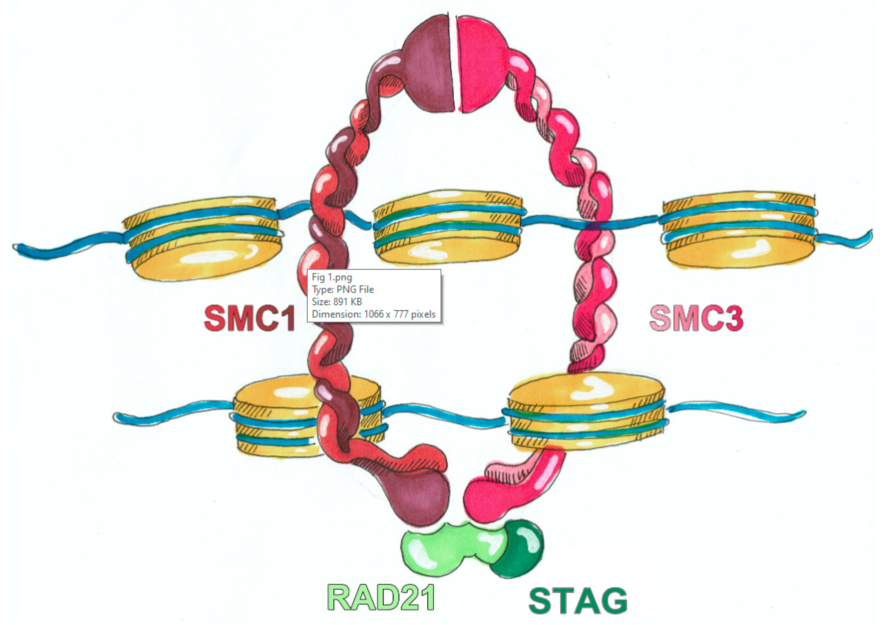
2. Biological Basis of CdLS

To date, six genes of the cohesin complex have been identified and associated with CdLS: nipped-B like protein (NIPBL), structural maintenance of chromosomes 1A (SMC1A) and 3 (SMC3), double strand break repair protein rad21 homolog (RAD21), bromodomain-containing protein 4 (BRD4), histone deacetylase 8 (HDAC8) [17]. Most of the affected individuals show de novo pathogenic variants in one of these genes, with the most prevalent variant in NIPBL (60–70%) [14]. The proteins encoded by CdLS causative genes have been identified as structural or regulatory components of the cohesin complex. The core of cohesin complex (Figure 1) is a multimeric system composed by different subunits SMC1A, SMC3, RAD21 and STAG, it is evolutionarily conserved and exerts its canonical role in sister chromatid cohesion, and non-canonical role in gene expression regulation. The other misregulated proteins, that are not comprised in the core, are cohesin- or chromatin-associated factors genetically different from cohesin but whose variants were found in patients presenting features of CdLSp phenotype through multiple next-generation sequencing (NGS) technologies [18]. The ring-shaped cohesin complex was initially identified for its key role in chromosomal integrity maintenance during the cell cycle phases but now it is known that the interplay with the chromosome loader NIPBL and the sequence-specific DNA binding protein CTCF (CCCTC-binding factor insulator protein) organizes the genome into TADs (topologically associated domains), chromatin loops, and contact domains [17]. Thus, the 3D DNA organization driven by cohesin complex and its associated regulatory proteins helps the contact of sequence elements, including enhancers and promoters, finally orchestrating gene expression [19]. In accordance with this fundamental regulatory role, CdLS cell lines do not display abnormalities in sister chromatid cohesion [20,21] but rather in genes and proteins expression and production [22,23,24,25,26], suggesting that CdLS etiopathology is due to altered transcriptional regulation derived from an impaired function of the cohesin complex in 3D chromatin organization. Furthermore, many genes have also been found weakly transcriptionally dysregulated in CdLS animal models [27].
Besides sister chromatid cohesion and gene expression regulation, a role has recently been proposed for the cohesin complex in DNA damage signaling and repair [28], raising the possibility of novel molecular mechanisms for CdLS. Specifically, Singh et al. demonstrated that insufficient cohesin levels in Nipbl and Hdac8 heterozygous mouse models led to persistent DNA damage in placental cells, resulting in increased senescence and cytokine secretion and ultimately in diminished embryo health and viability [29]. Furthermore, Olley and colleagues showed that CdLS-associated variant in BRD4 delays the cell cycle by perturbing regulation of DNA repair and increasing DNA damage signaling. Similarly, NIPBL deficient cells derived from CdLS patients exhibited an increase in DNA damage response, suggesting the importance of this signaling in the etiology of CdLS [30]. In addition, the RNA-sequencing of patient specific NIPBL+/− iPSC lines identified an altered expression of many mRNAs, pseudogenes, and non-coding RNAs leading to upregulation of gene sets with functions in epigenetic control and downregulation of WNT pathway [31].
WNT signaling alterations have been associated with a plethora of CNS abnormalities and canonical WNT pathway was found to be perturbed in CdLS models [32,33,34,35]. In the contest of embryonic development, the WNT signaling plays a fundamental role in all steps of development by regulating cell proliferation, differentiation, migration, genetic stability and apoptosis, as well as by maintaining adult stem cells and pluripotent state [36]. The many malformations characterizing the CdLSp phenotype have been hypothesized to be derived from developmental WNT/β catenin pathways deregulation.

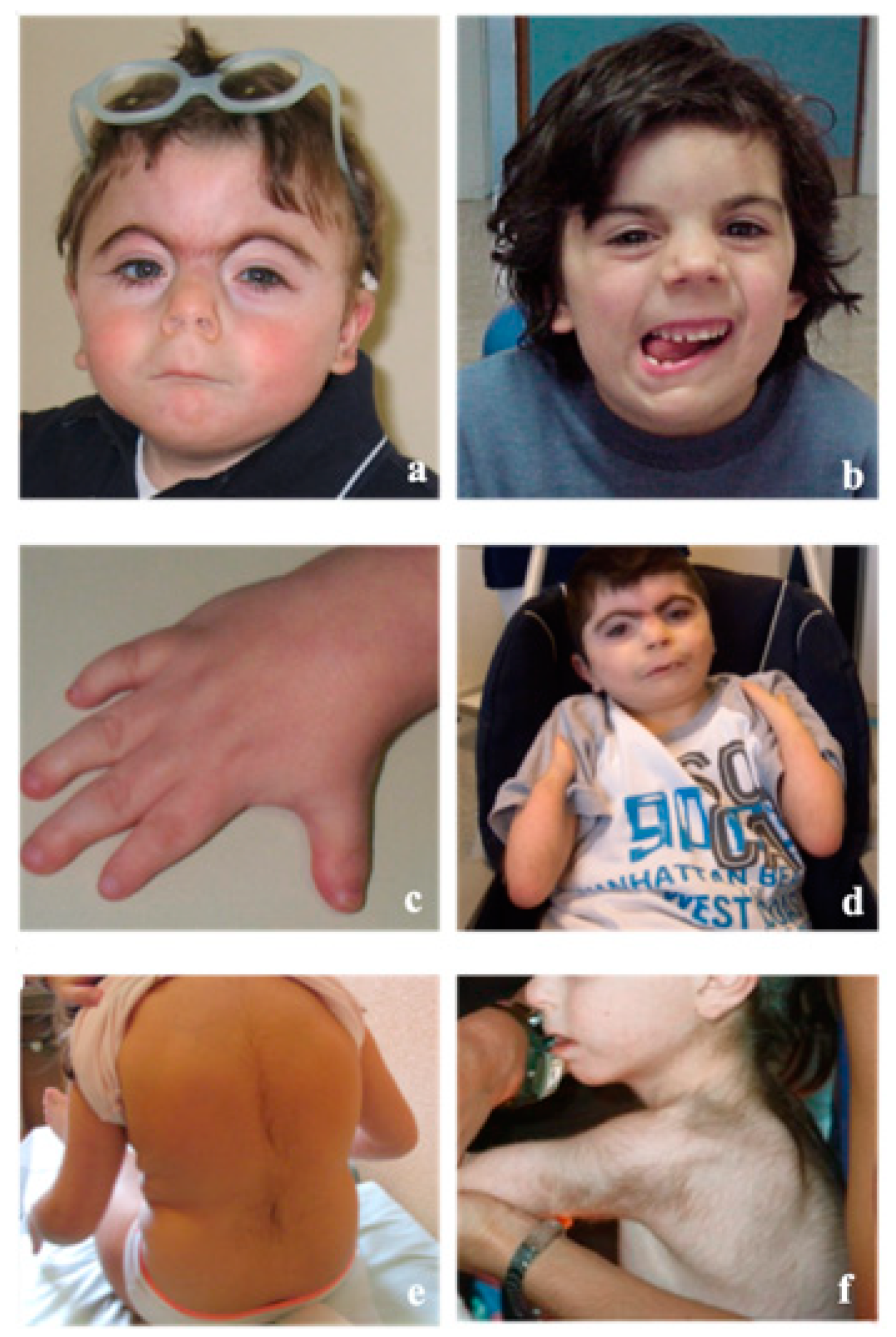
3. Clinical Features

Patients affected with classic CdLS show a quite unique clinical “gestalt” (Figure 2) which can be recognized in the neonatal period. They can present a short neck with low posterior hairline and hirsute forehead. The eyebrows are with synophrys and eyelashes are thick and long; palpebral ptosis is quite frequent with different level of severity. Ears are lowset, midface can be flattened, the nose is short and the philtrum long and featureless. The mouth has a thin upper lip with down-turned corners; a high palate, widely spaced teeth and micrognathia can be observed. Typically, hands and feet are small with shortening of the first metacarpal, proximally placed thumb, brachydactyly, clinodactyly of the fifth finger and single palmar creases. One third of patients can show reduction defects of the upper limbs of variable severity (from oligodactyly to ulnar deficiency to absent forearm). Some degree of reduction in elbow mobility is quite frequent only rarely due to a real radioulnar synostosis. Hirsutism, particularly of the face, lower back and forearms, cutis marmorata and small nipples are observed in the majority of patients.
Patients with CdLSp classically show a prenatal and postnatal growth delay; specific growth charts have been published to properly clinically follow these patients [37]. Such charts were designed before the discovery of the genetic basis of the disease, and hence they should be updated on the basis of genotypic differences. Notably, data from teenagers and young adults underline that CdLSp patients tend with time to develop overweight and/or outright obesity in a fair percentage of cases.
Major malformations have been observed in various organs and systems. Table 2 describes the different percentage of involvement. Apart from upper limb anomalies, none of these malformations is crucial from a diagnostic point of view. It is important to keep in mind that presence of intestinal malrotation, reported in between 5% and 10% of patients, is related to the risk of acute cecal volvulus, which represents a real surgical emergency and one of the most frequent causes of mortality in patients.
Psychomotor and intellectual development are classically delayed, showing a variable degree of severity, from mild to profound, with the majority of patients showing moderate to severe retardation. Communication skills are particularly impaired with expressive language that appears to be more compromised than the receptive one [38]. The behavioral phenotype of CdLSp patients has been deeply analyzed. It is well known that anxiety, mood disorders, impulsivity, challenging and self-injurious behavior, repetitive behavior are present in many patients. Self-injurious behavior, evident in about 56% of patients, seems to be related to severity of intellectual impairment, impulsivity, more compromised communication and NIPBL variant. Pain related to various medical comorbidities should always be considered as a possible reason for the onset of a self-injurious behavior.
As previously stated, natural history of CdLSp patients is characterized by the presence of many possible medical comorbidities that should be considered by pediatricians in the clinical follow-up. Their detailed description is beyond the scope of this review, so they are listed in Table 3.
Quite recently, data on nutritional issues in CdLSp patients have been published, showing that their diets are frequently unbalanced (76%) both from a quantitative and qualitative point of view. However, the authors did not find a direct correlation between patients’ BMI and the nutritional imbalance, hypothesizing an increased metabolic rate for explaining the growth pattern [39]. Similarly, Matute-Llorente et al. performed a detailed research on bone health and body composition in a small number of NIPBL CdLS patients presenting a normal adiposity but a reduced lean mass if quantified by DEXA [40]. Both these papers underline the need for further studies for elucidating the metabolic basis of the growth pattern of CdLSp patients.
Definition of an assessment procedure for the severity of single patient has been attempted by several groups. Kline et al. [14], Selicorni et al. [41], and Bhuiyan et al. [42] suggested specific severity scores based on the combination of different clinical features that classified CdLSp patients in mild/moderate/severe [14,41] or in classical/mild and possible [42]. Moreover, Cereda et al. [43] generated a prognostic score with the aim of early predicting the degree of severity of intellectual disability of a single patient based on a combination of clinical and genetic elements that can be easily defined in the first months of life. Currently none of these scores is considered sufficiently satisfactory on the base of the expectations of the parents.
Table 3. Prevalence of medical problems in CdLS patients [14].
Medical ProblemPrevalence
Gastrointestinal problems
-Feeding problemsfrequent
-Enteral nutrition40%
-Gastro esophageal reflux60–75%
-Batter esophagus9%
-Eosinophilic esophagitis16%
-Constipation10–15%
-Gassiness48%
Neurology
-Seizures45% (SMC1A gene) 15% (NIPBL gene)
-Autonomic nervous system disfunction81% (mild) 26% (severe)
-Sleep problems12–72%
Orthopedics
-Perthes disease4%
-Leg length differences46%
-Congenital hip dislocation10%
-ScoliosisOne third of patients after 10 years
-KyphosisA quarter of patients
-Joint contractures18–25%
-Bunions75%
Visual problems
-Palpebral ptosis37% (unilateral) 44% (bilateral)
-Blepharitis25%
-Nystagmus14–17%
-Strabismus16–26%
-Visual impairment44–53%
-Pigmented peripapillary ring83%
Hearing problems
-Conductive hearing loss75%
-Neurosensorial hearing loss25%
-Otitis media with effusion80–85%
Immunological defects33% (only one specific report) [44]
Thrombocytopeniarare
CancerNo increase of risk

4. Genotype–Phenotype Correlations

From the beginning of the genomic history of CdLS, the aim of researchers has been that of finding possible correlations between clinical phenotype and molecular data as soon as new genes have been discovered. Currently, despite the growing amount of information, major limits in this effort are represented by the enormous difference, in numerical terms, between patients with variants in the major gene (NIPBL) and patients carrying variants in the other genes of the cohesin complex. In fact, it is well known that the latter represent a minority (5–10%) among those who are now classified as CdLSp subjects [14].
NIPBL gene: about 70% of CdLS patients show variants in this gene. It is responsible for the great majority of the more classical and more severely affected cases. This is particularly true for those subjects with the loss of function variants (microdeletions, exon deletions, truncating, non-sense, splicing and frameshift). These patients are the only ones with classical limb reduction defects. Patients with missense variants usually show a better prognosis from both clinical and developmental issues [41,42,45]. Parenti et al. recently described a patient with a severe phenotype and typical CdLS facial dysmorphism carrying a pathogenic variant in MAU2, interactor of NIPBL. This variant appears to affect NIPBL coding sequence and therefore causing NIPBL haploinsufficiency [46].
SMC1A gene: variants in this gene represent about 5% of genetically determined subjects [47]. SMC1A patients show some differences from a dysmorphic point of view compared with NIPBL ones. Their auxological data at birth are frequently normal. Motor development milestones are achieved earlier than in NIPBL patients and their intellectual performance is generally higher [48]. However, about 40% of patients with variants in SMC1A show a completely different phenotype, characterized by a severe epileptic encephalopathy with Rett-like features [49,50].
SMC3 gene: this represents a very rare cause of CdLS. Patients bearing variants in this gene show an atypical CdLS phenotype characterized by short stature, intellectual disability, and quite high prevalence of heart malformation (50–60%) [51].
RAD21 gene: variants in RAD21 gene are rarely observed as a cause of CdLS, and the clinical phenotype is considered atypical [52,53]. Krab et al. reported clinical and molecular data of a wide cohort of RAD21 patients confirming that they show a quite attenuate CdLSp phenotype particularly concerning facial morphology, limb anomalies, behavior and cognitive development [54].
BRD4 gene: the reported number of patients showing haploinsufficiency in this gene so far is too low to describe a specific phenotype [55].
HDAC8 gene: patients with variants in this gene represent about 5% of the global CdLS cohort. These subjects can have both a classical and an atypical phenotype [24,56]. Male patients are more severely affected as the gene locus is on the X chromosome. At a clinical level, these patients quite frequently show hypertelorism, wide anterior fontanel and a happy personality. The severity of the cognitive involvement is generally worse than that SMC1A, SMC3, BDR4 and RAD21 patients [57].
Last, the ANKRD11 gene should be counted, as it was firstly associated with KBG syndrome and then associated with CdLS. It is known that the phenotypic overlapping between these two syndromes can sometimes be very strong [58]. Parenti et al. reported clinical and molecular data of 23 patients with ANKRD11 variants, showing that all but two of them received an initial clinical diagnosis of either CdLS or KBG syndrome; in only five patients did the phenotype at later stages of child development allow a final diagnosis of KBG syndrome [59].
In 2010, Castronovo et al. described a CdLS patient presenting a somatic mosaicism for a NIPBL truncating variant discussing whether mosaicism could be the reason for the less severe phenotype of the child [60]. Later (2013), Huisman et al. described a high prevalence of somatic mosaicism (23%) in their cohort of CdLS patients, suggesting that somatic mosaicism could be more frequent than expected in CdLS. Interestingly, signs and symptoms did not differ between patients with mosaicism and those with germline variants [61]. The real prevalence and the phenotypic consequences of somatic mosaicism in CdLS is still unclear and it needs further study.
A CdLS-like phenotype have been observed also in patients harboring variants in other genes associated with CdLSp overlapping phenotypes. Table 4 summarizes such information.

5. From Biological Basis to Future Treatment Options

To date, no definitive cure exists for CdLSp patients; only specific and localized symptomatic treatments, sometimes alongside surgery. CdLSp patients are characterized by intellectual disabilities, which range from profound to mild, and behavior of the autism-spectrum disorders indicating neural development alterations [14]. The disruption of gene regulation during critical steps of early embryonic development contributes to a WNT signaling alterations that has been associated with a plethora of CNS abnormalities. To date, different studies have shown that the WNT canonical pathway is altered in CdLS models [32,33,34,35], suggesting it and its downstream effectors as a treatment target option.

5.1. Lithium

Lithium is a well-known drug and an effective therapeutic agent for bipolar disorders [62], recently implemented for amyotrophic lateral sclerosis treatment as well, and acts as a specific inhibitor of the conserved WNT transducer GSK3 [63]. Notably, it has been shown that lithium treatment, by modulating the WNT pathway, was able to rescue phenotypes associated with CdLS in nipblb-loss-of-function zebrafish embryos [32] as well as in Drosophila nipbl model and murine and human in vitro models [35]. These data suggest that lithium might be used to at least partially ameliorate CdLS phenotype also in CdLS human fetuses, by maternal administration, since it is frequently prescribed to pregnant women for the treatment of bipolar disorder [64]. Yet, the use of lithium during pregnancy raises concerns, mainly with respect to the negative effects on embryo development. Two independent groups have shown the consequences of in utero lithium exposure on embryos by meta-analysis studies. Lithium administration in pregnant women during the first trimester increased the risk of fetus malformations and neonatal readmission within one month postpartum [65]. On the other hand, while most of preclinical studies reported adverse effects of prenatal exposure to lithium on neurodevelopment or behavior, clinical studies regarding intra uterine lithium exposure do not show abnormal fetus development [66]. Alternatively, a possible use of lithium in the treatment of CdLS patients might be considered in the first years of life, when newborn brain is still undergoing developmental and plasticity processes [67], with the aim of ameliorating cognitive and behavioral features. Lithium has also been proposed for a clinical trial in the context of fragile X syndrome for the treatment of behavioral aspects [68]. A selective inhibitor of GSK3α has been recently developed to avoid the toxic effects derived lithium-dependent inhibition of both GSK3 paralogs, and it was shown to successfully correct some phenotypes features of fragile X syndrome mouse models [69].

5.2. L-Leucin

Besides lithium treatment, L-leucine administration has been proposed. The reduced translation has been suggested to be a common feature in cohesinopathies animal models. L-leucine treatment during zebrafish development has been shown to rescue translation through mTOR pathway stimulation. Specifically, Xu and colleagues showed that in zebrafish embryos, the downregulation of cohesin complex genes by morpholino injection, resulted in multiple developmental defects typical of the CdLS phenotype, which were partially corrected by treatment with L-leucin. These data suggest that translation could be an interesting therapeutic target and that CdLS patients could benefit from this [70].

5.3. Antioxidant

At a cellular level, CdLS presents cell growth arrest, apoptosis, genome instability and premature aging, hallmarks of high levels of oxidative stress [71]. Thus, the reduction of reactive radical oxygen molecules presents a potential therapeutic target, and antioxidants have been suggested for drug repurposing with a clinical potential in improving CdLS phenotype. Treatment with ascorbic acid and riboceine drugs led to the extension of in vitro lifespan of CdLS cell lines and zebrafish model by reducing both the level of oxidative stress and genome instability [26]. In addition, it has been shown that N-acetyl-cysteine protects the DNA from damage and prevent cell death, in cerebellar cells [72,73]. A randomized clinical trial for the use of N-acetyl-cysteine in the treatment of neurodevelopmental disorders in CdLSp patients is estimated to start within this year (https://clinicaltrials.gov/ct2/show/NCT04381, accessed on 10 September 2019).

6. Conclusions

CdLS could be considered to be an archetype of the successful interlink between bench and bedside research. Indeed, the two lanes have developed in parallel while bridging the gaps in knowledge over the decades and, as a result of this shared effort, CdLS is now a well-defined rare genetic disorder. Furthermore, thanks to the advancement in understanding of CdLS, the information gained has led to the hypothesizing of future therapeutic approaches, while at the same time sustaining the improvement of daily care and planned follow-up.

Author Contributions

Conceptualization, A.S. and V.M.; resources, A.S., M.M., A.L. and V.M.; writing—original draft preparation, A.S., M.M, A.L and V.M.; writing—review and editing, A.S., M.M., A.L. and V.M.; visualization, M.M. and A.L.; supervision, A.S. and V.M; project administration, A.S. All authors have read and agreed to the published version of the manuscript.

Funding

This research did not receive any specific grant from funding agencies in the public, commercial, or not-for-profit sectors.

Institutional Review Board Statement

Not applicable.

Informed Consent Statement

Not applicable.

Acknowledgments

The authors are grateful to Susanna Brusa for assistance with drawings. We thank the Mariani Foundation and the Associazione Silvia Onlus for the support to the clinical activity of the Mariani Foundation Center for Fragile Child, Pediatric Unit ASST Lariana Como. The authors would also like to express their deepest gratitude to CdLS Family Italian Association for constant support and inspiration. The authors acknowledge support from Università degli Studi di Milano through the APC initiative.

Conflicts of Interest

The authors declare no conflict of interest.

References

  1. Mannini, L.; Cucco, F.; Quarantotti, V.; Krantz, I.D.; Musio, A. Mutation spectrum and genotype-phenotype correlation in Cornelia de Lange syndrome. Hum. Mutat. 2013, 34, 1589–1596. [Google Scholar] [CrossRef] [Green Version]
  2. De Lange, C.C. Sur un type nouveau de degenerescence (typus Amstelodamensis). Arch. Med. Enfants 1933, 36, 713–719. [Google Scholar]
  3. Meinecke, P.; Hayek, H. Brief historical note on the Brachmann-de Lange syndrome: A patient closely resembling the case described by Brachmann in 1916. Am. J. Med. Genet. 1990, 35, 449–450. [Google Scholar] [CrossRef] [PubMed]
  4. Brachmann, W.R.C. Ein Fall von symmetrischer Monodaktylie durch Ulnadefekt, mit symmetrischer Flughautbildung in den Ellenbeugen, sowie anderen Abnormitäten (Zwerghaftigkeit, Halsrippen, Behaarung) (A case of symmetrical monodactyly, representing ulnar deficiency, with symmetrical antecubital webbing and other abnormalities, (dwarfism, cervical ribs, hirsutism)). Jarb. Kinder Phys. Erzie 1916, 84, 225–235. [Google Scholar]
  5. Vrolik, W. Tabulae ad Illustrandam Embryogenesin Hominis et Mammalium Tam Naturalem Quam Abnormem; Amstelodami: London, UK, 1849. [Google Scholar]
  6. Berg, J.; McCreary, B.; Ridler, M.A.; Smith, G. The De Lange Syndrome; Pergamon Press: Oxford, UK, 1970. [Google Scholar]
  7. Preus, M.; Rex, A.P. Definition and diagnosis of the Brachmann-De Lange syndrome. Am. J. Med. Genet. 1983, 16, 301–312. [Google Scholar] [CrossRef]
  8. Kline, A.D.; Krantz, I.D.; Sommer, A.; Kliewer, M.; Jackson, L.G.; FitzPatrick, D.R.; Levin, A.V.; Selicorni, A. Cornelia de Lange syndrome: Clinical review, diagnostic and scoring systems, and anticipatory guidance. Am. J. Med. Genet. Part A 2007, 143, 1287–1296. [Google Scholar] [CrossRef]
  9. Van Allen, M.I.; Filippi, G.; Siegel-Bartelt, J.; Yong, S.-L.; McGillivray, B.; Zuker, R.M.; Smith, C.R.; Magee, J.F.; Ritchie, S.; Toi, A.; et al. Clinical variability within Brachmann-de Lange syndrome: A proposed classification system. Am. J. Med. Genet. 1993, 47, 947–958. [Google Scholar] [CrossRef]
  10. Selicorni, A.; Lalatta, F.; Livini, E.; Briscioli, V.; Piguzzi, T.; Bagozzi, D.C.; Mastroiacovo, P.; Zampino, G.; Gaeta, G.; Pugliese, A.; et al. Variability of the Brachmann-de Lange Syndrome. Am. J. Med. Genet. 1993, 47, 977–982. [Google Scholar] [CrossRef] [PubMed]
  11. Bay, C.; Mauk, J.; Radcliffe, J.; Kaplan, P. Mild Brachmann-de Lange syndrome. Delineation of the clinical phenotype, and characteristic behaviors in a six-year-old boy. Am. J. Med. Genet. 1993, 47, 965–968. [Google Scholar] [CrossRef] [PubMed]
  12. Clericuzio, C.L. Mild mental retardation with classic somatic phenotype in the Brachmann-de Lange syndrome. Am. J. Med. Genet. 1993, 47, 992–994. [Google Scholar] [CrossRef]
  13. Saal, H.M.; Samango-Sprouse, C.A.; Rodnan, L.A.; Rosenbaum, K.N.; Custer, D.A. Brachmann-de Lange syndrome with normal IQ. Am. J. Med. Genet. 1993, 47, 995–998. [Google Scholar] [CrossRef]
  14. Kline, A.D.; Moss, J.F.; Selicorni, A.; Bisgaard, A.M.; Deardorff, M.A.; Gillett, P.M.; Ishman, S.L.; Kerr, L.M.; Levin, A.V.; Mulder, P.A.; et al. Diagnosis and management of Cornelia de Lange syndrome: First international consensus statement. Nat. Rev. Genet. 2018, 19, 649–666. [Google Scholar] [CrossRef] [PubMed] [Green Version]
  15. Gurovich, Y.; Hanani, Y.; Bar, O.; Nadav, G.; Fleischer, N.; Gelbman, D.; Basel-Salmon, L.; Krawitz, P.M.; Kamphausen, S.B.; Zenker, M.; et al. Identifying facial phenotypes of genetic disorders using deep learning. Nat. Med. 2019, 25, 60–64. [Google Scholar] [CrossRef] [PubMed]
  16. Latorre-Pellicer, A.; Ascaso, Á.; Trujillano, L.; Gil-Salvador, M.; Arnedo, M.; Lucia-Campos, C.; Antoñanzas-Pérez, R.; Marcos-Alcalde, I.; Parenti, I.; Bueno-Lozano, G.; et al. Evaluating Face2Gene as a Tool to Identify Cornelia de Lange Syndrome by Facial Phenotypes. Int. J. Mol. Sci. 2020, 21, 1042. [Google Scholar] [CrossRef] [PubMed] [Green Version]
  17. Panaitescu, A.M.; Duta, S.; Gica, N.; Botezatu, R.; Nedelea, F.; Peltecu, G.; Veduta, A. A broader perspective on the prenatal diagnosis of cornelia de lange syndrome: Review of the literature and case presentation. Diagnostics 2021, 11, 142. [Google Scholar] [CrossRef]
  18. Avagliano, L.; Parenti, I.; Grazioli, P.; Di Fede, E.; Parodi, C.; Mariani, M.; Kaiser, F.J.; Selicorni, A.; Gervasini, C.; Massa, V. Chromatinopathies: A focus on Cornelia de Lange syndrome. Clin. Genet. 2020, 97, 3–11. [Google Scholar] [CrossRef] [PubMed] [Green Version]
  19. Weiss, F.D.; Calderon, L.; Wang, Y.F.; Georgieva, R.; Guo, Y.; Cvetesic, N.; Kaur, M.; Dharmalingam, G.; Krantz, I.D.; Lenhard, B.; et al. Neuronal genes deregulated in Cornelia de Lange Syndrome respond to removal and re-expression of cohesin. Nat. Commun. 2021, 12, 1–13. [Google Scholar] [CrossRef]
  20. Revenkova, E.; Focarelli, M.L.; Susani, L.; Paulis, M.; Bassi, M.T.; Mannini, L.; Frattini, A.; Delia, D.; Krantz, I.; Vezzoni, P.; et al. Cornelia de Lange syndrome mutations in SMC1A or SMC3 affect binding to DNA. Hum. Mol. Genet. 2009, 18, 418–427. [Google Scholar] [CrossRef] [Green Version]
  21. Castronovo, P.; Gervasini, C.; Cereda, A.; Masciadri, M.; Milani, D.; Russo, S.; Selicorni, A.; Larizza, L. Premature chromatid separation is not a useful diagnostic marker for Cornelia de Lange syndrome. Chromosom. Res. 2009, 17, 763–771. [Google Scholar] [CrossRef] [PubMed]
  22. Liu, J.; Zhang, Z.; Bando, M.; Itoh, T.; Deardorff, M.A.; Clark, D.; Kaur, M.; Tandy, S.; Kondoh, T.; Rappaport, E.; et al. Transcriptional Dysregulation in NIPBLand Cohesin Mutant Human Cells. PLoS Biol. 2009, 7, e1000119. [Google Scholar] [CrossRef] [PubMed]
  23. Gimigliano, A.; Mannini, L.; Bianchi, L.; Puglia, M.; Deardorff, M.A.; Menga, S.; Krantz, I.D.; Musio, A.; Bini, L. Proteomic profile identifies dysregulated pathways in Cornelia de Lange syndrome cells with distinct mutations in SMC1A and SMC3 genes. J. Proteome Res. 2012, 11, 6111–6123. [Google Scholar] [CrossRef] [PubMed] [Green Version]
  24. Deardorff, M.A.; Bando, M.; Nakato, R.; Watrin, E.; Itoh, T.; Minamino, M.; Saitoh, K.; Komata, M.; Katou, Y.; Clark, D.; et al. HDAC8 mutations in Cornelia de Lange syndrome affect the cohesin acetylation cycle. Nature 2012, 489, 313–317. [Google Scholar] [CrossRef] [PubMed]
  25. Mannini, L.; Lamaze, F.C.; Cucco, F.; Amato, C.; Quarantotti, V.; Rizzo, I.M.; Krantz, I.D.; Bilodeau, S.; Musio, A. Mutant cohesin affects RNA polymerase II regulation in Cornelia de Lange syndrome. Sci. Rep. 2015, 5, 16803. [Google Scholar] [CrossRef] [Green Version]
  26. Cukrov, D.; Newman, T.A.C.; Leask, M.; Leeke, B.; Sarogni, P.; Patimo, A.; Kline, A.D.; Krantz, I.D.; Horsfield, J.A.; Musio, A. Antioxidant treatment ameliorates phenotypic features of SMC1A-mutated Cornelia de Lange syndrome in vitro and in vivo. Hum. Mol. Genet. 2018, 27, 3002–3011. [Google Scholar] [CrossRef] [PubMed]
  27. Cucco, F.; Sarogni, P.; Rossato, S.; Alpa, M.; Patimo, A.; Latorre, A.; Magnani, C.; Puisac, B.; Ramos, F.J.; Pié, J.; et al. Pathogenic variants in EP300 and ANKRD11 in patients with phenotypes overlapping Cornelia de Lange syndrome. Am. J. Med. Genet. Part A 2020, 182, 1690–1696. [Google Scholar] [CrossRef] [PubMed]
  28. Litwin, I.; Pilarczyk, E.; Wysocki, R. The Emerging Role of Cohesin in the DNA Damage Response. Genes 2018, 9, 581. [Google Scholar] [CrossRef] [Green Version]
  29. Singh, V.P.; McKinney, S.; Gerton, J.L. Persistent DNA Damage and Senescence in the Placenta Impacts Developmental Outcomes of Embryos. Dev. Cell 2020, 54, 333.e7–347.e7. [Google Scholar] [CrossRef]
  30. Olley, G.; Pradeepa, M.M.; Grimes, G.R.; Piquet, S.; Polo, S.E.; FitzPatrick, D.R.; Bickmore, W.A.; Boumendil, C. Cornelia de Lange syndrome-associated mutations cause a DNA damage signalling and repair defect. Nat. Commun. 2021, 12, 3127. [Google Scholar] [CrossRef]
  31. Mills, J.A.; Herrera, P.S.; Kaur, M.; Leo, L.; McEldrew, D.; Tintos-Hernandez, J.A.; Rajagopalan, R.; Gagne, A.; Zhang, Z.; Ortiz-Gonzalez, X.R.; et al. NIPBL+/− haploinsufficiency reveals a constellation of transcriptome disruptions in the pluripotent and cardiac states. Sci. Rep. 2018, 8, 1056. [Google Scholar] [CrossRef] [Green Version]
  32. Pistocchi, A.; Fazio, G.; Cereda, A.; Ferrari, L.; Bettini, L.R.; Messina, G.; Cotelli, F.; Biondi, A.; Selicorni, A.; Massa, V. Cornelia de Lange Syndrome: NIPBL haploinsufficiency downregulates canonical Wnt pathway in zebrafish embryos and patients fibroblasts. Cell Death Dis. 2013, 4, e866. [Google Scholar] [CrossRef] [Green Version]
  33. Fazio, G.; Gaston-Massuet, C.; Bettini, L.R.; Graziola, F.; Scagliotti, V.; Cereda, A.; Ferrari, L.; Mazzola, M.; Cazzaniga, G.; Giordano, A.; et al. CyclinD1 Down-Regulation and Increased Apoptosis Are Common Features of Cohesinopathies. J. Cell. Physiol. 2016, 231, 613–622. [Google Scholar] [CrossRef]
  34. Avagliano, L.; Grazioli, P.; Mariani, M.; Bulfamante, G.P.; Selicorni, A.; Massa, V. Integrating molecular and structural findings: Wnt as a possible actor in shaping cognitive impairment in Cornelia de Lange syndrome. Orphanet J. Rare Dis. 2017, 12, 174. [Google Scholar] [CrossRef] [PubMed] [Green Version]
  35. Grazioli, P.; Parodi, C.; Mariani, M.; Bottai, D.; Di Fede, E.; Zulueta, A.; Avagliano, L.; Cereda, A.; Tenconi, R.; Wierzba, J.; et al. Lithium as a possible therapeutic strategy for Cornelia de Lange syndrome. Cell Death Discov. 2021, 7. [Google Scholar] [CrossRef]
  36. Miyabayashi, T.; Teo, J.-L.; Yamamoto, M.; McMillan, M.; Nguyen, C.; Kahn, M. Wnt/β-catenin/CBP signaling maintains long-term murine embryonic stem cell pluripotency. Proc. Natl. Acad. Sci. USA 2007, 104, 5668–5673. [Google Scholar] [CrossRef] [PubMed] [Green Version]
  37. Kline, A.D.; Barr, M.; Jackson, L.G. Growth manifestations in the Brachmann-de Lange syndrome. Am. J. Med. Genet. 1993, 47, 1042–1049. [Google Scholar] [CrossRef] [PubMed]
  38. Ajmone, P.F.; Rigamonti, C.; Dall’Ara, F.; Monti, F.; Vizziello, P.; Milani, D.; Cereda, A.; Selicorni, A.; Costantino, A. Communication, cognitive development and behavior in children with cornelia de lange syndrome (CdLS): Preliminary results. Am. J. Med. Genet. Part B Neuropsychiatr. Genet. 2014, 165, 223–229. [Google Scholar] [CrossRef]
  39. Parma, B.; Cianci, P.; Decimi, V.; Mariani, M.; Provero, M.C.; Funari, C.; Tajè, S.; Apuril, E.; Cereda, A.; Panceri, R.; et al. Complex nutritional deficiencies in a large cohort of Italian patients with Cornelia de Lange syndrome spectrum. Am. J. Med. Genet. Part A 2020, 182, 2094–2101. [Google Scholar] [CrossRef] [PubMed]
  40. Matute-Llorente, Á.; Ascaso, Á.; Latorre-Pellicer, A.; Puisac, B.; Trujillano, L.; Llorente, E.; Puente-Lanzarote, J.J.; Ayerza-Casas, A.; Arnedo, M.; Moreno, L.A.; et al. Targeted Gene Sequencing, Bone Health, and Body Composition in Cornelia de Lange Syndrome. Appl. Sci. 2021, 11, 710. [Google Scholar] [CrossRef]
  41. Selicorni, A.; Russo, S.; Gervasini, C.; Castronovo, P.; Milani, D.; Cavalleri, F.; Bentivegna, A.; Masciadri, M.; Domi, A.; Divizia, M.T.; et al. Clinical score of 62 Italian patients with Cornelia de Lange syndrome and correlations with the presence and type of NIPBL mutation. Clin. Genet. 2007, 72, 98–108. [Google Scholar] [CrossRef]
  42. Bhuiyan, Z.A.; Klein, M.; Hammond, P.; Van Haeringen, A.; Mannens, M.M.A.M.; Van Berckelaer-Onnes, I.; Hennekam, R.C.M. Genotype-phenotype correlations of 39 patients with Cornelia de Lange syndrome: The Dutch experience. J. Med. Genet. 2006, 43, 568–575. [Google Scholar] [CrossRef] [PubMed] [Green Version]
  43. Cereda, A.; Mariani, M.; Rebora, P.; Sajeva, A.; Ajmone, P.F.; Gervasini, C.; Russo, S.; Kullmann, G.; Valsecchi, G.; Selicorni, A. A new prognostic index of severity of intellectual disabilities in Cornelia de Lange syndrome. Am. J. Med. Genet. Part C Semin. Med. Genet. 2016, 172, 179–189. [Google Scholar] [CrossRef]
  44. Jyonouchi, S.; Orange, J.; Sullivan, K.E.; Krantz, I.; Deardorff, M. Immunologic Features of Cornelia de Lange Syndrome. Pediatrics 2013, 132, e484–e489. [Google Scholar] [CrossRef] [PubMed] [Green Version]
  45. Gillis, L.A.; McCallum, J.; Kaur, M.; DeScipio, C.; Yaeger, D.; Mariani, A.; Kline, A.D.; Li, H.H.; Devoto, M.; Jackson, L.G.; et al. NIPBL mutational analysis in 120 individuals with Cornelia de Lange syndrome and evaluation of genotype-phenotype correlations. Am. J. Hum. Genet. 2004, 75, 610–623. [Google Scholar] [CrossRef] [PubMed] [Green Version]
  46. Parenti, I.; Diab, F.; Gil, S.R.; Mulugeta, E.; Casa, V.; Berutti, R.; Brouwer, R.W.W.; Dupé, V.; Eckhold, J.; Graf, E.; et al. MAU2 and NIPBL Variants Impair the Heterodimerization of the Cohesin Loader Subunits and Cause Cornelia de Lange Syndrome. Cell Rep. 2020, 31, 107647. [Google Scholar] [CrossRef]
  47. Huisman, S.; Mulder, P.A.; Redeker, E.; Bader, I.; Bisgaard, A.M.; Brooks, A.; Cereda, A.; Cinca, C.; Clark, D.; Cormier-Daire, V.; et al. Phenotypes and genotypes in individuals with SMC1A variants. Am. J. Med. Genet. Part A 2017, 173, 2108–2125. [Google Scholar] [CrossRef] [Green Version]
  48. Borck, G.; Zarhrate, M.; Bonnefont, J.P.; Munnich, A.; Cormier-Daire, V.; Colleaux, L. Incidence and clinical features of X-linked Cornelia de Lange syndrome due to SMC1L1 mutations. Hum. Mutat. 2007, 28, 205–206. [Google Scholar] [CrossRef] [PubMed]
  49. Symonds, J.D.; Joss, S.; Metcalfe, K.A.; Somarathi, S.; Cruden, J.; Devlin, A.M.; Donaldson, A.; DiDonato, N.; Fitzpatrick, D.; Kaiser, F.J.; et al. Heterozygous truncation mutations of the SMC1A gene cause a severe early onset epilepsy with cluster seizures in females: Detailed phenotyping of 10 new cases. Epilepsia 2017, 58, 565–575. [Google Scholar] [CrossRef] [Green Version]
  50. Jansen, S.; Kleefstra, T.; Willemsen, M.H.; de Vries, P.; Pfundt, R.; Hehir-Kwa, J.Y.; Gilissen, C.; Veltman, J.A.; de Vries, B.B.A.; Vissers, L.E.L.M. De novo loss-of-function mutations in X-linked SMC1A cause severe ID and therapy-resistant epilepsy in females: Expanding the phenotypic spectrum. Clin. Genet. 2016, 90, 413–419. [Google Scholar] [CrossRef]
  51. Gil-Rodríguez, M.C.; Deardorff, M.A.; Ansari, M.; Tan, C.A.; Parenti, I.; Baquero-Montoya, C.; Ousager, L.B.; Puisac, B.; Hernández-Marcos, M.; Teresa-Rodrigo, M.E.; et al. De novo heterozygous mutations in SMC3 cause a range of cornelia de lange syndrome-overlapping phenotypes. Hum. Mutat. 2015, 36, 454–462. [Google Scholar] [CrossRef] [Green Version]
  52. Boyle, M.I.; Jespersgaard, C.; Nazaryan, L.; Bisgaard, A.-M.; Tümer, Z. A novel RAD21 variant associated with intrafamilial phenotypic variation in Cornelia de Lange syndrome—Review of the literature. Clin. Genet. 2017, 91, 647–649. [Google Scholar] [CrossRef]
  53. Deardorff, M.A.; Wilde, J.J.; Albrecht, M.; Dickinson, E.; Tennstedt, S.; Braunholz, D.; Mönnich, M.; Yan, Y.; Xu, W.; Gil-Rodríguez, M.C.; et al. RAD21 mutations cause a human cohesinopathy. Am. J. Hum. Genet. 2012, 90, 1014–1027. [Google Scholar] [CrossRef] [PubMed] [Green Version]
  54. Krab, L.C.; Marcos-Alcalde, I.; Assaf, M.; Balasubramanian, M.; Andersen, J.B.; Bisgaard, A.-M.; Fitzpatrick, D.R.; Gudmundsson, S.; Huisman, S.A.; Kalayci, T.; et al. Delineation of phenotypes and genotypes related to cohesin structural protein RAD21. Hum. Genet. 2020, 139, 575–592. [Google Scholar] [CrossRef] [PubMed] [Green Version]
  55. Olley, G.; Ansari, M.; Bengani, H.; Grimes, G.R.; Rhodes, J.; Von Kriegsheim, A.; Blatnik, A.; Stewart, F.J.; Wakeling, E.; Carroll, N.; et al. BRD4 interacts with NIPBL and BRD4 is mutated in a Cornelia de Lange-like syndrome. Nat. Genet. 2018, 50, 329–332. [Google Scholar] [CrossRef] [PubMed]
  56. Harakalova, M.; van den Boogaard, M.J.; Sinke, R.; van Lieshout, S.; van Tuil, M.C.; Duran, K.; Renkens, I.; Terhal, P.A.; de Kovel, C.; Nijman, I.J.; et al. X-exome sequencing identifies a HDAC8 variant in a large pedigree with X-linked intellectual disability, truncal obesity, gynaecomastia, hypogonadism and unusual face. J. Med. Genet. 2012, 49, 539–543. [Google Scholar] [CrossRef]
  57. Parenti, I.; Gervasini, C.; Pozojevic, J.; Wendt, K.S.; Watrin, E.; Azzollini, J.; Braunholz, D.; Buiting, K.; Cereda, A.; Engels, H.; et al. Expanding the clinical spectrum of the “HDAC8-phenotype”—Implications for molecular diagnostics, counseling and risk prediction. Clin. Genet. 2016, 89, 564–573. [Google Scholar] [CrossRef]
  58. Parenti, I.; Gervasini, C.; Pozojevic, J.; Graul-Neumann, L.; Azzollini, J.; Braunholz, D.; Watrin, E.; Wendt, K.S.; Cereda, A.; Cittaro, D.; et al. Broadening of cohesinopathies: Exome sequencing identifies mutations in ANKRD11 in two patients with Cornelia de Lange-overlapping phenotype. Clin. Genet. 2016, 89, 74–81. [Google Scholar] [CrossRef]
  59. Parenti, I.; Mallozzi, M.B.; Hüning, I.; Gervasini, C.; Kuechler, A.; Agolini, E.; Albrecht, B.; Baquero-Montoya, C.; Bohring, A.; Bramswig, N.C.; et al. ANKRD11 variants: KBG syndrome and beyond. Clin. Genet. 2021, cge.13977. [Google Scholar] [CrossRef]
  60. Castronovo, P.; Delahaye-Duriez, A.; Gervasini, C.; Azzollini, J.; Minier, F.; Russo, S.; Masciadri, M.; Selicorni, A.; Verloes, A.; Larizza, L. Somatic mosaicism in Cornelia de Lange syndrome: A further contributor to the wide clinical expressivity? Clin. Genet. 2010, 78, 560–564. [Google Scholar] [CrossRef]
  61. Huisman, S.A.; Redeker, E.J.W.; Maas, S.M.; Mannens, M.M.; Hennekam, R.C.M. High rate of mosaicism in individuals with Cornelia de Lange syndrome. J. Med. Genet. 2013, 50, 339–344. [Google Scholar] [CrossRef]
  62. Volkmann, C.; Bschor, T.; Köhler, S. Lithium Treatment Over the Lifespan in Bipolar Disorders. Front. Psychiatry 2020, 11, 377. [Google Scholar] [CrossRef]
  63. Stambolic, V.; Ruel, L.; Woodgett, J.R. Lithium inhibits glycogen synthase kinase-3 activity and mimics Wingless signalling in intact cells. Curr. Biol. 1996, 6, 1664–1669. [Google Scholar] [CrossRef] [Green Version]
  64. Poels, E.M.P.; Bijma, H.H.; Galbally, M.; Bergink, V. Lithium during pregnancy and after delivery: A review. Int. J. Bipolar Disord. 2018, 6, 26. [Google Scholar] [CrossRef] [PubMed] [Green Version]
  65. Munk-Olsen, T.; Liu, X.; Viktorin, A.; Brown, H.K.; Di Florio, A.; D’Onofrio, B.M.; Gomes, T.; Howard, L.M.; Khalifeh, H.; Krohn, H.; et al. Maternal and infant outcomes associated with lithium use in pregnancy: An international collaborative meta-analysis of six cohort studies. Lancet Psychiatry 2018, 5, 644–652. [Google Scholar] [CrossRef] [Green Version]
  66. Poels, E.M.P.; Schrijver, L.; Kamperman, A.M.; Hillegers, M.H.J.; Hoogendijk, W.J.G.; Kushner, S.A.; Roza, S.J. Long-term neurodevelopmental consequences of intrauterine exposure to lithium and antipsychotics: A systematic review and meta-analysis. Eur. Child. Adolesc. Psychiatry 2018, 27, 1209–1230. [Google Scholar] [CrossRef] [PubMed] [Green Version]
  67. Van Dyck, L.I.; Morrow, E.M. Genetic control of postnatal human brain growth. Curr. Opin. Neurol. 2017, 30, 114–124. [Google Scholar] [CrossRef] [PubMed] [Green Version]
  68. Liu, Z.; Smith, C.B. Lithium: A promising treatment for fragile X syndrome. ACS Chem. Neurosci. 2014, 5, 477–483. [Google Scholar] [CrossRef]
  69. McCamphill, P.K.; Stoppel, L.J.; Senter, R.K.; Lewis, M.C.; Heynen, A.J.; Stoppel, D.C.; Sridhar, V.; Collins, K.A.; Shi, X.; Pan, J.Q.; et al. Selective inhibition of glycogen synthase kinase 3α corrects pathophysiology in a mouse model of fragile X syndrome. Sci. Transl. Med. 2020, 12, eaam8572. [Google Scholar] [CrossRef]
  70. Xu, B.; Sowa, N.; Cardenas, M.E.; Gerton, J.L. L-leucine partially rescues translational and developmental defects associated with zebrafish models of Cornelia de Lange syndrome. Hum. Mol. Genet. 2015, 24, 1540–1555. [Google Scholar] [CrossRef]
  71. Sarogni, P.; Pallotta, M.M.; Musio, A. Cornelia de Lange syndrome: From molecular diagnosis to therapeutic approach. J. Med. Genet. 2020, 57, 289–295. [Google Scholar] [CrossRef]
  72. Reliene, R.; Fischer, E.; Schiestl, R.H. Effect of N -Acetyl Cysteine on Oxidative DNA Damage and the Frequency of DNA Deletions in Atm -Deficient Mice. Cancer Res. 2004, 64, 5148–5153. [Google Scholar] [CrossRef] [Green Version]
  73. Arakawa, M.; Ushimaru, N.; Osada, N.; Oda, T.; Ishige, K.; Ito, Y. N-acetylcysteine selectively protects cerebellar granule cells from 4-hydroxynonenal-induced cell death. Neurosci. Res. 2006, 55, 255–263. [Google Scholar] [CrossRef] [PubMed]
Figure 1. The ring-shaped cohesin complex is formed by a multi-subunit core: structural maintenance of chromosomes (SMC3 and SMC1A), RAD cohesin complex component (RAD21), and cohesin subunit SA (STAG). Cohesin complex can entrap the sister chromatids and regulates the separation of sister chromatids; furthermore, can modulate the chromatin conformation and affects normal gene expression.
Genes 12 01075 g001
Figure 2. Main clinical features in CdLSp. Facial phenotypes: classic phenotype (a) and mild phenotype (b). Typical small hand with brachydactyly, proximal thumb and clinodactyly of the fifth finger (c) or limb reduction defect (d). Typical hirsutism of the back localized (e) and diffuse (f).
Genes 12 01075 g002
Table 1. Diagnostic algorithm as suggested by the Consensus Statement.
Cardinal FeaturesScore
Synophrys (HP:0000664) and/or thick eyebrows (HP:0000574)2
Short nose (HP:0003196), concave nasal ridge (HP:0011120) and/or upturned nasal tip (HP:0000463)2
Long (HP:0000343) and/or smooth philtrum (HP:0000319)2
Thin upper lip vermilion (HP:0000219) and/or downturned corners of mouth (HP:0002714)2
Hand oligodactyly (HP:0001180) and/or adactyly (HP:0009776)2
Congenital diaphragmatic hernia (HP:0000776)2
Suggestive Features
Global developmental delay (HP:0001263) and/or intellectual disability (HP:0001249)1
Prenatal growth retardation (<2 SD) (HP:0001511)1
Postnatal growth retardation (<2 SD) (HP:0008897)1
Microcephaly (prenatally and/or postnatally) (HP:0000252)1
Small hands (HP:0200055) and/or feet (HP:0001773)1
Short fifth finger (HP:0009237)1
Hirsutism (HP:0001007)1
Interpretation of the Score
Score ≥11 points, of which at least 3 are cardinal features classic CdLS
Score between 9 or 10 points, of which at least 2 are cardinal featuresnon-classic CdLS
Score between 4–8 points, of which at least 1 is cardinal featuremolecular testing for CdLS indicated
Score <4 pointsinsufficient to indicate molecular testing for CdLS
Table 2. Major malformations described in CdLS [14].
MalformationPrevalence
Heart malformations (no specific defect)25%
Palate20%
Eyes
Unilateral or bilateral nasolacrimal duct obstruction
60–80%
Central Nervous System47% (in the wider cohort reported) [35]
Limb defectsAbout one third
Urinary tract10%
Genitalia
Cryptorchidism80%
Micropenis37%
Hypospadias9%
Bicornuate uterus19%
Gastrointestinal system
Intestinal malrotation5–10%
Pyloric stenosis7%
Diaphragmatic Herniarare
Table 4. CdLS-like phenotypes.
GeneOverlapping Syndrome
ANKRD11KBG syndrome
AFF4CHOP syndrome
EP300Rubinstein-Taybi syndrome
KMT2AWiedemann-Steiner syndrome
TAF6Alazami-Yuan syndrome
SETD5Mental retardation, autosomal dominant 23
ARID1BCoffin-Siris syndrome
Publisher’s Note: MDPI stays neutral with regard to jurisdictional claims in published maps and institutional affiliations.

