Review

Plasmid Transfer by Conjugation in Gram-Negative Bacteria: From the Cellular to the Community Level

Microbiologie Moléculaire et Biochimie Structurale (MMSB), Université Lyon 1, CNRS, Inserm, UMR5086, 69007 Lyon, France
*
Author to whom correspondence should be addressed.
These authors contributed equally to this work.
Genes 2020, 11(11), 1239; https://doi.org/10.3390/genes11111239
Submission received: 16 September 2020 / Revised: 15 October 2020 / Accepted: 20 October 2020 / Published: 22 October 2020
(This article belongs to the Special Issue Horizontal Gene Transfer in Bacteria)

Abstract

:
Bacterial conjugation, also referred to as bacterial sex, is a major horizontal gene transfer mechanism through which DNA is transferred from a donor to a recipient bacterium by direct contact. Conjugation is universally conserved among bacteria and occurs in a wide range of environments (soil, plant surfaces, water, sewage, biofilms, and host-associated bacterial communities). Within these habitats, conjugation drives the rapid evolution and adaptation of bacterial strains by mediating the propagation of various metabolic properties, including symbiotic lifestyle, virulence, biofilm formation, resistance to heavy metals, and, most importantly, resistance to antibiotics. These properties make conjugation a fundamentally important process, and it is thus the focus of extensive study. Here, we review the key steps of plasmid transfer by conjugation in Gram-negative bacteria, by following the life cycle of the F factor during its transfer from the donor to the recipient cell. We also discuss our current knowledge of the extent and impact of conjugation within an environmentally and clinically relevant bacterial habitat, bacterial biofilms.

Graphical Abstract

1. Introduction

Conjugation was first discovered in 1946 by Edward Tatum and Joshua Lederberg, who showed that bacteria could exchange genetic information through the unidirectional transfer of DNA, mediated by a so-called F (Fertility) factor [1]. It was later realized that the F factor is a replicative extra-chromosomal genetic element, for which they later coined the term plasmid, that can be transferred across the cell membranes of the parental strains. Since this seminal discovery, the identification of a plethora of conjugative elements, including plasmids, conjugative transposons, and integrative conjugative elements (ICEs), has revealed that conjugation is a universally conserved DNA transfer mechanism among Gram-negative and Gram-positive bacteria [2,3]. Conjugation was also shown to be a ubiquitous process that occurs in bacterial communities present in environments such as the soil, on plant surfaces, and in water and sewage, as well as in biofilms and bacterial communities associated with plant or animal hosts [4]. Within these niches, conjugation facilitates the adaptation of bacterial strains by mediating the propagation of advantageous metabolic properties, such as symbiotic lifestyle, virulence, or resistance to heavy metals and antimicrobials. Conjugation is therefore a major driver of the rapid evolution of bacterial genomes [5,6]. This fundamental importance has made conjugation the focus of extensive study over the last decades. Experimental approaches have provided a detailed understanding of the molecular mechanism of conjugational DNA transfer, while systematic sequencing has uncovered the extent of conjugation at the ecological scale.
Conjugative plasmids generally carry all the genes required for their maintenance during the vertical transfer from the mother to the daughter cells, as well as the genes necessary for horizontal transfer during conjugation from the donor to the recipient cell. These functions are encoded by different regions or modules that compose what is generally referred to as the plasmid backbone. Isolation and sequence analysis of an increasing number of conjugative plasmids has revealed considerable diversity in terms of genetic properties and organization. This diversity also indicates that different plasmids might use various regulations, molecular reactions, and strategies to achieve productive conjugational transfer and maintenance.
In this article, we review the key steps of conjugation by following the life cycle of the plasmid during its transfer from the donor to the recipient cell (Figure 1). We focus on the first discovered and extensively described F plasmid, which we use as a paradigm to discuss other conjugative systems in Gram-negative bacteria. The first section describes events occurring within the donor cell, i.e., the expression regulation of the plasmid tra genes required for conjugation, the processing of the plasmid by the relaxosome prior to transfer, the composition and function of the conjugative pilus in the mating pair formation process, the central role of type IV coupling proteins (T4CPs), and transfer by the type IV secretion system (T4SS). The second section focuses on the dynamics of the newly acquired plasmid within the recipient cell, i.e., plasmid establishment, which includes protection against host systems dedicated to foreign DNA elimination, early expression of leading genes, and the conversion of the ssDNA plasmid into dsDNA; plasmid maintenance, which includes plasmid replication and segregation; and the eventual phenotypic conversion of the transconjugant into a new donor cell with novel metabolic properties. In the third section, we review our current knowledge of the extent and impact of conjugation within an environmentally and clinically relevant bacterial habitat, bacterial biofilms.

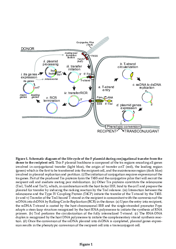
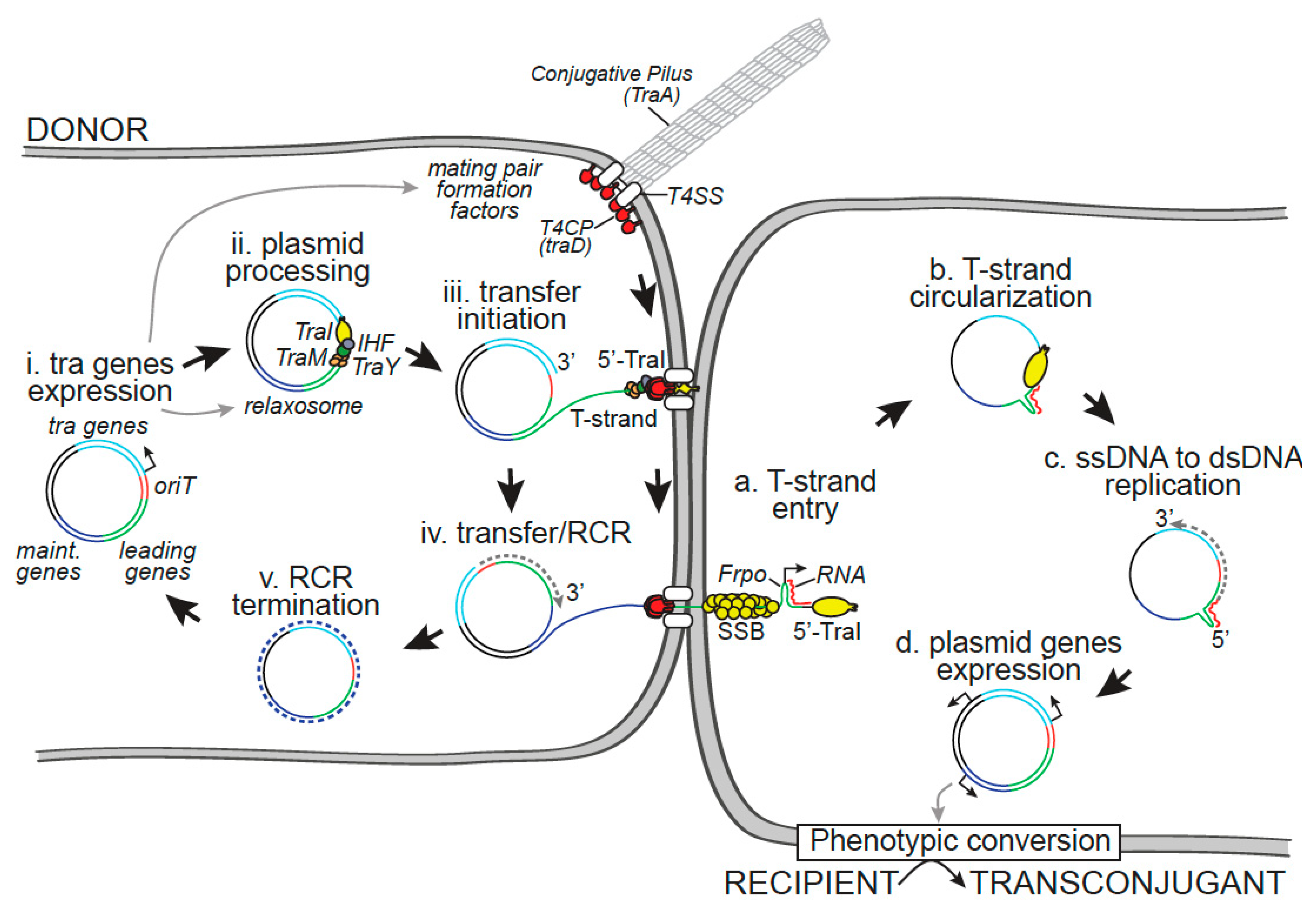
2. Within the Donor Cell

2.1. Transfer Gene Expression

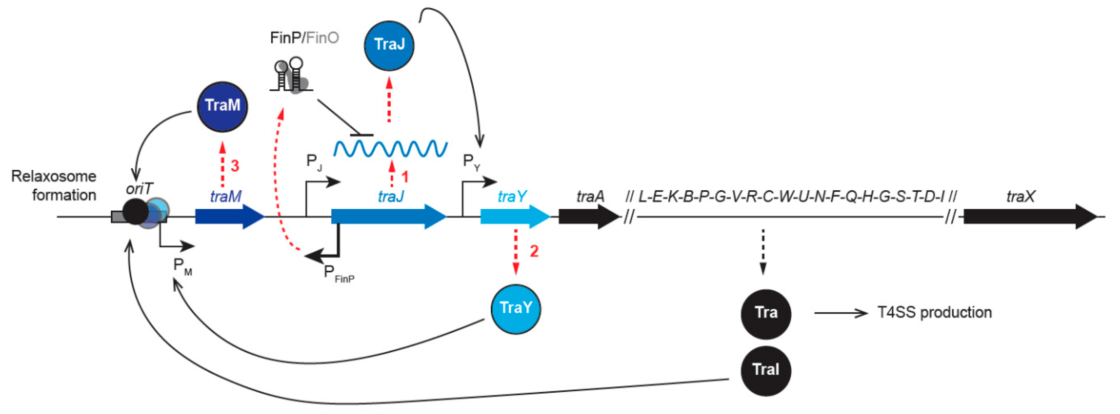
2.1.1. Regulation of tra Gene Expression

The ability of the donor strain to perform conjugation requires the expression of the transfer genes clustered in the tra region of the plasmid. The transfer genes encode all the protein factors involved in the elaboration of the conjugative pilus and the T4SS required for the formation of the mating pair, as well as the relaxosome components needed for the processing of the plasmid prior to transfer (Figure 1, step i). The expression of tra genes is regulated by several factors, including plasmid and host proteins, cell cycle progression, and environmental conditions. Most tra genes are gathered in one operon under the control of the PY promoter, while traJ and traM genes are located upstream and controlled by independent promoters (Figure 2) [7]. Transfer gene expression follows a specific regulation cascade that starts with the production of the TraJ protein (Figure 2, Step 1), which activates the PY promoter and the transcription of the tra operon (Figure 2, Step 2). The first gene to be transcribed, traY, encodes the TraY regulator protein that activates the PM promoter, resulting in the production of the relaxosome accessory protein TraM (Figure 2, Step 3) [8]. Therefore, this regulation cascade results in the expression of all genes involved in the elaboration of the conjugative pilus, the T4SS, and the relaxosome, which is composed of TraY, TraM, and TraI. It is observed that tra genes are normally repressed, presumably to avoid the fitness cost that would be associated with their constitutive expression [9]. It is important to remark that most regulation systems act by modulating the cellular levels or activity of the primary activator TraJ. In most F-like plasmids (R100, R1, R6-5, and ColB2-K77), the expression of traJ, and therefore that of other transfer genes, is repressed at the post-transcriptional level by the fertility inhibition system FinOP (Figure 2) [10,11]. FinP is an antisense RNA that is complementary to the stem-loop structures of traJ mRNA. FinP binding hides the ribosome binding site and prevents TraJ translation [12,13]. FinO is an RNA chaperon that protects FinP from degradation by RNase E and stabilizes the formation of the FinP–traJ mRNA duplex [14,15,16]. Moreover, tra gene expression is also regulated by chromosomal-encoded host factors [17]. One such regulation involves the silencing of PY, PM, and PJ promoters by the chromosome-encoded histone-like nucleoid structuring protein (H-NS) [18,19]. The H-NS copy number per cell varies during growth [20], thus rendering the F plasmid transfer rate growth phase-dependent, i.e., maximum in the exponential phase, reduced in the mid-exponential phase, and mostly abolished in the stationary phase [21,22]. However, during the exponential phase, H-NS repression activity is itself counteracted by the cooperative binding of TraJ and the host protein ArcA (aerobic respiration control of anoxic redox control) to the PY promoter [23]. In the case of the virulence plasmid pSLT of Salmonella enterica, H-NS repression activity also reportedly depends on Dam (DNA adenine methylase) methylation of the DNA [24]. Other examples of host factor-mediated regulation of tra gene expression include repression by the RNA binding protein Hfq, which destabilizes both traJ and traM transcripts [18], and by GroEL chaperone proteins that directly activate proteolysis of plasmid R1 TraJ during the cellular heat shock response [25].
For some other Gram-negative conjugation systems, tra gene expression is regulated by quorum-sensing (QS) mechanisms. This is the case for the conjugative tumor-inducing plasmid (pTi), which allows Agrobacterium to infect and disseminate within plant hosts. At high cellular density, Agrobacterium produces agrocinopine molecules that activate different operons, including the arc operon that encodes TraR (unrelated to the F TraR protein), a LuxR-like protein. The binding of TraR to the QS molecule 3-oxo-octanoylhomoserine lactone (OOHL) triggers the transcription of the trb and tra operons, resulting in the production of the T4SS and relaxosome proteins. The QS lactonase BlcC is also produced, resulting in the degradation of OOHL molecules in the stationary phase or during the carbon and nitrogen starvation associated with host plant death. This regulation provides coordination between the pTi conjugation proficiency and the host state or to the bacterial density within plant tissues during infection [26]. Pseudomonas aeruginosa uses QS as a defense mechanism against interspecies conjugation through the production of the QS molecule N-acyl homoserine lactone (AHL), which is involved in the regulation of mechanisms such as virulence, biofilm formation, and metabolism in the P. aeruginosa population [27]. AHL produced by P. aeruginosa can bind to the E. coli LuxR-like transcriptional factor SdiA, which then represses traI gene expression and prevents the conjugation of the RP4 broad host range plasmid that is integrated into the chromosome of E. coli donors.
Regulating the expression of transfer genes is the chief strategy used to modulate the transfer efficiency of conjugative plasmids. The above examples illustrate that tra gene expression is controlled by complex regulatory circuits, which involve the combined activities of plasmids and chromosomal host factors. This tight regulation allows for the control of the transfer efficiency in connection with the plasmid life cycle and the host physiology in response to environmental conditions and populational interactions.

2.1.2. Superspreader Mutations

Over the years, several studies have revealed genetic modifications, so-called superspreader mutations, that dramatically enhanced the conjugation efficiency of conjugative plasmids belonging to diverse incompatibility groups. The first superspreader mutation was characterized in the F plasmid, which carries an IS3 insertion sequence into the finO gene. FinO inactivation destabilizes the FinP-traJ mRNA duplex, thus resulting in the upregulation of traJ and the constitutive expression of tra genes [28]. This naturally occurring mutation accounts for the enhanced transfer efficiency of the F plasmid compared with the related IncF plasmids R100, R6-5, and R1, in which the FinOP regulatory system is still active [29]. More recently, genetically induced superspreader mutations of several resistance plasmids have been isolated in laboratory settings. In the IncI plasmid pESBL, which is associated with extended-spectrum β-lactamase production in Enterobacteria, inactivation of the Hft locus triggered the overexpression of conjugative pili and 20-fold enhancement of the transfer efficiency [30,31]. In the Citrobacter freundii IncM group plasmid pCTX-M3 that carries the blaCTX-M-3 gene, the deletion of two genes (orf35 and orf36) resulted in the enhanced expression of tra genes and increased plasmid transfer [32]. Another example was reported in the Gram-positive broad host range (Inc18) plasmid pIP501, which is involved in the propagation of vancomycin resistance from Enterococci to methicillin-resistant strains of Staphylococcus aureus. In this case, the deletion of the traN gene encoding the small cytosolic protein TraN (unrelated to the F TraN protein) resulted in the upregulation of transfer factors and the enhancement of the transfer efficiency [33].
Inducing the overexpression of plasmid transfer genes might not be the only way through which superspreader mutations increase the transfer efficiency of conjugative plasmids. It was shown that insertion of the Tn1999 transposon into the tir (transfer inhibition of RP4) gene of the IncL/M-type plasmid pOXA-48a, responsible for the dissemination of specific extended-spectrum β-lactamase genes in Enterobacteriaceae, increases the transfer efficiency by 50–100-fold without affecting traM expression levels [34]. The mechanism by which the inactivation of the Tir protein enhances transfer efficiency remains to be elucidated. These studies show that superspreader mutations can emerge by various mechanisms in different conjugative plasmids and have the potential to aggravate the spread of drug resistance plasmids among bacterial organisms.

2.2. Conjugative Pilus, Mating Pair Formation, and Stabilization

2.2.1. F-Pilus Structure and Biosynthesis

Bacterial conjugation is a contact-dependent horizontal gene transfer mechanism that involves a conjugative pilus associated with a T4SS. Electron microscopy imaging was instrumental in analyzing the morphology of numerous conjugation pili encoded by plasmids belonging to different incompatibility groups [35,36,37,38]. These studies revealed that pili fall into two main morphological categories—thin flexible and thick rigid, which influence the ability to support conjugation in liquid or on a solid surface. F encodes a thin flexible pilus, which has a tubular structure ~8 µm in diameter and up to ~20 µm in length and which is constituted by a helicoidal arrangement of a unique protein subunit, the F pilin or TraA [39,40,41,42,43,44] (Figure 1, step i). The traA gene encodes a 121 amino acid pro-pilin peptide that subsequently processed into a 70 amino acid F Pilin [45,46]. The maturation process involves TraQ and TraX proteins [47]. The TraQ chaperone-like protein binds transiently to the TraA pro-pilin precursor, thus allowing its accumulation in the inner membrane by an ATP-dependent pathway [48] and imparting it with the right conformation for a signal peptide cleavage. The processing of pro-pilin into mature pilin requires both cleavage by the host leader peptidase B (LepB) and N-terminal acetylation by TraX [49,50]. This maturation process ensures the availability of pilin subunits in the inner membrane before the assembly of the pilus by TraE, TraK, TraB, TraV, TraC, TraW, TraG, TraF, TraH, TraL, and TrbC encoded by the transfer region [51,52,53]. Mutational experiments have shown that this set of proteins can separate into different functions. Briefly, TraE, K, C, G, and L are responsible for the assembly of the tip, while TraB, V, W, F, and H are important for the pilus extension, and TrbI is required for pilus retraction [52,53]. The pathway of F-pilus biosynthesis has been extensively reviewed [52,54] and is not detailed here (Table 1).

2.2.2. Pilus Biological Function

The role of the F-pilus in conjugation has been actively debated. It was first proposed that the F-pilus extends to contact the recipient cell and then retracts to bring together the donor and recipient cells and form the mating pair [41,67,68]. This idea was convincingly supported by the direct visualization of the F-pilus dynamics in live cells, using a fluorescently labeled R17 bacteriophage that specifically binds along the pilus sides [69]. This work showed that donor cells produce flexible pili that continuously undergo cycles of extension and retracting, thereby probing the surroundings, regardless of the absence or presence of recipient cells. However, when contact is established with a recipient, pilus retraction draws the cells together, resulting in the formation of a mating pair [69]. In liquid culture, the pili mediate the formation of larger mating aggregates that contain donor and recipient cells in tight wall-to-wall contact [70,71].
Allowing the formation of wall-to-wall contact between mating partners might not be the only role of the F-pilus, which was also proposed to serve as a channel through which single-stranded DNA is transferred during conjugation between distant donor and recipient cells [72]. Undeniably, the pilus axial hole has a diameter (30 Å) that is large enough to accommodate the DNA molecule [41,44]. However, only a few reports provide evidence for conjugational DNA transfer between mating partners that are spatially separated from each other. It was shown that DNA transfer could occur between a donor and recipient that are separated by a 6 micron membrane with pores 0.01–0.1 micron in diameter [73]. Furthermore, microscopy imaging provided some evidence that DNA can be acquired by recipient cells that are not in direct contact with a donor cell [74]. Nevertheless, the F-pilus’ ability to transport DNA is still questioned and awaits the clear visualization of DNA transfer between distant donor and recipient cells that are only connected by a pilus.

2.2.3. Factors Involved in the Specificity of Donor–Recipient Interactions

The ability of the pilus to establish contact between donor and recipient cells can be considered the first rate-limiting step in the conjugation process and a key determinant of plasmid host range specificity. In the 1970s, numerous studies attempted to identify the specific recipient receptor required for F plasmid transfer [70,75,76,77,78,79,80]. The results revealed that mutations localized in the fourth external loop of the major porin OmpA or those that alter the inner core composition of the lipopolysaccharide (LPS) affect the transfer of the F plasmid and other IncF-like plasmids, such as R386, R538-1drd, and R1-19, but not the IncFII-type plasmid R100-1 [77,80,81,82]. Analysis of several ompA and LPS mutants revealed that they do not affect pili binding but result in defective mating pair stabilization [83]. Further investigation excluded that TraA is the donor component responsible for specific recognition of the recipient receptors [84] and uncovered the mating pair stabilization function of the outer membrane protein TraN, whose three external loops have been reported to interact with OmpA and LPS [85,86,87]. These findings indicate that ompA and LPS mutations do not alter the conjugation efficiency of the closely related IncFII-type plasmid R100, in which the amino acid sequence of the TraN central region is highly divergent from that of F-encoded TraN [59,77,80]. Mating pair stabilization additionally involves the multifunctional inner membrane protein TraG, of which the N-terminal part also plays a role in piliation and surface exclusion [59,88,89,90].
OmpA or LPS receptor factors cannot be considered strictly essential to F conjugation since their mutation only decreases transfer efficiency by 2–3 log. Moreover, mating pair stabilization defects can be overridden by performing mating on solid media, suggesting that interactions with OmpA or LPS are needed to stabilize the mating pair formation only in liquid mating [77,81,83]. One might then ask, is the F plasmid an exception to the need for recipient factors in some conditions? A recent study in Klebsiella pneumoniae identified the outer membrane OmpK36 homolog of the E. coli outer membrane OmpC as a receptor that mediates conjugation of the IncFII plasmid pKpQIL [91]. As observed in the case of the F plasmid, TraNpKpQIL determines the OmpK36-receptor specificity, while complementing a traNpKpQIL mutant with TraNR100 abolished this dependence, demonstrating that recipient receptors might be highly specific to the transferred plasmid. In addition to the IncF-type, the conjugation process of IncI-type plasmids was also shown to be sensitive to LPS mutations, and interestingly, some LPS mutations that affect IncI plasmid transfer do not impact the entry of the F plasmid, while others affect both types of plasmid [76,92]. Recently, the PilV adhesin encoded by the IncI1-type plasmid R64 was identified as the donor factor that binds to LPS in the recipient cell [93]. This adhesin is thought to localize to the tip of the thin type IV pilus that is required only in liquid mating conditions, rendering the interaction between PilV and LPS important only under these conditions, as observed for TraN and OmpA or LPS interactions in the case of the F plasmid. In contrast, no such recipient receptors could be identified for the broad host range plasmids RP4 and R388. Indeed, the ompA and LPS recipient mutants, which drastically decrease the efficiency of F conjugation, do not affect RP4 conjugation efficiency [94]. Large-scale mutant screening using the E. coli Keio collection or random E. coli insertion mutant library failed to identify recipient mutants that affect the transfer of the plasmids RP4 [95] or R388 [96].
Remarkably, some broad host range IncP-like plasmids can also be transferred in archaea [97] and eukaryotes such as yeast [98] and mammalian cells [99]. Although the efficiency of conjugation varies among types of recipient cells, these findings strongly suggest that plasmid transfer does not require any specific factors or active mechanisms on the recipient side. Alternatively, a “shoot and pump” conjugation model envisages that the type IV secretion apparatus could act like a syringe that is able to inject DNA into any kind of membrane, using the pilus as a needle [100]. Perforation of the cell wall bilayer of the recipient could be achieved by force or by dedicated enzymatic activity exposed at the pilus tip. The lack of a requirement for specific receptors on the surface of the target cell is not an exception to the conjugative T4SS. Indeed, although the structural components of the type VI secretion system machinery have been widely documented, no studies have yet characterized genetic factors that can act as target receptors on the prey cell surface.

2.3. Plasmid Processing by the Relaxosome

The initiation of conjugation requires the assembly and activity of a protein complex, the relaxosome, that allows the processing of the plasmid before DNA transfer (Figure 1, step ii). Plasmid processing involves a site- and strand-specific DNA cut (nick) at the nic site located in the origin of transfer (oriT) and the extrusion of the single-stranded DNA that will be transferred (T-strand) [101,102,103]. In the F system, these two reactions are performed by the multifunctional TraI relaxase protein, which has both a transesterase domain that catalyzes the nic reaction and a DNA helicase domain that unwinds the plasmid DNA [64,104,105,106,107,108,109]. Crucially, TraI recruitment and activity are governed by auxiliary proteins, including the integration host factor (IHF) and the plasmid-encoded TraY and TraM proteins, which have distinct roles in the relaxosome formation and activity at oriT [110,111,112]. The binding of IHF and TraY to their respective cognate binding sites ihfA/ihfB and sbyA located in oriT modulates the architecture of the DNA, thereby stimulating the loading of TraI [113,114,115,116,117]. The TraM protein regulates its own expression by binding to the sbmA and sbmB sites, located in the PM promoter, and stimulates the DNA relaxation reaction through direct interaction with TraI after binding to the sbmC site located in the oriT region [118,119,120,121,122]. The TraI nicking reaction involves a catalytically active Tyr residue [65,123] and results in the relaxation of the plasmid dsDNA, where the 5′-phosphate (P) end of the nicked strand (or T-strand) remains covalently bound to TraI (Figure 1, step iii) [106,108,124,125,126,127]. After the nicking reaction, the circular ssDNA conjugative plasmid is converted into dsDNA by Rolling Circle Replication (RCR) in the donor, while the linearized T-strand DNA bound to TraI at the 5′ end is transferred into the recipient cell through the conjugative pore (Figure 1, step iv).

2.4. Initiation of Rolling Circle Replication in the Donor Cell

The rolling circle replication (RCR) mechanism is employed for the vegetative replication of some bacterial plasmids and has been very well reviewed [128,129,130]. RCR is key to the transfer process of many Gram-negative and Gram-positive conjugative plasmids but also to the infection cycle of other mobile genetic elements, such as DNA/RNA viruses and bacteriophages [123,131]. The RCR reactions involved in vegetative replication or in plasmid transfer are very similar. The initiation and termination of RCR reactions, performed by the Rep protein during vegetative plasmid replication, are achieved by the relaxase protein during conjugation. Indeed, Rep and relaxases serve closely related functions, primarily allowing RCR initiation by nicking the double-strand DNA at the dso site (double-stranded origin) or the oriT site, respectively [132]. The nicking reaction generates a 5′-P end that remains covalently bound to Rep or TraI and a 3′OH end used as a primer for the host DNA polymerase III. While DNA polymerase III performs leading strand elongation, the parental double helix is unwound, and RCR ends with a second nicking reaction that releases the newly synthesized DNA strand (Figure 1, step v). In the case of vegetative RCR, DNA unwinding is performed by a host DNA helicase recruited by the Rep protein, while Rep itself ensures termination and the second nicking reaction. One major aspect of conjugation-associated RCR is that replication of the two ssDNA strands occurs in different cells, i.e., the leading strand is replicated in the donor, while the T-strand (lagging strand) is transferred and replicated in the recipient cell (Figure 1). Because the relaxase that initiates the nicking reaction is transferred together with the T-strand [65,133,134], a second relaxase protein is required in the donor to perform DNA unwinding as well as the second nicking reaction [3,65,123]. Consistently, biochemical assays show that two relaxase molecules bind to oriT: one associated with the 5′ end that is in an open transesterase conformation and one associated with the 3′ end that is in a closed helicase conformation [135].

2.5. T4CP Connects the Relaxosome to the T4SS

After processing by the relaxosome complex, the nucleoprotein complex, composed of the T-strand and the covalently bound TraI, needs to be recruited to the conjugative pore for transfer (Figure 1, step iii). This connection is mediated by the interaction between the relaxosome and the Type IV Coupling Protein (T4CP) located at the cell membrane [3,100]. All conjugative systems have their own T4CPs, such as TraD, TraG, and TrwB for the F, RP4, and R388 plasmids, respectively. T4CPs are not required for pilus production or DNA processing, yet they are key to substrate specificity [136]. Our understanding of the molecular interactions required for specific substrate recognition and translocation is still incomplete. A great deal of information has been provided by comparing the structure of F-like T4SS [137] to various T4SSs involved in protein or nucleoprotein transport [138,139,140,141]. It appears likely that conjugation systems are derived from ancestral protein translocation machinery that evolved to coincidently translocate DNA. In this view, the T4CP would serve as the substrate receptor that interacts with one or several relaxosome components to recruit the T-strand to the T4SS. In the case of the F plasmid and some other plasmid systems, it is well established that TraD interacts with the TraM relaxosome protein [142,143,144,145,146]. Interaction between the T4CP and the relaxase has been demonstrated for RP4 [147], R1 [1] and R388 plasmids [148]. Such interaction has been speculated in the F system but remains elusive [107,149].
T4CPs are DNA-dependent ATPases anchored to the cell membrane via their N-terminal domain and have been shown to interact with the T4SS components in R27 [150] and R388 plasmid systems [148]. T4CPs show similarities to membrane-anchored ring DNA translocases, such as SpoIIIE and FtsK, which are involved in chromosome DNA translocation during sporulation and cell division, respectively [100,151,152]. T4CP binds non-specifically to DNA, with a higher affinity for ssDNA [147,153,154], on which it forms oligomers with enhanced ATPase activity [155,156].
Altogether, these findings led to a model in which membrane-anchored T4CPs interact directly with the relaxosome and form hexameric structures on the T-strand, which actively translocate through the conjugation pore during transfer. However, it remains unclear whether a signal is required to activate the coupling function of the T4CP in a donor cell in which the T4SS and the relaxosome are already assembled and functional [3]. It has been suggested that the stability of the TraD oligomeric complex depends on an as yet unidentified F-encoded protein, which could then be a key regulator of plasmid transfer activation [155]. It has also been suggested that the formation of the mating pair could transduce a signal to activate the T4CP and trigger the transfer of the processed T-strand [3]. Notably, it was shown that the relaxase has to be unfolded to be translocated into recipient cells [134]. In A. tumefaciens, as in many other Gram-negative and Gram-positive systems, the unfolding of translocated proteins is proposed to be performed by the VirB11-like ATPase, which is absent from the F plasmid system [52,140,157]. However, one can reasonably presume that TraI unfolding also requires ATPase activity that involves one of the ATPases of the F T4SS.

3. Within the Recipient Cell

3.1. Plasmid Circularization by TraI

The relaxase is transferred to the recipient, where it is refolded and able to perform several activities required for the completion of the conjugation process (Figure 1, step a) [65,133,158]. The helicase activity of the internalized relaxase is thought to perform 5′ to 3′ tracking of the T-strand. Pulling by the relaxase from the recipient together with pushing by the T4CP from the donor presumably facilitates the transport of the T-strand through the conjugation pore [3]. Once both extremities of the oriT are brought together in the recipient, the relaxase performs the joining reaction, resulting in the recircularization of the ssDNA plasmid (Figure 1, step b) [65,133,158,159]. An alternative model proposes that the nicking of the newly synthesized oriT occurs in the donor cell before transfer [160]. There is no evidence for the requirement of additional host or plasmid factors in the circularization of the internalized T-strand. After completion of the recircularization reaction, the recipient cell possesses a single-stranded circular copy of the conjugative plasmid.

3.2. Avoiding Host Defense Systems against Foreign DNA

The newly acquired ssDNA conjugative plasmid might be considered as foreign DNA, against which host bacteria have developed defense mechanisms, such as restriction modification, exonucleases, and recombination system or adaptive immunity, such as the CRISPR-Cas system [161]. Despite these defense mechanisms, horizontal gene transfer plays an important role in genomic evolution (5–6% of bacterial genomes and up to 20% in some organisms) [6,162,163], implying that transferrable plasmids have evolved adaptive mechanisms to counteract these host defenses.
The restriction modification system (RM) is a ubiquitous defense mechanism found in 90% of sequenced bacterial genomes and other prokaryotes and is based on restriction enzymes and methylation [164]. Restriction modification mechanisms are described as a “primitive immunity system” against exogenous DNA [165]. These systems are based on restriction enzymes that specifically target unmethylated dsDNA sequences located in the newly acquired mobile genetic elements, while the host DNA is protected by methyl groups added to specific adenine or cytosine residues [165]. Whether the RM system can target the ssDNA plasmid before the complementary strand synthesis remains unclear. However, plasmids have evolved various strategies to counteract enzymatic DNA degradation upon entry into the new host cell. IncN, IncI, and IncF plasmids encode ArdA and ArdB proteins (alleviation of restriction of DNA) that directly inhibit the REase (Restriction Endonuclease) by mimic DNA sequences, thus competing for enzyme targets [166,167,168]. IncW plasmids encode the ArdC protein that protects the transferred T-strand by transiently blocking the restriction sites [169]. More recently, it was shown that an hde operon (host defense evasion) of IncI plasmids encodes two genes involved in anti-RM (vcrx089 and vcrx090) [170]. In addition to the production of inhibitory proteins, some plasmids have completely lost these restriction sites, as in the case of the RP4 plasmid [171].
CRISPR-Cas immune systems (Clustered Regularly Interspaced Short Palindromic Repeats and the CRISPR-associated protein) represent another defense mechanism against foreign DNA. CRISPR-Cas systems, found in ~45% of bacterial and up to 84% of Archaea genomes [172,173], have been described as being protective against infection by bacteriophages and, more recently, against plasmid acquisition [174]. It was then discovered that some phages encode an anti-CRISPR Acr protein that inhibits the activity of the CRISPR-Cas system [175,176]. Importantly, anti-CRISPR Acr loci have been identified in conjugative elements and plasmids of Listeria, Enterococcus, Streptococcus, and Staphylococcus [177]. These loci encode the CRISPR-Cas inhibitors AcrIIA16–19, which prevent exogenous DNA nicking mediated by the Cas9 enzyme in vivo. Mahendra et al. have also shown that conjugation of a Cas9-targeted plasmid of E. faecalis was possible in the presence of these CRISPR-Cas inhibitors. Encoding Acr-like proteins is therefore an efficient strategy by which conjugative plasmids facilitate their dissemination by avoiding degradation by the host CRISPR-Cas immune system. Another strategy is for conjugative plasmids to encode a Bet/Exo system able to repair double-strand breaks caused by CRISPR-Cas during conjugation, as recently reported for the IncC plasmid pVCR94 [170]. Expression of these genes inside recipient bacteria after the acquisition of the V. cholerae pCVR94 plasmid enables survival against exogenous DNA defense mechanisms without the involvement of anti-CRISPR-Cas proteins.

3.3. Role of the Leading Region Genes and Conversion of the ssDNA Plasmid into dsDNA

3.3.1. Early Expression of the Leading Region Genes

The leading region of conjugative plasmids is the first to be transferred into the recipient cell during conjugation (Figure 1, step a) [57]. The F plasmid leading region is well conserved, with a size of 13 kb and a location that is directly adjacent to the oriT, and encodes for at least eight proteins [178], including a homolog to the chromosomal single-strand binding protein SSB (SSBC), PsiB, and other proteins of unknown function. Importantly, plasmid ssb (ssbP) and psiB genes are expressed early upon entry of the plasmid into the recipient bacteria but not in the donor cells [179]. Similarly, RT-PCR studies showed that the expression of the psiB and ardA genes of the IncI1 plasmid begins 5 min after transfer initiation [180]. These observations suggested that leading region genes could be expressed rapidly from the newly acquired plasmid in single-stranded form, before its conversion into dsDNA. It was later shown that the leading region contains a specific 328 bp Frpo region (for F plasmid RNA polymerase), which, when single-stranded, can form a stem-loop structure presenting −10 and −35 double-stranded boxes that are recognized by the host RNA polymerase, which initiates the synthesis of RNA primers in vitro [181]. It was therefore proposed that Frpo may serve as a single-stranded promotor that allows the early expression of the leading region genes (Figure 1, step a).
Frpo was also proposed to direct the single-strand to double-strand conversion of the F plasmid. In vitro assays showed that the RNA primers synthesized by the RNA polymerase from Frpo persist as an RNA–DNA duplex that is recognized by host DNA polymerase III to initiate complementary strand synthesis [181]. Frpo-type sequences, also termed ssi for single-strand initiation sequence, are found on various conjugative plasmids, including R6K, R100, ColE1, ColE2, Col1B, and RSF1010 [182,183,184,185], and are functionally comparable to sso sequences (single-stranded origin) involved in the rolling circle replication mechanism [123,129,186]. These findings are consistent with the previous observation that complementary strand synthesis of the ssDNA F plasmid inside the recipient bacteria involves a cooperative mechanism between host RNA polymerase and DNA polymerase III [187].
Altogether, these findings led to a model in which Frpo can help to initiate early gene expression and the DNA synthesis reaction that converts the ssDNA plasmid into dsDNA duplex immediately upon entry of the T-strand into the recipient cell (Figure 1, step a–c). Whether Frpo performs these functions during conjugation in vivo remains to be demonstrated.

3.3.2. PsiB Inhibits the SOS Response

In the recipient cell, the presence of abnormal amounts of ssDNA, usually associated with DNA damage, results in the induction of the SOS response [188,189]. More precisely, the loading of the RecA recombination protein onto ssDNA results in the formation of the presynaptic filament, which stimulates the autocatalytic cleavage of LexA, the repressor of the SOS regulon. The SOS response triggers the induction of the division inhibitor SulA, resulting in cell filamentation and potentially the death of the transconjugant cell. SOS also induces the production of nucleases and other DNA processing factors that could provoke the degradation or mutation of the transferred ssDNA or its processing as a recombination intermediate [190]. To counteract these effects, several conjugative plasmids, including F, encode the PsiB protein (plasmid SOS inhibition), which inhibits SOS induction [191]. The depletion of psiB has mild effects on the efficiency of conjugation but increases the host SOS response by up to six-fold [179]. PsiB interacts directly with RecA, thereby inhibiting several activities, such as DNA binding, LexA cleavage, and the strand exchange reaction [192,193]. SOS response inhibition by PsiB is even more potent in the presence of the SSBC protein that coats the ssDNA. PsiB is well conserved among conjugative plasmids and is considered important for the early steps of plasmid establishment in the recipient, consistent with its early production in the transconjugant cells [57,191].

3.3.3. Roles of the Host and Plasmidic SSB Proteins in Plasmid Establishment

Upon entry into the recipient cell, the transferred ssDNA plasmid is coated by the host SSBC protein. SSBC is a universally conserved essential protein that binds non-specifically to ssDNA. It is involved in various mechanisms, including DNA replication, repair, and recombination; SOS induction; and other DNA metabolic processes [194,195]. Upon binding, SSB protects the ssDNA against enzymatic degradation and increases the processivity of DNA polymerases II and III [196] during the replication reaction that converts the ssDNA strand into the dsDNA helix. The rapid recruitment of the host SSBC protein to the transferred ssDNA has recently been visualized by fluorescence microscopy [197]. This work revealed that SSBC proteins are rapidly recruited to the ssDNA that penetrates the recipient cell, presumably protecting it and facilitating its processing (Figure 1, step a). Interestingly, the F plasmid, as in many other conjugative plasmids, encodes its own SSBP protein, which is homologous to the E. coli SSBC [198]. One might then ask, what is the benefit gained by conjugative plasmids encoding their own SSBP, and what is the specific function of SSBP compared with the chromosomal SSBC?
SSBP binds ssDNA non-specifically, and SSBP of different incompatibility groups (IncF, IncI, IncY, Inc9, IncT, and IncB/O) can partially complement conditional mutations of the E. coli ssbC gene [197,199,200,201]. Although the expression of the F plasmid-encoded SSBP protein in trans enables the growth of the ssbC deletion mutant, complemented mutants exhibit some filamentation and growth rate reduction [202]. Moreover, a reduced affinity to ssDNA is observed for the F plasmid SSBP in comparison with the E. coli SSBC, and the F plasmid SSBP cannot stimulate the reaction of DNA synthesis by DNA polymerase III in vitro [203]. Sequence alignment revealed that SSBP proteins that complement the E. coli ssbC mutant share high homology only with the N-terminal part of E. coli SSBC [203]. The SSBC N-terminal region contains the domains for ssDNA binding and monomer–monomer interactions to cooperatively maintain the binding of the tetrameric structure of SSBC to ssDNA. This structural conservation would explain the ability of SSBP to bind ssDNA. However, the C-terminal domains of SSBP are much more homologous to each other than to SSBC [203]. However, as this domain interacts with partner proteins that constitute the SSBC interactome, one possibility would be that the interactome of SSBP and the reaction it is involved in might be different from that of SSBC.
Not all SSBP proteins have been shown to complement E. coli ssbC mutants [199]. The latter study found that the inability of SSBP from IncP-like RK2 to complement the E. coli ssb1 mutation could be attributed to ssbP gene repression by the RK2 kor genes and that a derepressed plasmid indeed complemented the thermosensitive growth of E. coli ssb1 mutations [204]. It is thus reasonable to consider the possibility that abundant SSBP could also complement the ssbC mutant.
To date, the function of the F plasmid SSBP in the context of conjugation is still unclear. The SSBP protein could contribute to the protection of the transferred ssDNA by inhibiting enzymatic degradation or the recruitment of inhibiting proteins of the host. However, its expression timing instead supports the idea that SSBP could be involved in the complementary strand synthesis of the transferred DNA or could simply increase the pool of available single-strand binding protein, which is required for the first cycle of vegetative replication of the plasmid.

3.4. Plasmid Maintenance: Replication and Segregation

Maintenance of the newly acquired dsDNA conjugative plasmid in the recipient cell lineage depends on two main active mechanisms: plasmid replication and segregation of the plasmid copies into daughter cells over generations.
The mechanisms of plasmid DNA replication in bacteria have been extensively studied and are the focus of well-referenced reviews [123,128,205,206,207,208]. Here, we stress the role of replication in conjugation host-range specificity. The F plasmid is efficiently transferred to E. coli and relatively close enterobacteria, while no transconjugant can be recovered after mating with more distant bacteria, such as Vibrio or Pseudomonas. As early as 1982, host range restriction was attributed to the plasmid’s inability to replicate in the recipient bacteria rather than to the inefficiency of plasmid transfer per se [188]. This was demonstrated by showing that mobilizable plasmids containing a cis-acting origin of transfer of the F plasmid and an origin of replication that is active in the tested recipient can then be transferred by the F conjugation machinery in Pseudomonas [188], V. cholerae [209], and yeast [98]. The same approach was employed to show that pCTX-M3, an IncI-like plasmid, was able to use its conjugation machinery to transfer a mobilizable plasmid to host recipients in which pCTX-M3 does not replicate [32]. The failure to replicate the F plasmid in P. aeruginosa comes in part from an inability of the plasmid replication protein RepE in the RepFIA replicon in complex with host DnaA-oriS to form a stable interaction with the host helicase DnaB [210]. In contrast, broad host range plasmids such as RK2 regulate their maintenance by modulating alternative strategies of replication depending on the host [211]. These findings indicate that the host range of the conjugation machinery and replication origins belonging to the same plasmid differ and can be mechanistically uncoupled. The specificity of narrow host range plasmids appears to be limited by the specificity of their replicon rather than by their transfer range.
Maintenance of the newly acquired dsDNA plasmid also requires the segregation of plasmid copies into the daughter cells during transconjugant cell division. To do so, low copy number plasmids encode active partition systems, of which the mechanism, biological functions, and conservation have been extensively reviewed [212,213,214,215,216]. In the context of conjugation, it is worth mentioning another maintenance strategy that involves the integration of the newly acquired conjugative DNA into the chromosome. Chromosome integration ensures the stable inheritance of the conjugative elements by vertical gene transfer over generations. The first characterized example was again the F plasmid, which uses insertion sequences (IS) and RecA-dependent homologous recombination to integrate into the E. coli genome [217]. The integrated plasmid can still initiate the conjugative transfer of the whole chromosome of the resulting “Hfr” strain (high frequency of recombination). This process of transfer is both progressive and oriented, and the order in which chromosomic genes are transferred depends on the position where the F plasmid was integrated. Alternatively, the integrated F plasmid can be excised out of the chromosome and recover its original autonomous form [218]. Chromosome integration is widely used by mobile DNA, such as ICE (Integrative Conjugative Elements), transposons, and phages, in both Gram-negative and Gram-positive bacteria; all of these systems have their particularities, especially regarding the recombination systems used for their integration/excision [219,220].

3.5. Phenotypic Conversion of the Transconjugant

The expression of genes carried by the newly acquired genetic element results in the phenotypic conversion of the recipient cell into a transconjugant that exhibits additional metabolic properties (Figure 1, step d). The expression of plasmid genes involved in DNA transfer converts the transconjugant into a new donor that is able to further transfer the plasmid to the population, thus accounting for the exponential rate of conjugative plasmid dissemination (see tra gene expression, Section 2.1). Not all genes encoded within the tra region of the plasmid backbone are directly involved in the process of DNA transfer. Indeed, conjugative plasmids often carry immunity (or exclusion) gene systems that are widespread in Gram-negative and Gram-positive organisms [90,221,222,223,224,225,226,227,228]. These immunity systems limit the ability of plasmid-carrying cells to serve as a recipient for the same plasmid [42,54,62,229]. Preventing self-mating by surface exclusion is thought to avoid the metabolic cost and potential cell death associated with repeated plasmid transfer but also to be important for plasmid stability and evolution [62,229].
The F plasmid immunity system relies on two factors—TraT and TraS proteins, neither of which is required for F-pilus synthesis or DNA transfer [56,230,231,232]. These two exclusion factors work at different levels. TraT is an abundant outer membrane protein that is thought to span the cell surface [230,231]. TraT production inhibits the formation of stable mating aggregates, presumably by interfering with the interaction between the pilus and recipient surface receptors. Consistent with this idea, it was reported that TraT interacts with OmpA, further suggesting that it could compete with TraN, which is key to mating pair stabilization [85,86,233]. TraS is an inner membrane protein, the production of which only slightly reduced the aggregation of mating populations but reduced DNA transfer frequencies by 100–200-fold. It is thus proposed that TraS acts by preventing DNA transfer when stable mating aggregates have already formed [230,231,234]. In F-like plasmids, it is proposed that TraS interacts with TraG to achieve the entry exclusion process [89,90]. For these reasons, TraS is referred to as an entry exclusion protein (Eex), and TraT as a surface exclusion protein (Sfx). Like other tra genes, traS and traT expression is controlled by TraJ, implying that transconjugant cells concomitantly acquire plasmid transfer ability and immunity to self-transfer during the phenotypic conversion.
In addition to genes located on the plasmid backbone, conjugative plasmids may carry additional genes that are not directly involved in conjugation but in a variety of biological functions, such as virulence, biofilm formation, symbiotic lifestyle, membrane trafficking, resistance to heavy metals, and, most importantly, resistance to antibiotics. Acquisition of these metabolic functions potentially facilitates bacterial adaptation and survival in changing environments and makes conjugation a major driver of the evolution of bacterial genomes. The successful maintenance of conjugative elements in bacterial populations shows that this selective advantage compensates for the metabolic burden associated with the metabolism of the newly acquired genetic information (highjacking of the host replication, transcription, and translation machineries) [235]. The most prominent example is the acquisition of conjugative drug resistance plasmids, which enable bacterial proliferation in microbial communities that contain antibiotic-producing organisms or in antibiotic-polluted and clinical environments. Indeed, analysis of commensal, environmental, and clinical antibiotic-resistant pathogenic strains revealed a multitude of conjugative plasmids that carry one or more genes for resistance to most, if not all, classes of antibiotics currently used in clinical treatments. Conjugation is considered to be the most widespread intra- and interspecies resistance transfer mechanism, accounting for 80% of acquired resistance [236].

4. Conjugation in Natural Habitats: The Example of Bacterial Biofilms

Gene transfer by conjugation is known to contribute to the genetic dynamics of bacterial populations living in a variety of environments, including the soil, on plant surfaces, and in water and sewage, as well as in bacterial communities associated with plant or animal hosts [4]. Bacteria are generally considered to be planktonic unicellular organisms, yet in natural and clinical environments, they often live in complex structures called biofilms. Biofilms shelter bacteria from external hazards but have also been proposed to offer a niche that facilitates the dissemination of drug resistance determinants by conjugation. Below, we review our current understanding of the interplay between biofilms and bacterial conjugation.

4.1. The Biofilm as a Niche Promoting Bacterial Conjugation

In natural environments, bacteria predominantly live in spatially structured communities termed biofilms, in which a self-produced extracellular matrix holds the cells together [237]. Planktonic cells that initiate biofilm formation can adhere to a living or inert surface (surface-attached biofilms) or can be present at the air–liquid interface as a free-floating community (pellicle). Bacterial biofilms are found in virtually every ecosystem on Earth, from aquatic systems (sludge, rocks, and wastewater) to terrestrial environments (rhizosphere) and human organisms (skin, intestinal, urogenital, and respiratory tracts). Furthermore, biofilms are associated with persistent and severe infections because of their ability to colonize medical devices and implants. Indeed, biofilm structures offer protection against hostile environments and, more worryingly, against antibiotic treatments. The biofilm architecture depends on the bacterial species, the surface colonized, and environmental conditions, but this lifestyle is characterized by the production of extracellular polymeric substances (EPSs) mainly composed of polysaccharides, proteins, lipids, and extracellular DNA. EPS matrix production is dynamic and continuous and mediates the formation of the biofilm architecture in which aggregates of microorganisms are trapped.
Many studies on bacterial conjugation have shown that plasmid transfer can occur in both natural and artificial biofilms, from the aquatic environment [238], phytosphere [239], animal and human hosts [240,241], or reactor-associated biofilms [242,243]. These papers studied plasmid transfer at the population level and mainly relied on limited cultivation-based assays that probably underestimate the extent of conjugation in natural biofilms. Further works have studied conjugative plasmids expressing fluorescent markers, allowing the direct in situ visualization of donors, recipients, and transconjugants at the single-cell level within several types of biofilms formed at the liquid–air surface [244], on a semisolid agar surface [245,246,247], on a filter [248], in flow chambers [246,249,250], or in a reactor [251].
The biofilm environment provides a high cell density and close cell-to-cell proximity that may facilitate HGT through bacterial conjugation. In line with the view that biofilm is a hot spot niche of conjugation, several studies have shown that the frequency of plasmid transfer is higher in the biofilm than in the planktonic mode of growth [252,253,254,255]. Although the biofilm appears to be a favorable environment for HGT by conjugation, many studies at the single-cell level have reported limited plasmid propagation inside a preformed established biofilm beyond the contact zone between the donors and recipients [256]. These observations are directly linked to the complex spatial structure of the biofilm, which might have a critical impact on horizontal plasmid spread within a biofilm.

4.2. Impact of the Biofilm Structure on Conjugation

The biofilm is a complex structure and sometimes composed of mixed bacterial species. The matrix shapes the spatial organization by clustering cells in microcolonies in an architecture characterized by nonuniform cell arrangements, open channels, pores, cavities, and different layers of living cells [257,258]. Such an organization determines the formation of cell clusters/aggregates and could therefore influence the efficiency of conjugative transfer [244]. This possibility can be addressed using microscopy to analyze the distribution of cells that are active in conjugation within biofilms. To date, most studies have investigated the spread of the GFP-tagged Pseudomonas putida TOL plasmid. Analysis of P. putida recipient biofilms established in flow chambers revealed that transconjugants appeared at the top surface of the biofilm but not in the deeper layers, reflecting a limited invasion of the transferred plasmid [246,250]. On an agar plate, transconjugants only appeared at the contact zone between the TOL plasmid donors and the recipient colonies [245,246,259]. Limited transfer to the outer layers of the biofilm was also observed for IncF, IncI, and IncW plasmids in E. coli [247]. It has been proposed that transfer can only be efficient over a short period between metabolically active cells growing at the donor–recipient interface [246,260]. Consistently, it was shown that plasmid invasion stops in non-dividing cells [259].
Within biofilm structures, chemical gradients of oxygen, nutrients, temperature, and pH create microenvironments that influence the metabolic activity of bacterial cells [261]. This results in physiological heterogeneity between the cells that surround the border of the biofilm and those that are embedded deep inside. Furthermore, variations in the spontaneous mutation frequency within parts of the biofilm result in the emergence of variant subpopulations with genetic heterogeneity [262]. Whether and how these factors impact the spatial pattern of conjugation within biofilms remain unknown.

4.3. Impact of Conjugative Plasmids on Biofilm Formation

Several studies have investigated the implication of the presence of conjugative plasmids from diverse incompatibility groups for biofilm formation ability [263,264,265,266,267]. To initiate biofilm formation, planktonic cells produce cell appendages like flagella and adhesion factors such as type IV pili and type 1 and curli fimbriae [268,269]. Genes coding for these types of accessory factors that promote attachment to biotic or abiotic surfaces are often found in conjugative plasmids, resulting in increased host biofilm formation [253]. Examples include type 3 fimbriae of the IncX1 plasmid pOLA52 [270], non-conjugative type IV pili of the IncI1 plasmid pSERB1 [271], or pilus-like structure and surface adhesins of the Enterococcus faecalis plasmids pBEE99 and pCF10, respectively [272,273,274]. In 2001, Ghigo made the unexpected observation that the conjugative pilus itself of a derepressed F plasmid can promote the biofilm of E. coli cells that are initially unable to form such a structure and revealed that the pilin TraA is the main adhesion factor that induces biofilm formation [263]. Microscopic structure analysis of derepressed IncF plasmid R1drd19- and F-carrying E. coli biofilm showed the rapid formation of a dense and mature 3D mushroom-type biofilm similar to the P. aeruginosa biofilm architecture [275]. The formation of this peculiar architecture and the biofilm maturation generated by derepressed plasmids override the need for cell surface appendages such as flagella, type 1 fimbriae, Ag43, or curli, which are essential to E. coli biofilm [275]. In contrast, maturation of the 3D mushroom-type biofilm structure depends on curli production, induced in E. coli by the natural F plasmid, which does not constitutively express F-pili [276]. The presence of the plasmid R1drd19 also increases E. coli biofilm formation by decreasing the motility and increasing the level of quorum-sensing inducer AI-2 [277,278], and the IncP-9 TOL plasmid in P. putida increases the production of extracellular DNA known to play a role in the structure of the biofilm [279] and thus the biofilm formation capacity [280]. However, the genetic mechanism by which conjugative plasmids increase biofilm formation has not been elucidated, but the presence of repressed or natural conjugative plasmids affects the global host chromosomal gene expression [276,278].
While the role of the conjugative pili has been mainly studied in E. coli biofilms, one may speculate that their impact differs depending on the host. Indeed, Røder et al. observed that the conjugative pili of the IncP-1 plasmid pKJK5 reduced the surface attachment of P. putida by increasing cell–cell adhesion, resulting in reduced biofilm formation [281]. Further investigation will be necessary to decipher the complex interconnections between the conjugative plasmid and biofilm formation.

4.4. Influence of Antibiotic Treatment on Conjugation within Biofilms

Interactions between conjugation and biofilms have been proposed to promote both community-building and gene transfer. This synergic interaction raises serious questions about the contribution of HGT to the evolution and adaptation of biofilm-forming pathogens. Because of the increase in antibiotic-resistant infections, recent investigations have aimed to provide a new understanding of biofilm responses to antimicrobial treatments.
Subminimal inhibitory concentrations (sub-MICs) of aminoglycosides enhance the biofilm biomass of the P. aeruginosa strain PAO1 and clinical isolates of E. coli through the response regulator Arr, a predicted phosphodiesterase that alters cyclic di-guanosine monophosphate (c-di-GMP) levels [282]. Linares et al. further demonstrated that, in addition to aminoglycosides, sub-MICs of tetracycline and norfloxacin also increase the formation of P. aeruginosa biofilm. However, no clear causal factors were identified [283]. Interestingly, a combination of tetracycline and cephradine has a synergistic effect on the biofilm formation of a mixed culture of E. coli and P. aeruginosa [284]. Tetracycline also promotes the biofilm formation of the pathogen Acinetobacter baumannii, and whole-genome sequence analysis revealed an increase in the rate of mutations such as SNPs, as well as insertions and deletions, under subinhibitory drug exposition [285]. In addition to the accumulation of genotypic variation, biofilm treatment with a low level of antibiotics produces changes in the gene expression profile, some of which may be linked to increased biofilm formation [283,285,286].
Recently, Diaz-Pascual et al. investigated Vibrio cholerae biofilm at the community scale using a single-cell imaging system, revealing changes in biofilm dynamics and architecture in response to antibiotic treatment [287]. After tetracycline exposure, they observed modifications in biofilm architecture and cell morphology, including a 2.5-fold increase in cell volume and a 29% decrease in cell density. This cell density decrease reflected a considerable alteration of the multicellular arrangement and the breakdown of the matrix within the biofilm. Furthermore, biofilms became susceptible to the colonization of their interior by new cells, and the colonizer population increased until they invaded the resident biofilm. Clearly, a sublethal dose of antibiotics influences the biofilm lifestyle, inducing significant modifications to the entire population. The biofilm matrix forms a shield to prevent the penetration and diffusion of antimicrobials, and the increase in biofilm formation in response to antibiotics, illustrated by an enhancement of the biofilm matrix, seems to be a defense mechanism of the bacterial community.
In parallel, some antibiotics have also been recognized as signaling molecules that increase conjugative transfer [288,289,290,291,292,293,294,295,296]. Interestingly, when conjugation occurs in an antibiotic-free environment with a donor strain that is pretreated with subconcentrations of antibiotics, the conjugation frequency increases significantly [297,298]. The mechanisms by which antibiotics affect plasmid transfer remain unclear. In the literature, it is proposed that sub-MIC antibiotic treatment enhances the frequency of conjugation through the upregulation of tra gene expression in donors [291,297,298,299]. However, in many studies, the increase in conjugation frequency was evaluated using an antibiotic whose resistance gene is carried by the tested conjugative plasmid itself [288,290,291,293,294,299,300]. This approach makes it difficult to distinguish between the selection bias induced by the antibiotic in the mating population and the actual effect on conjugation frequencies. Two studies instead support that antibiotics primarily act through the differential selection of the donor, recipient, and transconjugant once gene transfer has occurred, rather than stimulating conjugation per se [197,301]. By using living cell microscopy, they were able to visualize conjugation dynamics in real time. Nolivos et al. showed that the transfer frequency of an F plasmid harboring the gene for tetracycline resistance was not increased by the presence of tetracycline. Lopatkin et al. also demonstrated that antibiotics from six major classes had no effect on the conjugation efficiency of plasmids from five different incompatibility groups. These reports suggested that the direct contribution of antibiotics to gene transfer has been overestimated and proposed that antibiotics may act only as post-transfer selection drivers, favoring the growth of transconjugants over recipients. Although a number of studies have advanced the potential stimulating effect of antibiotics on conjugation, their real impact needs to be further explored, and antibiotics with different modes of action must be tested.
Undoubtedly, antibiotics play a role that must not be overlooked in the emergence of new multi-resistant pathogenic strains. It is troubling that antibiotic treatments amplify biofilm formation, increasing the difficulty in healing biofilm-associated infections. Antibiotics not only induce biofilm formation but also improve gene transfer within the community. Furthermore, profound changes induced by antibiotics allow for the invasion of the biofilm by external microorganisms [287]. As biofilms are suitable environments for conjugative transfer, we can easily imagine that antibiotics could potentiate the invasion by a potential donor that harbors gene resistance and that its dissemination within the biofilm could act as a synergistic factor instead of an antagonist one. Microfluidic technology represents a promising method to investigate, in real-time and without disrupting biofilm structure, the dynamics of conjugation within communities. Recent studies have used microfluidic devices combined with confocal microscopy to monitor real-time plasmid RP4 transfer in mixed P. putida and E. coli biofilms and in activated sludge [302]. They were able to show that the structure and composition of the biofilm could modulate gene transfer routes. Indeed, in E. coli biofilms, the explosive spread of transconjugants illustrated the significant role of plasmid transfer, while in the sludge community, vertical gene transfer was more predominant. Using these advanced techniques, it is now more necessary to understand how antibiotics can influence gene dissemination within these complex structures.

5. Conclusions

Our current knowledge of the sequence of reactions required for plasmid conjugation is well documented, especially for model plasmids such as the F factor, but also for other plasmids like RP4, R388, or pTi. The combination of genetic and biochemistry approaches has allowed the function of key Tra proteins in these reactions to be described. However, even for these systems, the mechanistic functions of most Tra proteins remain elusive. They have mainly been described in terms of their essentiality for mating pair formation or stabilization, DNA transfer, and immunity, and a further understanding of their activity at the molecular scale is lacking. Furthermore, as emphasized in this review, a number of major fundamental questions remains, such as the pilus’ ability to transport DNA during distant transfer, the existence and nature of a potential signal that is triggered by mating pair formation that would activate conjugation, or the role of the leading genes in the early steps of plasmid establishment, for instance. Because of its intimate connection with the dissemination of drug resistance, conjugation has reemerged as the focus of a global research effort. Modern experimental approaches should help to gain new insights into the mechanism of conjugation at the molecular and cellular scales, as well as those regarding the extent of conjugation in natural bacterial communities and its impact on the dissemination of bacterial metabolic traits.

Author Contributions

Conceptualization, S.B. and C.L.; writing—original draft preparation, C.V., K.G., S.D. and S.B.; writing—review and editing, S.B. and C.L.; figure preparation, S.B. and C.L.; table preparation, C.V., K.G.; supervision, S.B. and C.L.; funding acquisition, C.L. All authors have read and agreed to the published version of the manuscript.

Funding

This research was funded by French National Research Agency, grant number ANR-18-CE35-0008 to C.L. and K.G. and ANR-19-ARMB-0006-01 to C.L., S.B., and S.D., and the University of Lyon through funding to C.V. C.L. also acknowledges the Schlumberger Foundation for Education and Research (FSER 2019) and the Foundation for Innovation in Infectiology FINOVI (AO-2014) for financial support.

Acknowledgments

The authors thank Yoshiharu Yamaichi, Nelly Dubarry, and Erwan Gueguen for their helpful discussions. Annick Dedieu for proof-reading of the manuscript. We are also grateful to all research groups contributing to improving our understanding of bacterial DNA conjugation, and we apologize for the potentially incomplete citation list.

Conflicts of Interest

The authors declare no conflict of interest.

References

  1. Lederberg, J.; Tatum, E.L. Gene recombination in Escherichia coli. Nature 1946, 158, 558. [Google Scholar] [CrossRef] [PubMed]
  2. Grohmann, E.; Muth, G.; Espinosa, M. Conjugative plasmid transfer in gram-positive bacteria. Microbiol. Mol. Biol. Rev. 2003, 67, 277–301. [Google Scholar] [CrossRef] [PubMed] [Green Version]
  3. Cruz, F.D.L.; Frost, L.S.; Meyer, R.J.; Zechner, E.L. Conjugative DNA metabolism in Gram-negative bacteria. FEMS Microbiol. Rev. 2010, 34, 18–40. [Google Scholar] [CrossRef] [PubMed]
  4. Davison, J. Genetic exchange between bacteria in the environment. Plasmid 1999, 42, 73–91. [Google Scholar] [CrossRef]
  5. Lawrence, J.G. Gene transfer, speciation, and the evolution of bacterial genomes. Curr. Opin. Microbiol. 1999, 2, 519–523. [Google Scholar] [CrossRef]
  6. Ochman, H.; Lawrence, J.G.; Groisman, E.A. Lateral gene transfer and the nature of bacterial innovation. Nature 2000, 405, 299–304. [Google Scholar] [CrossRef]
  7. Ippen-Ihler, K.; Achtman, M.; Willetts, N. Deletion map of the Escherichia coli K-12 sex factor F: The order of eleven transfer cistrons. J. Bacteriol. 1972, 110, 857–863. [Google Scholar] [CrossRef] [Green Version]
  8. Penfold, S.S.; Simon, J.; Frost, L.S. Regulation of the expression of the traM gene of the F sex factor of Escherichia coli. Mol. Microbiol. 1996, 20, 549–558. [Google Scholar] [CrossRef] [PubMed]
  9. Koraimann, G.; Wagner, M.A. Social behavior and decision making in bacterial conjugation. Front. Cell. Infect. Microbiol. 2014, 4. [Google Scholar] [CrossRef] [Green Version]
  10. Timmis, K.N.; Andrés, I.; Achtman, M. Fertility repression of F-like conjugative plasmids: Physical mapping of the R6--5 finO and finP cistrons and identification of the finO protein. Proc. Natl. Acad. Sci. USA 1978, 75, 5836–5840. [Google Scholar] [CrossRef] [Green Version]
  11. Willetts, N. The transcriptional control of fertility in F-like plasmids. J. Mol. Biol. 1977, 112, 141–148. [Google Scholar] [CrossRef]
  12. Frost, L.; Lee, S.; Yanchar, N.; Paranchych, W. finP and fisO mutations in FinP anti-sense RNA suggest a model for FinOP action in the repression of bacterial conjugation by the Flac plasmid JCFL0. Mol. Gen. Genet. 1989, 218, 152–160. [Google Scholar] [CrossRef] [PubMed]
  13. Koraimann, G.; Teferle, K.; Markolin, G.; Woger, W.; Högenauer, G. The FinOP repressor system of plasmid R1: Analysis of the antisense RNA control of traJ expression and conjugative DNA transfer. Mol. Microbiol. 1996, 21, 811–821. [Google Scholar] [CrossRef] [PubMed]
  14. van Biesen, T.; Frost, L.S. Differential levels of fertility inhibition among F-like plasmids are related to the cellular concentration of finO mRNA. Mol. Microbiol. 1992, 6, 771–780. [Google Scholar] [CrossRef] [PubMed]
  15. van Biesen, T.; Frost, L.S. The FinO protein of IncF plasmids binds FinP antisense RNA and its target, traJ mRNA, and promotes duplex formation. Mol. Microbiol. 1994, 14, 427–436. [Google Scholar] [CrossRef]
  16. Jerome, L.J.; van Biesen, T.; Frost, L.S. Degradation of FinP antisense RNA from F-like plasmids: The RNA-binding protein, FinO, protects FinP from ribonuclease E. J. Mol. Biol. 1999, 285, 1457–1473. [Google Scholar] [CrossRef] [PubMed]
  17. Wong, J.J.W.; Lu, J.; Glover, J.N.M. Relaxosome function and conjugation regulation in F-like plasmids—A structural biology perspective. Mol. Microbiol. 2012, 85, 602–617. [Google Scholar] [CrossRef]
  18. Will, W.R.; Frost, L.S. Characterization of the opposing roles of H-NS and TraJ in transcriptional regulation of the F-Plasmid tra Operon. J. Bacteriol. 2006, 188, 507–514. [Google Scholar] [CrossRef] [Green Version]
  19. Will, W.R.; Lu, J.; Frost, L.S. The role of H-NS in silencing F transfer gene expression during entry into stationary phase. Mol. Microbiol. 2004, 54, 769–782. [Google Scholar] [CrossRef]
  20. Ali Azam, T.; Iwata, A.; Nishimura, A.; Ueda, S.; Ishihama, A. Growth phase-dependent variation in protein composition of the Escherichia coli nucleoid. J. Bacteriol. 1999, 181, 6361–6370. [Google Scholar] [CrossRef] [Green Version]
  21. Frost, L.S.; Manchak, J. F-phenocopies: Characterization of expression of the F transfer region in stationary phase. Microbiology 1998, 144 Pt 9, 2579–2587. [Google Scholar] [CrossRef] [Green Version]
  22. Headd, B.; Bradford, S.A. The conjugation window in an Escherichia coli K-12 strain with an IncFII Plasmid. Appl. Environ. Microbiol. 2020, 86. [Google Scholar] [CrossRef] [PubMed]
  23. Lu, J.; Peng, Y.; Wan, S.; Frost, L.S.; Raivio, T.; Glover, J.N.M. Cooperative function of TraJ and ArcA in regulating the F Plasmid tra Operon. J. Bacteriol. 2019, 201. [Google Scholar] [CrossRef] [PubMed] [Green Version]
  24. Camacho, E.M.; Serna, A.; Madrid, C.; Marqués, S.; Fernández, R.; de la Cruz, F.; Juárez, A.; Casadesús, J. Regulation of finP transcription by DNA Adenine Methylation in the virulence Plasmid of Salmonella enterica. J. Bacteriol. 2005, 187, 5691–5699. [Google Scholar] [CrossRef] [PubMed] [Green Version]
  25. Zahrl, D.; Wagner, A.; Tscherner, M.; Koraimann, G. GroEL plays a central role in stress-induced negative regulation of bacterial conjugation by promoting proteolytic degradation of the activator protein TraJ. J. Bacteriol. 2007, 189, 5885–5894. [Google Scholar] [CrossRef] [Green Version]
  26. Dessaux, Y.; Faure, D. Quorum sensing and Quorum quenching in Agrobacterium: A “Go/No Go system”? Genes 2018, 9, 210. [Google Scholar] [CrossRef] [Green Version]
  27. Lu, Y.; Zeng, J.; Wu, B.; Wang, L.; Cai, R.; Zhang, N.; Li, Y.; Huang, X.; Huang, B.; Chen, C.; et al. Quorum sensing N-acyl homoserine Lactones-SdiA suppresses Escherichia coli-Pseudomonas aeruginosa conjugation through Inhibiting traI expression. Front. Cell. Infect. Microbiol. 2017, 7. [Google Scholar] [CrossRef] [Green Version]
  28. Cheah, K.C.; Skurray, R. The F plasmid carries an IS3 insertion within finO. J. Gen. Microbiol. 1986, 132, 3269–3275. [Google Scholar] [CrossRef] [Green Version]
  29. Yoshioka, Y.; Ohtsubo, H.; Ohtsubo, E. Repressor gene finO in plasmids R100 and F: Constitutive transfer of plasmid F is caused by insertion of IS3 into F finO. J. Bacteriol. 1987, 169, 619–623. [Google Scholar] [CrossRef] [Green Version]
  30. Poidevin, M.; Sato, M.; Altinoglu, I.; Delaplace, M.; Sato, C.; Yamaichi, Y. Mutation in ESBL Plasmid from Escherichia coli O104:H4 leads autoagglutination and enhanced Plasmid dissemination. Front. Microbiol. 2018, 9, 130. [Google Scholar] [CrossRef] [Green Version]
  31. Yamaichi, Y.; Chao, M.C.; Sasabe, J.; Clark, L.; Davis, B.M.; Yamamoto, N.; Mori, H.; Kurokawa, K.; Waldor, M.K. High-Resolution genetic analysis of the requirements for horizontal transmission of the ESBL plasmid from Escherichia coli O104:H4. Nucleic Acids Res. 2015, 43, 348–360. [Google Scholar] [CrossRef] [Green Version]
  32. Dmowski, M.; Gołębiewski, M.; Kern-Zdanowicz, I. Characteristics of the conjugative transfer system of the IncM Plasmid pCTX-M3 and identification of Its putative regulators. J. Bacteriol. 2018, 200. [Google Scholar] [CrossRef] [Green Version]
  33. Kohler, V.; Goessweiner-Mohr, N.; Aufschnaiter, A.; Fercher, C.; Probst, I.; Pavkov-Keller, T.; Hunger, K.; Wolinski, H.; Büttner, S.; Grohmann, E.; et al. TraN: A novel repressor of an Enterococcus conjugative type IV secretion system. Nucleic Acids Res. 2018, 46, 9201–9219. [Google Scholar] [CrossRef] [PubMed]
  34. Potron, A.; Poirel, L.; Nordmann, P. Derepressed transfer properties leading to the efficient spread of the plasmid encoding carbapenemase OXA-48. Antimicrob. Agents Chemother. 2014, 58, 467–471. [Google Scholar] [CrossRef] [PubMed] [Green Version]
  35. Bradley, D.E. Derepressed plasmids of incompatibility group I1 determine two different morphological forms of pilus. Plasmid 1983, 9, 331–334. [Google Scholar] [CrossRef]
  36. Bradley, D.E. Specification of the conjugative pili and surface mating systems of Pseudomonas plasmids. J. Gen. Microbiol. 1983, 129, 2545–2556. [Google Scholar] [CrossRef] [Green Version]
  37. Bradley, D.E. Characteristics and function of thick and thin conjugative pili determined by transfer-derepressed plasmids of incompatibility groups I1, I2, I5, B, K and Z. J. Gen. Microbiol. 1984, 130, 1489–1502. [Google Scholar] [CrossRef] [Green Version]
  38. Bradley, D.E.; Taylor, D.E.; Cohen, D.R. Specification of surface mating systems among conjugative drug resistance plasmids in Escherichia coli K-12. J. Bacteriol. 1980, 143, 1466–1470. [Google Scholar] [CrossRef] [Green Version]
  39. Costa, T.R.D.; Ilangovan, A.; Ukleja, M.; Redzej, A.; Santini, J.M.; Smith, T.K.; Egelman, E.H.; Waksman, G. Structure of the bacterial sex F Pilus reveals an assembly of a stoichiometric protein-phospholipid complex. Cell 2016, 166, 1436–1444.e10. [Google Scholar] [CrossRef] [Green Version]
  40. Date, T.; Inuzuka, M.; Tomoeda, M. Purification and characterization of F pili from Escherichia coli. Biochemistry 1977, 16, 5579–5585. [Google Scholar] [CrossRef]
  41. Folkhard, W.; Leonard, K.R.; Malsey, S.; Marvin, D.A.; Dubochet, J.; Engel, A.; Achtman, M.; Helmuth, R. X-ray diffraction and electron microscope studies on the structure of bacterial F pili. J. Mol. Biol. 1979, 130, 145–160. [Google Scholar] [CrossRef]
  42. Ippen-Ihler, K.A.; Minkley, E.G. The conjugation system of F, the fertility factor of Escherichia coli. Annu. Rev. Genet. 1986, 20, 593–624. [Google Scholar] [CrossRef] [PubMed]
  43. Silverman, P.M.; Clarke, M.B. New insights into F-pilus structure, dynamics, and function. Integr. Biol. 2010, 2, 25–31. [Google Scholar] [CrossRef] [PubMed]
  44. Wang, Y.A.; Yu, X.; Silverman, P.M.; Harris, R.L.; Egelman, E.H. The structure of F-pili. J. Mol. Biol. 2009, 385, 22–29. [Google Scholar] [CrossRef]
  45. Frost, L.S.; Paranchych, W.; Willetts, N.S. DNA sequence of the F traALE region that includes the gene for F pilin. J. Bacteriol. 1984, 160, 395–401. [Google Scholar] [CrossRef] [Green Version]
  46. Minkley, E.G.; Polen, S.; Brinton, C.C.; Ippen-Ihler, K. Identification of the structural gene for F-pilin. J. Mol. Biol. 1976, 108, 111–121. [Google Scholar] [CrossRef]
  47. Maneewannakul, K.; Maneewannakul, S.; Ippen-Ihler, K. Synthesis of F pilin. J. Bacteriol. 1993, 175, 1384–1391. [Google Scholar] [CrossRef] [Green Version]
  48. Harris, R.L.; Sholl, K.A.; Conrad, M.N.; Dresser, M.E.; Silverman, P.M. Interaction between the F plasmid TraA (F-pilin) and TraQ proteins. Mol. Microbiol. 1999, 34, 780–791. [Google Scholar] [CrossRef]
  49. Majdalani, N.; Ippen-Ihler, K. Membrane insertion of the F-pilin subunit is Sec independent but requires leader peptidase B and the proton motive force. J. Bacteriol. 1996, 178, 3742–3747. [Google Scholar] [CrossRef] [Green Version]
  50. Moore, D.; Hamilton, C.M.; Maneewannakul, K.; Mintz, Y.; Frost, L.S.; Ippen-Ihler, K. The Escherichia coli K-12 F plasmid gene traX is required for acetylation of F pilin. J. Bacteriol. 1993, 175, 1375–1383. [Google Scholar] [CrossRef] [Green Version]
  51. Firth, N.; Berg, T.; Skurray, R.A. Evolution of conjugative plasmids from gram-positive bacteria. Mol. Microbiol. 1999, 31, 1598–1600. [Google Scholar] [PubMed]
  52. Lawley, T.D.; Klimke, W.A.; Gubbins, M.J.; Frost, L.S. F factor conjugation is a true type IV secretion system. FEMS Microbiol. Lett. 2003, 224, 1–15. [Google Scholar] [CrossRef] [Green Version]
  53. Manchak, J.; Anthony, K.G.; Frost, L.S. Mutational analysis of F-pilin reveals domains for pilus assembly, phage infection and DNA transfer. Mol. Microbiol. 2002, 43, 195–205. [Google Scholar] [CrossRef] [PubMed]
  54. Firth, N.; Ippen-Ihler, K.; Skurray, R.A. Gene Transfer: Conjugation Structure and Function of the F Factor and Mechanism of Conjugation; American Society of Microbiology: Washington, DC, USA, 1999. [Google Scholar]
  55. Koraimann, G. Spread and persistence of virulence and antibiotic resistance genes: A ride on the F Plasmid conjugation module. EcoSal Plus 2018, 8. [Google Scholar] [CrossRef] [PubMed] [Green Version]
  56. Arutyunov, D.; Frost, L.S. F conjugation: Back to the beginning. Plasmid 2013, 70, 18–32. [Google Scholar] [CrossRef]
  57. Frost, L.S.; Ippen-Ihler, K.; Skurray, R.A. Analysis of the sequence and gene products of the transfer region of the F sex factor. Microbiol. Rev. 1994, 58, 162–210. [Google Scholar] [CrossRef] [Green Version]
  58. Anthony, K.G.; Kathir, P.; Moore, D.; Ippen-Ihler, K.; Frost, L.S. Analysis of the traLEKBP sequence and the TraP protein from three F-like plasmids: F, R100-1 and ColB2. J. Bacteriol. 1996, 178, 3194–3200. [Google Scholar] [CrossRef] [Green Version]
  59. Anthony, K.G.; Klimke, W.A.; Manchak, J.; Frost, L.S. Comparison of proteins involved in Pilus synthesis and mating pair stabilization from the related Plasmids F and R100-1: Insights into the mechanism of conjugation. J. Bacteriol. 1999, 181, 5149–5159. [Google Scholar] [CrossRef] [Green Version]
  60. Gopalkrishnan, S.; Ross, W.; Chen, A.Y.; Gourse, R.L. TraR directly regulates transcription initiation by mimicking the combined effects of the global regulators DksA and ppGpp. Proc. Natl. Acad. Sci. USA 2017, 114, E5539–E5548. [Google Scholar] [CrossRef] [Green Version]
  61. Shala-Lawrence, A.; Bragagnolo, N.; Nowroozi-Dayeni, R.; Kheyson, S.; Audette, G.F. The interaction of TraW and TrbC is required to facilitate conjugation in F-like plasmids. Biochem. Biophys. Res. Commun. 2018, 503, 2386–2392. [Google Scholar] [CrossRef]
  62. Garcillán-Barcia, M.P.; de la Cruz, F. Why is entry exclusion an essential feature of conjugative plasmids? Plasmid 2008, 60, 1–18. [Google Scholar] [CrossRef] [PubMed]
  63. Achtman, M. Mating aggregates in Escherichia coli conjugation. J. Bacteriol. 1975, 123, 505–515. [Google Scholar] [CrossRef] [PubMed] [Green Version]
  64. Lanka, E.; Wilkins, B.M. DNA processing reactions in bacterial conjugation. Annu. Rev. Biochem. 1995, 31. [Google Scholar] [CrossRef]
  65. Dostál, L.; Shao, S.; Schildbach, J.F. Tracking F plasmid TraI relaxase processing reactions provides insight into F plasmid transfer. Nucleic Acids Res. 2011, 39, 2658–2670. [Google Scholar] [CrossRef] [PubMed] [Green Version]
  66. Matson, S.W.; Sampson, J.K.; Byrd, D.R.N. F Plasmid conjugative DNA transfer the TraI helicase activity is essential for DNA strand transfer. J. Biol. Chem. 2001, 276, 2372–2379. [Google Scholar] [CrossRef] [Green Version]
  67. Curtiss, R. Bacterial conjugation. Annu. Rev. Microbiol. 1969, 23, 69–136. [Google Scholar] [CrossRef]
  68. Marvin, D.A.; Hohn, B. Filamentous bacterial viruses. Bacteriol. Rev. 1969, 33, 172–209. [Google Scholar] [CrossRef] [Green Version]
  69. Clarke, M.; Maddera, L.; Harris, R.L.; Silverman, P.M. F-pili dynamics by live-cell imaging. Proc. Natl. Acad. Sci. USA 2008, 105, 17978–17981. [Google Scholar] [CrossRef] [Green Version]
  70. Achtman, M.; Morelli, G.; Schwuchow, S. Cell-Cell interactions in conjugating Escherichia coli: Role of F pili and fate of mating aggregates. J. Bacteriol. 1978, 135, 1053–1061. [Google Scholar] [CrossRef] [Green Version]
  71. Dürrenberger, M.B.; Villiger, W.; Bächi, T. Conjugational junctions: Morphology of specific contacts in conjugating Escherichia coli bacteria. J. Struct. Biol. 1991, 107, 146–156. [Google Scholar] [CrossRef]
  72. Brinton, C.C. The structure, function, synthesis and genetic control of bacterial pili and a molecular model for DNA and RNA transport in gram negative bacteria. Trans. N. Y. Acad. Sci. 1965, 27, 1003–1054. [Google Scholar] [CrossRef] [PubMed]
  73. Harrington, L.C.; Rogerson, A.C. The F pilus of Escherichia coli appears to support stable DNA transfer in the absence of wall-to-wall contact between cells. J. Bacteriol. 1990, 172, 7263–7264. [Google Scholar] [CrossRef] [PubMed] [Green Version]
  74. Babić, A.; Lindner, A.B.; Vulić, M.; Stewart, E.J.; Radman, M. Direct visualization of horizontal gene transfer. Science 2008, 319, 1533–1536. [Google Scholar] [CrossRef] [PubMed]
  75. Davies, J.K.; Reeves, P. Colicin tolerance and map location of conjugation-deficient mutants. J. Bacteriol. 1975, 123, 372–373. [Google Scholar] [CrossRef] [Green Version]
  76. Falkinham, J.O.; Curtiss, R. Isolation and characterization of conjugation-deficient mutants of Escherichia coli K-12. J. Bacteriol. 1976, 126, 1194–1206. [Google Scholar] [CrossRef] [PubMed] [Green Version]
  77. Havekes, L.M.; Hoekstra, W.P. Characterization of an Escherichia coli K-12 F-Con-mutant. J. Bacteriol. 1976, 126, 593–600. [Google Scholar] [CrossRef] [Green Version]
  78. Manning, P.A.; Puspurs, A.; Reeves, P. Outer membrane of Escherichia coli K-12: Isolation of mutants with altered protein 3A by using host range mutants of bacteriophage K3. J. Bacteriol. 1976, 127, 1080–1084. [Google Scholar] [CrossRef] [PubMed] [Green Version]
  79. Monner, D.A.; Jonsson, S.; Boman, H.G. Ampicillin-Resistant mutants of Escherichia coli K-12 with lipopolysaccharide alterations affecting mating ability and susceptibility to sex-specific bacteriophages. J. Bacteriol. 1971, 107, 420–432. [Google Scholar] [CrossRef] [Green Version]
  80. Skurray, R.A.; Hancock, R.E.; Reeves, P. Con--mutants: Class of mutants in Escherichia coli K-12 lacking a major cell wall protein and defective in conjugation and adsorption of a bacteriophage. J. Bacteriol. 1974, 119, 726–735. [Google Scholar] [CrossRef] [Green Version]
  81. Manoil, C.; Rosenbusch, J.P. Conjugation-deficient mutants of Escherichia coli distinguish classes of functions of the outer membrane OmpA protein. Mol. Gen. Genet. 1982, 187, 148–156. [Google Scholar] [CrossRef]
  82. Morona, R.; Klose, M.; Henning, U. Escherichia coli K-12 outer membrane protein (OmpA) as a bacteriophage receptor: Analysis of mutant genes expressing altered proteins. J. Bacteriol. 1984, 159, 570–578. [Google Scholar] [CrossRef] [PubMed] [Green Version]
  83. Achtman, M.; Schwuchow, S.; Helmuth, R.; Morelli, G.; Manning, P.A. Cell-Cell interactions in conjugating Escherichia coli: Con− mutants and stabilization of mating aggregates. Mol. Gen. Genet. MGG 1978, 164, 171–183. [Google Scholar] [CrossRef]
  84. Anthony, K.G.; Sherburne, C.; Sherburne, R.; Frost, L.S. The role of the pilus in recipient cell recognition during bacterial conjugation mediated by F-like plasmids. Mol. Microbiol. 1994, 13, 939–953. [Google Scholar] [CrossRef] [PubMed]
  85. Klimke, W.A.; Frost, L.S. Genetic analysis of the role of the transfer gene, traN, of the F and R100-1 plasmids in mating pair stabilization during conjugation. J. Bacteriol. 1998, 180, 4036–4043. [Google Scholar] [CrossRef] [Green Version]
  86. Klimke, W.A.; Rypien, C.D.; Klinger, B.; Kennedy, R.A.; Rodriguez-Maillard, J.M.; Frost, L.S. The mating pair stabilization protein, TraN, of the F plasmid is an outer-membrane protein with two regions that are important for its function in conjugation. Microbiology 2005, 151, 3527–3540. [Google Scholar] [CrossRef] [PubMed] [Green Version]
  87. Maneewannakul, S.; Kathir, P.; Ippen-Ihler, K. Characterization of the F plasmid mating aggregation gene traN and of a new F transfer region locus trbE. J. Mol. Biol. 1992, 225, 299–311. [Google Scholar] [CrossRef]
  88. Willetts, N.; Achtman, M. Genetic analysis of transfer by the Escherichia coli sex factor F, using P1 transductional complementation. J. Bacteriol. 1972, 110, 843–851. [Google Scholar] [CrossRef] [Green Version]
  89. Audette, G.F.; Manchak, J.; Beatty, P.; Klimke, W.A.; Frost, L.S. Entry exclusion in F-like plasmids requires intact TraG in the donor that recognizes its cognate TraS in the recipient. Microbiology (Read. Engl.) 2007, 153, 442–451. [Google Scholar] [CrossRef] [Green Version]
  90. Marrero, J.; Waldor, M.K. Determinants of entry exclusion within Eex and TraG are cytoplasmic. J. Bacteriol. 2007, 189, 6469–6473. [Google Scholar] [CrossRef] [Green Version]
  91. Low, W.W.; Wong, J.; Pena, A.; Seddon, C.; Costa, T.; Beis, K.; Frankel, G. OmpK36 and TraN facilitate conjugal transfer of the Klebsiella pneumoniae carbapenem resistance plasmid pKpQIL. Microbiology 2020. [Google Scholar] [CrossRef]
  92. Havekes, L.; Tommassen, J.; Hoekstra, W.; Lugtenberg, B. Isolation and characterization of Escherichia coli K-12 F- mutants defective in conjugation with an I-type donor. J. Bacteriol. 1977, 129, 1–8. [Google Scholar] [CrossRef] [PubMed] [Green Version]
  93. Ishiwa, A.; Komano, T. PilV Adhesins of Plasmid R64 Thin Pili specifically bind to the lipopolysaccharides of recipient cells. J. Mol. Biol. 2004, 343, 615–625. [Google Scholar] [CrossRef] [PubMed]
  94. Frost, L.S.; Simon, J. Studies on the pili of the promiscuous plasmid RP4. In Molecular Mechanisms of Bacterial Virulence; Kado, C.I., Crosa, J.H., Eds.; Developments in Plant Pathology; Springer: Dordrecht, The Netherlands, 1994; Volume 3, pp. 47–65. ISBN 978-94-010-4322-9. [Google Scholar]
  95. Moriguchi, K.; Zoolkefli, F.I.R.M.; Abe, M.; Kiyokawa, K.; Yamamoto, S.; Suzuki, K. Targeting antibiotic resistance genes is a better approach to block acquisition of antibiotic resistance than blocking conjugal transfer by recipient cells: A genome-wide screening in Escherichia coli. Front. Microbiol. 2020, 10. [Google Scholar] [CrossRef] [PubMed] [Green Version]
  96. Pérez-Mendoza, D.; de la Cruz, F. Escherichia coli genes affecting recipient ability in plasmid conjugation: Are there any? BMC Genom. 2009, 10, 71. [Google Scholar] [CrossRef]
  97. Dodsworth, J.A.; Li, L.; Wei, S.; Hedlund, B.P.; Leigh, J.A.; de Figueiredo, P. Interdomain conjugal transfer of DNA from bacteria to Archaea. Appl. Environ. Microbiol. 2010, 76, 5644–5647. [Google Scholar] [CrossRef] [Green Version]
  98. Heinemann, J.A.; Sprague, G.F. Bacterial conjugative plasmids mobilize DNA transfer between bacteria and yeast. Nature 1989, 340, 205–209. [Google Scholar] [CrossRef]
  99. Waters, V.L. Conjugation between bacterial and mammalian cells. Nat. Genet. 2001, 29, 375–376. [Google Scholar] [CrossRef]
  100. Llosa, M.; Gomis-Rüth, F.X.; Coll, M.; de la Cruz Fd, F. Bacterial conjugation: A two-step mechanism for DNA transport. Mol. Microbiol. 2002, 45, 1–8. [Google Scholar] [CrossRef]
  101. Clewell, D.B.; Helinski, D.E. Existence of the colicinogenic factor-sex factor ColI-b-P9 as a supercoiled circular DNA-protein relaxation complex. Biochem. Biophys. Res. Commun. 1970, 41, 150–156. [Google Scholar] [CrossRef]
  102. Cohen, A.; Fisher, W.D.; Curtiss, R.; Adler, H.I. DNA isolated from Escherichia coli minicells mated with F+ cells. Proc. Natl. Acad. Sci. USA 1968, 61, 61–68. [Google Scholar] [CrossRef] [Green Version]
  103. Willetts, N.; Skurray, R. The conjugation system of F-like plasmids. Annu. Rev. Genet. 1980, 14, 41–76. [Google Scholar] [CrossRef] [PubMed]
  104. Dostál, L.; Schildbach, J.F. Single-Stranded DNA binding by F TraI relaxase and helicase domains is coordinately regulated. J. Bacteriol. 2010, 192, 3620–3628. [Google Scholar] [CrossRef] [PubMed] [Green Version]
  105. Everett, R.; Willetts, N. Characterisation of an In Vivo system for nicking at the origin of conjugal DNA transfer of the sex factor F. J. Mol. Biol. 1980, 136, 129–150. [Google Scholar] [CrossRef]
  106. Matson, S.W.; Morton, B.S. Escherichia coli DNA helicase I catalyzes a site- and strand-specific nicking reaction at the F plasmid oriT. J. Biol. Chem. 1991, 266, 16232–16237. [Google Scholar]
  107. Matson, S.W.; Ragonese, H. The F-plasmid TraI protein contains three functional domains required for conjugative DNA strand transfer. J. Bacteriol. 2005, 187, 697–706. [Google Scholar] [CrossRef] [Green Version]
  108. Reygers, U.; Wessel, R.; Müller, H.; Hoffmann-Berling, H. Endonuclease activity of Escherichia coli DNA helicase I directed against the transfer origin of the F factor. EMBO J. 1991, 10, 2689–2694. [Google Scholar] [CrossRef]
  109. Traxler, B.A.; Minkley, E.G. Evidence that DNA helicase I and oriT site-specific nicking are both functions of the F TraI protein. J. Mol. Biol. 1988, 204, 205–209. [Google Scholar] [CrossRef]
  110. Howard, M.T.; Nelson, W.C.; Matson, S.W. Stepwise assembly of a relaxosome at the F plasmid origin of transfer. J. Biol. Chem. 1995, 270, 28381–28386. [Google Scholar]
  111. Nelson, W.C.; Morton, B.S.; Lahue, E.E.; Matson, S.W. Characterization of the Escherichia coli F factor traY gene product and its binding sites. J. Bacteriol. 1993, 175, 2221–2228. [Google Scholar] [CrossRef] [Green Version]
  112. Schildbach, J.F.; Robinson, C.R.; Sauer, R.T. Biophysical characterization of the TraY protein of Escherichia coli F factor. J. Biol. Chem. 1998, 273, 1329–1333. [Google Scholar] [CrossRef] [Green Version]
  113. Luo, Y.; Gao, Q.; Deonier, R.C. Mutational and physical analysis of F plasmid traY protein binding to oriT. Mol. Microbiol. 1994, 11, 459–469. [Google Scholar] [CrossRef] [PubMed]
  114. Nelson, W.C.; Howard, M.T.; Sherman, J.A.; Matson, S.W. The traY gene product and integration host factor stimulate Escherichia coli DNA helicase I-catalyzed nicking at the F plasmid oriT. J. Biol. Chem. 1995, 270, 28374–28380. [Google Scholar] [PubMed] [Green Version]
  115. Rice, P.A.; Yang, S.; Mizuuchi, K.; Nash, H.A. Crystal structure of an IHF-DNA complex: A protein-induced DNA U-turn. Cell 1996, 87, 1295–1306. [Google Scholar] [CrossRef] [Green Version]
  116. Tsai, M.M.; Fu, Y.H.; Deonier, R.C. Intrinsic bends and integration host factor binding at F plasmid oriT. J. Bacteriol. 1990, 172, 4603–4609. [Google Scholar] [CrossRef] [Green Version]
  117. Williams, S.L.; Schildbach, J.F. TraY and integration host factor oriT binding sites and F conjugal transfer: Sequence variations, but not altered spacing, are tolerated. J. Bacteriol. 2007, 189, 3813–3823. [Google Scholar] [CrossRef] [Green Version]
  118. Di Laurenzio, L.; Frost, L.S.; Paranchych, W. The TraM protein of the conjugative plasmid F binds to the origin of transfer of the F and ColE1 plasmids. Mol. Microbiol. 1992, 6, 2951–2959. [Google Scholar] [CrossRef]
  119. Fu, Y.H.; Tsai, M.M.; Luo, Y.N.; Deonier, R.C. Deletion analysis of the F plasmid oriT locus. J. Bacteriol. 1991, 173, 1012–1020. [Google Scholar] [CrossRef] [Green Version]
  120. Kupelwieser, G.; Schwab, M.; Högenauer, G.; Koraimann, G.; Zechner, E.L. Transfer protein TraM stimulates TraI-catalyzed cleavage of the transfer origin of plasmid R1 In Vivo. J. Mol. Biol. 1998, 275, 81–94. [Google Scholar] [CrossRef]
  121. Mihajlovic, S.; Lang, S.; Sut, M.V.; Strohmaier, H.; Gruber, C.J.; Koraimann, G.; Cabezón, E.; Moncalián, G.; de la Cruz, F.; Zechner, E.L. Plasmid r1 conjugative DNA processing is regulated at the coupling protein interface. J. Bacteriol. 2009, 191, 6877–6887. [Google Scholar] [CrossRef] [PubMed] [Green Version]
  122. Ragonese, H.; Haisch, D.; Villareal, E.; Choi, J.-H.; Matson, S.W. The F plasmid-encoded TraM protein stimulates relaxosome-mediated cleavage at oriT through an interaction with TraI. Mol. Microbiol. 2007, 63, 1173–1184. [Google Scholar] [CrossRef]
  123. Wawrzyniak, P.; Płucienniczak, G.; Bartosik, D. The different faces of rolling-circle replication and its multifunctional initiator proteins. Front. Microbiol. 2017, 8, 2353. [Google Scholar] [CrossRef] [PubMed] [Green Version]
  124. Byrd, D.R.; Matson, S.W. Nicking by transesterification: The reaction catalysed by a relaxase. Mol. Microbiol. 1997, 25, 1011–1022. [Google Scholar] [CrossRef] [PubMed] [Green Version]
  125. Guiney, D.G.; Helinski, D.R. Relaxation complexes of poasmid DNA and protein. III. Association of protein with the 5′ terminus of the broken DNA strand in the relaxed complex of plasmid ColE1. J. Biol. Chem. 1975, 250, 8796–8803. [Google Scholar]
  126. Matson, S.W.; Nelson, W.C.; Morton, B.S. Characterization of the reaction product of the oriT nicking reaction catalyzed by Escherichia coli DNA helicase I. J. Bacteriol. 1993, 175, 2599–2606. [Google Scholar] [CrossRef] [PubMed] [Green Version]
  127. Pansegrau, W.; Ziegelin, G.; Lanka, E. Covalent association of the traI gene product of plasmid RP4 with the 5′-terminal nucleotide at the relaxation nick site. J. Biol. Chem. 1990, 265, 10637–10644. [Google Scholar] [PubMed]
  128. Khan, S.A. Rolling-Circle replication of bacterial plasmids. Microbiol. Mol. Biol. Rev. 1997, 61, 442–455. [Google Scholar] [CrossRef] [PubMed]
  129. Khan, S.A. Plasmid rolling-circle replication: Highlights of two decades of research. Plasmid 2005, 53, 126–136. [Google Scholar] [CrossRef]
  130. Ruiz-Masó, J.A.; MachóN, C.; Bordanaba-Ruiseco, L.; Espinosa, M.; Coll, M.; Del Solar, G. Plasmid rolling-circle replication. Microbiol. Spectr. 2015, 3. [Google Scholar] [CrossRef] [Green Version]
  131. Waters, V.L.; Guiney, D.G. Processes at the nick region link conjugation, T-DNA transfer and rolling circle replication. Mol. Microbiol. 1993, 9, 1123–1130. [Google Scholar] [CrossRef]
  132. Lorenzo-Díaz, F.; Fernández-López, C.; Garcillán-Barcia, M.P.; Espinosa, M. Bringing them together: Plasmid pMV158 rolling circle replication and conjugation under an evolutionary perspective. Plasmid 2014, 74, 15–31. [Google Scholar] [CrossRef] [Green Version]
  133. Draper, O.; César, C.E.; Machón, C.; de la Cruz, F.; Llosa, M. Site-Specific recombinase and integrase activities of a conjugative relaxase in recipient cells. Proc. Natl. Acad. Sci. USA 2005, 102, 16385–16390. [Google Scholar] [CrossRef] [PubMed] [Green Version]
  134. Trokter, M.; Waksman, G. Translocation through the conjugative type IV secretion system requires unfolding of its protein substrate. J. Bacteriol. 2018, 200. [Google Scholar] [CrossRef] [PubMed] [Green Version]
  135. Ilangovan, A.; Kay, C.W.M.; Roier, S.; El Mkami, H.; Salvadori, E.; Zechner, E.L.; Zanetti, G.; Waksman, G. Cryo-EM structure of a relaxase reveals the molecular basis of DNA unwinding during bacterial conjugation. Cell 2017, 169, 708–721.e12. [Google Scholar] [CrossRef] [PubMed] [Green Version]
  136. Cabezón, E.; Sastre, J.I.; de la Cruz, F. Genetic evidence of a coupling role for the TraG protein family in bacterial conjugation. Mol. Gen. Genet. 1997, 254, 400–406. [Google Scholar] [CrossRef] [PubMed]
  137. Cascales, E.; Christie, P.J. Definition of a bacterial type IV secretion pathway for a DNA substrate. Science 2004, 304, 1170–1173. [Google Scholar] [CrossRef] [PubMed] [Green Version]
  138. Christie, P.J. Type IV secretion: The Agrobacterium VirB/D4 and related conjugation systems. Biochim. Biophys. Acta 2004, 1694, 219–234. [Google Scholar] [CrossRef]
  139. Christie, P.J. The mosaic type IV secretion systems. EcoSal Plus 2016, 7. [Google Scholar] [CrossRef] [PubMed] [Green Version]
  140. Christie, P.J.; Whitaker, N.; González-Rivera, C. BBA review revised mechanism and structure of the bacterial type IV secretion systems. Biochim. Biophys. Acta 2014, 1843, 1578. [Google Scholar] [CrossRef] [Green Version]
  141. Beranek, A.; Zettl, M.; Lorenzoni, K.; Schauer, A.; Manhart, M.; Koraimann, G. Thirty-Eight C-terminal amino acids of the coupling protein TraD of the F-like conjugative resistance plasmid R1 are required and sufficient to confer binding to the substrate selector protein TraM. J. Bacteriol. 2004, 186, 6999–7006. [Google Scholar] [CrossRef] [Green Version]
  142. Disqué-Kochem, C.; Dreiseikelmann, B. The cytoplasmic DNA-binding protein TraM binds to the inner membrane protein TraD In Vitro. J. Bacteriol. 1997, 179, 6133–6137. [Google Scholar] [CrossRef] [Green Version]
  143. Lu, J.; Frost, L.S. Mutations in the C-terminal region of TraM provide evidence for In Vivo TraM-TraD interactions during F-plasmid conjugation. J. Bacteriol. 2005, 187, 4767–4773. [Google Scholar] [CrossRef] [Green Version]
  144. Lu, J.; Zhao, W.; Frost, L.S. Mutational analysis of TraM correlates oligomerization and DNA binding with autoregulation and conjugative DNA transfer. J. Biol. Chem. 2004, 279, 55324–55333. [Google Scholar] [CrossRef] [Green Version]
  145. Lu, J.; Wong, J.J.W.; Edwards, R.A.; Manchak, J.; Frost, L.S.; Glover, J.N.M. Structural basis of specific TraD-TraM recognition during F plasmid-mediated bacterial conjugation. Mol. Microbiol. 2008, 70, 89–99. [Google Scholar] [CrossRef] [PubMed]
  146. Schröder, G.; Krause, S.; Zechner, E.L.; Traxler, B.; Yeo, H.-J.; Lurz, R.; Waksman, G.; Lanka, E. TraG-like proteins of DNA transfer systems and of the Helicobacter pylori type IV secretion system: Inner membrane gate for exported substrates? J. Bacteriol. 2002, 184, 2767–2779. [Google Scholar] [CrossRef] [PubMed] [Green Version]
  147. Llosa, M.; Zunzunegui, S.; de la Cruz, F. Conjugative coupling proteins interact with cognate and heterologous VirB10-like proteins while exhibiting specificity for cognate relaxosomes. Proc. Natl. Acad. Sci. USA 2003, 100, 10465–10470. [Google Scholar] [CrossRef] [Green Version]
  148. Haft, R.J.F.; Palacios, G.; Nguyen, T.; Mally, M.; Gachelet, E.G.; Zechner, E.L.; Traxler, B. General mutagenesis of F plasmid TraI reveals its role in conjugative regulation. J. Bacteriol. 2006, 188, 6346–6353. [Google Scholar] [CrossRef] [Green Version]
  149. Gilmour, M.W.; Gunton, J.E.; Lawley, T.D.; Taylor, D.E. Interaction between the IncHI1 plasmid R27 coupling protein and type IV secretion system: TraG associates with the coiled-coil mating pair formation protein TrhB. Mol. Microbiol. 2003, 49, 105–116. [Google Scholar] [CrossRef]
  150. Egelman, E.H. Structural biology. Pumping DNA. Nature 2001, 409, 573–575. [Google Scholar] [CrossRef]
  151. Gomis-Rüth, F.X.; Moncalián, G.; Pérez-Luque, R.; González, A.; Cabezón, E.; de la Cruz, F.; Coll, M. The bacterial conjugation protein TrwB resembles ring helicases and F1-ATPase. Nature 2001, 409, 637–641. [Google Scholar] [CrossRef] [PubMed]
  152. Moncalián, G.; Cabezón, E.; Alkorta, I.; Valle, M.; Moro, F.; Valpuesta, J.M.; Goñi, F.M.; de La Cruz, F. Characterization of ATP and DNA binding activities of TrwB, the coupling protein essential in plasmid R388 conjugation. J. Biol. Chem. 1999, 274, 36117–36124. [Google Scholar] [CrossRef] [Green Version]
  153. Schröder, G.; Lanka, E. TraG-like proteins of type IV secretion systems: Functional dissection of the multiple activities of TraG (RP4) and TrwB (R388). J. Bacteriol. 2003, 185, 4371–4381. [Google Scholar] [CrossRef] [PubMed] [Green Version]
  154. Haft, R.J.F.; Gachelet, E.G.; Nguyen, T.; Toussaint, L.; Chivian, D.; Traxler, B. In Vivo oligomerization of the F conjugative coupling protein TraD. J. Bacteriol. 2007, 189, 6626–6634. [Google Scholar] [CrossRef] [PubMed] [Green Version]
  155. Tato, I.; Zunzunegui, S.; de la Cruz, F.; Cabezon, E. TrwB, the coupling protein involved in DNA transport during bacterial conjugation, is a DNA-dependent ATPase. Proc. Natl. Acad. Sci. USA 2005, 102, 8156–8161. [Google Scholar] [CrossRef] [PubMed] [Green Version]
  156. Christie, P.J.; Atmakuri, K.; Krishnamoorthy, V.; Jakubowski, S.; Cascales, E. Biogenesis, architecture, and function of bacterial type IV secretion systems. Annu. Rev. Microbiol. 2005, 59, 451–485. [Google Scholar] [CrossRef] [PubMed] [Green Version]
  157. Garcillán-Barcia, M.P.; Jurado, P.; González-Pérez, B.; Moncalián, G.; Fernández, L.A.; de la Cruz, F. Conjugative transfer can be inhibited by blocking relaxase activity within recipient cells with intrabodies. Mol. Microbiol. 2007, 63, 404–416. [Google Scholar] [CrossRef] [PubMed]
  158. Chandler, M.; de la Cruz, F.; Dyda, F.; Hickman, A.B.; Moncalian, G.; Ton-Hoang, B. Breaking and joining single-stranded DNA: The HUH endonuclease superfamily. Nat. Rev. Microbiol. 2013, 11, 525–538. [Google Scholar] [CrossRef]
  159. Becker, E.C.; Meyer, R. Origin and fate of the 3′ ends of single-stranded DNA generated by conjugal transfer of plasmid R1162. J. Bacteriol. 2012, 194, 5368–5376. [Google Scholar] [CrossRef] [Green Version]
  160. Forsberg, K.J.; Malik, H.S. Microbial genomics: The expanding universe of bacterial defense systems. Curr. Biol. 2018, 28, R361–R364. [Google Scholar] [CrossRef] [Green Version]
  161. Casjens, S. Prophages and bacterial genomics: What have we learned so far? Mol. Microbiol. 2003, 49, 277–300. [Google Scholar] [CrossRef]
  162. Narra, H.P.; Ochman, H. Of what use is sex to bacteria? Curr. Biol. 2006, 16, R705–R710. [Google Scholar] [CrossRef] [Green Version]
  163. Johnston, C.D.; Cotton, S.L.; Rittling, S.R.; Starr, J.R.; Borisy, G.G.; Dewhirst, F.E.; Lemon, K.P. Systematic evasion of the restriction-modification barrier in bacteria. Proc. Natl. Acad. Sci. USA 2019, 116, 11454–11459. [Google Scholar] [CrossRef] [PubMed] [Green Version]
  164. Bickle, T.A. Restricting restriction. Mol. Microbiol. 2004, 51, 3–5. [Google Scholar] [CrossRef] [PubMed]
  165. Chen, K.; Reuter, M.; Sanghvi, B.; Roberts, G.A.; Cooper, L.P.; Tilling, M.; Blakely, G.W.; Dryden, D.T.F. ArdA proteins from different mobile genetic elements can bind to the EcoKI Type I DNA methyltransferase of E. coli K12. Biochim. Biophys. Acta 2014, 1844, 505–511. [Google Scholar] [CrossRef] [PubMed] [Green Version]
  166. McMahon, S.A.; Roberts, G.A.; Johnson, K.A.; Cooper, L.P.; Liu, H.; White, J.H.; Carter, L.G.; Sanghvi, B.; Oke, M.; Walkinshaw, M.D.; et al. Extensive DNA mimicry by the ArdA anti-restriction protein and its role in the spread of antibiotic resistance. Nucleic Acids Res. 2009, 37, 4887–4897. [Google Scholar] [CrossRef]
  167. Wilkins, B.M. Plasmid promiscuity: Meeting the challenge of DNA immigration control. Environ. Microbiol. 2002, 4, 495–500. [Google Scholar] [CrossRef]
  168. Belogurov, A.A.; Delver, E.P.; Agafonova, O.V.; Belogurova, N.G.; Lee, L.Y.; Kado, C.I. Antirestriction protein Ard (Type C) encoded by IncW plasmid pSa has a high similarity to the “protein transport” domain of TraC1 primase of promiscuous plasmid RP4. J. Mol. Biol. 2000, 296, 969–977. [Google Scholar] [CrossRef]
  169. Roy, D.; Huguet, K.T.; Grenier, F.; Burrus, V. IncC conjugative plasmids and SXT/R391 elements repair double-strand breaks caused by CRISPR-Cas during conjugation. Nucleic Acids Res. 2020, 8815–8827. [Google Scholar] [CrossRef]
  170. Wilkins, B.M.; Chilley, P.M.; Thomas, A.T.; Pocklington, M.J. Distribution of restriction enzyme recognition sequences on broad host range plasmid RP4: Molecular and evolutionary implications. J. Mol. Biol. 1996, 258, 447–456. [Google Scholar] [CrossRef]
  171. Grissa, I.; Vergnaud, G.; Pourcel, C. The CRISPRdb database and tools to display CRISPRs and to generate dictionaries of spacers and repeats. BMC Bioinform. 2007, 8, 172. [Google Scholar] [CrossRef] [Green Version]
  172. Makarova, K.S.; Wolf, Y.I.; Alkhnbashi, O.S.; Costa, F.; Shah, S.A.; Saunders, S.J.; Barrangou, R.; Brouns, S.J.J.; Charpentier, E.; Haft, D.H.; et al. An updated evolutionary classification of CRISPR-Cas systems. Nat. Rev. Microbiol. 2015, 13, 722–736. [Google Scholar] [CrossRef] [Green Version]
  173. Garneau, J.E.; Dupuis, M.-È.; Villion, M.; Romero, D.A.; Barrangou, R.; Boyaval, P.; Fremaux, C.; Horvath, P.; Magadán, A.H.; Moineau, S. The CRISPR/Cas bacterial immune system cleaves bacteriophage and plasmid DNA. Nature 2010, 468, 67–71. [Google Scholar] [CrossRef] [PubMed]
  174. Bondy-Denomy, J.; Pawluk, A.; Maxwell, K.L.; Davidson, A.R. Bacteriophage genes that inactivate the CRISPR/Cas bacterial immune system. Nature 2013, 493, 429–432. [Google Scholar] [CrossRef] [PubMed] [Green Version]
  175. Bondy-Denomy, J.; Garcia, B.; Strum, S.; Du, M.; Rollins, M.F.; Hidalgo-Reyes, Y.; Wiedenheft, B.; Maxwell, K.L.; Davidson, A.R. Multiple mechanisms for CRISPR-Cas inhibition by anti-CRISPR proteins. Nature 2015, 526, 136–139. [Google Scholar] [CrossRef] [PubMed]
  176. Mahendra, C.; Christie, K.A.; Osuna, B.A.; Pinilla-Redondo, R.; Kleinstiver, B.P.; Bondy-Denomy, J. Broad-Spectrum anti-CRISPR proteins facilitate horizontal gene transfer. Nat. Microbiol. 2020, 5, 620–629. [Google Scholar] [CrossRef] [PubMed]
  177. Cram, D.; Ray, A.; O’Gorman, L.; Skurray, R. Transcriptional analysis of the leading region in F plasmid DNA transfer. Plasmid 1984, 11, 221–233. [Google Scholar] [CrossRef]
  178. Jones, A.L.; Barth, P.T.; Wilkins, B.M. Zygotic induction of plasmid ssb and psiB genes following conjugative transfer of Incl1 plasmid Collb-P9. Mol. Microbiol. 1992, 6, 605–613. [Google Scholar] [CrossRef] [PubMed]
  179. Althorpe, N.J.; Chilley, P.M.; Thomas, A.T.; Brammar, W.J.; Wilkins, B.M. Transient transcriptional activation of the Incl1 plasmid anti-restriction gene (ardA) and SOS inhibition gene (psiB) early in conjugating recipient bacteria. Mol. Microbiol. 1999, 31, 133–142. [Google Scholar] [CrossRef]
  180. Masai, H.; Arai, K. Frpo: A novel single-stranded DNA promoter for transcription and for primer RNA synthesis of DNA replication. Cell 1997, 89, 897–907. [Google Scholar] [CrossRef] [Green Version]
  181. Honda, Y.; Sakai, H.; Komano, T. Two single-strand DNA initiation signals located in the oriV region of plasmid RSF1010. Gene 1988, 68, 221–228. [Google Scholar] [CrossRef]
  182. Masai, H.; Arai, K. Mechanisms of primer RNA synthesis and D-loop/R-loop-dependent DNA replication in Escherichia coli. Biochimie 1996, 78, 1109–1117. [Google Scholar] [CrossRef]
  183. Nomura, N.; Low, R.L.; Ray, D.S. Selective cloning of Co1E1 DNA initiation sequences using the cloning vector M13 delta E101. Gene 1982, 18, 239–246. [Google Scholar] [CrossRef]
  184. Nomura, N.; Masai, H.; Inuzuka, M.; Miyazaki, C.; Ohtsubo, E.; Itoh, T.; Sasamoto, S.; Matsui, M.; Ishizaki, R.; Arai, K. Identification of eleven single-strand initiation sequences (ssi) for priming of DNA replication in the F, R6K, R100 and ColE2 plasmids. Gene 1991, 108, 15–22. [Google Scholar] [CrossRef] [PubMed]
  185. Díaz, A.; Lacks, S.A.; López, P. Multiple roles for DNA polymerase I in establishment and replication of the promiscuous plasmid pLS1. Mol. Microbiol. 1994, 14, 773–783. [Google Scholar] [CrossRef] [PubMed]
  186. Wilkins, B.M.; Hollom, S.E. Conjugational synthesis of F lac+ and Col I DNA in the presence of rifampicin and in Escherichia coli K12 mutants defective in DNA synthesis. Mol. Gen. Genet. 1974, 134, 143–156. [Google Scholar] [CrossRef]
  187. Baharoglu, Z.; Mazel, D. SOS, the formidable strategy of bacteria against aggressions. FEMS Microbiol. Rev. 2014, 38, 1126–1145. [Google Scholar] [CrossRef] [Green Version]
  188. Baharoglu, Z.; Bikard, D.; Mazel, D. Conjugative DNA transfer induces the bacterial SOS response and promotes antibiotic resistance development through Integron activation. PLoS Genet. 2010, 6, e1001165. [Google Scholar] [CrossRef] [Green Version]
  189. Maslowska, K.H.; Makiela-Dzbenska, K.; Fijalkowska, I.J. The SOS system: A complex and tightly regulated response to DNA damage. Environ. Mol. Mutagen. 2019, 60, 368–384. [Google Scholar] [CrossRef] [Green Version]
  190. Golub, E.; Bailone, A.; Devoret, R. A gene encoding an SOS inhibitor is present in different conjugative plasmids. J. Bacteriol. 1988, 170, 4392–4394. [Google Scholar] [CrossRef] [Green Version]
  191. Bailone, A.; Bäckman, A.; Sommer, S.; Célérier, J.; Bagdasarian, M.M.; Bagdasarian, M.; Devoret, R. PsiB polypeptide prevents activation of RecA protein in Escherichia coli. Mol. Gen. Genet. 1988, 214, 389–395. [Google Scholar] [CrossRef]
  192. Petrova, V.; Chitteni-Pattu, S.; Drees, J.C.; Inman, R.B.; Cox, M.M. An SOS inhibitor that binds to free RecA protein: The PsiB protein. Mol. Cell 2009, 36, 121–130. [Google Scholar] [CrossRef] [Green Version]
  193. Meyer, R.R.; Laine, P.S. The single-stranded DNA-binding protein of Escherichia coli. Microbiol. Rev. 1990, 54, 342–380. [Google Scholar] [CrossRef] [PubMed]
  194. Shereda, R.D.; Kozlov, A.G.; Lohman, T.M.; Cox, M.M.; Keck, J.L. SSB as an organizer/mobilizer of genome maintenance complexes. Crit. Rev. Biochem. Mol. Biol. 2008, 43, 289–318. [Google Scholar] [CrossRef] [Green Version]
  195. Sigal, N.; Delius, H.; Kornberg, T.; Gefter, M.L.; Alberts, B. A DNA-unwinding protein isolated from Escherichia coli: Its interaction with DNA and with DNA polymerases. Proc. Natl. Acad. Sci. USA 1972, 69, 3537–3541. [Google Scholar] [CrossRef] [PubMed] [Green Version]
  196. Nolivos, S.; Cayron, J.; Dedieu, A.; Page, A.; Delolme, F.; Lesterlin, C. Role of AcrAB-TolC multidrug efflux pump in drug-resistance acquisition by plasmid transfer. Science 2019, 364, 778–782. [Google Scholar] [CrossRef]
  197. Golub, E.I.; Low, K.B. Conjugative plasmids of enteric bacteria from many different incompatibility groups have similar genes for single-stranded DNA-binding proteins. J. Bacteriol. 1985, 162, 235–241. [Google Scholar] [CrossRef] [PubMed] [Green Version]
  198. Kolodkin, A.L.; Capage, M.A.; Golub, E.I.; Low, K.B. F sex factor of Escherichia coli K-12 codes for a single-stranded DNA binding protein. Proc. Natl. Acad. Sci. USA 1983, 80, 4422–4426. [Google Scholar] [CrossRef] [Green Version]
  199. Golub, E.I.; Low, K.B. Derepression of single-stranded DNA-binding protein genes on plasmids derepressed for conjugation, and complementation of an E. coli ssb-mutation by these genes. Mol. Gen. Genet. 1986, 204, 410–416. [Google Scholar] [CrossRef]
  200. Howland, C.J.; Rees, C.E.; Barth, P.T.; Wilkins, B.M. The ssb gene of plasmid ColIb-P9. J. Bacteriol. 1989, 171, 2466–2473. [Google Scholar] [CrossRef] [Green Version]
  201. Porter, R.D.; Black, S. The single-stranded-DNA-binding protein encoded by the Escherichia coli F factor can complement a deletion of the chromosomal ssb gene. J. Bacteriol. 1991, 173, 2720–2723. [Google Scholar] [CrossRef] [Green Version]
  202. Ruvolo, P.P.; Keating, K.M.; Williams, K.R.; Chase, J.W. Single-Stranded DNA binding proteins (SSBs) from prokaryotic transmissible plasmids. Proteins 1991, 9, 120–134. [Google Scholar] [CrossRef]
  203. Jovanovic, O.S.; Ayres, E.K.; Figurski, D.H. The replication initiator operon of promiscuous plasmid RK2 encodes a gene that complements an Escherichia coli mutant defective in single-stranded DNA-binding protein. J. Bacteriol. 1992, 174, 4842–4846. [Google Scholar] [CrossRef] [PubMed] [Green Version]
  204. del Solar, G.; Giraldo, R.; Ruiz-Echevarría, M.J.; Espinosa, M.; Díaz-Orejas, R. Replication and control of circular bacterial plasmids. Microbiol. Mol. Biol. Rev. 1998, 62, 434–464. [Google Scholar] [CrossRef] [Green Version]
  205. Pinto, U.M.; Pappas, K.M.; Winans, S.C. The ABCs of plasmid replication and segregation. Nat. Rev. Microbiol. 2012, 10, 755–765. [Google Scholar] [CrossRef] [PubMed]
  206. Rakowski, S.A.; Filutowicz, M. Plasmid R6K replication control. Plasmid 2013, 69, 231–242. [Google Scholar] [CrossRef] [PubMed] [Green Version]
  207. Nordström, K. Plasmid R1--replication and its control. Plasmid 2006, 55, 1–26. [Google Scholar] [CrossRef]
  208. Guiney, D.G. Host range of conjugation and replication functions of the Escherichia coli sex plasmid Flac. J. Mol. Biol. 1982, 162, 699–703. [Google Scholar] [CrossRef]
  209. Zhong, Z.; Helinski, D.; Toukdarian, A. Plasmid host-range: Restrictions to F replication in Pseudomonas. Plasmid 2005, 54, 48–56. [Google Scholar] [CrossRef]
  210. Adamczyk, M.; Jagura-Burdzy, G. Spread and survival of promiscuous IncP-1 plasmids. Acta Biochim. Pol. 2003, 50, 425–453. [Google Scholar] [CrossRef] [Green Version]
  211. Baxter, J.C.; Funnell, B.E. Plasmid partition mechanisms. Microbiol. Spectr. 2014, 2. [Google Scholar] [CrossRef] [Green Version]
  212. Ebersbach, G.; Gerdes, K. Plasmid segregation mechanisms. Annu. Rev. Genet. 2005, 39, 453–479. [Google Scholar] [CrossRef] [Green Version]
  213. Gerdes, K.; Howard, M.; Szardenings, F. Pushing and pulling in prokaryotic DNA segregation. Cell 2010, 141, 927–942. [Google Scholar] [CrossRef] [PubMed] [Green Version]
  214. Møller-Jensen, J.; Jensen, R.B.; Gerdes, K. Plasmid and chromosome segregation in prokaryotes. Trends Microbiol. 2000, 8, 313–320. [Google Scholar] [CrossRef]
  215. Schumacher, M.A. Bacterial plasmid partition machinery: A minimalist approach to survival. Curr. Opin. Struct. Biol. 2012, 22, 72–79. [Google Scholar] [CrossRef] [Green Version]
  216. Deonier, R.C.; Davidson, N. The sequence organization of the integrated F plasmid in two Hfr strains of Escherichia coli. J. Mol. Biol. 1976, 107, 207–222. [Google Scholar] [CrossRef]
  217. Deonier, R.C.; Mirels, L. Excision of F plasmid sequences by recombination at directly repeated insertion sequence 2 elements: Involvement of recA. Proc. Natl. Acad. Sci. USA 1977, 74, 3965–3969. [Google Scholar] [CrossRef] [PubMed] [Green Version]
  218. Johnson, C.M.; Grossman, A.D. Integrative and Conjugative Elements (ICEs): What they do and how they work. Annu. Rev. Genet. 2015, 49, 577–601. [Google Scholar] [CrossRef] [Green Version]
  219. Shao, Q.; Hawkins, A.; Zeng, L. Phage DNA dynamics in cells with different fates. Biophys. J. 2015, 108, 2048–2060. [Google Scholar] [CrossRef] [PubMed] [Green Version]
  220. Gago-Córdoba, C.; Val-Calvo, J.; Miguel-Arribas, A.; Serrano, E.; Singh, P.K.; Abia, D.; Wu, L.J.; Meijer, W.J.J. Surface exclusion revisited: Function related to differential expression of the surface exclusion system of Bacillus subtilis Plasmid pLS20. Front. Microbiol. 2019, 10, 1502. [Google Scholar] [CrossRef] [PubMed]
  221. Galli, D.M.; Chen, J. Entry exclusion activity on conjugative plasmid pVT745. Plasmid 2006, 55, 158–163. [Google Scholar] [CrossRef]
  222. Gunton, J.E.; Ussher, J.E.R.; Rooker, M.M.; Wetsch, N.M.; Alonso, G.; Taylor, D.E. Entry exclusion in the IncHI1 plasmid R27 is mediated by EexA and EexB. Plasmid 2008, 59, 86–101. [Google Scholar] [CrossRef]
  223. Haase, J.; Kalkum, M.; Lanka, E. TrbK, a small cytoplasmic membrane lipoprotein, functions in entry exclusion of the IncP α plasmid RP4. J. Bacteriol. 1996, 178, 6720–6729. [Google Scholar] [CrossRef] [PubMed] [Green Version]
  224. Humbert, M.; Huguet, K.T.; Coulombe, F.; Burrus, V. Entry exclusion of conjugative Plasmids of the IncA, IncC, and related untyped incompatibility groups. J. Bacteriol. 2019, 201. [Google Scholar] [CrossRef] [Green Version]
  225. Kraushaar, B.; Appel, B.; Lanka, E.; Strauch, E. Entry exclusion and oriT of a conjugative system encoded by the cryptic plasmid p29930 of Yersinia enterocolitica. Plasmid 2010, 64, 79–84. [Google Scholar] [CrossRef] [PubMed]
  226. Pohlman, R.F.; Genetti, H.D.; Winans, S.C. Entry exclusion of the IncN plasmid pKM101 is mediated by a single hydrophilic protein containing a lipid attachment motif. Plasmid 1994, 31, 158–165. [Google Scholar] [CrossRef]
  227. Possoz, C.; Gagnat, J.; Sezonov, G.; Guérineau, M.; Pernodet, J.-L. Conjugal immunity of Streptomyces strains carrying the integrative element pSAM2 is due to the pif gene (pSAM2 immunity factor). Mol. Microbiol. 2003, 47, 1385–1393. [Google Scholar] [CrossRef] [PubMed] [Green Version]
  228. Thomas, C.M.; Nielsen, K.M. Mechanisms of, and barriers to, horizontal gene transfer between bacteria. Nat. Rev. Microbiol. 2005, 3, 711–721. [Google Scholar] [CrossRef] [PubMed]
  229. Achtman, M.; Kennedy, N.; Skurray, R. Cell-Cell interactions in conjugating Escherichia coli: Role of traT protein in surface exclusion. Proc. Natl. Acad. Sci. USA 1977, 74, 5104–5108. [Google Scholar] [CrossRef] [Green Version]
  230. Manning, P.A.; Beutin, L.; Achtman, M. Outer membrane of Escherichia coli: Properties of the F sex factor traT protein which is involved in surface exclusion. J. Bacteriol. 1980, 142, 285–294. [Google Scholar] [CrossRef] [Green Version]
  231. Minkley, E.G.; Ippen-Ihler, K. Identification of a membrane protein associated with expression of the surface exclusion region of the F transfer operon. J. Bacteriol. 1977, 129, 1613–1622. [Google Scholar] [CrossRef] [Green Version]
  232. Riede, I.; Eschbach, M.L. Evidence that TraT interacts with OmpA of Escherichia coli. FEBS Lett. 1986, 205, 241–245. [Google Scholar] [CrossRef] [Green Version]
  233. Jalajakumari, M.B.; Guidolin, A.; Buhk, H.J.; Manning, P.A.; Ham, L.M.; Hodgson, A.L.; Cheah, K.C.; Skurray, R.A. Surface exclusion genes traS and traT of the F sex factor of Escherichia coli K-12. Determination of the nucleotide sequence and promoter and terminator activities. J. Mol. Biol. 1987, 198, 1–11. [Google Scholar] [CrossRef]
  234. San Millan, A.; MacLean, R.C. Fitness costs of Plasmids: A limit to Plasmid transmission. Microbiol. Spectr. 2017, 5. [Google Scholar] [CrossRef] [Green Version]
  235. Barlow, M. What antimicrobial resistance has taught us about horizontal gene transfer. Methods Mol. Biol. 2009, 532, 397–411. [Google Scholar] [CrossRef] [PubMed]
  236. Hall-Stoodley, L.; Costerton, J.W.; Stoodley, P. Bacterial biofilms: From the natural environment to infectious diseases. Nat. Rev. Microbiol. 2004, 2, 95–108. [Google Scholar] [CrossRef] [PubMed]
  237. Bale, M.J.; Fry, J.C.; Day, M.J. Plasmid transfer between strains of Pseudomonas aeruginosa on membrane filters attached to river stones. J. Gen. Microbiol. 1987, 133, 3099–3107. [Google Scholar] [CrossRef] [Green Version]
  238. Lilley, A.K.; Bailey, M.J. Impact of Plasmid pQBR103 acquisition and carriage on the phytosphere fitness of Pseudomonas fluorescens SBW25: Burden and benefit. Appl. Environ. Microbiol. 1997, 63, 1584–1587. [Google Scholar] [CrossRef] [Green Version]
  239. Munck, C.; Sheth, R.U.; Freedberg, D.E.; Wang, H.H. Recording mobile DNA in the gut microbiota using an Escherichia coli CRISPR-Cas spacer acquisition platform. Nat. Commun. 2020, 11, 95. [Google Scholar] [CrossRef] [Green Version]
  240. Ronda, C.; Chen, S.P.; Cabral, V.; Yaung, S.J.; Wang, H.H. Metagenomic engineering of the mammalian gut microbiome in situ. Nat. Methods 2019, 16, 167–170. [Google Scholar] [CrossRef]
  241. Ehlers, L.J.; Bouwer, E.J. RP4 plasmid transfer among species of pseudomonas in a biofilm reactor. Water Sci. Technol. 1999, 39, 163–171. [Google Scholar] [CrossRef]
  242. Yang, D.; Wang, J.; Qiu, Z.; Jin, M.; Shen, Z.; Chen, Z.; Wang, X.; Zhang, B.; Li, J.-W. Horizontal transfer of antibiotic resistance genes in a membrane bioreactor. J. Biotechnol. 2013, 167, 441–447. [Google Scholar] [CrossRef]
  243. Hausner, M.; Wuertz, S. High rates of conjugation in bacterial biofilms as determined by quantitative in situ analysis. Appl. Environ. Microbiol. 1999, 65, 3710–3713. [Google Scholar] [CrossRef] [PubMed] [Green Version]
  244. Christensen, B.B.; Sternberg, C.; Molin, S. Bacterial plasmid conjugation on semi-solid surfaces monitored with the green fluorescent protein (GFP) from Aequorea victoria as a marker. Gene 1996, 173, 59–65. [Google Scholar] [CrossRef]
  245. Haagensen, J.A.J.; Hansen, S.K.; Johansen, T.; Molin, S. In situ detection of horizontal transfer of mobile genetic elements. FEMS Microbiol. Ecol. 2002, 42, 261–268. [Google Scholar] [CrossRef] [PubMed]
  246. Reisner, A.; Wolinski, H.; Zechner, E.L. In situ monitoring of IncF plasmid transfer on semi-solid agar surfaces reveals a limited invasion of plasmids in recipient colonies. Plasmid 2012, 67, 155–161. [Google Scholar] [CrossRef] [Green Version]
  247. Lilley, A.K.; Bailey, M.J. The transfer dynamics of Pseudomonas sp. plasmid pQBR11 in biofilms. FEMS Microbiol. Ecol. 2002, 42, 243–250. [Google Scholar] [CrossRef] [PubMed]
  248. Aspray, T.J.; Hansen, S.K.; Burns, R.G. A soil-based microbial biofilm exposed to 2,4-D: Bacterial community development and establishment of conjugative plasmid pJP4. FEMS Microbiol. Ecol. 2005, 54, 317–327. [Google Scholar] [CrossRef]
  249. Christensen, B.B.; Sternberg, C.; Andersen, J.B.; Eberl, L.; Moller, S.; Givskov, M.; Molin, S. Establishment of new genetic traits in a microbial biofilm community. Appl. Environ. Microbiol. 1998, 64, 2247–2255. [Google Scholar] [CrossRef] [Green Version]
  250. Nancharaiah, Y.V.; Wattiau, P.; Wuertz, S.; Bathe, S.; Mohan, S.V.; Wilderer, P.A.; Hausner, M. Dual labeling of Pseudomonas putida with fluorescent proteins for in situ monitoring of conjugal transfer of the TOL plasmid. Appl. Environ. Microbiol. 2003, 69, 4846–4852. [Google Scholar] [CrossRef] [Green Version]
  251. Abe, K.; Nomura, N.; Suzuki, S. Biofilms: Hot spots of horizontal gene transfer (HGT) in aquatic environments, with a focus on a new HGT mechanism. FEMS Microbiol. Ecol. 2020, 96. [Google Scholar] [CrossRef]
  252. Madsen, J.S.; Burmølle, M.; Hansen, L.H.; Sørensen, S.J. The interconnection between biofilm formation and horizontal gene transfer. FEMS Immunol. Med. Microbiol. 2012, 65, 183–195. [Google Scholar] [CrossRef] [Green Version]
  253. Molin, S.; Tolker-Nielsen, T. Gene transfer occurs with enhanced efficiency in biofilms and induces enhanced stabilisation of the biofilm structure. Curr. Opin. Biotechnol. 2003, 14, 255–261. [Google Scholar] [CrossRef]
  254. Nesse, L.L.; Simm, R. Biofilm: A hotspot for emerging bacterial genotypes. Adv. Appl. Microbiol. 2018, 103, 223–246. [Google Scholar] [CrossRef] [PubMed]
  255. Stalder, T.; Top, E. Plasmid transfer in biofilms: A perspective on limitations and opportunities. NPJ Biofilms Microbiomes 2016, 2. [Google Scholar] [CrossRef] [PubMed] [Green Version]
  256. Fuchs, F.M.; Holland, G.; Moeller, R.; Laue, M. Directed freeze-fracturing of Bacillus subtilis biofilms for conventional scanning electron microscopy. J. Microbiol. Methods 2018, 152, 165–172. [Google Scholar] [CrossRef] [PubMed]
  257. Serra, D.O.; Richter, A.M.; Klauck, G.; Mika, F.; Hengge, R. Microanatomy at cellular resolution and spatial order of physiological differentiation in a bacterial biofilm. mBio 2013, 4, e00103–00113. [Google Scholar] [CrossRef] [Green Version]
  258. Seoane, J.; Yankelevich, T.; Dechesne, A.; Merkey, B.; Sternberg, C.; Smets, B.F. An individual-based approach to explain plasmid invasion in bacterial populations. FEMS Microbiol. Ecol. 2011, 75, 17–27. [Google Scholar] [CrossRef] [Green Version]
  259. Licht, T.R.; Christensen, B.B.; Krogfelt, K.A.; Molin, S. Plasmid transfer in the animal intestine and other dynamic bacterial populations: The role of community structure and environment. Microbiology 1999, 145 Pt 9, 2615–2622. [Google Scholar] [CrossRef] [Green Version]
  260. Serra, D.O.; Hengge, R. Stress responses go three dimensional—The spatial order of physiological differentiation in bacterial macrocolony biofilms. Environ. Microbiol. 2014, 16, 1455–1471. [Google Scholar] [CrossRef] [Green Version]
  261. Stewart, P.S.; Franklin, M.J. Physiological heterogeneity in biofilms. Nat. Rev. Microbiol. 2008, 6, 199–210. [Google Scholar] [CrossRef]
  262. Ghigo, J.M. Natural conjugative plasmids induce bacterial biofilm development. Nature 2001, 412, 442–445. [Google Scholar] [CrossRef]
  263. Lim, J.Y.; La, H.J.; Sheng, H.; Forney, L.J.; Hovde, C.J. Influence of plasmid pO157 on Escherichia coli O157:H7 Sakai biofilm formation. Appl. Environ. Microbiol. 2010, 76, 963–966. [Google Scholar] [CrossRef] [PubMed] [Green Version]
  264. Liu, Z.; Que, F.; Liao, L.; Zhou, M.; You, L.; Zhao, Q.; Li, Y.; Niu, H.; Wu, S.; Huang, R. Study on the promotion of bacterial biofilm formation by a Salmonella conjugative plasmid and the underlying mechanism. PLoS ONE 2014, 9, e109808. [Google Scholar] [CrossRef] [PubMed] [Green Version]
  265. Reisner, A.; Höller, B.M.; Molin, S.; Zechner, E.L. Synergistic effects in mixed Escherichia coli biofilms: Conjugative plasmid transfer drives biofilm expansion. J. Bacteriol. 2006, 188, 3582–3588. [Google Scholar] [CrossRef] [PubMed] [Green Version]
  266. Shi, H.; Zhou, X.; Zou, W.; Wang, Y.; Lei, C.; Xiang, R.; Zhou, L.; Liu, B.; Zhang, A.; Wang, H. Co-Occurrence of biofilm formation and quinolone resistance in Salmonella enterica serotype typhimurium carrying an IncHI2-type oqxAB-positive plasmid. Microb. Pathog. 2018, 123, 68–73. [Google Scholar] [CrossRef]
  267. Beloin, C.; Roux, A.; Ghigo, J.M. Escherichia coli biofilms. Curr. Top. Microbiol. Immunol. 2008, 322, 249–289. [Google Scholar] [CrossRef] [Green Version]
  268. Craig, L.; Forest, K.T.; Maier, B. Type IV pili: Dynamics, biophysics and functional consequences. Nat. Rev. Microbiol. 2019, 17, 429–440. [Google Scholar] [CrossRef]
  269. Burmølle, M.; Bahl, M.I.; Jensen, L.B.; Sørensen, S.J.; Hansen, L.H. Type 3 fimbriae, encoded by the conjugative plasmid pOLA52, enhance biofilm formation and transfer frequencies in Enterobacteriaceae strains. Microbiology 2008, 154, 187–195. [Google Scholar] [CrossRef] [Green Version]
  270. Dudley, E.G.; Abe, C.; Ghigo, J.-M.; Latour-Lambert, P.; Hormazabal, J.C.; Nataro, J.P. An IncI1 plasmid contributes to the adherence of the atypical enteroaggregative Escherichia coli strain C1096 to cultured cells and abiotic surfaces. Infect. Immun. 2006, 74, 2102–2114. [Google Scholar] [CrossRef] [Green Version]
  271. Bhatty, M.; Cruz, M.R.; Frank, K.L.; Gomez, J.A.L.; Andrade, F.; Garsin, D.A.; Dunny, G.M.; Kaplan, H.B.; Christie, P.J. Enterococcus faecalis pCF10-encoded surface proteins PrgA, PrgB (aggregation substance) and PrgC contribute to plasmid transfer, biofilm formation and virulence. Mol. Microbiol. 2015, 95, 660–677. [Google Scholar] [CrossRef] [Green Version]
  272. Coburn, P.S.; Baghdayan, A.S.; Craig, N.; Burroughs, A.; Tendolkar, P.; Miller, K.; Najar, F.Z.; Roe, B.A.; Shankar, N. A novel conjugative plasmid from Enterococcus faecalis E99 enhances resistance to ultraviolet radiation. Plasmid 2010, 64, 18–25. [Google Scholar] [CrossRef] [Green Version]
  273. Tendolkar, P.M.; Baghdayan, A.S.; Shankar, N. Putative surface proteins encoded within a novel transferable locus confer a high-biofilm phenotype to Enterococcus faecalis. J. Bacteriol. 2006, 188, 2063–2072. [Google Scholar] [CrossRef] [PubMed] [Green Version]
  274. Reisner, A.; Haagensen, J.A.J.; Schembri, M.A.; Zechner, E.L.; Molin, S. Development and maturation of Escherichia coli K-12 biofilms. Mol. Microbiol. 2003, 48, 933–946. [Google Scholar] [CrossRef] [PubMed]
  275. May, T.; Okabe, S. Escherichia coli harboring a natural IncF conjugative F plasmid develops complex mature biofilms by stimulating synthesis of colanic acid and Curli. J. Bacteriol. 2008, 190, 7479–7490. [Google Scholar] [CrossRef] [PubMed] [Green Version]
  276. Barrios, A.F.G.; Zuo, R.; Ren, D.; Wood, T.K. Hha, YbaJ, and OmpA regulate Escherichia coli K12 biofilm formation and conjugation plasmids abolish motility. Biotechnol. Bioeng. 2006, 93, 188–200. [Google Scholar] [CrossRef]
  277. Yang, X.; Ma, Q.; Wood, T.K. The R1 conjugative plasmid increases Escherichia coli biofilm formation through an envelope stress response. Appl. Environ. Microbiol. 2008, 74, 2690–2699. [Google Scholar] [CrossRef] [Green Version]
  278. Okshevsky, M.; Meyer, R.L. The role of extracellular DNA in the establishment, maintenance and perpetuation of bacterial biofilms. Crit. Rev. Microbiol. 2015, 41, 341–352. [Google Scholar] [CrossRef]
  279. D’Alvise, P.W.; Sjøholm, O.R.; Yankelevich, T.; Jin, Y.; Wuertz, S.; Smets, B.F. TOL plasmid carriage enhances biofilm formation and increases extracellular DNA content in Pseudomonas putida KT2440. FEMS Microbiol. Lett. 2010, 312, 84–92. [Google Scholar] [CrossRef] [Green Version]
  280. Røder, H.L.; Hansen, L.H.; Sørensen, S.J.; Burmølle, M. The impact of the conjugative IncP-1 plasmid pKJK5 on multispecies biofilm formation is dependent on the plasmid host. FEMS Microbiol. Lett. 2013, 344, 186–192. [Google Scholar] [CrossRef] [Green Version]
  281. Hoffman, L.R.; D’Argenio, D.A.; MacCoss, M.J.; Zhang, Z.; Jones, R.A.; Miller, S.I. Aminoglycoside antibiotics induce bacterial biofilm formation. Nature 2005, 436, 1171–1175. [Google Scholar] [CrossRef]
  282. Linares, J.F.; Gustafsson, I.; Baquero, F.; Martinez, J.L. Antibiotics as intermicrobial signaling agents instead of weapons. Proc. Natl. Acad. Sci. USA 2006, 103, 19484–19489. [Google Scholar] [CrossRef] [Green Version]
  283. Salcedo, D.E.; Lee, J.H.; Ha, U.H.; Kim, S.P. The effects of antibiotics on the biofilm formation and antibiotic resistance gene transfer. Desalin. Water Treat. 2015, 54, 3582–3588. [Google Scholar] [CrossRef]
  284. Penesyan, A.; Nagy, S.S.; Kjelleberg, S.; Gillings, M.R.; Paulsen, I.T. Rapid microevolution of biofilm cells in response to antibiotics. NPJ Biofilms Microbiomes 2019, 5, 34. [Google Scholar] [CrossRef] [Green Version]
  285. Bagge, N.; Schuster, M.; Hentzer, M.; Ciofu, O.; Givskov, M.; Greenberg, E.P.; Høiby, N. Pseudomonas aeruginosa biofilms exposed to imipenem exhibit changes in global gene expression and β-lactamase and alginate production. Antimicrob. Agents Chemother. 2004, 48, 1175–1187. [Google Scholar] [CrossRef] [PubMed] [Green Version]
  286. Díaz-Pascual, F.; Hartmann, R.; Lempp, M.; Vidakovic, L.; Song, B.; Jeckel, H.; Thormann, K.M.; Yildiz, F.H.; Dunkel, J.; Link, H.; et al. Breakdown of Vibrio cholerae biofilm architecture induced by antibiotics disrupts community barrier function. Nat. Microbiol. 2019, 4, 2136–2145. [Google Scholar] [CrossRef]
  287. Al-Masaudi, S.B.; Day, M.J.; Russell, A.D. Effect of some antibiotics and biocides on plasmid transfer in Staphylococcus aureus. J. Appl. Bacteriol. 1991, 71, 239–243. [Google Scholar] [CrossRef]
  288. Barr, V.; Barr, K.; Millar, M.R.; Lacey, R.W. β-Lactam antibiotics increase the frequency of plasmid transfer in Staphylococcus aureus. J. Antimicrob. Chemother. 1986, 17, 409–413. [Google Scholar] [CrossRef] [PubMed]
  289. Feld, L.; Schjørring, S.; Hammer, K.; Licht, T.R.; Danielsen, M.; Krogfelt, K.; Wilcks, A. Selective pressure affects transfer and establishment of a Lactobacillus plantarum resistance plasmid in the gastrointestinal environment. J. Antimicrob. Chemother. 2008, 61, 845–852. [Google Scholar] [CrossRef] [PubMed] [Green Version]
  290. Liu, G.; Bogaj, K.; Bortolaia, V.; Olsen, J.E.; Thomsen, L.E. Antibiotic-Induced, increased conjugative transfer is common to diverse naturally occurring ESBL Plasmids in Escherichia coli. Front. Microbiol. 2019, 10, 2119. [Google Scholar] [CrossRef]
  291. Lu, Y.; Zeng, J.; Wang, L.; Lan, K.; Shunmei, E.; Wang, L.; Xiao, Q.; Luo, Q.; Huang, X.; Huang, B.; et al. Antibiotics promote Escherichia coli-Pseudomonas aeruginosa conjugation through inhibiting quorum sensing. Antimicrob. Agents Chemother. 2017, 61. [Google Scholar] [CrossRef] [PubMed] [Green Version]
  292. Ma, H.; Bryers, J.D. Non-Invasive determination of conjugative transfer of plasmids bearing antibiotic-resistance genes in biofilm-bound bacteria: Effects of substrate loading and antibiotic selection. Appl. Microbiol. Biotechnol. 2013, 97, 317–328. [Google Scholar] [CrossRef] [PubMed] [Green Version]
  293. Ohlsen, K.; Ternes, T.; Werner, G.; Wallner, U.; Löffler, D.; Ziebuhr, W.; Witte, W.; Hacker, J. Impact of antibiotics on conjugational resistance gene transfer in Staphylococcus aureus in sewage. Environ. Microbiol. 2003, 5, 711–716. [Google Scholar] [CrossRef] [Green Version]
  294. Xia, Z.-J.; Wang, J.; Hu, W.; Liu, H.; Gao, X.-Z.; Wu, Z.-H.; Zhang, P.-Y.; Li, Y.-Z. Improving conjugation efficacy of Sorangium cellulosum by the addition of dual selection antibiotics. J. Ind. Microbiol. Biotechnol. 2008, 35, 1157–1163. [Google Scholar] [CrossRef] [PubMed]
  295. Zhang, P.-Y.; Xu, P.-P.; Xia, Z.-J.; Wang, J.; Xiong, J.; Li, Y.-Z. Combined treatment with the antibiotics kanamycin and streptomycin promotes the conjugation of Escherichia coli. FEMS Microbiol. Lett. 2013, 348, 149–156. [Google Scholar] [CrossRef] [Green Version]
  296. Møller, T.S.B.; Liu, G.; Boysen, A.; Thomsen, L.E.; Lüthje, F.L.; Mortensen, S.; Møller-Jensen, J.; Olsen, J.E. Treatment with Cefotaxime affects expression of conjugation associated proteins and conjugation transfer frequency of an IncI1 Plasmid in Escherichia coli. Front. Microbiol. 2017, 8, 2365. [Google Scholar] [CrossRef] [Green Version]
  297. Shun-Mei, E.; Zeng, J.-M.; Yuan, H.; Lu, Y.; Cai, R.-X.; Chen, C. Sub-Inhibitory concentrations of fluoroquinolones increase conjugation frequency. Microb. Pathog. 2018, 114, 57–62. [Google Scholar] [CrossRef] [PubMed]
  298. Cantas, L.; Midtlyng, P.J.; Sørum, H. Impact of antibiotic treatments on the expression of the R plasmid tra genes and on the host innate immune activity during pRAS1 bearing Aeromonas hydrophila infection in zebrafish (Danio rerio). BMC Microbiol. 2012, 12, 37. [Google Scholar] [CrossRef] [PubMed] [Green Version]
  299. Kim, S.; Yun, Z.; Ha, U.-H.; Lee, S.; Park, H.; Kwon, E.E.; Cho, Y.; Choung, S.; Oh, J.; Medriano, C.A.; et al. Transfer of antibiotic resistance plasmids in pure and activated sludge cultures in the presence of environmentally representative micro-contaminant concentrations. Sci. Total Environ. 2014, 468–469, 813–820. [Google Scholar] [CrossRef] [PubMed]
  300. Lopatkin, A.J.; Huang, S.; Smith, R.P.; Srimani, J.K.; Sysoeva, T.A.; Bewick, S.; Karig, D.K.; You, L. Antibiotics as a selective driver for conjugation dynamics. Nat. Microbiol. 2016, 1, 16044. [Google Scholar] [CrossRef] [Green Version]
  301. Li, B.; Qiu, Y.; Zhang, J.; Huang, X.; Shi, H.; Yin, H. Real-Time study of rapid spread of antibiotic resistance plasmid in biofilm using microfluidics. Environ. Sci. Technol. 2018, 52, 11132–11141. [Google Scholar] [CrossRef]
  302. Qiu, Y.; Zhang, J.; Li, B.; Wen, X.; Liang, P.; Huang, X. A novel microfluidic system enables visualization and analysis of antibiotic resistance gene transfer to activated sludge bacteria in biofilm. Sci. Total Environ. 2018, 642, 582–590. [Google Scholar] [CrossRef]
Figure 1. Schematic diagram of the life cycle of the F plasmid during conjugational transfer from the donor to the recipient cell. This F plasmid backbone is composed of the tra regions encoding all genes involved in conjugational transfer (light blue); the origin of transfer oriT (red); the leading region (green), which is the first to be transferred into the recipient cell; and the maintenance region (dark blue) involved in plasmid replication and partition. (i) The initiation of conjugation requires the expression of the tra genes. Some of the produced Tra proteins form the T4SS and the conjugative pilus that will recruit the recipient cell and mediate mating pair stabilization. (ii) Other Tra proteins constitute the relaxosome (TraI, TraM, and TraY), which, in combination with the integration host factor (IHF), bind to the oriT and prepare the plasmid for transfer by inducing the nicking reaction by the TraI relaxase. (iii) Interaction between the relaxosome and the Type IV Coupling Protein (T4CP) initiates the transfer of the T-strand by the T4SS. (iv, v) Transfer of the TraI-bound T-strand in the recipient is concomitant with the conversion of the ssDNA into dsDNA by Rolling Circle Replication (RCR) in the donor. (a) Upon entry into the recipient, the ssDNA T-strand is coated by the host chromosomal SSB, and the single-stranded promotor Frpo adopts a stem-loop structure recognized by the host RNA polymerase to initiate the synthesis of RNA primers. (b) TraI performs the circularization of the fully internalized T-strand. (c) The RNA–DNA duplex is recognized by the host DNA polymerase to initiate the complementary strand synthesis reaction. (d) Once the conversion of the ssDNA plasmid into dsDNA is completed, plasmid gene expression results in the phenotypic conversion of the recipient cell into a transconjugant cell.
Genes 11 01239 g001
Figure 2. Activation cascade of tra gene expression. The PJ promoter first drives traJ expression (1). Translated TraJ protein binds to the PY promoter to notably produce TraY, which activates the PM promoter (2), other Tra proteins constituting the T4SS, and the relaxase TraI. Once produced (3), TraM autoregulates its own expression through the PM promoter and, in combination with TraY and TraI, forms the relaxosome bound to oriT. The activation of this regulatory cascade is modulated by the FinP/FinO complex, which represses the translation of TraJ at the post-transcriptional level. Dotted red arrows illustrate the transcription–translation process.
Genes 11 01239 g002
Table 1. Description of Tra proteins. Proteins are presented following the order of the corresponding tra genes in the tra region of the F plasmid. The proposed function, the description of their biological activity, the subcellular localization (IM: Inner membrane; OM: Outer membrane; C: Cytoplasm; P: Periplasm), and the homologs in RP4, pTI, or R388 plasmids are shown.
ProteinProposed FunctionDescriptionLocalizationHomologReference
TraMRelaxosomeoriT binding, TraI stimulation,C [17,52,55,56]
Interaction with TraD
TraJRegulationTranscription factor (anti-silencer/activator of PY)C [17,55,57]
TraYRelaxosome RegulationoriT binding, Transcription factor (activator of PM)C [17,52,55,57]
TraAPilinMajor subunit of the pilusIMVirB2 (pTi)[52,55,56]
TrbC (RP4)
TraLPilus assemblyPilus assemblyOMVirB3 (pTi)[52,53,55,57,58]
TrbD (RP4)
TraEPilus assemblyPilus assemblyIM/PVirB5 (pTi)[52,53,55]
TraKPilus assemblyCell envelope-spanning channelIM/PVirB9 (pTi)[52,53,55,56]
TraBPilus extensionCell envelope-spanning channelIMVirB10 (pTi)[52,53,55,56,58]
TrbI (RP4)
TraPPilus extensionExtended pilus stabilizationIM [55,57]
TraGPilus assemblyPilus tip assemblyIMVirB6/VirB8 (pTi)[52,53,55,57,59]
Mating pair stabilizationStabilization via C-terminal Interaction with TraN,
ExclusionInteraction with TraS
TraVPilus extensionLipoproteinOM/PVirB7 (pTi)[52,53,55,56]
TraRRegulationTranscription regulator by binding to RNA polymeraseC [58,60]
TraCPilus assemblyNTPaseIMVirB4 (pTi)[52,53,55,56]
TrbE (RP4)
TraWPilus extensionPilus synthesisP [52,53,56,57,61]
TraUDNA transferDNA transferP [52,55,56]
TraNMating pair stabilizationStabilization of OmpA and Lps bindingOM [52,55,56]
Exclusion systemInteraction with TraG
TraFPilus extensionDisulfide bonds for T4SS assemblyP [52,53,55,56]
TraQPilin maturationChaperone-likeIM [55,56,57]
TraHPilus extensionInteraction with TraF and TraUP [52,55]
TraGPilus assemblyPilus tip assemblyIMVirB6/VirB8 (pTi)[52,55,57,59]
Mating pair stabilizationStabilization via C-terminal Interaction with TraN,
ExclusionInteraction with TraS
TraSEntry Exclusion (Eex)Interaction with TraGIM [55,56]
TraTSurface exclusion (Sfx)Disaggregation of mating pair after DNA transfer, Interferes with TraN–OmpA interactionOM [55,56,62,63]
TraDT4CPCoupling protein/DNA dependent ATPase
Interaction with TraM
IMVirD4 (pTi)[55,56,57,64]
TraG (RP4)
TrwB (R388)
TraIRelaxosomeRelaxase, transesterase and helicaseCVirD2 (pTi)[55,57,65,66]
TrwC (R388)
TraXPilin maturationN-terminal acetylaseIMTrbP (RP4)[55,56,57](1)(3)(8)
Publisher’s Note: MDPI stays neutral with regard to jurisdictional claims in published maps and institutional affiliations.

