Next Issue
Volume 9, October
Previous Issue
Volume 9, August


Cells, Volume 9, Issue 9 (September 2020) – 225 articles

Cover Story (view full-size image): 
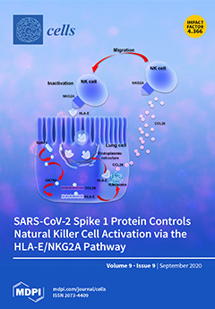
Natural killer cells are important in the control of viral infection, though their role during the SARS-CoV-2 infection had not previously been identified. Peripheral blood NK cells from SARS-CoV-2-naïve subjects were evaluated for their activation, degranulation, interferon-gamma expression in the presence of SARS-CoV-2 spike proteins. We show, for the first time, that NK cells are affected by SARS-CoV-2 spike 1 protein (SP1) expression in lung epithelial cells via HLA-E/NKG2A interaction. The internalization of the viral SP1 induces a cellular stress condition in lung epithelial cells and consequent upmodulation of HLA-E molecules via GATA3 transcription factor. The resulting interaction between HLA-E/NKG2A induces NK cell exhaustion that might contribute to immunopathogenesis in SARS-CoV-2 infection. View this paper
  • Issues are regarded as officially published after their release is announced to the table of contents alert mailing list.
  • You may sign up for e-mail alerts to receive table of contents of newly released issues.
  • PDF is the official format for papers published in both, html and pdf forms. To view the papers in pdf format, click on the "PDF Full-text" link, and use the free Adobe Reader to open them.
Order results
Result details
Section
Select all
Export citation of selected articles as:
15 pages, 1879 KiB  
Article
miR-9-Mediated Inhibition of EFEMP1 Contributes to the Acquisition of Pro-Tumoral Properties in Normal Fibroblasts
by Giulia Cosentino, Sandra Romero-Cordoba, Ilaria Plantamura, Alessandra Cataldo and Marilena V. Iorio
Cells 2020, 9(9), 2143; https://doi.org/10.3390/cells9092143 - 22 Sep 2020
Cited by 15 | Viewed by 2676
Abstract
Tumor growth and invasion occurs through a dynamic interaction between cancer and stromal cells, which support an aggressive niche. MicroRNAs are thought to act as tumor messengers to “corrupt” stromal cells. We previously demonstrated that miR-9, a known metastamiR, is released by triple [...] Read more.
Tumor growth and invasion occurs through a dynamic interaction between cancer and stromal cells, which support an aggressive niche. MicroRNAs are thought to act as tumor messengers to “corrupt” stromal cells. We previously demonstrated that miR-9, a known metastamiR, is released by triple negative breast cancer (TNBC) cells to enhance the transition of normal fibroblasts (NFs) into cancer-associated fibroblast (CAF)-like cells. EGF containing fibulin extracellular matrix protein 1 (EFEMP1), which encodes for the ECM glycoprotein fibulin-3, emerged as a miR-9 putative target upon miRNA’s exogenous upmodulation in NFs. Here we explored the impact of EFEMP1 downmodulation on fibroblast’s acquisition of CAF-like features, and how this phenotype influences neoplastic cells to gain chemoresistance. Indeed, upon miR-9 overexpression in NFs, EFEMP1 resulted downmodulated, both at RNA and protein levels. The luciferase reporter assay showed that miR-9 directly targets EFEMP1 and its silencing recapitulates miR-9-induced pro-tumoral phenotype in fibroblasts. In particular, EFEMP1 siRNA-transfected (si-EFEMP1) fibroblasts have an increased ability to migrate and invade. Moreover, TNBC cells conditioned with the supernatant of NFs transfected with miR-9 or si-EFEMP1 became more resistant to cisplatin. Overall, our results demonstrate that miR-9/EFEMP1 axis is crucial for the conversion of NFs to CAF-like cells under TNBC signaling. Full article
(This article belongs to the Special Issue Micro- and Macro-Environmental Factors in Solid Cancers)
Show Figures

Figure 1

18 pages, 3369 KiB  
Article
Human Mesenchymal Stromal Cell Secretome Promotes the Immunoregulatory Phenotype and Phagocytosis Activity in Human Macrophages
by Minna Holopainen, Ulla Impola, Petri Lehenkari, Saara Laitinen and Erja Kerkelä
Cells 2020, 9(9), 2142; https://doi.org/10.3390/cells9092142 - 22 Sep 2020
Cited by 6 | Viewed by 3008
Abstract
Human mesenchymal stromal/stem cells (hMSCs) show great promise in cell therapy due to their immunomodulatory properties. The overall immunomodulatory response of hMSCs resembles the resolution of inflammation, in which lipid mediators and regulatory macrophages (Mregs) play key roles. We investigated the effect of [...] Read more.
Human mesenchymal stromal/stem cells (hMSCs) show great promise in cell therapy due to their immunomodulatory properties. The overall immunomodulatory response of hMSCs resembles the resolution of inflammation, in which lipid mediators and regulatory macrophages (Mregs) play key roles. We investigated the effect of hMSC cell-cell contact and secretome on macrophages polarized and activated toward Mreg phenotype. Moreover, we studied the effect of supplemented polyunsaturated fatty acids (PUFAs): docosahexaenoic acid (DHA) and arachidonic acid, the precursors of lipid mediators, on hMSC immunomodulation. Our results show that unlike hMSC cell-cell contact, the hMSC secretome markedly increased the CD206 expression in both Mreg-polarized and Mreg-activated macrophages. Moreover, the secretome enhanced the expression of programmed death-ligand 1 on Mreg-polarized macrophages and Mer receptor tyrosine kinase on Mreg-activated macrophages. Remarkably, these changes were translated into improved Candida albicans phagocytosis activity of macrophages. Taken together, these results demonstrate that the hMSC secretome promotes the immunoregulatory and proresolving phenotype of Mregs. Intriguingly, DHA supplementation to hMSCs resulted in a more potentiated immunomodulation with increased CD163 expression and decreased gene expression of matrix metalloproteinase 2 in Mreg-polarized macrophages. These findings highlight the potential of PUFA supplementations as an easy and safe method to improve the hMSC therapeutic potential. Full article
(This article belongs to the Special Issue Enhancing Mesenchymal Stem Cells (MSCs) for Therapeutic Purposes)
Show Figures

Figure 1

19 pages, 8300 KiB  
Article
Pimozide Suppresses the Growth of Brain Tumors by Targeting STAT3-Mediated Autophagy
by Alok Ranjan, Itishree Kaushik and Sanjay K. Srivastava
Cells 2020, 9(9), 2141; https://doi.org/10.3390/cells9092141 - 22 Sep 2020
Cited by 24 | Viewed by 3427
Abstract
Brain tumors are considered as one of the most aggressive and incurable forms of cancer. The majority of the patients with brain tumors have a median survival rate of 12%. Brain tumors are lethal despite the availability of advanced treatment options such as [...] Read more.
Brain tumors are considered as one of the most aggressive and incurable forms of cancer. The majority of the patients with brain tumors have a median survival rate of 12%. Brain tumors are lethal despite the availability of advanced treatment options such as surgical removal, chemotherapy, and radiotherapy. In this study, we have evaluated the anti-cancer effects of pimozide, which is a neuroleptic drug used for the treatment of schizophrenia and chronic psychosis. Pimozide significantly reduced the proliferation of U-87MG, Daoy, GBM 28, and U-251MG brain cancer cell lines by inducing apoptosis with IC50 (Inhibitory concentration 50) ranging from 12 to 16 μM after 48 h of treatment. Our Western blotting analysis indicated that pimozide suppressed the phosphorylation of STAT3 at Tyr705 and Src at Tyr416, and it inhibited the expression of anti-apoptotic markers c-Myc, Mcl-1, and Bcl-2. Significant autophagy induction was observed with pimozide treatment. LC3B, Beclin-1, and ATG5 up-regulation along with autolysosome formation confirmed the induction of autophagy with pimozide treatment. Inhibiting autophagy using 3-methyladenine or LC3B siRNA significantly blocked the apoptosis-inducing effects of pimozide, suggesting that pimozide mediated its apoptotic effects by inducing autophagy. Oral administration of 25 mg/kg pimozide suppressed the intracranially implanted U-87MG tumor growth by 45% in athymic nude mice. The chronic administration of pimozide showed no general signs of toxicity, and the behavioral activity of the mice remained unchanged. Taken together, these results indicate that pimozide inhibits the growth of brain cancer by autophagy-mediated apoptosis. Full article
(This article belongs to the Special Issue Recent Advances in Cancer Therapy)
Show Figures

Figure 1

15 pages, 1833 KiB  
Review
The Role of Chaperone-Mediated Autophagy in Cell Cycle Control and Its Implications in Cancer
by Marina Andrade-Tomaz, Izadora de Souza, Clarissa Ribeiro Reily Rocha and Luciana Rodrigues Gomes
Cells 2020, 9(9), 2140; https://doi.org/10.3390/cells9092140 - 22 Sep 2020
Cited by 55 | Viewed by 6625
Abstract
The cell cycle involves a network of proteins that modulate the sequence and timing of proliferation events. Unregulated proliferation is the most fundamental hallmark of cancer; thus, changes in cell cycle control are at the heart of malignant transformation processes. Several cellular processes [...] Read more.
The cell cycle involves a network of proteins that modulate the sequence and timing of proliferation events. Unregulated proliferation is the most fundamental hallmark of cancer; thus, changes in cell cycle control are at the heart of malignant transformation processes. Several cellular processes can interfere with the cell cycle, including autophagy, the catabolic pathway involved in degradation of intracellular constituents in lysosomes. According to the mechanism used to deliver cargo to the lysosome, autophagy can be classified as macroautophagy (MA), microautophagy (MI), or chaperone-mediated autophagy (CMA). Distinct from other autophagy types, CMA substrates are selectively recognized by a cytosolic chaperone, one-by-one, and then addressed for degradation in lysosomes. The function of MA in cell cycle control, and its influence in cancer progression, are already well-established. However, regulation of the cell cycle by CMA, in the context of tumorigenesis, has not been fully addressed. This review aims to present and debate the molecular mechanisms by which CMA can interfere in the cell cycle, in the context of cancer. Thus, cell cycle modulators, such as MYC, hypoxia-inducible factor-1 subunit alpha (HIF-1α), and checkpoint kinase 1 (CHK1), regulated by CMA activity will be discussed. Finally, the review will focus on how CMA dysfunction may impact the cell cycle, and as consequence promote tumorigenesis. Full article
(This article belongs to the Special Issue Cell Cycle Control and Cancer)
Show Figures

Graphical abstract

14 pages, 2290 KiB  
Article
Neutrophil Extracellular Traps Promote the Development and Growth of Human Salivary Stones
by Mirco Schapher, Michael Koch, Daniela Weidner, Michael Scholz, Stefan Wirtz, Aparna Mahajan, Irmgard Herrmann, Jeeshan Singh, Jasmin Knopf, Moritz Leppkes, Christine Schauer, Anika Grüneboom, Christoph Alexiou, Georg Schett, Heinrich Iro, Luis E. Muñoz and Martin Herrmann
Cells 2020, 9(9), 2139; https://doi.org/10.3390/cells9092139 - 22 Sep 2020
Cited by 23 | Viewed by 3279
Abstract
Salivary gland stones, or sialoliths, are the most common cause of the obstruction of salivary glands. The mechanism behind the formation of sialoliths has been elusive. Symptomatic sialolithiasis has a prevalence of 0.45% in the general population, is characterized by recurrent painful periprandial [...] Read more.
Salivary gland stones, or sialoliths, are the most common cause of the obstruction of salivary glands. The mechanism behind the formation of sialoliths has been elusive. Symptomatic sialolithiasis has a prevalence of 0.45% in the general population, is characterized by recurrent painful periprandial swelling of the affected gland, and often results in sialadenitis with the need for surgical intervention. Here, we show by the use of immunohistochemistry, immunofluorescence, computed tomography (CT) scans and reconstructions, special dye techniques, bacterial genotyping, and enzyme activity analyses that neutrophil extracellular traps (NETs) initiate the formation and growth of sialoliths in humans. The deposition of neutrophil granulocyte extracellular DNA around small crystals results in the dense aggregation of the latter, and the subsequent mineralization creates alternating layers of dense mineral, which are predominantly calcium salt deposits and DNA. The further agglomeration and appositional growth of these structures promotes the development of macroscopic sialoliths that finally occlude the efferent ducts of the salivary glands, causing clinical symptoms and salivary gland dysfunction. These findings provide an entirely novel insight into the mechanism of sialolithogenesis, in which an immune system-mediated response essentially participates in the physicochemical process of concrement formation and growth. Full article
(This article belongs to the Special Issue NET Formation in Health and Disease)
Show Figures

Figure 1

17 pages, 1040 KiB  
Review
Oxidoreductases in Glycoprotein Glycosylation, Folding, and ERAD
by Chaitanya Patel, Haddas Saad, Marina Shenkman and Gerardo Z. Lederkremer
Cells 2020, 9(9), 2138; https://doi.org/10.3390/cells9092138 - 22 Sep 2020
Cited by 14 | Viewed by 5183
Abstract
N-linked glycosylation and sugar chain processing, as well as disulfide bond formation, are among the most common post-translational protein modifications taking place in the endoplasmic reticulum (ER). They are essential modifications that are required for membrane and secretory proteins to achieve their correct [...] Read more.
N-linked glycosylation and sugar chain processing, as well as disulfide bond formation, are among the most common post-translational protein modifications taking place in the endoplasmic reticulum (ER). They are essential modifications that are required for membrane and secretory proteins to achieve their correct folding and native structure. Several oxidoreductases responsible for disulfide bond formation, isomerization, and reduction have been shown to form stable, functional complexes with enzymes and chaperones that are involved in the initial addition of an N-glycan and in folding and quality control of the glycoproteins. Some of these oxidoreductases are selenoproteins. Recent studies also implicate glycan machinery–oxidoreductase complexes in the recognition and processing of misfolded glycoproteins and their reduction and targeting to ER-associated degradation. This review focuses on the intriguing cooperation between the glycoprotein-specific cell machineries and ER oxidoreductases, and highlights open questions regarding the functions of many members of this large family. Full article
(This article belongs to the Section Intracellular and Plasma Membranes)
Show Figures

Figure 1

26 pages, 9594 KiB  
Article
Choice of Differentiation Media Significantly Impacts Cell Lineage and Response to CFTR Modulators in Fully Differentiated Primary Cultures of Cystic Fibrosis Human Airway Epithelial Cells
by Vinciane Saint-Criq, Livia Delpiano, John Casement, Jennifer C. Onuora, JinHeng Lin and Michael A. Gray
Cells 2020, 9(9), 2137; https://doi.org/10.3390/cells9092137 - 21 Sep 2020
Cited by 25 | Viewed by 5096
Abstract
In vitro cultures of primary human airway epithelial cells (hAECs) grown at air–liquid interface have become a valuable tool to study airway biology under normal and pathologic conditions, and for drug discovery in lung diseases such as cystic fibrosis (CF). An increasing number [...] Read more.
In vitro cultures of primary human airway epithelial cells (hAECs) grown at air–liquid interface have become a valuable tool to study airway biology under normal and pathologic conditions, and for drug discovery in lung diseases such as cystic fibrosis (CF). An increasing number of different differentiation media, are now available, making comparison of data between studies difficult. Here, we investigated the impact of two common differentiation media on phenotypic, transcriptomic, and physiological features of CF and non-CF epithelia. Cellular architecture and density were strongly impacted by the choice of medium. RNA-sequencing revealed a shift in airway cell lineage; one medium promoting differentiation into club and goblet cells whilst the other enriched the growth of ionocytes and multiciliated cells. Pathway analysis identified differential expression of genes involved in ion and fluid transport. Physiological assays (intracellular/extracellular pH, Ussing chamber) specifically showed that ATP12A and CFTR function were altered, impacting pH and transepithelial ion transport in CF hAECs. Importantly, the two media differentially affected functional responses to CFTR modulators. We argue that the effect of growth conditions should be appropriately determined depending on the scientific question and that our study can act as a guide for choosing the optimal growth medium for specific applications. Full article
Show Figures

Graphical abstract

23 pages, 509 KiB  
Review
Cytokine Profiling in Myeloproliferative Neoplasms: Overview on Phenotype Correlation, Outcome Prediction, and Role of Genetic Variants
by Elena Masselli, Giulia Pozzi, Giuliana Gobbi, Stefania Merighi, Stefania Gessi, Marco Vitale and Cecilia Carubbi
Cells 2020, 9(9), 2136; https://doi.org/10.3390/cells9092136 - 21 Sep 2020
Cited by 26 | Viewed by 4198
Abstract
Among hematologic malignancies, the classic Philadelphia-negative chronic myeloproliferative neoplasms (MPNs) are considered a model of inflammation-related cancer development. In this context, the use of immune-modulating agents has recently expanded the MPN therapeutic scenario. Cytokines are key mediators of an auto-amplifying, detrimental cross-talk between [...] Read more.
Among hematologic malignancies, the classic Philadelphia-negative chronic myeloproliferative neoplasms (MPNs) are considered a model of inflammation-related cancer development. In this context, the use of immune-modulating agents has recently expanded the MPN therapeutic scenario. Cytokines are key mediators of an auto-amplifying, detrimental cross-talk between the MPN clone and the tumor microenvironment represented by immune, stromal, and endothelial cells. This review focuses on recent advances in cytokine-profiling of MPN patients, analyzing different expression patterns among the three main Philadelphia-negative (Ph-negative) MPNs, as well as correlations with disease molecular profile, phenotype, progression, and outcome. The role of the megakaryocytic clone as the main source of cytokines, particularly in myelofibrosis, is also reviewed. Finally, we report emerging intriguing evidence on the contribution of host genetic variants to the chronic pro-inflammatory state that typifies MPNs. Full article
Show Figures

Figure 1

17 pages, 1436 KiB  
Review
Mitochondrial Calcium Deregulation in the Mechanism of Beta-Amyloid and Tau Pathology
by Noemi Esteras and Andrey Y. Abramov
Cells 2020, 9(9), 2135; https://doi.org/10.3390/cells9092135 - 21 Sep 2020
Cited by 62 | Viewed by 5625
Abstract
Aggregation and deposition of β-amyloid and/or tau protein are the key neuropathological features in neurodegenerative disorders such as Alzheimer’s disease (AD) and other tauopathies including frontotemporal dementia (FTD). The interaction between oxidative stress, mitochondrial dysfunction and the impairment of calcium ions (Ca2+ [...] Read more.
Aggregation and deposition of β-amyloid and/or tau protein are the key neuropathological features in neurodegenerative disorders such as Alzheimer’s disease (AD) and other tauopathies including frontotemporal dementia (FTD). The interaction between oxidative stress, mitochondrial dysfunction and the impairment of calcium ions (Ca2+) homeostasis induced by misfolded tau and β-amyloid plays an important role in the progressive neuronal loss occurring in specific areas of the brain. In addition to the control of bioenergetics and ROS production, mitochondria are fine regulators of the cytosolic Ca2+ homeostasis that induce vital signalling mechanisms in excitable cells such as neurons. Impairment in the mitochondrial Ca2+ uptake through the mitochondrial Ca2+ uniporter (MCU) or release through the Na+/Ca2+ exchanger may lead to mitochondrial Ca2+ overload and opening of the permeability transition pore inducing neuronal death. Recent evidence suggests an important role for these mechanisms as the underlying causes for neuronal death in β-amyloid and tau pathology. The present review will focus on the mechanisms that lead to cytosolic and especially mitochondrial Ca2+ disturbances occurring in AD and tau-induced FTD, and propose possible therapeutic interventions for these disorders. Full article
Show Figures

Figure 1

25 pages, 2553 KiB  
Review
The Rationale of Neprilysin Inhibition in Prevention of Myocardial Ischemia-Reperfusion Injury during ST-Elevation Myocardial Infarction
by Alessandro Bellis, Ciro Mauro, Emanuele Barbato, Giuseppe Di Gioia, Daniela Sorriento, Bruno Trimarco and Carmine Morisco
Cells 2020, 9(9), 2134; https://doi.org/10.3390/cells9092134 - 21 Sep 2020
Cited by 12 | Viewed by 3690
Abstract
During the last three decades, timely myocardial reperfusion using either thrombolytic therapy or primary percutaneous intervention (pPCI) has allowed amazing improvements in outcomes with a more than halving in 1-year ST-elevation myocardial infarction (STEMI) mortality. However, mortality and left ventricle (LV) remodeling remain [...] Read more.
During the last three decades, timely myocardial reperfusion using either thrombolytic therapy or primary percutaneous intervention (pPCI) has allowed amazing improvements in outcomes with a more than halving in 1-year ST-elevation myocardial infarction (STEMI) mortality. However, mortality and left ventricle (LV) remodeling remain substantial in these patients. As such, novel therapeutic interventions are required to reduce myocardial infarction size, preserve LV systolic function, and improve survival in reperfused-STEMI patients. Myocardial ischemia-reperfusion injury (MIRI) prevention represents the main goal to reach in order to reduce STEMI mortality. There is currently no effective therapy for MIRI prevention in STEMI patients. A significant reason for the weak and inconsistent results obtained in this field may be the presence of multiple, partially redundant, mechanisms of cell death during ischemia-reperfusion, whose relative importance may depend on the conditions. Therefore, it is always more recognized that it is important to consider a “multi-targeted cardioprotective therapy”, defined as an additive or synergistic cardioprotective agents or interventions directed to distinct targets with different timing of application (before, during, or after pPCI). Given that some neprilysin (NEP) substrates (natriuretic peptides, angiotensin II, bradykinin, apelins, substance P, and adrenomedullin) exert a cardioprotective effect against ischemia-reperfusion injury, it is conceivable that antagonism of proteolytic activity by this enzyme may be considered in a multi-targeted strategy for MIRI prevention. In this review, by starting from main pathophysiological mechanisms promoting MIRI, we discuss cardioprotective effects of NEP substrates and the potential benefit of NEP pharmacological inhibition in MIRI prevention. Full article
Show Figures

Figure 1

20 pages, 5719 KiB  
Article
NRF2 Is an Upstream Regulator of MYC-Mediated Osteoclastogenesis and Pathological Bone Erosion
by Peter Sang Uk Park, Se Hwan Mun, Steven L. Zeng, Haemin Kim, Seyeon Bae and Kyung-Hyun Park-Min
Cells 2020, 9(9), 2133; https://doi.org/10.3390/cells9092133 - 21 Sep 2020
Cited by 11 | Viewed by 3355
Abstract
Osteoclasts are the sole bone-resorbing cells that play an essential role in homeostatic bone remodeling and pathogenic bone destruction such as inflammatory arthritis. Pharmacologically targeting osteoclasts has been a promising approach to alleviating bone disease, but there remains room for improvement in mitigating [...] Read more.
Osteoclasts are the sole bone-resorbing cells that play an essential role in homeostatic bone remodeling and pathogenic bone destruction such as inflammatory arthritis. Pharmacologically targeting osteoclasts has been a promising approach to alleviating bone disease, but there remains room for improvement in mitigating drug side effects and enhancing cell specificity. Recently, we demonstrated the crucial role of MYC and its downstream effectors in driving osteoclast differentiation. Despite these advances, upstream regulators of MYC have not been well defined. In this study, we identify nuclear factor erythroid 2-related factor 2 (NRF2), a transcription factor known to regulate the expression of phase II antioxidant enzymes, as a novel upstream regulator of MYC. NRF2 negatively regulates receptor activator of nuclear factor-κB ligand (RANKL)-induced osteoclastogenesis through the ERK and p38 signaling-mediated suppression of MYC transcription. Furthermore, the ablation of MYC in osteoclasts reverses the enhanced osteoclast differentiation and activity in NRF2 deficiency in vivo and in vitro in addition to protecting NRF2-deficient mice from pathological bone loss in a murine model of inflammatory arthritis. Our findings indicate that this novel NRF2-MYC axis could be instrumental for the fine-tuning of osteoclast formation and provides additional ways in which osteoclasts could be therapeutically targeted to prevent pathological bone erosion. Full article
(This article belongs to the Special Issue Molecular Basis of Osteoclast Differentiation and Activation)
Show Figures

Figure 1

21 pages, 2507 KiB  
Review
Microglia and Macrophages in the Pathological Central and Peripheral Nervous Systems
by Naoki Abe, Tasuku Nishihara, Toshihiro Yorozuya and Junya Tanaka
Cells 2020, 9(9), 2132; https://doi.org/10.3390/cells9092132 - 21 Sep 2020
Cited by 46 | Viewed by 7108
Abstract
Microglia, the immunocompetent cells in the central nervous system (CNS), have long been studied as pathologically deteriorating players in various CNS diseases. However, microglia exert ameliorating neuroprotective effects, which prompted us to reconsider their roles in CNS and peripheral nervous system (PNS) pathophysiology. [...] Read more.
Microglia, the immunocompetent cells in the central nervous system (CNS), have long been studied as pathologically deteriorating players in various CNS diseases. However, microglia exert ameliorating neuroprotective effects, which prompted us to reconsider their roles in CNS and peripheral nervous system (PNS) pathophysiology. Moreover, recent findings showed that microglia play critical roles even in the healthy CNS. The microglial functions that normally contribute to the maintenance of homeostasis in the CNS are modified by other cells, such as astrocytes and infiltrated myeloid cells; thus, the microglial actions on neurons are extremely complex. For a deeper understanding of the pathophysiology of various diseases, including those of the PNS, it is important to understand microglial functioning. In this review, we discuss both the favorable and unfavorable roles of microglia in neuronal survival in various CNS and PNS disorders. We also discuss the roles of blood-borne macrophages in the pathogenesis of CNS and PNS injuries because they cooperatively modify the pathological processes of resident microglia. Finally, metabolic changes in glycolysis and oxidative phosphorylation, with special reference to the pro-/anti-inflammatory activation of microglia, are intensively addressed, because they are profoundly correlated with the generation of reactive oxygen species and changes in pro-/anti-inflammatory phenotypes. Full article
(This article belongs to the Special Issue Neuron-Glia Interactions)
Show Figures

Figure 1

18 pages, 1427 KiB  
Review
Schwann Cell Role in Selectivity of Nerve Regeneration
by Sara Bolívar, Xavier Navarro and Esther Udina
Cells 2020, 9(9), 2131; https://doi.org/10.3390/cells9092131 - 20 Sep 2020
Cited by 57 | Viewed by 7660
Abstract
Peripheral nerve injuries result in the loss of the motor, sensory and autonomic functions of the denervated segments of the body. Neurons can regenerate after peripheral axotomy, but inaccuracy in reinnervation causes a permanent loss of function that impairs complete recovery. Thus, understanding [...] Read more.
Peripheral nerve injuries result in the loss of the motor, sensory and autonomic functions of the denervated segments of the body. Neurons can regenerate after peripheral axotomy, but inaccuracy in reinnervation causes a permanent loss of function that impairs complete recovery. Thus, understanding how regenerating axons respond to their environment and direct their growth is essential to improve the functional outcome of patients with nerve lesions. Schwann cells (SCs) play a crucial role in the regeneration process, but little is known about their contribution to specific reinnervation. Here, we review the mechanisms by which SCs can differentially influence the regeneration of motor and sensory axons. Mature SCs express modality-specific phenotypes that have been associated with the promotion of selective regeneration. These include molecular markers, such as L2/HNK-1 carbohydrate, which is differentially expressed in motor and sensory SCs, or the neurotrophic profile after denervation, which differs remarkably between SC modalities. Other important factors include several molecules implicated in axon-SC interaction. This cell–cell communication through adhesion (e.g., polysialic acid) and inhibitory molecules (e.g., MAG) contributes to guiding growing axons to their targets. As many of these factors can be modulated, further research will allow the design of new strategies to improve functional recovery after peripheral nerve injuries. Full article
(This article belongs to the Special Issue Schwann Cells: From Formation to Clinical Significance)
Show Figures

Figure 1

4 pages, 194 KiB  
Editorial
Neutrophils and Neutrophil Extracellular Traps Regulate Immune Responses in Health and Disease
by Shrikant R. Mulay and Hans-Joachim Anders
Cells 2020, 9(9), 2130; https://doi.org/10.3390/cells9092130 - 20 Sep 2020
Cited by 12 | Viewed by 2754
Abstract
Neutrophils are first responders of antimicrobial host defense and sterile inflammation, and therefore, play important roles during health and disease [...] Full article
16 pages, 4880 KiB  
Article
High Sensitivity of Circulating Tumor Cells Derived from a Colorectal Cancer Patient for Dual Inhibition with AKT and mTOR Inhibitors
by Daniel J. Smit, Laure Cayrefourcq, Marie-Therese Haider, Nico Hinz, Klaus Pantel, Catherine Alix-Panabières and Manfred Jücker
Cells 2020, 9(9), 2129; https://doi.org/10.3390/cells9092129 - 20 Sep 2020
Cited by 24 | Viewed by 3880
Abstract
Circulating tumor cells (CTCs) are cells shed from the primary tumor into the bloodstream. While many studies on solid tumor cells exist, data on CTCs are scarce. The mortality of cancer is mostly associated with metastasis and recent research identified CTCs as initiators [...] Read more.
Circulating tumor cells (CTCs) are cells shed from the primary tumor into the bloodstream. While many studies on solid tumor cells exist, data on CTCs are scarce. The mortality of cancer is mostly associated with metastasis and recent research identified CTCs as initiators of metastasis. The PI3K/AKT/mTOR signaling pathway is an intracellular pathway that regulates essential functions including protein biosynthesis, cell growth, cell cycle control, survival and migration. Importantly, activating oncogenic mutations and amplifications in this pathway are frequently observed in a wide variety of cancer entities, underlining the significance of this signaling pathway. In this study, we analyzed the functional role of the PI3K/AKT/mTOR signaling pathway in the CTC-MCC-41 line, derived from a patient with metastatic colorectal cancer. One striking finding in our study was the strong sensitivity of this CTC line against AKT inhibition using MK2206 and mTOR inhibition using RAD001 within the nanomolar range. This suggests that therapies targeting AKT and mTOR could have been beneficial for the patient from which the CTC line was isolated. Additionally, a dual targeting approach of AKT/mTOR inside the PI3K/AKT/mTOR signaling pathway in the colorectal CTCs showed synergistic effects in vitro. Depending on the phenotypical behavior of CTC-MCC-41 in cell culture (adherent vs. suspension), we identified altered phosphorylation levels inside the PI3K/AKT/mTOR pathway. We observed a downregulation of the PI3K/AKT/mTOR signaling pathway, but not of the RAS/RAF/MAPK pathway, in CTCs growing in suspension in comparison to adherent CTCs. Our results highlight distinct functions of AKT isoforms in CTC-MCC-41 cells with respect to cell proliferation. Knockdown of AKT1 and AKT2 leads to significantly impaired proliferation of CTC-MCC-41 cells in vitro. Therefore, our data demonstrate that the PI3K/AKT/mTOR signaling pathway plays a key role in the proliferation of CTC-MCC-41. Full article
(This article belongs to the Section Cell Signaling)
Show Figures

Graphical abstract

15 pages, 1138 KiB  
Review
EpCAM as Modulator of Tissue Plasticity
by François Fagotto
Cells 2020, 9(9), 2128; https://doi.org/10.3390/cells9092128 - 19 Sep 2020
Cited by 7 | Viewed by 3274
Abstract
The Epithelial Cell Adhesion Molecule or EpCAM is a well-known marker highly expressed in carcinomas and showing a strong correlation with poor cancer prognosis. While its name relates to its proposed function as a cell adhesion molecule, EpCAM has been shown to have [...] Read more.
The Epithelial Cell Adhesion Molecule or EpCAM is a well-known marker highly expressed in carcinomas and showing a strong correlation with poor cancer prognosis. While its name relates to its proposed function as a cell adhesion molecule, EpCAM has been shown to have various signalling functions. In particular, it has been identified as an important positive regulator of cell adhesion and migration, playing an essential role in embryonic morphogenesis as well as intestinal homeostasis. This activity is not due to its putative adhesive function, but rather to its ability to repress myosin contractility by impinging on a PKC signalling cascade. This mechanism confers EpCAM the unique property of favouring tissue plasticity. I review here the currently available data, comment on possible connections with other properties of EpCAM, and discuss the potential significance in the context of cancer invasion. Full article
Show Figures

Graphical abstract

22 pages, 4392 KiB  
Article
Juniperonic Acid Biosynthesis is Essential in Caenorhabditis elegans Lacking Δ6 Desaturase (fat-3) and Generates New ω-3 Endocannabinoids
by Sujay Guha, Serafina Calarco, M. Salomé Gachet and Jürg Gertsch
Cells 2020, 9(9), 2127; https://doi.org/10.3390/cells9092127 - 19 Sep 2020
Cited by 5 | Viewed by 3812
Abstract
In eukaryotes, the C20:4 polyunsaturated fatty acid arachidonic acid (AA) plays important roles as a phospholipid component, signaling molecule and precursor of the endocannabinoid-prostanoid axis. Accordingly, the absence of AA causes detrimental effects. Here, compensatory mechanisms involved in AA deficiency in Caenorhabditis elegans [...] Read more.
In eukaryotes, the C20:4 polyunsaturated fatty acid arachidonic acid (AA) plays important roles as a phospholipid component, signaling molecule and precursor of the endocannabinoid-prostanoid axis. Accordingly, the absence of AA causes detrimental effects. Here, compensatory mechanisms involved in AA deficiency in Caenorhabditis elegans were investigated. We show that the ω-3 C20:4 polyunsaturated fatty acid juniperonic acid (JuA) is generated in the C. elegans fat-3(wa22) mutant, which lacks Δ6 desaturase activity and cannot generate AA and ω-3 AA. JuA partially rescued the loss of function of AA in growth and development. Additionally, we observed that supplementation of AA and ω-3 AA modulates lifespan of fat-3(wa22) mutants. We described a feasible biosynthetic pathway that leads to the generation of JuA from α-linoleic acid (ALA) via elongases ELO-1/2 and Δ5 desaturase which is rate-limiting. Employing liquid chromatography mass spectrometry (LC-MS/MS), we identified endocannabinoid-like ethanolamine and glycerol derivatives of JuA and ω-3 AA. Like classical endocannabinoids, these lipids exhibited binding interactions with NPR-32, a G protein coupled receptor (GPCR) shown to act as endocannabinoid receptor in C. elegans. Our study suggests that the eicosatetraenoic acids AA, ω-3 AA and JuA share similar biological functions. This biosynthetic plasticity of eicosatetraenoic acids observed in C. elegans uncovers a possible biological role of JuA and associated ω-3 endocannabinoids in Δ6 desaturase deficiencies, highlighting the importance of ALA. Full article
(This article belongs to the Section Cellular Pathology)
Show Figures

Graphical abstract

14 pages, 2211 KiB  
Article
FRET-Based Sorting of Live Cells Reveals Shifted Balance between PLK1 and CDK1 Activities During Checkpoint Recovery
by Lorenzo Lafranchi, Erik Müllers, Dorothea Rutishauser and Arne Lindqvist
Cells 2020, 9(9), 2126; https://doi.org/10.3390/cells9092126 - 19 Sep 2020
Cited by 1 | Viewed by 3711
Abstract
Cells recovering from the G2/M DNA damage checkpoint rely more on Aurora A-PLK1 signaling than cells progressing through an unperturbed G2 phase, but the reason for this discrepancy is not known. Here, we devised a method based on a FRET reporter for PLK1 [...] Read more.
Cells recovering from the G2/M DNA damage checkpoint rely more on Aurora A-PLK1 signaling than cells progressing through an unperturbed G2 phase, but the reason for this discrepancy is not known. Here, we devised a method based on a FRET reporter for PLK1 activity to sort cells in distinct populations within G2 phase. We employed mass spectroscopy to characterize changes in protein levels through an unperturbed G2 phase and validated that ATAD2 levels decrease in a proteasome-dependent manner. Comparing unperturbed cells with cells recovering from DNA damage, we note that at similar PLK1 activities, recovering cells contain higher levels of Cyclin B1 and increased phosphorylation of CDK1 targets. The increased Cyclin B1 levels are due to continuous Cyclin B1 production during a DNA damage response and are sustained until mitosis. Whereas partial inhibition of PLK1 suppresses mitotic entry more efficiently when cells recover from a checkpoint, partial inhibition of CDK1 suppresses mitotic entry more efficiently in unperturbed cells. Our findings provide a resource for proteome changes during G2 phase, show that the mitotic entry network is rewired during a DNA damage response, and suggest that the bottleneck for mitotic entry shifts from CDK1 to PLK1 after DNA damage. Full article
(This article belongs to the Special Issue Cell Cycle Control and Cancer)
Show Figures

Figure 1

29 pages, 2653 KiB  
Review
Transcriptional Regulation of Wnt/β-Catenin Pathway in Colorectal Cancer
by Jia Bian, Marius Dannappel, Chunhua Wan and Ron Firestein
Cells 2020, 9(9), 2125; https://doi.org/10.3390/cells9092125 - 19 Sep 2020
Cited by 124 | Viewed by 12762
Abstract
The Wnt/β-catenin signaling pathway exerts integral roles in embryogenesis and adult homeostasis. Aberrant activation of the pathway is implicated in growth-associated diseases and cancers, especially as a key driver in the initiation and progression of colorectal cancer (CRC). Loss or inactivation of Adenomatous [...] Read more.
The Wnt/β-catenin signaling pathway exerts integral roles in embryogenesis and adult homeostasis. Aberrant activation of the pathway is implicated in growth-associated diseases and cancers, especially as a key driver in the initiation and progression of colorectal cancer (CRC). Loss or inactivation of Adenomatous polyposis coli (APC) results in constitutive activation of Wnt/β-catenin signaling, which is considered as an initiating event in the development of CRC. Increased Wnt/β-catenin signaling is observed in virtually all CRC patients, underscoring the importance of this pathway for therapeutic intervention. Prior studies have deciphered the regulatory networks required for the cytoplasmic stabilisation or degradation of the Wnt pathway effector, β-catenin. However, the mechanism whereby nuclear β-catenin drives or inhibits expression of Wnt target genes is more diverse and less well characterised. Here, we describe a brief synopsis of the core canonical Wnt pathway components, set the spotlight on nuclear mediators and highlight the emerging role of chromatin regulators as modulators of β-catenin-dependent transcription activity and oncogenic output. Full article
(This article belongs to the Special Issue The Role of Mediator Kinase in Cancer )
Show Figures

Figure 1

17 pages, 2363 KiB  
Article
Attempts at the Characterization of In-Cell Biophysical Processes Non-Invasively—Quantitative NMR Diffusometry of a Model Cellular System
by Weronika Mazur and Artur T. Krzyżak
Cells 2020, 9(9), 2124; https://doi.org/10.3390/cells9092124 - 19 Sep 2020
Cited by 7 | Viewed by 2151
Abstract
In the literature, diffusion studies of cell systems are usually limited to two water pools that are associated with the extracellular space and the entire interior of the cell. Therefore, the time-dependent diffusion coefficient contains information about the geometry of these two water [...] Read more.
In the literature, diffusion studies of cell systems are usually limited to two water pools that are associated with the extracellular space and the entire interior of the cell. Therefore, the time-dependent diffusion coefficient contains information about the geometry of these two water regions and the water exchange through their boundary. This approach is due to the fact that most of these studies use pulse techniques and relatively low gradients, which prevents the achievement of high b-values. As a consequence, it is not possible to register the signal coming from proton populations with a very low bulk or apparent self-diffusion coefficient, such as cell organelles. The purpose of this work was to obtain information on the geometry and dynamics of water at a level lower than the cell size, i.e., in cellular structures, using the time-dependent diffusion coefficient method. The model of the cell system was made of baker’s yeast (Saccharomyces cerevisiae) since that is commonly available and well-characterized. We measured characteristic fresh yeast properties with the application of a compact Nuclear Magnetic Resonance (NMR)-Magritek Mobile Universal Surface Explorer (MoUSE) device with a very high, constant gradient (~24 T/m), which enabled us to obtain a sufficient stimulated echo attenuation even for very short diffusion times (0.2–40 ms) and to apply very short diffusion encoding times. In this work, due to a very large diffusion weighting (b-values), splitting the signal into three components was possible, among which one was associated only with cellular structures. Time-dependent diffusion coefficient analysis allowed us to determine the self-diffusion coefficients of extracellular fluid, cytoplasm and cellular organelles, as well as compartment sizes. Cellular organelles contributing to each compartment were identified based on the random walk simulations and approximate volumes of water pools calculated using theoretical sizes or molar fractions. Information about different cell structures is contained in different compartments depending on the diffusion regime, which is inherent in studies applying extremely high gradients. Full article
Show Figures

Figure 1

15 pages, 2034 KiB  
Article
Human Amnion Epithelial Cells Impair T Cell Proliferation: The Role of HLA-G and HLA-E Molecules
by Fabio Morandi, Danilo Marimpietri, Andre Görgens, Alessia Gallo, Raghuraman Chittor Srinivasan, Samir El-Andaloussi and Roberto Gramignoli
Cells 2020, 9(9), 2123; https://doi.org/10.3390/cells9092123 - 19 Sep 2020
Cited by 19 | Viewed by 3573
Abstract
The immunoprivilege status characteristic of human amnion epithelial cells (hAECs) has been recently highlighted in the context of xenogenic transplantation. However, the mechanism(s) involved in such regulatory functions have been so far only partially been clarified. Here, we have analyzed the expression of [...] Read more.
The immunoprivilege status characteristic of human amnion epithelial cells (hAECs) has been recently highlighted in the context of xenogenic transplantation. However, the mechanism(s) involved in such regulatory functions have been so far only partially been clarified. Here, we have analyzed the expression of HLA-Ib molecules in isolated hAEC obtained from full term placentae. Moreover, we asked whether these molecules are involved in the immunoregulatory functions of hAEC. Human amnion-derived cells expressed surface HLA-G and HLA-F at high levels, whereas the commonly expressed HLA-E molecule has been measured at a very low level or null on freshly isolated cells. HLA-Ib molecules can be expressed as membrane-bound and soluble forms, and in all hAEC batches analyzed we measured high levels of sHLA-G and sHLA-E when hAEC were maintained in culture, and such a release was time-dependent. Moreover, HLA-G was present in extracellular vesicles (EVs) released by hAEC. hAEC suppressed T cell proliferation in vitro at different hAEC:T cell ratios, as previously reported. Moreover, inhibition of T cell proliferation was partially reverted by pretreating hAEC with anti-HLA-G, anti-HLA-E and anti-β2 microglobulin, thus suggesting that HLA-G and -E molecules are involved in hAEC-mediated suppression of T cell proliferation. Finally, either large-size EV (lsEV) or small-size EV (ssEV) derived from hAEC significantly modulated T-cell proliferation. In conclusion, we have here characterized one of the mechanism(s) underlying immunomodulatory functions of hAEC, related to the expression and release of HLA-Ib molecules. Full article
(This article belongs to the Special Issue Immuno-Pathology in Organ and Cell Transplantation)
Show Figures

Graphical abstract

11 pages, 10614 KiB  
Article
NIR-Triggered Hyperthermal Effect of Polythiophene Nanoparticles Synthesized by Surfactant-Free Oxidative Polymerization Method on Colorectal Carcinoma Cells
by Deval Prasad Bhattarai and Beom Su Kim
Cells 2020, 9(9), 2122; https://doi.org/10.3390/cells9092122 - 18 Sep 2020
Cited by 7 | Viewed by 2342
Abstract
In this work, polythiophene nanoparticles (PTh–NPs) were synthesized by a surfactant-free oxidative chemical polymerization method at 60 °C, using ammonium persulphate as an oxidant. Various physicochemical properties were studied in terms of field emission scanning electron microscopy (FESEM), X-ray diffraction (XRD), Fourier transform [...] Read more.
In this work, polythiophene nanoparticles (PTh–NPs) were synthesized by a surfactant-free oxidative chemical polymerization method at 60 °C, using ammonium persulphate as an oxidant. Various physicochemical properties were studied in terms of field emission scanning electron microscopy (FESEM), X-ray diffraction (XRD), Fourier transform infra-red (FT-IR) spectroscopy, and differential scanning calorimetry (DSC)/thermogravimetric analysis (TGA). Photothermal performance of the as-synthesized PTh–NPs was studied by irradiating near infra-red of 808 nm under different concentration of the substrate and power supply. The photothermal stability of PTh–NPs was also studied. Photothermal effects of the as-synthesized PTh–NPs on colorectal cancer cells (CT-26) were studied at 100 µg/mL concentration and 808 nm NIR irradiation of 2.0 W/cm2 power. Our in vitro results showed remarkable NIR laser-triggered photothermal apoptotic cell death by PTh–NPs. Based on the experimental findings, it is revealed that PTh–NPs can act as a heat mediator and can be an alternative material for photothermal therapy in cancer treatment. Full article
Show Figures

Figure 1

2 pages, 177 KiB  
Editorial
Regulation of Apoptosis by the Bcl-2 Family of Proteins: Field on a Brink
by Alexey S. Ladokhin
Cells 2020, 9(9), 2121; https://doi.org/10.3390/cells9092121 - 18 Sep 2020
Cited by 17 | Viewed by 1895
Abstract
Apoptosis, a form of programmed cell death, is a highly regulated process critical for tissue development, homeostasis, and pathogenesis of various diseases [...] Full article
(This article belongs to the Special Issue Regulation of Apoptosis by the Bcl-2 Family of Proteins)
16 pages, 3252 KiB  
Article
Therapeutic Benefit of the Association of Lodenafil with Mesenchymal Stem Cells on Hypoxia-induced Pulmonary Hypertension in Rats
by Marina de Moraes Carvalho da Silva, Allan Kardec Nogueira de Alencar, Jaqueline Soares da Silva, Tadeu Lima Montagnoli, Grazielle Fernandes da Silva, Bruna de Souza Rocha, Guilherme Carneiro Montes, Rosália Mendez-Otero, Pedro Moreno Pimentel-Coelho, Juliana F. Vasques, Margarete Manhães Trahez, Roberto Takashi Sudo and Gisele Zapata-Sudo
Cells 2020, 9(9), 2120; https://doi.org/10.3390/cells9092120 - 18 Sep 2020
Cited by 4 | Viewed by 2765
Abstract
Pulmonary arterial hypertension (PAH) is characterized by the remodeling of pulmonary arteries, with an increased pulmonary arterial pressure and right ventricle (RV) overload. This work investigated the benefit of the association of human umbilical cord mesenchymal stem cells (hMSCs) with lodenafil, a phosphodiesterase-5 [...] Read more.
Pulmonary arterial hypertension (PAH) is characterized by the remodeling of pulmonary arteries, with an increased pulmonary arterial pressure and right ventricle (RV) overload. This work investigated the benefit of the association of human umbilical cord mesenchymal stem cells (hMSCs) with lodenafil, a phosphodiesterase-5 inhibitor, in an animal model of PAH. Male Wistar rats were exposed to hypoxia (10% O2) for three weeks plus a weekly i.p. injection of a vascular endothelial growth factor receptor inhibitor (SU5416, 20 mg/kg, SuHx). After confirmation of PAH, animals received intravenous injection of 5.105 hMSCs or vehicle, followed by oral treatment with lodenafil carbonate (10 mg/kg/day) for 14 days. The ratio between pulmonary artery acceleration time and RV ejection time reduced from 0.42 ± 0.01 (control) to 0.24 ± 0.01 in the SuHx group, which was not altered by lodenafil alone but was recovered to 0.31 ± 0.01 when administered in association with hMSCs. RV afterload was confirmed in the SuHx group with an increased RV systolic pressure (mmHg) of 52.1 ± 8.8 normalized to 29.6 ± 2.2 after treatment with the association. Treatment with hMSCs + lodenafil reversed RV hypertrophy, fibrosis and interstitial cell infiltration in the SuHx group. Combined therapy of lodenafil and hMSCs may be a strategy for PAH treatment. Full article
(This article belongs to the Section Stem Cells)
Show Figures

Figure 1

16 pages, 3724 KiB  
Article
Genetic Variation in CNS Myelination and Functional Brain Connectivity in Recombinant Inbred Mice
by Andrea Goudriaan, Maarten Loos, Sabine Spijker, August B. Smit and Mark H. G. Verheijen
Cells 2020, 9(9), 2119; https://doi.org/10.3390/cells9092119 - 18 Sep 2020
Cited by 4 | Viewed by 2204
Abstract
Myelination greatly increases the speed of action potential propagation of neurons, thereby enhancing the efficacy of inter-neuronal communication and hence, potentially, optimizing the brain’s signal processing capability. The impact of genetic variation on the extent of axonal myelination and its consequences for brain [...] Read more.
Myelination greatly increases the speed of action potential propagation of neurons, thereby enhancing the efficacy of inter-neuronal communication and hence, potentially, optimizing the brain’s signal processing capability. The impact of genetic variation on the extent of axonal myelination and its consequences for brain functioning remain to be determined. Here we investigated this question using a genetic reference panel (GRP) of mouse BXD recombinant inbred (RI) strains, which partly model genetic diversity as observed in human populations, and which show substantial genetic differences in a variety of behaviors, including learning, memory and anxiety. We found coherent differences in the expression of myelin genes in brain tissue of RI strains of the BXD panel, with the largest differences in the hippocampus. The parental C57BL/6J (C57) and DBA/2J (DBA) strains were on opposite ends of the expression spectrum, with C57 showing higher myelin transcript expression compared with DBA. Our experiments showed accompanying differences between C57 and DBA in myelin protein composition, total myelin content, and white matter conduction velocity. Finally, the hippocampal myelin gene expression of the BXD strains correlated significantly with behavioral traits involving anxiety and/or activity. Taken together, our data indicate that genetic variation in myelin gene expression translates to differences observed in myelination, axonal conduction speed, and possibly in anxiety/activity related behaviors. Full article
(This article belongs to the Special Issue Neuron-Glia Interactions)
Show Figures

Figure 1

19 pages, 1278 KiB  
Article
How Relevant Are Bone Marrow-Derived Mast Cells (BMMCs) as Models for Tissue Mast Cells? A Comparative Transcriptome Analysis of BMMCs and Peritoneal Mast Cells
by Srinivas Akula, Aida Paivandy, Zhirong Fu, Michael Thorpe, Gunnar Pejler and Lars Hellman
Cells 2020, 9(9), 2118; https://doi.org/10.3390/cells9092118 - 17 Sep 2020
Cited by 25 | Viewed by 5045
Abstract
Bone marrow-derived mast cells (BMMCs) are often used as a model system for studies of the role of MCs in health and disease. These cells are relatively easy to obtain from total bone marrow cells by culturing under the influence of IL-3 or [...] Read more.
Bone marrow-derived mast cells (BMMCs) are often used as a model system for studies of the role of MCs in health and disease. These cells are relatively easy to obtain from total bone marrow cells by culturing under the influence of IL-3 or stem cell factor (SCF). After 3 to 4 weeks in culture, a nearly homogenous cell population of toluidine blue-positive cells are often obtained. However, the question is how relevant equivalents these cells are to normal tissue MCs. By comparing the total transcriptome of purified peritoneal MCs with BMMCs, here we obtained a comparative view of these cells. We found several important transcripts that were expressed at very high levels in peritoneal MCs, but were almost totally absent from the BMMCs, including the major chymotryptic granule protease Mcpt4, the neurotrophin receptor Gfra2, the substance P receptor Mrgprb2, the metalloprotease Adamts9 and the complement factor 2 (C2). In addition, there were a number of other molecules that were expressed at much higher levels in peritoneal MCs than in BMMCs, including the transcription factors Myb and Meis2, the MilR1 (Allergin), Hdc (Histidine decarboxylase), Tarm1 and the IL-3 receptor alpha chain. We also found many transcripts that were highly expressed in BMMCs but were absent or expressed at low levels in the peritoneal MCs. However, there were also numerous MC-related transcripts that were expressed at similar levels in the two populations of cells, but almost absent in peritoneal macrophages and B cells. These results reveal that the transcriptome of BMMCs shows many similarities, but also many differences to that of tissue MCs. BMMCs can thereby serve as suitable models in many settings concerning the biology of MCs, but our findings also emphasize that great care should be taken when extrapolating findings from BMMCs to the in vivo function of tissue-resident MCs. Full article
Show Figures

Figure 1

5 pages, 188 KiB  
Editorial
Seeking and Exploring Efficient Ways to Target Cancer
by Tuula Kallunki
Cells 2020, 9(9), 2117; https://doi.org/10.3390/cells9092117 - 17 Sep 2020
Viewed by 1651
Abstract
Anti-cancer treatments have never been so numerous and so efficient [...] Full article
(This article belongs to the Special Issue Killing Cancer: Discovery and Selection of New Target Molecules)
12 pages, 483 KiB  
Review
Incorporating Prognostic Biomarkers into Risk Assessment Models and TNM Staging for Prostate Cancer
by Ragheed Saoud, Nassib Abou Heidar, Alessia Cimadamore and Gladell P. Paner
Cells 2020, 9(9), 2116; https://doi.org/10.3390/cells9092116 - 17 Sep 2020
Cited by 13 | Viewed by 3373
Abstract
In current practice, prostate cancer staging alone is not sufficient to adequately assess the patient’s prognosis and plan the management strategies. Multiple clinicopathological parameters and risk tools for prostate cancer have been developed over the past decades to better characterize the disease and [...] Read more.
In current practice, prostate cancer staging alone is not sufficient to adequately assess the patient’s prognosis and plan the management strategies. Multiple clinicopathological parameters and risk tools for prostate cancer have been developed over the past decades to better characterize the disease and provide an enhanced assessment of prognosis. Herein, we review novel prognostic biomarkers and their integration into risk assessment models for prostate cancer focusing on their capability to help avoid unnecessary imaging studies, biopsies and diagnosis of low risk prostate cancers, to help in the decision-making process between active surveillance and treatment intervention, and to predict recurrence after radical prostatectomy. There is an imperative need of reliable biomarkers to stratify prostate cancer patients that may benefit from different management approaches. The integration of biomarkers panel with risk assessment models appears to improve prostate cancer diagnosis and management. However, integration of novel genomic biomarkers in future prognostic models requires further validation in their clinical efficacy, standardization, and cost-effectiveness in routine application. Full article
Show Figures

Figure 1

19 pages, 1220 KiB  
Review
Role of GPER-Mediated Signaling in Testicular Functions and Tumorigenesis
by Adele Chimento, Arianna De Luca, Marta Claudia Nocito, Paola Avena, Davide La Padula, Lucia Zavaglia and Vincenzo Pezzi
Cells 2020, 9(9), 2115; https://doi.org/10.3390/cells9092115 - 17 Sep 2020
Cited by 30 | Viewed by 4174
Abstract
Estrogen signaling plays important roles in testicular functions and tumorigenesis. Fifteen years ago, it was discovered that a member of the G protein-coupled receptor family, GPR30, which binds also with high affinity to estradiol and is responsible, in part, for the rapid non-genomic [...] Read more.
Estrogen signaling plays important roles in testicular functions and tumorigenesis. Fifteen years ago, it was discovered that a member of the G protein-coupled receptor family, GPR30, which binds also with high affinity to estradiol and is responsible, in part, for the rapid non-genomic actions of estrogens. GPR30, renamed as GPER, was detected in several tissues including germ cells (spermatogonia, spermatocytes, spermatids) and somatic cells (Sertoli and Leydig cells). In our previous review published in 2014, we summarized studies that evidenced a role of GPER signaling in mediating estrogen action during spermatogenesis and testis development. In addition, we evidenced that GPER seems to be involved in modulating estrogen-dependent testicular cancer cell growth; however, the effects on cell survival and proliferation depend on specific cell type. In this review, we update the knowledge obtained in the last years on GPER roles in regulating physiological functions of testicular cells and its involvement in neoplastic transformation of both germ and somatic cells. In particular, we will focus our attention on crosstalk among GPER signaling, classical estrogen receptors and other nuclear receptors involved in testis physiology regulation. Full article
Show Figures

Figure 1

31 pages, 1445 KiB  
Review
Hedgehog Signaling and Truncated GLI1 in Cancer
by Daniel Doheny, Sara G. Manore, Grace L. Wong and Hui-Wen Lo
Cells 2020, 9(9), 2114; https://doi.org/10.3390/cells9092114 - 17 Sep 2020
Cited by 99 | Viewed by 10863
Abstract
The hedgehog (HH) signaling pathway regulates normal cell growth and differentiation. As a consequence of improper control, aberrant HH signaling results in tumorigenesis and supports aggressive phenotypes of human cancers, such as neoplastic transformation, tumor progression, metastasis, and drug resistance. Canonical activation of [...] Read more.
The hedgehog (HH) signaling pathway regulates normal cell growth and differentiation. As a consequence of improper control, aberrant HH signaling results in tumorigenesis and supports aggressive phenotypes of human cancers, such as neoplastic transformation, tumor progression, metastasis, and drug resistance. Canonical activation of HH signaling occurs through binding of HH ligands to the transmembrane receptor Patched 1 (PTCH1), which derepresses the transmembrane G protein-coupled receptor Smoothened (SMO). Consequently, the glioma-associated oncogene homolog 1 (GLI1) zinc-finger transcription factors, the terminal effectors of the HH pathway, are released from suppressor of fused (SUFU)-mediated cytoplasmic sequestration, permitting nuclear translocation and activation of target genes. Aberrant activation of this pathway has been implicated in several cancer types, including medulloblastoma, rhabdomyosarcoma, basal cell carcinoma, glioblastoma, and cancers of lung, colon, stomach, pancreas, ovarian, and breast. Therefore, several components of the HH pathway are under investigation for targeted cancer therapy, particularly GLI1 and SMO. GLI1 transcripts are reported to undergo alternative splicing to produce truncated variants: loss-of-function GLI1ΔN and gain-of-function truncated GLI1 (tGLI1). This review covers the biochemical steps necessary for propagation of the HH activating signal and the involvement of aberrant HH signaling in human cancers, with a highlight on the tumor-specific gain-of-function tGLI1 isoform. Full article
(This article belongs to the Special Issue Hedgehog Signaling in Development and Cancer)
Show Figures

Figure 1

Previous Issue
Next Issue
Back to TopTop