Next Issue
Volume 9, September
Previous Issue
Volume 9, July


Cells, Volume 9, Issue 8 (August 2020) – 171 articles

Cover Story (view full-size image): 
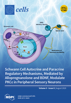
Crosstalk between Schwann cells and neurons, involving protein kinase type C-ε (PKCε), plays an important role in the sensitization of primary afferent nociceptors. Schwann cells possess the enzymatic machinery able to synthetize the progesterone metabolite allopregnanolone (ALLO) and tonically release it. ALLO autocrinally increases the expression and protein levels of the brain-derived neurotrophic factor (BDNF). Successively, being released by Schwann cells, BDNF paracrinally activates PKCε in dorsal root ganglia neurons. PKCε is renowned for increasing neuronal excitability via the phosphorylation of several ion channels, promoting the hyperalgesic priming of peripheral fibers, in turn leading to the modulation of noxious stimuli to the central nervous system. These mechanisms emphasize promising targets for inhibiting the onset and chronification of neuropathic pain. View this paper
  • Issues are regarded as officially published after their release is announced to the table of contents alert mailing list.
  • You may sign up for e-mail alerts to receive table of contents of newly released issues.
  • PDF is the official format for papers published in both, html and pdf forms. To view the papers in pdf format, click on the "PDF Full-text" link, and use the free Adobe Reader to open them.
Order results
Result details
Section
Select all
Export citation of selected articles as:
5 pages, 13741 KiB  
Correction
Correction: Yao, Y., et al. Activation of Slit2/Robo1 Signaling Promotes Tumor Metastasis in Colorectal Carcinoma through Activation of the TGF-β/Smads Pathway. Cells 2019, 8, 635
by Yuying Yao, Zijun Zhou, Liuyou Li, Junchen Li, Lixun Huang, Jiangchao Li, Cuiling Qi, Lingyun Zheng, Lijing Wang and Qian-Qian Zhang
Cells 2020, 9(8), 1918; https://doi.org/10.3390/cells9081918 - 18 Aug 2020
Cited by 3 | Viewed by 1888
Abstract
The author wishes to make the following correction to this paper [...] Full article
15 pages, 4601 KiB  
Article
MiRNA let-7 from TPO(+) Extracellular Vesicles is a Potential Marker for a Differential Diagnosis of Follicular Thyroid Nodules
by Lidia Zabegina, Inga Nazarova, Margarita Knyazeva, Nadezhda Nikiforova, Maria Slyusarenko, Sergey Titov, Dmitry Vasilyev, Ilya Sleptsov and Anastasia Malek
Cells 2020, 9(8), 1917; https://doi.org/10.3390/cells9081917 - 18 Aug 2020
Cited by 15 | Viewed by 3887
Abstract
Background: The current approaches to distinguish follicular adenomas (FA) and follicular thyroid cancer (FTC) at the pre-operative stage have low predictive value. Liquid biopsy-based analysis of circulating extracellular vesicles (EVs) presents a promising diagnostic method. However, the extreme heterogeneity of plasma EV population [...] Read more.
Background: The current approaches to distinguish follicular adenomas (FA) and follicular thyroid cancer (FTC) at the pre-operative stage have low predictive value. Liquid biopsy-based analysis of circulating extracellular vesicles (EVs) presents a promising diagnostic method. However, the extreme heterogeneity of plasma EV population hampers the development of new diagnostic tests. We hypothesize that the isolation of EVs with thyroid-specific surface molecules followed by miRNA analysis, may have improved diagnostic potency. Methods: The total population of EVs was isolated from the plasma of patients with FA (n = 30) and FTC (n = 30). Thyroid peroxidase (TPO)-positive EVs were isolated from the total populations using immune-beads. The miRNA from the TPO(+)EVs obtained from the plasma of FA and FTC patients was assayed by RT-PCR. The diagnostic potency of the selected miRNAs was estimated by the receiver operating characteristic (ROC) analysis. Results: TPO(+)EVs can be efficiently isolated by immunobeads. The analysis of Let-7 family members in TPO(+)EVs allows one to distinguish FA and FTC with high accuracy (area under curve defined by ROC = 0.77–0.84). Conclusion: The isolation of TPO(+)EVs, followed by RT-qPCR analysis of Let-7 family members, may present a helpful approach to manage follicular nodules in the thyroid gland. Full article
(This article belongs to the Special Issue Liquid Biopsy)
Show Figures

Graphical abstract

19 pages, 1725 KiB  
Review
Stemness Potency of Human Gingival Cells—Application in Anticancer Therapies and Clinical Trials
by Katarzyna Stefańska, Katarzyna Mehr, Maria Wieczorkiewicz, Magdalena Kulus, Ana Angelova Volponi, Jamil A. Shibli, Paul Mozdziak, Mariusz T. Skowroński, Paweł Antosik, Jędrzej M. Jaśkowski, Hanna Piotrowska-Kempisty, Bartosz Kempisty and Marta Dyszkiewicz-Konwińska
Cells 2020, 9(8), 1916; https://doi.org/10.3390/cells9081916 - 18 Aug 2020
Cited by 12 | Viewed by 4141
Abstract
Gingivae, as the part of periodontium, are involved in tooth support and possess the ability to heal rapidly, without scar formation. Recently, dental tissues have been identified as a potential source of mesenchymal stem cells (MSCs) and several populations of MSCs were isolated [...] Read more.
Gingivae, as the part of periodontium, are involved in tooth support and possess the ability to heal rapidly, without scar formation. Recently, dental tissues have been identified as a potential source of mesenchymal stem cells (MSCs) and several populations of MSCs were isolated from the orofacial region, including gingival mesenchymal stem cells (GMSCs). GMSCs exhibit robust immunomodulatory and differentiation potential and are easily obtainable, which make them promising candidates for cellular therapies. Apart from being tested for application in immunologic- and inflammatory-related disorders and various tissue regeneration, GMSCs promise to be a valuable tool in cancer treatment, especially in tongue squamous cell carcinoma (TSCC) with the use of targeted therapy, since GMSCs are able to selectively migrate towards the cancerous cells both in vitro and in vivo. In addition to their ability to uptake and release anti-neoplastic drugs, GMSCs may be transduced with apoptosis-inducing factors and used for cancer growth inhibition. Moreover, GMSCs, as most mammalian cells, secrete exosomes, which are a subset of extracellular vesicles with a diameter of 40–160 nm, containing DNA, RNA, lipids, metabolites, and proteins. Such GMSCs-derived exosomes may be useful therapeutic tool in cell-free therapy, as well as their culture medium. GMSCs exhibit molecular and stem-cell properties that make them well suited in preclinical and clinical studies. Full article
Show Figures

Figure 1

15 pages, 1251 KiB  
Article
Impact of Anticoagulation and Sample Processing on the Quantification of Human Blood-Derived microRNA Signatures
by Marion Mussbacher, Teresa L. Krammer, Stefan Heber, Waltraud C. Schrottmaier, Stephan Zeibig, Hans-Peter Holthoff, David Pereyra, Patrick Starlinger, Matthias Hackl and Alice Assinger
Cells 2020, 9(8), 1915; https://doi.org/10.3390/cells9081915 - 18 Aug 2020
Cited by 20 | Viewed by 2723
Abstract
Blood-derived microRNA signatures have emerged as powerful biomarkers for predicting and diagnosing cardiovascular disease, cancer, and metabolic disorders. Platelets and platelet-derived microvesicles are a major source of microRNAs. We have previously shown that the inappropriate anticoagulation and storage of blood samples causes substantial [...] Read more.
Blood-derived microRNA signatures have emerged as powerful biomarkers for predicting and diagnosing cardiovascular disease, cancer, and metabolic disorders. Platelets and platelet-derived microvesicles are a major source of microRNAs. We have previously shown that the inappropriate anticoagulation and storage of blood samples causes substantial platelet activation that is associated with the release of platelet-stored molecules into the plasma. However, it is currently unclear if circulating microRNA levels are affected by artificial platelet activation due to suboptimal plasma preparation. To address this issue, we used a standardized RT-qPCR test for 12 microRNAs (thrombomiR®, TAmiRNA GmbH, Vienna, Austria) that have been associated with cardiovascular and thrombotic diseases and were detected in platelets and/other hematopoietic cells. Blood was prevented from coagulating with citrate–theophylline–adenosine–dipyridamole (CTAD), sodium citrate, or ethylenediaminetetraacetic acid (EDTA) and stored for different time periods either at room temperature or at 4 °C prior to plasma preparation and the subsequent quantification of microRNAs. We found that five microRNAs (miR-191-5p, miR-320a, miR-21-5p, miR-23a-3p, and miR-451a) were significantly increased in the EDTA plasma. Moreover, we observed a time-dependent increase in plasma microRNAs that was most pronounced in the EDTA blood stored at room temperature for 24 h. Furthermore, significant correlations between microRNA levels and plasma concentrations of platelet-stored molecules pointed towards in vitro platelet activation. Therefore, we strongly recommend to (i) use CTAD as an anticoagulant, (ii) process blood samples as quickly as possible, and (iii) store blood samples at 4 °C whenever immediate plasma preparation is not feasible to generate reliable data on blood-derived microRNA signatures. Full article
(This article belongs to the Section Cellular Immunology)
Show Figures

Graphical abstract

16 pages, 2428 KiB  
Article
Decreased Equilibrative Nucleoside Transporter 1 (ENT1) Activity Contributes to the High Extracellular Adenosine Levels in Mesenchymal Glioblastoma Stem-Like Cells
by Sebastián Alarcón, María de los Ángeles Toro, Carolina Villarreal, Rómulo Melo, Rodrigo Fernández, Angel Ayuso Sacido, Daniel Uribe, Rody San Martín and Claudia Quezada
Cells 2020, 9(8), 1914; https://doi.org/10.3390/cells9081914 - 18 Aug 2020
Cited by 12 | Viewed by 3539
Abstract
Glioblastoma multiforme is one of the most malignant types of cancer. This is mainly due to a cell subpopulation with an extremely aggressive potential, called glioblastoma stem-like cells (GSCs). These cells produce high levels of extracellular adenosine which has been associated with increased [...] Read more.
Glioblastoma multiforme is one of the most malignant types of cancer. This is mainly due to a cell subpopulation with an extremely aggressive potential, called glioblastoma stem-like cells (GSCs). These cells produce high levels of extracellular adenosine which has been associated with increased chemoresistance, migration, and invasion in glioblastoma. In this study, we attempted to elucidate the mechanisms that control extracellular adenosine levels in GSC subtypes. By using primary and U87MG-derived GSCs, we associated increased extracellular adenosine with the mesenchymal phenotype. [3H]-adenosine uptake occurred mainly through the equilibrative nucleoside transporters (ENTs) in GSCs, but mesenchymal GSCs have lower expression and ENT1-mediated uptake activity than proneural GSCs. By analyzing expression and enzymatic activity, we determined that ecto-5′-nucleotidase (CD73) is predominantly expressed in proneural GSCs, driving AMPase activity. While in mesenchymal GSCs, both CD73 and Prostatic Acid Phosphatase (PAP) contribute to the AMP (adenosine monophosphate) hydrolysis. We did not observe significant differences between the expression of proteins involved in the metabolization of adenosine among the GCSs subtypes. In conclusion, the lower expression and activity of the ENT1 transporter in mesenchymal GSCs contributes to the high level of extracellular adenosine that these GSCs present. Full article
(This article belongs to the Section Stem Cells)
Show Figures

Figure 1

16 pages, 2592 KiB  
Review
The Intrinsically Disordered W Protein Is Multifunctional during Henipavirus Infection, Disrupting Host Signalling Pathways and Nuclear Import
by Sofiya Tsimbalyuk, Emily M. Cross, Mikayla Hoad, Camilla M. Donnelly, Justin A. Roby and Jade K. Forwood
Cells 2020, 9(8), 1913; https://doi.org/10.3390/cells9081913 - 18 Aug 2020
Cited by 10 | Viewed by 3183 | Correction
Abstract
Nipah and Hendra viruses are highly pathogenic, zoonotic henipaviruses that encode proteins that inhibit the host’s innate immune response. The W protein is one of four products encoded from the P gene and binds a number of host proteins to regulate signalling pathways. [...] Read more.
Nipah and Hendra viruses are highly pathogenic, zoonotic henipaviruses that encode proteins that inhibit the host’s innate immune response. The W protein is one of four products encoded from the P gene and binds a number of host proteins to regulate signalling pathways. The W protein is intrinsically disordered, a structural attribute that contributes to its diverse host protein interactions. Here, we review the role of W in innate immune suppression through inhibition of both pattern recognition receptor (PRR) pathways and interferon (IFN)-responsive signalling. PRR stimulation leading to activation of IRF-3 and IFN release is blocked by henipavirus W, and unphosphorylated STAT proteins are sequestered within the nucleus of host cells by W, thereby inhibiting the induction of IFN stimulated genes. We examine the critical role of nuclear transport in multiple functions of W and how specific binding of importin-alpha (Impα) isoforms, and the 14-3-3 group of regulatory proteins suggests further modulation of these processes. Overall, the disordered nature and multiple functions of W warrant further investigation to understand henipavirus pathogenesis and may reveal insights aiding the development of novel therapeutics. Full article
Show Figures

Figure 1

17 pages, 2885 KiB  
Article
Exposure of Human Skin Organoids to Low Genotoxic Stress Can Promote Epithelial-to-Mesenchymal Transition in Regenerating Keratinocyte Precursor Cells
by Sophie Cavallero, Renata Neves Granito, Daniel Stockholm, Peggy Azzolin, Michèle T. Martin and Nicolas O. Fortunel
Cells 2020, 9(8), 1912; https://doi.org/10.3390/cells9081912 - 18 Aug 2020
Cited by 7 | Viewed by 4028
Abstract
For the general population, medical diagnosis is a major cause of exposure to low genotoxic stress, as various imaging techniques deliver low doses of ionizing radiation. Our study investigated the consequences of low genotoxic stress on a keratinocyte precursor fraction that includes stem [...] Read more.
For the general population, medical diagnosis is a major cause of exposure to low genotoxic stress, as various imaging techniques deliver low doses of ionizing radiation. Our study investigated the consequences of low genotoxic stress on a keratinocyte precursor fraction that includes stem and progenitor cells, which are at risk for carcinoma development. Human skin organoids were bioengineered according to a clinically-relevant model, exposed to a single 50 mGy dose of γ rays, and then xeno-transplanted in nude mice to follow full epidermis generation in an in vivo context. Twenty days post-xenografting, mature skin grafts were sampled and analyzed by semi-quantitative immuno-histochemical methods. Pre-transplantation exposure to 50 mGy of immature human skin organoids did not compromise engraftment, but half of xenografts generated from irradiated precursors exhibited areas displaying focal dysplasia, originating from the basal layer of the epidermis. Characteristics of epithelial-to-mesenchymal transition (EMT) were documented in these dysplastic areas, including loss of basal cell polarity and cohesiveness, epithelial marker decreases, ectopic expression of the mesenchymal marker α-SMA and expression of the EMT promoter ZEB1. Taken together, these data show that a very low level of radiative stress in regenerating keratinocyte stem and precursor cells can induce a micro-environment that may constitute a favorable context for long-term carcinogenesis. Full article
(This article belongs to the Special Issue Stem Cells and Irradiation)
Show Figures

Figure 1

14 pages, 559 KiB  
Review
Zebrafish: A Suitable Tool for the Study of Cell Signaling in Bone
by Maria Teresa Valenti, Giulia Marchetto, Monica Mottes and Luca Dalle Carbonare
Cells 2020, 9(8), 1911; https://doi.org/10.3390/cells9081911 - 17 Aug 2020
Cited by 16 | Viewed by 5630
Abstract
In recent decades, many studies using the zebrafish model organism have been performed. Zebrafish, providing genetic mutants and reporter transgenic lines, enable a great number of studies aiming at the investigation of signaling pathways involved in the osteoarticular system and at the identification [...] Read more.
In recent decades, many studies using the zebrafish model organism have been performed. Zebrafish, providing genetic mutants and reporter transgenic lines, enable a great number of studies aiming at the investigation of signaling pathways involved in the osteoarticular system and at the identification of therapeutic tools for bone diseases. In this review, we will discuss studies which demonstrate that many signaling pathways are highly conserved between mammals and teleost and that genes involved in mammalian bone differentiation have orthologs in zebrafish. We will also discuss as human diseases, such as osteogenesis imperfecta, osteoarthritis, osteoporosis and Gaucher disease can be investigated in the zebrafish model. Full article
(This article belongs to the Special Issue Signaling Pathway Analysis and Disease Modeling in Zebrafish)
Show Figures

Figure 1

19 pages, 3640 KiB  
Article
TLR3-Dependent Activation of TLR2 Endogenous Ligands via the MyD88 Signaling Pathway Augments the Innate Immune Response
by Hellen S. Teixeira, Jiawei Zhao, Ethan Kazmierski, Denis F. Kinane and Manjunatha R. Benakanakere
Cells 2020, 9(8), 1910; https://doi.org/10.3390/cells9081910 - 17 Aug 2020
Cited by 8 | Viewed by 3370
Abstract
The role of the adaptor molecule MyD88 is thought to be independent of Toll-like receptor 3 (TLR3) signaling. In this report, we demonstrate a previously unknown role of MyD88 in TLR3 signaling in inducing endogenous ligands of TLR2 to elicit innate immune responses. [...] Read more.
The role of the adaptor molecule MyD88 is thought to be independent of Toll-like receptor 3 (TLR3) signaling. In this report, we demonstrate a previously unknown role of MyD88 in TLR3 signaling in inducing endogenous ligands of TLR2 to elicit innate immune responses. Of the various TLR ligands examined, the TLR3-specific ligand polyinosinic:polycytidylic acid (poly I:C), significantly induced TNF production and the upregulation of other TLR transcripts, in particular, TLR2. Accordingly, TLR3 stimulation also led to a significant upregulation of endogenous TLR2 ligands mainly, HMGB1 and Hsp60. By contrast, the silencing of TLR3 significantly downregulated MyD88 and TLR2 gene expression and pro-inflammatory IL1β, TNF, and IL8 secretion. The silencing of MyD88 similarly led to the downregulation of TLR2, IL1β, TNF and IL8, thus suggesting MyD88 to somehow act downstream of TLR3. Corroborating in vitro data, Myd88−/− knockout mice downregulated TNF, CXCL1; and phospho-p65 and phospho-IRF3 nuclear localization, upon poly I:C treatment in a mouse model of skin infection. Taken together, we identified a previously unknown role for MyD88 in the TLR3 signaling pathway, underlying the importance of TLRs and adapter protein interplay in modulating endogenous TLR ligands culminating in pro-inflammatory cytokine regulation. Full article
(This article belongs to the Section Cell Signaling)
Show Figures

Figure 1

31 pages, 1435 KiB  
Review
The Intestinal Barrier and Current Techniques for the Assessment of Gut Permeability
by Ida Schoultz and Åsa V. Keita
Cells 2020, 9(8), 1909; https://doi.org/10.3390/cells9081909 - 17 Aug 2020
Cited by 220 | Viewed by 20901
Abstract
The intestinal barrier is essential in human health and constitutes the interface between the outside and the internal milieu of the body. A functional intestinal barrier allows absorption of nutrients and fluids but simultaneously prevents harmful substances like toxins and bacteria from crossing [...] Read more.
The intestinal barrier is essential in human health and constitutes the interface between the outside and the internal milieu of the body. A functional intestinal barrier allows absorption of nutrients and fluids but simultaneously prevents harmful substances like toxins and bacteria from crossing the intestinal epithelium and reaching the body. An altered intestinal permeability, a sign of a perturbed barrier function, has during the last decade been associated with several chronic conditions, including diseases originating in the gastrointestinal tract but also diseases such as Alzheimer and Parkinson disease. This has led to an intensified interest from researchers with diverse backgrounds to perform functional studies of the intestinal barrier in different conditions. Intestinal permeability is defined as the passage of a solute through a simple membrane and can be measured by recording the passage of permeability markers over the epithelium via the paracellular or the transcellular route. The methodological tools to investigate the gut barrier function are rapidly expanding and new methodological approaches are being developed. Here we outline and discuss, in vivo, in vitro and ex vivo techniques and how these methods can be utilized for thorough investigation of the intestinal barrier. Full article
(This article belongs to the Special Issue Epithelial Cell Mechanics: From Physiology to Pathology)
Show Figures

Graphical abstract

20 pages, 2480 KiB  
Review
Nuclear Envelope Proteins Modulating the Heterochromatin Formation and Functions in Fission Yeast
by Yasuhiro Hirano, Haruhiko Asakawa, Takeshi Sakuno, Tokuko Haraguchi and Yasushi Hiraoka
Cells 2020, 9(8), 1908; https://doi.org/10.3390/cells9081908 - 16 Aug 2020
Cited by 10 | Viewed by 5506
Abstract
The nuclear envelope (NE) consists of the inner and outer nuclear membranes (INM and ONM), and the nuclear pore complex (NPC), which penetrates the double membrane. ONM continues with the endoplasmic reticulum (ER). INM and NPC can interact with chromatin to regulate the [...] Read more.
The nuclear envelope (NE) consists of the inner and outer nuclear membranes (INM and ONM), and the nuclear pore complex (NPC), which penetrates the double membrane. ONM continues with the endoplasmic reticulum (ER). INM and NPC can interact with chromatin to regulate the genetic activities of the chromosome. Studies in the fission yeast Schizosaccharomyces pombe have contributed to understanding the molecular mechanisms underlying heterochromatin formation by the RNAi-mediated and histone deacetylase machineries. Recent studies have demonstrated that NE proteins modulate heterochromatin formation and functions through interactions with heterochromatic regions, including the pericentromeric and the sub-telomeric regions. In this review, we first introduce the molecular mechanisms underlying the heterochromatin formation and functions in fission yeast, and then summarize the NE proteins that play a role in anchoring heterochromatic regions and in modulating heterochromatin formation and functions, highlighting roles for a conserved INM protein, Lem2. Full article
(This article belongs to the Special Issue Heterochromatin Formation and Function)
Show Figures

Figure 1

27 pages, 1657 KiB  
Review
Phosphorylation Targets of DNA-PK and Their Role in HIV-1 Replication
by Andrey Anisenko, Marina Kan, Olga Shadrina, Anna Brattseva and Marina Gottikh
Cells 2020, 9(8), 1907; https://doi.org/10.3390/cells9081907 - 16 Aug 2020
Cited by 12 | Viewed by 4502
Abstract
The DNA dependent protein kinase (DNA-PK) is a trimeric nuclear complex consisting of a large protein kinase and the Ku heterodimer. The kinase activity of DNA-PK is required for efficient repair of DNA double-strand breaks (DSB) by non-homologous end joining (NHEJ). We also [...] Read more.
The DNA dependent protein kinase (DNA-PK) is a trimeric nuclear complex consisting of a large protein kinase and the Ku heterodimer. The kinase activity of DNA-PK is required for efficient repair of DNA double-strand breaks (DSB) by non-homologous end joining (NHEJ). We also showed that the kinase activity of DNA-PK is essential for post-integrational DNA repair in the case of HIV-1 infection. Besides, DNA-PK is known to participate in such cellular processes as protection of mammalian telomeres, transcription, and some others where the need for its phosphorylating activity is not clearly elucidated. We carried out a systematic search and analysis of DNA-PK targets described in the literature and identified 67 unique DNA-PK targets phosphorylated in response to various in vitro and/or in vivo stimuli. A functional enrichment analysis of DNA-PK targets and determination of protein–protein associations among them were performed. For 27 proteins from these 67 DNA-PK targets, their participation in the HIV-1 life cycle was demonstrated. This information may be useful for studying the functioning of DNA-PK in various cellular processes, as well as in various stages of HIV-1 replication. Full article
(This article belongs to the Special Issue Double-Strand DNA Break Repair and Human Disease)
Show Figures

Figure 1

16 pages, 2342 KiB  
Article
Lysophosphatidic Acid Receptor 1- and 3-Mediated Hyperalgesia and Hypoalgesia in Diabetic Neuropathic Pain Models in Mice
by Hiroshi Ueda, Hiroyuki Neyama and Yosuke Matsushita
Cells 2020, 9(8), 1906; https://doi.org/10.3390/cells9081906 - 16 Aug 2020
Cited by 8 | Viewed by 3175
Abstract
Lysophosphatidic acid (LPA) signaling is known to play key roles in the initiation and maintenance of various chronic pain models. Here we examined whether LPA signaling is also involved in diabetes-induced abnormal pain behaviors. The high-fat diet (HFD) showing elevation of blood glucose [...] Read more.
Lysophosphatidic acid (LPA) signaling is known to play key roles in the initiation and maintenance of various chronic pain models. Here we examined whether LPA signaling is also involved in diabetes-induced abnormal pain behaviors. The high-fat diet (HFD) showing elevation of blood glucose levels and body weight caused thermal, mechanical hyperalgesia, hypersensitivity to 2000 or 250 Hz electrical-stimulation and hyposensitivity to 5 Hz stimulation to the paw in wild-type (WT) mice. These HFD-induced abnormal pain behaviors and body weight increase, but not elevated glucose levels were abolished in LPA1−/− and LPA3−/− mice. Repeated daily intrathecal (i.t.) treatments with LPA1/3 antagonist AM966 reversed these abnormal pain behaviors. Similar abnormal pain behaviors and their blockade by daily AM966 (i.t.) or twice daily Ki16425, another LPA1/3 antagonist was also observed in db/db mice which show high glucose levels and body weight. Furthermore, streptozotocin-induced similar abnormal pain behaviors, but not elevated glucose levels or body weight loss were abolished in LPA1−/− and LPA3−/− mice. These results suggest that LPA1 and LPA3 play key roles in the development of both type I and type II diabetic neuropathic pain. Full article
Show Figures

Figure 1

14 pages, 2664 KiB  
Article
In Vivo siRNA Delivery to Immunosuppressive Liver Macrophages by α-Mannosyl-Functionalized Cationic Nanohydrogel Particles
by Leonard Kaps, Nadine Leber, Adrian Klefenz, Niklas Choteschovsky, Rudolf Zentel, Lutz Nuhn and Detlef Schuppan
Cells 2020, 9(8), 1905; https://doi.org/10.3390/cells9081905 - 15 Aug 2020
Cited by 34 | Viewed by 4450
Abstract
Macrophages are the front soldiers of the innate immune system and are vital for immune defense, tumor surveillance, and tissue homeostasis. In chronic diseases, including cancer and liver fibrosis, macrophages can be forced into an immunosuppressive and profibrotic M2 phenotype. M2-type macrophages overexpress [...] Read more.
Macrophages are the front soldiers of the innate immune system and are vital for immune defense, tumor surveillance, and tissue homeostasis. In chronic diseases, including cancer and liver fibrosis, macrophages can be forced into an immunosuppressive and profibrotic M2 phenotype. M2-type macrophages overexpress the mannose receptor CD206. Targeting these cells via CD206 and macrophage repolarization towards an immune stimulating and antifibrotic M1 phenotype through RNA interference represents an appealing therapeutic approach. We designed nanohydrogel particles equipped with mannose residues on the surface (ManNP) that delivered siRNA more efficiently to M2 polarized macrophages compared to their untargeted counterparts (NonNP) in vitro. The ManNP were then assessed for their in vivo targeting potential in mice with experimental liver fibrosis that is characterized by increased profibrotic (and immunosuppressive) M2-type macrophages. Double-labelled siRNA-loaded ManNP carrying two different near infrared labels for siRNA and ManNP showed good biocompatibility and robust uptake in fibrotic livers as assessed by in vivo near infrared imaging. siRNA–ManNP were highly colocalized with CD206+ M2-type macrophages on a cellular level, while untargeted NP (NonNP) showed little colocalization and were non-specifically taken up by other liver cells. ManNP did not induce hepatic inflammation or kidney dysfunction, as demonstrated by serological analysis. In conclusion, α-mannosyl-functionalized ManNP direct NP towards M2-type macrophages in diseased livers and prevent unspecific uptake in non-target cells. ManNP are promising vehicles for siRNA and other drugs for immunomodulatory treatment of liver fibrosis and liver cancer. Full article
(This article belongs to the Special Issue Nanoparticles in Cancer Immunotherapy)
Show Figures

Graphical abstract

37 pages, 1316 KiB  
Review
Oncology Therapeutics Targeting the Metabolism of Amino Acids
by Nefertiti Muhammad, Hyun Min Lee and Jiyeon Kim
Cells 2020, 9(8), 1904; https://doi.org/10.3390/cells9081904 - 15 Aug 2020
Cited by 26 | Viewed by 6984
Abstract
Amino acid metabolism promotes cancer cell proliferation and survival by supporting building block synthesis, producing reducing agents to mitigate oxidative stress, and generating immunosuppressive metabolites for immune evasion. Malignant cells rewire amino acid metabolism to maximize their access to nutrients. Amino acid transporter [...] Read more.
Amino acid metabolism promotes cancer cell proliferation and survival by supporting building block synthesis, producing reducing agents to mitigate oxidative stress, and generating immunosuppressive metabolites for immune evasion. Malignant cells rewire amino acid metabolism to maximize their access to nutrients. Amino acid transporter expression is upregulated to acquire amino acids from the extracellular environment. Under nutrient depleted conditions, macropinocytosis can be activated where proteins from the extracellular environment are engulfed and degraded into the constituent amino acids. The demand for non-essential amino acids (NEAAs) can be met through de novo synthesis pathways. Cancer cells can alter various signaling pathways to boost amino acid usage for the generation of nucleotides, reactive oxygen species (ROS) scavenging molecules, and oncometabolites. The importance of amino acid metabolism in cancer proliferation makes it a potential target for therapeutic intervention, including via small molecules and antibodies. In this review, we will delineate the targets related to amino acid metabolism and promising therapeutic approaches. Full article
(This article belongs to the Special Issue New Aspects of Targeting Cancer Metabolism in Therapeutic Approach)
Show Figures

Figure 1

15 pages, 2025 KiB  
Review
Myosin XVI in the Nervous System
by Elek Telek, András Kengyel and Beáta Bugyi
Cells 2020, 9(8), 1903; https://doi.org/10.3390/cells9081903 - 15 Aug 2020
Cited by 5 | Viewed by 3243
Abstract
The myosin family is a large inventory of actin-associated motor proteins that participate in a diverse array of cellular functions. Several myosin classes are expressed in neural cells and play important roles in neural functioning. A recently discovered member of the myosin superfamily, [...] Read more.
The myosin family is a large inventory of actin-associated motor proteins that participate in a diverse array of cellular functions. Several myosin classes are expressed in neural cells and play important roles in neural functioning. A recently discovered member of the myosin superfamily, the vertebrate-specific myosin XVI (Myo16) class is expressed predominantly in neural tissues and appears to be involved in the development and proper functioning of the nervous system. Accordingly, the alterations of MYO16 has been linked to neurological disorders. Although the role of Myo16 as a generic actin-associated motor is still enigmatic, the N-, and C-terminal extensions that flank the motor domain seem to confer unique structural features and versatile interactions to the protein. Recent biochemical and physiological examinations portray Myo16 as a signal transduction element that integrates cell signaling pathways to actin cytoskeleton reorganization. This review discusses the current knowledge of the structure-function relation of Myo16. In light of its prevalent localization, the emphasis is laid on the neural aspects. Full article
(This article belongs to the Special Issue Actin-Myosin Cytoskeleton Regulation and Function)
Show Figures

Figure 1

15 pages, 2236 KiB  
Review
Highlights on Genomics Applications for Lysosomal Storage Diseases
by Valentina La Cognata, Maria Guarnaccia, Agata Polizzi, Martino Ruggieri and Sebastiano Cavallaro
Cells 2020, 9(8), 1902; https://doi.org/10.3390/cells9081902 - 14 Aug 2020
Cited by 14 | Viewed by 12486
Abstract
Lysosomal storage diseases (LSDs) are a heterogeneous group of rare multisystem genetic disorders occurring mostly in infancy and childhood, characterized by a gradual accumulation of non-degraded substrates inside the lysosome. Although the cellular pathogenesis of LSDs is complex and still not fully understood, [...] Read more.
Lysosomal storage diseases (LSDs) are a heterogeneous group of rare multisystem genetic disorders occurring mostly in infancy and childhood, characterized by a gradual accumulation of non-degraded substrates inside the lysosome. Although the cellular pathogenesis of LSDs is complex and still not fully understood, the approval of disease-specific therapies and the rapid emergence of novel diagnostic methods led to the implementation of extensive national newborn screening (NBS) programs in several countries. In the near future, this will help the development of standardized workflows aimed to more timely diagnose these conditions. Hereby, we report an overview of LSD diagnostic process and treatment strategies, provide an update on the worldwide NBS programs, and discuss the opportunities and challenges arising from genomics applications in screening, diagnosis, and research. Full article
(This article belongs to the Special Issue Lysosomal Storage Disorders)
Show Figures

Figure 1

32 pages, 4197 KiB  
Review
MPN: The Molecular Drivers of Disease Initiation, Progression and Transformation and their Effect on Treatment
by Julian Grabek, Jasmin Straube, Megan Bywater and Steven W. Lane
Cells 2020, 9(8), 1901; https://doi.org/10.3390/cells9081901 - 14 Aug 2020
Cited by 26 | Viewed by 5685
Abstract
Myeloproliferative neoplasms (MPNs) constitute a group of disorders identified by an overproduction of cells derived from myeloid lineage. The majority of MPNs have an identifiable driver mutation responsible for cytokine-independent proliferative signalling. The acquisition of coexisting mutations in chromatin modifiers, spliceosome complex components, [...] Read more.
Myeloproliferative neoplasms (MPNs) constitute a group of disorders identified by an overproduction of cells derived from myeloid lineage. The majority of MPNs have an identifiable driver mutation responsible for cytokine-independent proliferative signalling. The acquisition of coexisting mutations in chromatin modifiers, spliceosome complex components, DNA methylation modifiers, tumour suppressors and transcriptional regulators have been identified as major pathways for disease progression and leukemic transformation. They also confer different sensitivities to therapeutic options. This review will explore the molecular basis of MPN pathogenesis and specifically examine the impact of coexisting mutations on disease biology and therapeutic options. Full article
(This article belongs to the Special Issue Pathophysiology and Molecular Targets in Myeloid Neoplasia)
Show Figures

Figure 1

22 pages, 8386 KiB  
Article
A Scaffold-Free 3-D Co-Culture Mimics the Major Features of the Reverse Warburg Effect In Vitro
by Florian Keller, Roman Bruch, Richard Schneider, Julia Meier-Hubberten, Mathias Hafner and Rüdiger Rudolf
Cells 2020, 9(8), 1900; https://doi.org/10.3390/cells9081900 - 13 Aug 2020
Cited by 11 | Viewed by 4313
Abstract
Most tumors consume large amounts of glucose. Concepts to explain the mechanisms that mediate the achievement of this metabolic need have proposed a switch of the tumor mass to aerobic glycolysis. Depending on whether primarily tumor or stroma cells undergo such a commutation, [...] Read more.
Most tumors consume large amounts of glucose. Concepts to explain the mechanisms that mediate the achievement of this metabolic need have proposed a switch of the tumor mass to aerobic glycolysis. Depending on whether primarily tumor or stroma cells undergo such a commutation, the terms ‘Warburg effect’ or ‘reverse Warburg effect’ were coined to describe the underlying biological phenomena. However, current in vitro systems relying on 2-D culture, single cell-type spheroids, or basal-membrane extract (BME/Matrigel)-containing 3-D structures do not thoroughly reflect these processes. Here, we aimed to establish a BME/Matrigel-free 3-D microarray cancer model to recapitulate the metabolic interplay between cancer and stromal cells that allows mechanistic analyses and drug testing. Human HT-29 colon cancer and CCD-1137Sk fibroblast cells were used in mono- and co-cultures as 2-D monolayers, spheroids, and in a cell-chip format. Metabolic patterns were studied with immunofluorescence and confocal microscopy. In chip-based co-cultures, HT-29 cells showed facilitated 3-D growth and increased levels of hexokinase-2, TP53-induced glycolysis and apoptosis regulator (TIGAR), lactate dehydrogenase, and: translocase of outer mitochondrial membrane 20 (TOMM20), when compared with HT-29 mono-cultures. Fibroblasts co-cultured with HT-29 cells expressed higher levels of mono-carboxylate transporter 4, hexokinase-2, microtubule-associated proteins 1A/1B light chain 3, and ubiquitin-binding protein p62 than in fibroblast mono-cultures, in both 2-D cultures and chips. Tetramethylrhodamin-methylester (TMRM) live-cell imaging of chip co-cultures revealed a higher mitochondrial potential in cancer cells than in fibroblasts. The findings demonstrate a crosstalk between cancer cells and fibroblasts that affects cellular growth and metabolism. Chip-based 3-D co-cultures of cancer cells and fibroblasts mimicked features of the reverse Warburg effect. Full article
(This article belongs to the Section Cell Signaling)
Show Figures

Figure 1

12 pages, 5884 KiB  
Communication
A Novel Approach for Detecting Unique Variations among Infectious Bacterial Species in Endocarditic Cardiac Valve Vegetation
by Nadji Hannachi, Hubert Lepidi, Anthony Fontanini, Tatsuki Takakura, Jacques Bou-Khalil, Frédérique Gouriet, Gilbert Habib, Didier Raoult, Laurence Camoin-Jau and Jean-Pierre Baudoin
Cells 2020, 9(8), 1899; https://doi.org/10.3390/cells9081899 - 13 Aug 2020
Cited by 9 | Viewed by 2720
Abstract
Infectious endocarditis (IE) remains one of the deadliest heart diseases with a high death rate, generally following thrombo-embolic events. Today, therapy is based on surgery and antibiotic therapy. When thromboembolic complications in IE patients persist, this is often due to our lack of [...] Read more.
Infectious endocarditis (IE) remains one of the deadliest heart diseases with a high death rate, generally following thrombo-embolic events. Today, therapy is based on surgery and antibiotic therapy. When thromboembolic complications in IE patients persist, this is often due to our lack of knowledge regarding the pathophysiological development and organization of cells in the vegetation, most notably the primordial role of platelets and further triggered hemostasis, which is related to the diversity of infectious microorganisms involved. Our objective was to study the organization of IE vegetations due to different bacteria species in order to understand the related pathophysiological mechanism of vegetation development. We present an approach for ultrastructural analysis of whole-infected heart valve tissue based on scanning electron microscopy and energy-dispersive X-ray spectroscopy. Our approach allowed us to detect differences in cell organization between the analyzed vegetations and revealed a distinct chemical feature in viridans Streptococci ones. Our results illustrate the benefits that such an approach may bring for guiding therapy, considering the germ involved for each IE patient. Full article
Show Figures

Figure 1

19 pages, 5929 KiB  
Article
The N-Terminus Makes the Difference: Impact of Genotype-Specific Disparities in the N-Terminal Part of The Hepatitis B Virus Large Surface Protein on Morphogenesis of Viral and Subviral Particles
by Bingfu Jiang, Xingjian Wen, Qingyan Wu, Daniela Bender, Gert Carra, Michael Basic, Alica Kubesch, Kai-Henrik Peiffer, Klaus Boller and Eberhard Hildt
Cells 2020, 9(8), 1898; https://doi.org/10.3390/cells9081898 - 13 Aug 2020
Cited by 5 | Viewed by 2771
Abstract
The N-terminus of the hepatitis B virus (HBV) large surface protein (LHB) differs with respect to genotypes. Compared to the amino terminus of genotype (Gt)D, in GtA, GtB and GtC, an additional identical 11 amino acids (aa) are found, while GtE and GtG [...] Read more.
The N-terminus of the hepatitis B virus (HBV) large surface protein (LHB) differs with respect to genotypes. Compared to the amino terminus of genotype (Gt)D, in GtA, GtB and GtC, an additional identical 11 amino acids (aa) are found, while GtE and GtG share another similar 10 aa. Variants of GtB and GtC affecting this N-terminal part are associated with hepatoma formation. Deletion of these amino-terminal 11 aa in GtA reduces the amount of LHBs and changes subcellular accumulation (GtA-like pattern) to a dispersed distribution (GtD-like pattern). Vice versa, the fusion of the GtA-derived N-terminal 11 aa to GtD causes a GtA-like phenotype. However, insertion of the corresponding GtE-derived 10 aa to GtD has no effect. Deletion of these 11aa decreases filament size while neither the number of released viral genomes nor virion size and infectivity are affected. A negative regulatory element (aa 2–8) and a dominant positive regulatory element (aa 9–11) affecting the amount of LHBs were identified. The fusion of this motif to eGFP revealed that the effect on protein amount and subcellular distribution is not restricted to LHBs. These data identify a novel region in the N-terminus of LHBs affecting the amount and subcellular distribution of LHBs and identify release-promoting and -inhibiting aa residues within this motive. Full article
(This article belongs to the Special Issue Hepatitis B Virus and Host Interactions)
Show Figures

Figure 1

22 pages, 3028 KiB  
Review
Involvement of JNK1 in Neuronal Polarization During Brain Development
by Rubén Darío Castro-Torres, Oriol Busquets, Antoni Parcerisas, Ester Verdaguer, Jordi Olloquequi, Miren Ettcheto, Carlos Beas-Zarate, Jaume Folch, Antoni Camins and Carme Auladell
Cells 2020, 9(8), 1897; https://doi.org/10.3390/cells9081897 - 13 Aug 2020
Cited by 6 | Viewed by 3995
Abstract
The c-Jun N-terminal Kinases (JNKs) are a group of regulatory elements responsible for the control of a wide array of functions within the cell. In the central nervous system (CNS), JNKs are involved in neuronal polarization, starting from the cell division of neural [...] Read more.
The c-Jun N-terminal Kinases (JNKs) are a group of regulatory elements responsible for the control of a wide array of functions within the cell. In the central nervous system (CNS), JNKs are involved in neuronal polarization, starting from the cell division of neural stem cells and ending with their final positioning when migrating and maturing. This review will focus mostly on isoform JNK1, the foremost contributor of total JNK activity in the CNS. Throughout the text, research from multiple groups will be summarized and discussed in order to describe the involvement of the JNKs in the different steps of neuronal polarization. The data presented support the idea that isoform JNK1 is highly relevant to the regulation of many of the processes that occur in neuronal development in the CNS. Full article
Show Figures

Figure 1

38 pages, 1429 KiB  
Review
Advances in Therapeutic Targeting of Cancer Stem Cells within the Tumor Microenvironment: An Updated Review
by Kevin Dzobo, Dimakatso Alice Senthebane, Chelene Ganz, Nicholas Ekow Thomford, Ambroise Wonkam and Collet Dandara
Cells 2020, 9(8), 1896; https://doi.org/10.3390/cells9081896 - 13 Aug 2020
Cited by 76 | Viewed by 21747
Abstract
Despite great strides being achieved in improving cancer patients’ outcomes through better therapies and combinatorial treatment, several hurdles still remain due to therapy resistance, cancer recurrence and metastasis. Drug resistance culminating in relapse continues to be associated with fatal disease. The cancer stem [...] Read more.
Despite great strides being achieved in improving cancer patients’ outcomes through better therapies and combinatorial treatment, several hurdles still remain due to therapy resistance, cancer recurrence and metastasis. Drug resistance culminating in relapse continues to be associated with fatal disease. The cancer stem cell theory posits that tumors are driven by specialized cancer cells called cancer stem cells (CSCs). CSCs are a subpopulation of cancer cells known to be resistant to therapy and cause metastasis. Whilst the debate on whether CSCs are the origins of the primary tumor rages on, CSCs have been further characterized in many cancers with data illustrating that CSCs display great abilities to self-renew, resist therapies due to enhanced epithelial to mesenchymal (EMT) properties, enhanced expression of ATP-binding cassette (ABC) membrane transporters, activation of several survival signaling pathways and increased immune evasion as well as DNA repair mechanisms. CSCs also display great heterogeneity with the consequential lack of specific CSC markers presenting a great challenge to their targeting. In this updated review we revisit CSCs within the tumor microenvironment (TME) and present novel treatment strategies targeting CSCs. These promising strategies include targeting CSCs-specific properties using small molecule inhibitors, immunotherapy, microRNA mediated inhibitors, epigenetic methods as well as targeting CSC niche-microenvironmental factors and differentiation. Lastly, we present recent clinical trials undertaken to try to turn the tide against cancer by targeting CSC-associated drug resistance and metastasis. Full article
(This article belongs to the Section Stem Cells)
Show Figures

Graphical abstract

24 pages, 5609 KiB  
Article
The Hippo Pathway Effector YAP1 Regulates Intestinal Epithelial Cell Differentiation
by Sepideh Fallah and Jean-François Beaulieu
Cells 2020, 9(8), 1895; https://doi.org/10.3390/cells9081895 - 13 Aug 2020
Cited by 12 | Viewed by 3976
Abstract
The human intestine is covered by epithelium, which is continuously replaced by new cells provided by stem cells located at the bottom of the glands. The maintenance of intestinal stem cells is supported by a niche which is composed of several signaling proteins [...] Read more.
The human intestine is covered by epithelium, which is continuously replaced by new cells provided by stem cells located at the bottom of the glands. The maintenance of intestinal stem cells is supported by a niche which is composed of several signaling proteins including the Hippo pathway effectors YAP1/TAZ. The role of YAP1/TAZ in cell proliferation and regeneration is well documented but their involvement on the differentiation of intestinal epithelial cells is unclear. In the present study, the role of YAP1/TAZ on the differentiation of intestinal epithelial cells was investigated using the HT29 cell line, the only multipotent intestinal cell line available, with a combination of knockdown approaches. The expression of intestinal differentiation cell markers was tested by qPCR, Western blot, indirect immunofluorescence and electron microscopy analyses. The results show that TAZ is not expressed while the abolition of YAP1 expression led to a sharp increase in goblet and absorptive cell differentiation and reduction of some stem cell markers. Further studies using double knockdown experiments revealed that most of these effects resulting from YAP1 abolition are mediated by CDX2, a key intestinal cell transcription factor. In conclusion, our results indicate that YAP1/TAZ negatively regulate the differentiation of intestinal epithelial cells through the inhibition of CDX2 expression. Full article
(This article belongs to the Special Issue The Hippo Signaling Pathway in Development and Disease)
Show Figures

Figure 1

32 pages, 1421 KiB  
Review
Considering Cause and Effect of Immune Cell Aging on Cardiac Repair after Myocardial Infarction
by Stephanie W. Tobin, Faisal J. Alibhai, Richard D. Weisel and Ren-Ke Li
Cells 2020, 9(8), 1894; https://doi.org/10.3390/cells9081894 - 13 Aug 2020
Cited by 13 | Viewed by 4848
Abstract
The importance of the immune system for cardiac repair following myocardial infarction is undeniable; however, the complex nature of immune cell behavior has limited the ability to develop effective therapeutics. This limitation highlights the need for a better understanding of the function of [...] Read more.
The importance of the immune system for cardiac repair following myocardial infarction is undeniable; however, the complex nature of immune cell behavior has limited the ability to develop effective therapeutics. This limitation highlights the need for a better understanding of the function of each immune cell population during the inflammatory and resolution phases of cardiac repair. The development of reliable therapies is further complicated by aging, which is associated with a decline in cell and organ function and the onset of cardiovascular and immunological diseases. Aging of the immune system has important consequences on heart function as both chronic cardiac inflammation and an impaired immune response to cardiac injury are observed in older individuals. Several studies have suggested that rejuvenating the aged immune system may be a valid therapeutic candidate to prevent or treat heart disease. Here, we review the basic patterns of immune cell behavior after myocardial infarction and discuss the autonomous and nonautonomous manners of hematopoietic stem cell and immune cell aging. Lastly, we identify prospective therapies that may rejuvenate the aged immune system to improve heart function such as anti-inflammatory and senolytic therapies, bone marrow transplant, niche remodeling and regulation of immune cell differentiation. Full article
(This article belongs to the Special Issue Stem Cell-Immune Function and Cardiac Regeneration)
Show Figures

Figure 1

31 pages, 2005 KiB  
Review
Human Neural Stem Cell Systems to Explore Pathogen-Related Neurodevelopmental and Neurodegenerative Disorders
by Matteo Baggiani, Maria Teresa Dell’Anno, Mauro Pistello, Luciano Conti and Marco Onorati
Cells 2020, 9(8), 1893; https://doi.org/10.3390/cells9081893 - 12 Aug 2020
Cited by 13 | Viewed by 5987
Abstract
Building and functioning of the human brain requires the precise orchestration and execution of myriad molecular and cellular processes, across a multitude of cell types and over an extended period of time. Dysregulation of these processes affects structure and function of the brain [...] Read more.
Building and functioning of the human brain requires the precise orchestration and execution of myriad molecular and cellular processes, across a multitude of cell types and over an extended period of time. Dysregulation of these processes affects structure and function of the brain and can lead to neurodevelopmental, neurological, or psychiatric disorders. Multiple environmental stimuli affect neural stem cells (NSCs) at several levels, thus impairing the normal human neurodevelopmental program. In this review article, we will delineate the main mechanisms of infection adopted by several neurotropic pathogens, and the selective NSC vulnerability. In particular, TORCH agents, i.e., Toxoplasma gondii, others (including Zika virus and Coxsackie virus), Rubella virus, Cytomegalovirus, and Herpes simplex virus, will be considered for their devastating effects on NSC self-renewal with the consequent neural progenitor depletion, the cellular substrate of microcephaly. Moreover, new evidence suggests that some of these agents may also affect the NSC progeny, producing long-term effects in the neuronal lineage. This is evident in the paradigmatic example of the neurodegeneration occurring in Alzheimer’s disease. Full article
(This article belongs to the Special Issue Neural Stem Cell Systems to Study Brain Development and Diseases)
Show Figures

Figure 1

22 pages, 5282 KiB  
Article
Repetitive Intermittent Hyperglycemia Drives the M1 Polarization and Inflammatory Responses in THP-1 Macrophages Through the Mechanism Involving the TLR4-IRF5 Pathway
by Fatema Al-Rashed, Sardar Sindhu, Hossein Arefanian, Ashraf Al Madhoun, Shihab Kochumon, Reeby Thomas, Sarah Al-Kandari, Abdulwahab Alghaith, Texy Jacob, Fahd Al-Mulla and Rasheed Ahmad
Cells 2020, 9(8), 1892; https://doi.org/10.3390/cells9081892 - 12 Aug 2020
Cited by 32 | Viewed by 4637
Abstract
Repetitive intermittent hyperglycemia (RIH) is an independent risk factor for complications associated with type-2 diabetes (T2D). Glucose fluctuations commonly occur in T2D patients with poor glycemic control or following intensive therapy. Reducing blood glucose as well as glucose fluctuations is critical to the [...] Read more.
Repetitive intermittent hyperglycemia (RIH) is an independent risk factor for complications associated with type-2 diabetes (T2D). Glucose fluctuations commonly occur in T2D patients with poor glycemic control or following intensive therapy. Reducing blood glucose as well as glucose fluctuations is critical to the control of T2D and its macro-/microvascular complications. The interferon regulatory factor (IRF)-5 located downstream of the nutrient sensor toll-like receptor (TLR)-4, is emerging as a key metabolic regulator. It remains unclear how glucose fluctuations may alter the IRF5/TLR4 expression and inflammatory responses in monocytes/macrophages. To investigate this, first, we determined IRF5 gene expression by real-time qRT-PCR in the white adipose tissue samples from 39 T2D and 48 nondiabetic individuals. Next, we cultured THP-1 macrophages in hypo- and hyperglycemic conditions and compared, at the protein and transcription levels, the expressions of IRF5, TLR4, and M1/M2 polarization profile and inflammatory markers against control (normoglycemia). Protein expression was assessed using flow cytometry, ELISA, Western blotting, and/or confocal microscopy. IRF5 silencing was achieved by small interfering RNA (siRNA) transfection. The data show that adipose IRF5 gene expression was higher in T2D than nondiabetic counterparts (p = 0.006), which correlated with glycated hemoglobin (HbA1c) (r = 0.47/p < 0.001), homeostatic model assessment of insulin resistance (HOMA-IR) (r = 0.23/p = 0.03), tumor necrosis factor (TNF)-α (r = 0.56/p < 0.0001), interleukin (IL)-1β (r = 0.40/p = 0.0009), and C-C motif chemokine receptor (CCR)-2 (r = 0.49/p < 0.001) expression. IRF5 expression in macrophages was induced/upregulated (p < 0.05) by hypoglycemia (3 mM/L), persistent hyperglycemia (15 mM/L–25 mM/L), and RIH/glucose fluctuations (3–15 mM/L) as compared to normoglycemia (5 mM/L). RIH/glucose fluctuations also induced M1 polarization and an inflammatory profile (CD11c, IL-1β, TNF-α, IL-6, and monocyte chemoattractant protein (MCP)-1) in macrophages. RIH/glucose fluctuations also drove the expression of matrix metalloproteinase (MMP)-9 (p < 0.001), which is a known marker for cardiovascular complication in T2D patients. Notably, all these changes were counteracted by IRF5 silencing in macrophages. In conclusion, RIH/glucose fluctuations promote the M1 polarization and inflammatory responses in macrophages via the mechanism involving TLR4-IRF5 pathway, which may have significance for metabolic inflammation. Full article
Show Figures

Figure 1

19 pages, 1923 KiB  
Review
RNA-Binding Protein Rbm24 as a Multifaceted Post-Transcriptional Regulator of Embryonic Lineage Differentiation and Cellular Homeostasis
by Raphaëlle Grifone, Ming Shao, Audrey Saquet and De-Li Shi
Cells 2020, 9(8), 1891; https://doi.org/10.3390/cells9081891 - 12 Aug 2020
Cited by 23 | Viewed by 10502
Abstract
RNA-binding proteins control the metabolism of RNAs at all stages of their lifetime. They are critically required for the post-transcriptional regulation of gene expression in a wide variety of physiological and pathological processes. Rbm24 is a highly conserved RNA-binding protein that displays strongly [...] Read more.
RNA-binding proteins control the metabolism of RNAs at all stages of their lifetime. They are critically required for the post-transcriptional regulation of gene expression in a wide variety of physiological and pathological processes. Rbm24 is a highly conserved RNA-binding protein that displays strongly regionalized expression patterns and exhibits dynamic changes in subcellular localization during early development. There is increasing evidence that it acts as a multifunctional regulator to switch cell fate determination and to maintain tissue homeostasis. Dysfunction of Rbm24 disrupts cell differentiation in nearly every tissue where it is expressed, such as skeletal and cardiac muscles, and different head sensory organs, but the molecular events that are affected may vary in a tissue-specific, or even a stage-specific manner. Recent works using different animal models have uncovered multiple post-transcriptional regulatory mechanisms by which Rbm24 functions in key developmental processes. In particular, it represents a major splicing factor in muscle cell development, and plays an essential role in cytoplasmic polyadenylation during lens fiber cell terminal differentiation. Here we review the advances in understanding the implication of Rbm24 during development and disease, by focusing on its regulatory roles in physiological and pathological conditions. Full article
(This article belongs to the Section Stem Cells)
Show Figures

Figure 1

15 pages, 4266 KiB  
Article
Behavioral Changes in Stem-Cell Potency by HepG2-Exhausted Medium
by Francesca Balzano, Giuseppe Garroni, Sara Cruciani, Emanuela Bellu, Silvia Dei Giudici, Annalisa Oggiano, Giampiero Capobianco, Salvatore Dessole, Carlo Ventura and Margherita Maioli
Cells 2020, 9(8), 1890; https://doi.org/10.3390/cells9081890 - 12 Aug 2020
Cited by 7 | Viewed by 2885
Abstract
Wharton jelly mesenchymal stem cells (WJ-MSCs) are able to differentiate into different cell lineages upon stimulation. This ability is closely related to the perfect balance between the pluripotency-related genes, which control stem-cell proliferation, and genes able to orchestrate the appearance of a specific [...] Read more.
Wharton jelly mesenchymal stem cells (WJ-MSCs) are able to differentiate into different cell lineages upon stimulation. This ability is closely related to the perfect balance between the pluripotency-related genes, which control stem-cell proliferation, and genes able to orchestrate the appearance of a specific phenotype. Here we studied the expression of stemness-related genes, epigenetic regulators (DNMT1, SIRT1), miRNAs (miR-145, miR-148, and miR-185) related to stemness, exosomes, the cell-cycle regulators p21 (WAF1/CIP1) and p53, and the senescence-associated genes (p16, p19, and hTERT). Cells were cultured in the presence or absence of the human hepatocarcinoma cell line HepG2-exhausted medium, to evaluate changes in stemness, differentiation capability, and senescence sensibility. Our results showed the overexpression of SIRT1 and reduced levels of p21 mRNA. Moreover, we observed a downregulation of DNMT1, and a simultaneous overexpression of Oct-4 and c-Myc. These findings suggest that WJ-MSCs are more likely to retain a stem phenotype and sometimes to switch to a highly undifferentiable proliferative-like behavior if treated with medium exhausted by human HepG2 cell lines. Full article
Show Figures

Figure 1

25 pages, 4932 KiB  
Article
Hepatitis B Virus Exploits ERGIC-53 in Conjunction with COPII to Exit Cells
by Lisa Zeyen, Tatjana Döring and Reinhild Prange
Cells 2020, 9(8), 1889; https://doi.org/10.3390/cells9081889 - 12 Aug 2020
Cited by 15 | Viewed by 4213
Abstract
Several decades after its discovery, the hepatitis B virus (HBV) still displays one of the most successful pathogens in human populations worldwide. The identification and characterization of interactions between cellular and pathogenic components are essential for the development of antiviral treatments. Due to [...] Read more.
Several decades after its discovery, the hepatitis B virus (HBV) still displays one of the most successful pathogens in human populations worldwide. The identification and characterization of interactions between cellular and pathogenic components are essential for the development of antiviral treatments. Due to its small-sized genome, HBV highly depends on cellular functions to produce and export progeny particles. Deploying biochemical-silencing methods and molecular interaction studies in HBV-expressing liver cells, we herein identified the cellular ERGIC-53, a high-mannose-specific lectin, and distinct components of the endoplasmic reticulum (ER) export machinery COPII as crucial factors of viral trafficking and egress. Whereas the COPII subunits Sec24A, Sec23B and Sar1 are needed for both viral and subviral HBV particle exit, ERGIC-53 appears as an exclusive element of viral particle propagation, therefore interacting with the N146-glycan of the HBV envelope in a productive manner. Cell-imaging studies pointed to ER-derived, subcellular compartments where HBV assembly initiates. Moreover, our findings provide evidence that HBV exploits the functions of ERGIC-53 and Sec24A after the envelopment of nucleocapsids at these compartments in conjunction with endosomal sorting complexes required for transport (ESCRT) components. These data reveal novel insights into HBV assembly and trafficking, illustrating therapeutic prospects for intervening with the viral life cycle. Full article
(This article belongs to the Special Issue Hepatitis B Virus and Host Interactions)
Show Figures

Figure 1

Previous Issue
Next Issue
Back to TopTop