Next Issue
Volume 9, July
Previous Issue
Volume 9, May


Cells, Volume 9, Issue 6 (June 2020) – 243 articles

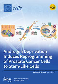
Cover Story (view full-size image): Due to their high cell plasticity, tumor cells may switch into a different histological subtype to escape from targeted therapy. In this study, we demonstrated that long-term androgen signal depletion of prostate LNCaP cells, either by incubation in charcoal-stripped medium or by 2-hidroxyflutamide treatment, induced a neuroendocrine phenotype followed by re-differentiation towards a “stem-like” state. The prostate stem-like cells had enhanced expression of the stem cell markers CD133, ALDH1A1, and ABCB1A and the pluripotent transcription factors Nanog and Oct4. Additionally, those cells were resistant to docetaxel and 2-hidroxyflutamide. Overexpression of AMPK in stem-like cells downregulated stem markers while restoring docetaxel sensitivity, providing a new regulatory mechanism of prostate cancer plasticity through AMPK. View this paper
  • Issues are regarded as officially published after their release is announced to the table of contents alert mailing list.
  • You may sign up for e-mail alerts to receive table of contents of newly released issues.
  • PDF is the official format for papers published in both, html and pdf forms. To view the papers in pdf format, click on the "PDF Full-text" link, and use the free Adobe Reader to open them.
Order results
Result details
Section
Select all
Export citation of selected articles as:
37 pages, 1278 KiB  
Review
Kynurenines in the Pathogenesis of Multiple Sclerosis: Therapeutic Perspectives
by Tamás Biernacki, Dániel Sandi, Krisztina Bencsik and László Vécsei
Cells 2020, 9(6), 1564; https://doi.org/10.3390/cells9061564 - 26 Jun 2020
Cited by 33 | Viewed by 4886
Abstract
Over the past years, an increasing amount of evidence has emerged in support of the kynurenine pathway’s (KP) pivotal role in the pathogenesis of several neurodegenerative, psychiatric, vascular and autoimmune diseases. Different neuroactive metabolites of the KP are known to exert opposite effects [...] Read more.
Over the past years, an increasing amount of evidence has emerged in support of the kynurenine pathway’s (KP) pivotal role in the pathogenesis of several neurodegenerative, psychiatric, vascular and autoimmune diseases. Different neuroactive metabolites of the KP are known to exert opposite effects on neurons, some being neuroprotective (e.g., picolinic acid, kynurenic acid, and the cofactor nicotinamide adenine dinucleotide), while others are toxic to neurons (e.g., 3-hydroxykynurenine, quinolinic acid). Not only the alterations in the levels of the metabolites but also disturbances in their ratio (quinolinic acid/kynurenic acid) have been reported in several diseases. In addition to the metabolites, the enzymes participating in the KP have been unearthed to be involved in modulation of the immune system, the energetic upkeep of neurons and have been shown to influence redox processes and inflammatory cascades, revealing a sophisticated, intertwined system. This review considers various methods through which enzymes and metabolites of the kynurenine pathway influence the immune system, the roles they play in the pathogenesis of neuroinflammatory diseases based on current evidence with a focus on their involvement in multiple sclerosis, as well as therapeutic approaches. Full article
Show Figures

Figure 1

11 pages, 2417 KiB  
Article
Polysaccharide Multilayer Films in Sensors for Detecting Prostate Tumor Cells Based on Hyaluronan-CD44 Interactions
by João Batista Maia Rocha Neto, Andrey Coatrini Soares, Rogério Aparecido Bataglioli, Olívia Carr, Carlos Alberto Rodrigues Costa, Osvaldo N. Oliveira, Jr., Marisa Masumi Beppu and Hernandes F. Carvalho
Cells 2020, 9(6), 1563; https://doi.org/10.3390/cells9061563 - 26 Jun 2020
Cited by 16 | Viewed by 3614
Abstract
The increasing need for point-of-care diagnosis has sparked the development of label-free sensing platforms, some of which are based on impedance measurements with biological cells. Here, interdigitated electrodes were functionalized with layer-by-layer (LbL) films of hyaluronan (HA) and chitosan (CHI) to detect prostatic [...] Read more.
The increasing need for point-of-care diagnosis has sparked the development of label-free sensing platforms, some of which are based on impedance measurements with biological cells. Here, interdigitated electrodes were functionalized with layer-by-layer (LbL) films of hyaluronan (HA) and chitosan (CHI) to detect prostatic tumor cells (PC3 line). The deposition of LbL films was confirmed with atomic force microscopy and polarization-modulated infrared reflection absorption spectroscopy (PM-IRRAS), which featured the vibrational modes of the HA top layer capable of interacting specifically with glycoprotein CD44 receptors overexpressed in tumor cells. Though the CHI/HA LbL films cannot be considered as a traditional biosensor due to their limited selectivity, it was possible to distinguish prostate tumor cells in the range from 50 to 600 cells/µL in in vitro experiments with impedance spectroscopy. This was achieved by treating the impedance data with information visualization methods, which confirmed the distinguishing ability of the films by observing the absence of false positives in a series of control experiments. The CD44–HA interactions may, therefore, be exploited in clinical analyses and point-of-care diagnostics for cancer, particularly if computational methods are used to process the data. Full article
Show Figures

Graphical abstract

19 pages, 2857 KiB  
Article
Characterization of Hspb8 in Zebrafish
by Magda Dubińska-Magiera, Joanna Niedbalska-Tarnowska, Marta Migocka-Patrzałek, Ewelina Posyniak and Małgorzata Daczewska
Cells 2020, 9(6), 1562; https://doi.org/10.3390/cells9061562 - 26 Jun 2020
Cited by 13 | Viewed by 3891
Abstract
Hspb8 is a member of the small heat shock protein (sHSP) family. Its expression is known to be upregulated under heat shock. This protein interacts with different partners and can, therefore, be involved in various processes relevant to tissue integrity and functioning. In [...] Read more.
Hspb8 is a member of the small heat shock protein (sHSP) family. Its expression is known to be upregulated under heat shock. This protein interacts with different partners and can, therefore, be involved in various processes relevant to tissue integrity and functioning. In humans, mutations in the gene encoding Hspb8 can lead to the development of various diseases such as myopathies and neuropathies. In our study, we aimed to perform an in-depth characterization of zebrafish Hspb8 during zebrafish development. We applied techniques such as RT-qPCR, Western blot, immunofluorescence, co-immunoprecipitation, LC-MS, and morpholino-mediated knockdown. We broadened the knowledge regarding zebrafish hspb8 expression during development under normal and heat shock conditions as well as its tissue- and subcellular-specific localization. A co-IP analysis allowed us to conclude that zebrafish Hspb8 can interact with proteins such as Bag3 and Hsc70, which are crucial for formation of an autophagy-inducing complex. We also demonstrated that hspb8 morpholino-mediated knockdown has an impact on zebrafish embryos’ morphology, muscle ultrastructure, and motility behavior. Our research provides a valuable resource for the potential use of the zebrafish as a model for studying pathological conditions associated with hspb8 disorders. Full article
Show Figures

Figure 1

13 pages, 1168 KiB  
Article
Cathelicidin-Related Antimicrobial Peptide Regulates CD73 Expression in Mouse Th17 Cells via p38
by Jeonghyun Lee, Kyong-Oh Shin, Yesol Kim, Jaewon Cho, Hyung W. Lim, Sung-Il Yoon, Geun-Shik Lee, Hyun-Jeong Ko, Pyeung-Hyeun Kim, Yoshikazu Uchida, Kyungho Park and Seung Goo Kang
Cells 2020, 9(6), 1561; https://doi.org/10.3390/cells9061561 - 26 Jun 2020
Cited by 4 | Viewed by 3632
Abstract
The effector function of tumor-infiltrated CD4+ T cells is readily suppressed by many types of immune regulators in the tumor microenvironment, which is one of the major mechanisms of immune tolerance against cancer. Cathelicidin-related antimicrobial peptide (CRAMP), the mouse analog of LL-37 [...] Read more.
The effector function of tumor-infiltrated CD4+ T cells is readily suppressed by many types of immune regulators in the tumor microenvironment, which is one of the major mechanisms of immune tolerance against cancer. Cathelicidin-related antimicrobial peptide (CRAMP), the mouse analog of LL-37 peptide in humans, is a cationic antimicrobial peptide belonging to the cathelicidin family; however, its secretion by cancer cells and role in the tumor microenvironment (TME) remain unclear. In this study, we explored the possibility of an interaction between effector CD4+ T cells and CRAMP using in vitro-generated mouse Th17 cells. We found that CRAMP stimulates Th17 cells to express the ectonucleotidase CD73, while simultaneously inducing cell death. This finding suggested that CD73-expressing Th17 cells may function as immune suppressor cells instead of effector cells. In addition, treatment of pharmacological inhibitors of the transforming growth factor-beta (TGF-β) signaling pathway showed that induction of CD73 expression is mediated by the p38 signaling pathway. Overall, our findings suggest that tumor-derived LL-37 likely functions as an immune suppressor that induces immune tolerance against tumors through shaping effector Th17 cells into suppressor Th17 cells, suggesting a new intervention target to improve cancer immunotherapy. Full article
(This article belongs to the Special Issue Immunomodulatory Factors in Host Defense)
Show Figures

Graphical abstract

14 pages, 2438 KiB  
Article
Mistletoe-Extract Drugs Stimulate Anti-Cancer Vγ9Vδ2 T Cells
by Ling Ma, Swati Phalke, Caroline Stévigny, Florence Souard and David Vermijlen
Cells 2020, 9(6), 1560; https://doi.org/10.3390/cells9061560 - 26 Jun 2020
Cited by 9 | Viewed by 3779
Abstract
Human phosphoantigen-reactive Vγ9Vδ2 T cells possess several characteristics, including MHC-independent recognition of tumor cells and potent killing potential, that make them attractive candidates for cancer immunotherapeutic approaches. Injectable preparations from the hemi-parasite plant Viscum album L. (European mistletoe) are commonly prescribed as complementary [...] Read more.
Human phosphoantigen-reactive Vγ9Vδ2 T cells possess several characteristics, including MHC-independent recognition of tumor cells and potent killing potential, that make them attractive candidates for cancer immunotherapeutic approaches. Injectable preparations from the hemi-parasite plant Viscum album L. (European mistletoe) are commonly prescribed as complementary cancer therapy in European countries such as Germany, but their mechanism of action remains poorly understood. Here, we investigated in-depth the in vitro response of human T cells towards mistletoe-extract drugs by analyzing their functional and T-cell-receptor (TCR) response using flow cytometry and high-throughput sequencing respectively. Non-fermented mistletoe-extract drugs (AbnobaViscum), but not their fermented counterparts (Iscador), induced specific expansion of Vγ9Vδ2 T cells among T cells. Furthermore, AbnobaViscum rapidly induced the release of cytotoxic granules and the production of the cytokines IFNγ and TNFα in Vγ9Vδ2 T cells. This stimulation of anti-cancer Vγ9Vδ2 T cells was mediated by the butyrophilin BTN3A, did not depend on the accumulation of endogenous phosphoantigens and involved the same Vγ9Vδ2 TCR repertoire as those of phosphoantigen-reactive Vγ9Vδ2 T cells. These insights highlight Vγ9Vδ2 T cells as a potential target for mistletoe-extract drugs and their role in cancer patients receiving these herbal drugs needs to be investigated. Full article
Show Figures

Figure 1

33 pages, 1119 KiB  
Review
Immunotherapy in Myeloproliferative Diseases
by Lukas M. Braun and Robert Zeiser
Cells 2020, 9(6), 1559; https://doi.org/10.3390/cells9061559 - 26 Jun 2020
Cited by 16 | Viewed by 4538
Abstract
Myeloproliferative diseases, including myeloproliferative neoplasms (MPN) and myelodysplastic syndromes (MDS), are driven by genetic abnormalities and increased inflammatory signaling and are at high risk to transform into acute myeloid leukemia (AML). Myeloid-derived suppressor cells were reported to enhance leukemia immune escape by suppressing [...] Read more.
Myeloproliferative diseases, including myeloproliferative neoplasms (MPN) and myelodysplastic syndromes (MDS), are driven by genetic abnormalities and increased inflammatory signaling and are at high risk to transform into acute myeloid leukemia (AML). Myeloid-derived suppressor cells were reported to enhance leukemia immune escape by suppressing an effective anti-tumor immune response. MPNs are a potentially immunogenic disease as shown by their response to interferon-α treatment and allogeneic hematopoietic stem-cell transplantation (allo-HSCT). Novel immunotherapeutic approaches such as immune checkpoint inhibition, tumor vaccination, or cellular therapies using target-specific lymphocytes have so far not shown strong therapeutic efficacy. Potential reasons could be the pro-inflammatory and immunosuppressive microenvironment in the bone marrow of patients with MPN, driving tumor immune escape. In this review, we discuss the biology of MPNs with respect to the pro-inflammatory milieu in the bone marrow (BM) and potential immunotherapeutic approaches. Full article
(This article belongs to the Special Issue Pathophysiology and Molecular Targets in Myeloid Neoplasia)
Show Figures

Figure 1

31 pages, 1971 KiB  
Review
Targeting Cardiac Stem Cell Senescence to Treat Cardiac Aging and Disease
by Eleonora Cianflone, Michele Torella, Flavia Biamonte, Antonella De Angelis, Konrad Urbanek, Francesco S. Costanzo, Marcello Rota, Georgina M. Ellison-Hughes and Daniele Torella
Cells 2020, 9(6), 1558; https://doi.org/10.3390/cells9061558 - 26 Jun 2020
Cited by 75 | Viewed by 7813
Abstract
Adult stem/progenitor are a small population of cells that reside in tissue-specific niches and possess the potential to differentiate in all cell types of the organ in which they operate. Adult stem cells are implicated with the homeostasis, regeneration, and aging of all [...] Read more.
Adult stem/progenitor are a small population of cells that reside in tissue-specific niches and possess the potential to differentiate in all cell types of the organ in which they operate. Adult stem cells are implicated with the homeostasis, regeneration, and aging of all tissues. Tissue-specific adult stem cell senescence has emerged as an attractive theory for the decline in mammalian tissue and organ function during aging. Cardiac aging, in particular, manifests as functional tissue degeneration that leads to heart failure. Adult cardiac stem/progenitor cell (CSC) senescence has been accordingly associated with physiological and pathological processes encompassing both non-age and age-related decline in cardiac tissue repair and organ dysfunction and disease. Senescence is a highly active and dynamic cell process with a first classical hallmark represented by its replicative limit, which is the establishment of a stable growth arrest over time that is mainly secondary to DNA damage and reactive oxygen species (ROS) accumulation elicited by different intrinsic stimuli (like metabolism), as well as external stimuli and age. Replicative senescence is mainly executed by telomere shortening, the activation of the p53/p16INK4/Rb molecular pathways, and chromatin remodeling. In addition, senescent cells produce and secrete a complex mixture of molecules, commonly known as the senescence-associated secretory phenotype (SASP), that regulate most of their non-cell-autonomous effects. In this review, we discuss the molecular and cellular mechanisms regulating different characteristics of the senescence phenotype and their consequences for adult CSCs in particular. Because senescent cells contribute to the outcome of a variety of cardiac diseases, including age-related and unrelated cardiac diseases like diabetic cardiomyopathy and anthracycline cardiotoxicity, therapies that target senescent cell clearance are actively being explored. Moreover, the further understanding of the reversibility of the senescence phenotype will help to develop novel rational therapeutic strategies. Full article
Show Figures

Figure 1

36 pages, 2770 KiB  
Article
Substantially Altered Expression Profile of Diabetes/Cardiovascular/Cerebrovascular Disease Associated microRNAs in Children Descending from Pregnancy Complicated by Gestational Diabetes Mellitus—One of Several Possible Reasons for an Increased Cardiovascular Risk
by Ilona Hromadnikova, Katerina Kotlabova, Lenka Dvorakova, Ladislav Krofta and Jan Sirc
Cells 2020, 9(6), 1557; https://doi.org/10.3390/cells9061557 - 26 Jun 2020
Cited by 21 | Viewed by 3387
Abstract
Gestational diabetes mellitus (GDM), one of the major pregnancy-related complications, characterized as a transitory form of diabetes induced by insulin resistance accompanied by a low/absent pancreatic beta-cell compensatory adaptation to the increased insulin demand, causes the acute, long-term, and transgenerational health complications. The [...] Read more.
Gestational diabetes mellitus (GDM), one of the major pregnancy-related complications, characterized as a transitory form of diabetes induced by insulin resistance accompanied by a low/absent pancreatic beta-cell compensatory adaptation to the increased insulin demand, causes the acute, long-term, and transgenerational health complications. The aim of the study was to assess if alterations in gene expression of microRNAs associated with diabetes/cardiovascular/cerebrovascular diseases are present in whole peripheral blood of children aged 3–11 years descending from GDM complicated pregnancies. A substantially altered microRNA expression profile was found in children descending from GDM complicated pregnancies. Almost all microRNAs with the exception of miR-92a-3p, miR-155-5p, and miR-210-3p were upregulated. The microRNA expression profile also differed between children after normal and GDM complicated pregnancies in relation to the presence of overweight/obesity, prehypertension/hypertension, and/or valve problems and heart defects. Always, screening based on the combination of microRNAs was superior over using individual microRNAs, since at 10.0% false positive rate it was able to identify a large proportion of children with an aberrant microRNA expression profile (88.14% regardless of clinical findings, 75.41% with normal clinical findings, and 96.49% with abnormal clinical findings). In addition, the higher incidence of valve problems and heart defects was found in children with a prior exposure to GDM. The extensive file of predicted targets of all microRNAs aberrantly expressed in children descending from GDM complicated pregnancies indicates that a large group of these genes is involved in ontologies of diabetes/cardiovascular/cerebrovascular diseases. In general, children with a prior exposure to GDM are at higher risk of later development of diabetes mellitus and cardiovascular/cerebrovascular diseases, and would benefit from dispensarisation as well as implementation of primary prevention strategies. Full article
(This article belongs to the Special Issue microRNA Bioinformatics)
Show Figures

Figure 1

14 pages, 1544 KiB  
Article
Circulating microRNA Signature Associated to Interstitial Lung Abnormalities in Respiratory Asymptomatic Subjects
by Blanca Ortiz-Quintero, Ivette Buendía-Roldán, Eric Gustavo Ramírez-Salazar, Yalbi I Balderas-Martínez, Sandra Lizbeth Ramírez-Rodríguez, Karen Martínez-Espinosa and Moisés Selman
Cells 2020, 9(6), 1556; https://doi.org/10.3390/cells9061556 - 26 Jun 2020
Cited by 13 | Viewed by 2714
Abstract
Interstitial lung abnormalities (ILA) are observed in around 9% of older respiratory asymptomatic subjects, mainly smokers. Evidence suggests that ILA may precede the development of interstitial lung diseases and may evolve to progressive fibrosis. Identifying biomarkers of this subclinical status is relevant for [...] Read more.
Interstitial lung abnormalities (ILA) are observed in around 9% of older respiratory asymptomatic subjects, mainly smokers. Evidence suggests that ILA may precede the development of interstitial lung diseases and may evolve to progressive fibrosis. Identifying biomarkers of this subclinical status is relevant for early diagnosis and to predict outcome. We aimed to identify circulating microRNAs (miRNAs) associated to ILA in a cohort of respiratory asymptomatic subjects older than 60 years. We identified 81 subjects with ILA from our Lung-Aging Program in Mexico City (n = 826). We randomly selected 112 subjects without ILA (Ctrl) from the same cohort. Using polymerase chain reaction PCR-Array technology (24 ILA and 24 Ctrl, screening cohort) and reverse-transcriptase quantitative polymerase chain reaction (RT-qPCR) (57 ILA and 88 Ctr, independent validation cohort) we identified seven up-regulated miRNAs in serum of ILA compared to Ctrl (miR-193a-5p, p < 0.0001; miR-502-3p, p < 0.0001; miR-200c-3p, p = 0.003; miR-16-5p, p = 0.003; miR-21-5p, p = 0.002; miR-126-3p, p = 0.004 and miR-34a-5p, p < 0.005). Pathways regulated by these miRNAs include transforming growth factor beta (TGF-β), Wnt, mammalian target of rapamycin (mTOR), Insulin, mitogen-activated protein kinase (MAPK) signaling, and senescence. Receiver operator characteristic (ROC) curve analysis indicated that miR-193a-5p (area under the curve AUC: 0.75) and miR-502-3p (AUC 0.71) have acceptable diagnostic value. This is the first identification of circulating miRNAs associated to ILA in respiratory asymptomatic subjects, providing potential non-invasive biomarkers and molecular targets to better understand the pathogenic mechanisms associated to ILA. Full article
(This article belongs to the Special Issue microRNA as Biomarker)
Show Figures

Figure 1

20 pages, 5288 KiB  
Article
Redox Regulation of NOX Isoforms on FAK(Y397)/SRC(Y416) Phosphorylation Driven Epithelial-to-Mesenchymal Transition in Malignant Cervical Epithelial Cells
by Young Mee Kim, Karthika Muthuramalingam and Moonjae Cho
Cells 2020, 9(6), 1555; https://doi.org/10.3390/cells9061555 - 26 Jun 2020
Cited by 8 | Viewed by 2840
Abstract
Epithelial-to-mesenchymal transition (EMT) promulgates epithelial cell associated disease-defining characteristics in tumorigenesis and organ fibrosis. Growth factors such as epidermal growth factor and fibroblast growth factor in addition to cytokines such as transforming growth factor-β1 (TGF-β1) is said to play a prominent role in [...] Read more.
Epithelial-to-mesenchymal transition (EMT) promulgates epithelial cell associated disease-defining characteristics in tumorigenesis and organ fibrosis. Growth factors such as epidermal growth factor and fibroblast growth factor in addition to cytokines such as transforming growth factor-β1 (TGF-β1) is said to play a prominent role in remodeling related pathological events of cancer progression such as invasion, metastasis, apoptosis, EMT, etc. through redox related cellular secondary messengers, in particular the reactive oxygen species (ROS). However, the signaling cascade underlying the redox mechanism and thereby the progression of EMT remains largely unknown. In this study, upon TGF-β1 treatment, we observed an induction in NOX isoforms—NOX2 and NOX4—that have time (early and late) and cellular localization (nucleus and autophagosome co-localized) dependent effects in mediating EMT associated cell proliferation and migration through activation of the focal adhesion kinase (FAK)/SRC pathway in HeLa, human cervical cancer cells. Upon silencing NOX2/4 gene expression and using the SRC inhibitor (AZD0530), progression of TGF-β1 induced EMT related cellular remodeling, extra cellular matrix (ECM) production, cell migration and invasion, got significantly reverted. Together, these results indicate that NOX2 and NOX4 play important, albeit distinct, roles in the activation of cytokine mediated EMT and its associated processes via tyrosine phosphorylation of the FAK/SRC pathway. Full article
Show Figures

Figure 1

27 pages, 1162 KiB  
Review
Moonlighting in Mitosis: Analysis of the Mitotic Functions of Transcription and Splicing Factors
by Maria Patrizia Somma, Evgeniya N. Andreyeva, Gera A. Pavlova, Claudia Pellacani, Elisabetta Bucciarelli, Julia V. Popova, Silvia Bonaccorsi, Alexey V. Pindyurin and Maurizio Gatti
Cells 2020, 9(6), 1554; https://doi.org/10.3390/cells9061554 - 26 Jun 2020
Cited by 16 | Viewed by 6293
Abstract
Moonlighting proteins can perform one or more additional functions besides their primary role. It has been posited that a protein can acquire a moonlighting function through a gradual evolutionary process, which is favored when the primary and secondary functions are exerted in different [...] Read more.
Moonlighting proteins can perform one or more additional functions besides their primary role. It has been posited that a protein can acquire a moonlighting function through a gradual evolutionary process, which is favored when the primary and secondary functions are exerted in different cellular compartments. Transcription factors (TFs) and splicing factors (SFs) control processes that occur in interphase nuclei and are strongly reduced during cell division, and are therefore in a favorable situation to evolve moonlighting mitotic functions. However, recently published moonlighting protein databases, which comprise almost 400 proteins, do not include TFs and SFs with secondary mitotic functions. We searched the literature and found several TFs and SFs with bona fide moonlighting mitotic functions, namely they localize to specific mitotic structure(s), interact with proteins enriched in the same structure(s), and are required for proper morphology and functioning of the structure(s). In addition, we describe TFs and SFs that localize to mitotic structures but cannot be classified as moonlighting proteins due to insufficient data on their biochemical interactions and mitotic roles. Nevertheless, we hypothesize that most TFs and SFs with specific mitotic localizations have either minor or redundant moonlighting functions, or are evolving towards the acquisition of these functions. Full article
(This article belongs to the Special Issue Frontiers in Cytoskeleton Research—From Development to Disease)
Show Figures

Figure 1

33 pages, 2197 KiB  
Review
Amyloid Proteins and Peripheral Neuropathy
by Mohammed M. H. Albariqi, Sjoukje Engelsman, Niels Eijkelkamp and Jo W. M. Höppener
Cells 2020, 9(6), 1553; https://doi.org/10.3390/cells9061553 - 26 Jun 2020
Cited by 12 | Viewed by 5141
Abstract
Painful peripheral neuropathy affects millions of people worldwide. Peripheral neuropathy develops in patients with various diseases, including rare familial or acquired amyloid polyneuropathies, as well as some common diseases, including type 2 diabetes mellitus and several chronic inflammatory diseases. Intriguingly, these diseases share [...] Read more.
Painful peripheral neuropathy affects millions of people worldwide. Peripheral neuropathy develops in patients with various diseases, including rare familial or acquired amyloid polyneuropathies, as well as some common diseases, including type 2 diabetes mellitus and several chronic inflammatory diseases. Intriguingly, these diseases share a histopathological feature—deposits of amyloid-forming proteins in tissues. Amyloid-forming proteins may cause tissue dysregulation and damage, including damage to nerves, and may be a common cause of neuropathy in these, and potentially other, diseases. Here, we will discuss how amyloid proteins contribute to peripheral neuropathy by reviewing the current understanding of pathogenic mechanisms in known inherited and acquired (usually rare) amyloid neuropathies. In addition, we will discuss the potential role of amyloid proteins in peripheral neuropathy in some common diseases, which are not (yet) considered as amyloid neuropathies. We conclude that there are many similarities in the molecular and cell biological defects caused by aggregation of the various amyloid proteins in these different diseases and propose a common pathogenic pathway for “peripheral amyloid neuropathies”. Full article
(This article belongs to the Special Issue Novel Insights into Molecular Mechanisms of Chronic Pain)
Show Figures

Figure 1

28 pages, 2264 KiB  
Review
The NLRP3 Inflammasome as a Critical Actor in the Inflammaging Process
by Maria Sebastian-Valverde and Giulio M. Pasinetti
Cells 2020, 9(6), 1552; https://doi.org/10.3390/cells9061552 - 26 Jun 2020
Cited by 37 | Viewed by 6228
Abstract
As a consequence of the considerable increase in the human lifespan over the last century, we are experiencing the appearance and impact of new age-related diseases. The causal relationships between aging and an enhanced susceptibility of suffering from a broad spectrum of diseases [...] Read more.
As a consequence of the considerable increase in the human lifespan over the last century, we are experiencing the appearance and impact of new age-related diseases. The causal relationships between aging and an enhanced susceptibility of suffering from a broad spectrum of diseases need to be better understood. However, one specific shared feature seems to be of capital relevance for most of these conditions: the low-grade chronic inflammatory state inherently associated with aging, i.e., inflammaging. Here, we review the molecular and cellular mechanisms that link aging and inflammaging, focusing on the role of the innate immunity and more concretely on the nucleotide-binding oligomerization domain (NOD)-like receptor family pyrin domain containing 3 (NLRP3) inflammasome, as well as how the chronic activation of this inflammasome has a detrimental effect on different age-related disorders. Full article
(This article belongs to the Special Issue Roles of Inflammasomes in Aging and Age-Related Diseases)
Show Figures

Figure 1

13 pages, 1678 KiB  
Article
Synthesis and Evaluation of Dodecaboranethiol Containing Kojic Acid (KA-BSH) as a Novel Agent for Boron Neutron Capture Therapy
by Koji Takeuchi, Yoshihide Hattori, Shinji Kawabata, Gen Futamura, Ryo Hiramatsu, Masahiko Wanibuchi, Hiroki Tanaka, Shin-ichiro Masunaga, Koji Ono, Shin-Ichi Miyatake and Mitsunori Kirihata
Cells 2020, 9(6), 1551; https://doi.org/10.3390/cells9061551 - 25 Jun 2020
Cited by 18 | Viewed by 3153
Abstract
Boron neutron capture therapy (BNCT) is a form of tumor-cell selective particle irradiation using low-energy neutron irradiation of boron-10 (10B) to produce high-linear energy transfer (LET) alpha particles and recoiling 7Li nuclei (10B [n, alpha] 7Li) in [...] Read more.
Boron neutron capture therapy (BNCT) is a form of tumor-cell selective particle irradiation using low-energy neutron irradiation of boron-10 (10B) to produce high-linear energy transfer (LET) alpha particles and recoiling 7Li nuclei (10B [n, alpha] 7Li) in tumor cells. Therefore, it is important to achieve the selective delivery of large amounts of 10B to tumor cells, with only small amounts of 10B to normal tissues. To develop practical materials utilizing 10B carriers, we designed and synthesized novel dodecaboranethiol (BSH)-containing kojic acid (KA-BSH). In the present study, we evaluated the effects of this novel 10B carrier on cytotoxicity, 10B concentrations in F98 rat glioma cells, and micro-distribution of KA-BSH in vitro. Furthermore, biodistribution studies were performed in a rat brain tumor model. The tumor boron concentrations showed the highest concentrations at 1 h after the termination of administration. Based on these results, neutron irradiation was evaluated at the Kyoto University Research Reactor Institute (KURRI) with KA-BSH. Median survival times (MSTs) of untreated and irradiated control rats were 29.5 and 30.5 days, respectively, while animals that received KA-BSH, followed by neutron irradiation, had an MST of 36.0 days (p = 0.0027, 0.0053). Based on these findings, further studies are warranted in using KA-BSH as a new B compound for malignant glioma. Full article
(This article belongs to the Special Issue Biology of Boron Neutron Capture Therapy (BNCT))
Show Figures

Figure 1

19 pages, 5444 KiB  
Article
Nicotinamide Supplementation during the In Vitro Maturation of Oocytes Improves the Developmental Competence of Preimplantation Embryos: Potential Link to SIRT1/AKT Signaling
by Marwa El Sheikh, Ahmed Atef Mesalam, Muhammad Idrees, Tabinda Sidrat, Ayman Mesalam, Kyeong-Lim Lee and Il-Keun Kong
Cells 2020, 9(6), 1550; https://doi.org/10.3390/cells9061550 - 25 Jun 2020
Cited by 15 | Viewed by 3211
Abstract
Nicotinamide (NAM), the amide form of vitamin B3, plays pivotal roles in regulating various cellular processes including energy production and maintenance of genomic stability. The current study aimed at deciphering the effect of NAM, when administered during in vitro maturation (IVM), on the [...] Read more.
Nicotinamide (NAM), the amide form of vitamin B3, plays pivotal roles in regulating various cellular processes including energy production and maintenance of genomic stability. The current study aimed at deciphering the effect of NAM, when administered during in vitro maturation (IVM), on the developmental competence of bovine preimplantation embryos. Our results showed that low NAM concentrations reduced the oxidative stress and improved mitochondrial profile, total cleavage and 8–16 cell stage embryo development whereas the opposite profile was observed upon exposure to high NAM concentrations (10 mM onward). Remarkably, the hatching rates of day-7 and day-8 blastocysts were significantly improved under 0.1 mM NAM treatment. Using RT-qPCR and immunofluorescence, the autophagy-related (Beclin-1 (BECN1), LC3B, and ATG5) and the apoptotic (Caspases; CASP3 and 9) markers were upregulated in oocytes exposed to high NAM concentration (40 mM), whereas only CASP3 was affected, downregulated, following 0.1 mM treatment. Additionally, the number of cells per blastocyst and the levels of SIRT1, PI3K, AKT, and mTOR were higher, while the inner cell mass-specific transcription factors GATA6, SOX2, and OCT4 were more abundant, in day-8 embryos of NAM-treated group. Taken together, to our knowledge, this is the first study reporting that administration of low NAM concentrations during IVM can ameliorate the developmental competence of embryos through the potential regulation of oxidative stress, apoptosis, and SIRT1/AKT signaling. Full article
(This article belongs to the Section Cell Signaling)
Show Figures

Figure 1

15 pages, 816 KiB  
Review
A Review on Notch Signaling and Colorectal Cancer
by Ashish Tyagi, Arun K. Sharma and Chendil Damodaran
Cells 2020, 9(6), 1549; https://doi.org/10.3390/cells9061549 - 25 Jun 2020
Cited by 86 | Viewed by 7071
Abstract
Colorectal cancer (CRC) has one of the highest mortality rates despite the advancement of treatment options. Aggressive CRC remains difficult to treat owing to the activation of oncogenic signaling pathways such as the Notch signaling pathway. The role of Notch receptors varies according [...] Read more.
Colorectal cancer (CRC) has one of the highest mortality rates despite the advancement of treatment options. Aggressive CRC remains difficult to treat owing to the activation of oncogenic signaling pathways such as the Notch signaling pathway. The role of Notch receptors varies according to the difference in their structures; in particular, aberrant activation of Notch1 has been attributed to the severity of CRC. Notch1 activation in CRC is inhibited by small molecule inhibitors that target γ-secretase, an enzyme responsible for the third and last cleavage step of Notch receptors. γ-Secretase also produces the intracellular domain that finally carries out cellular functions by activating downstream effectors. However, most inhibitors block γ-secretase non-selectively and cause severe toxicity. Plant-source-derived small molecules, monoclonal antibodies, biological molecules (such as SiRNAs), and compounds targeting the Notch1 receptor itself or the downstream molecules such as HES1 are some of the options that are in advanced stages of clinical trials. The Negative Regulatory Region (NRR), which plays a central role in the transduction of Notch1 signaling in the event of ligand-dependent and ligand-independent Notch1 processing is also being targeted specifically by monoclonal antibodies (mAbs) to prevent aberrant Notch1 activation. In this review, we discuss the role of Notch1 in CRC, particularly its metastatic phenotype, and how mutations in Notch1, specifically in its NRR region, contribute to the aberrant activation of Notch1 signaling, which, in turn, contributes to CRC pathogenesis. We also discuss prevailing and emerging therapies that target the Notch1 receptor and the NRR region, and we highlight the potential of these therapies in abrogating Notch signaling and, thus, CRC development and progression. Full article
(This article belongs to the Special Issue The Roles of Notch Signaling in Cancers)
Show Figures

Figure 1

17 pages, 2509 KiB  
Article
Molecular Basis for Ser/Thr Specificity in PKA Signaling
by Matthias J. Knape, Maximilian Wallbott, Nicole C. G. Burghardt, Daniela Bertinetti, Jan Hornung, Sven H. Schmidt, Robin Lorenz and Friedrich W. Herberg
Cells 2020, 9(6), 1548; https://doi.org/10.3390/cells9061548 - 25 Jun 2020
Cited by 4 | Viewed by 3499
Abstract
cAMP-dependent protein kinase (PKA) is the major receptor of the second messenger cAMP and a prototype for Ser/Thr-specific protein kinases. Although PKA strongly prefers serine over threonine substrates, little is known about the molecular basis of this substrate specificity. We employ classical enzyme [...] Read more.
cAMP-dependent protein kinase (PKA) is the major receptor of the second messenger cAMP and a prototype for Ser/Thr-specific protein kinases. Although PKA strongly prefers serine over threonine substrates, little is known about the molecular basis of this substrate specificity. We employ classical enzyme kinetics and a surface plasmon resonance (SPR)-based method to analyze each step of the kinase reaction. In the absence of divalent metal ions and nucleotides, PKA binds serine (PKS) and threonine (PKT) substrates, derived from the heat-stable protein kinase inhibitor (PKI), with similar affinities. However, in the presence of metal ions and adenine nucleotides, the Michaelis complex for PKT is unstable. PKA phosphorylates PKT with a higher turnover due to a faster dissociation of the product complex. Thus, threonine substrates are not necessarily poor substrates of PKA. Mutation of the DFG+1 phenylalanine to β-branched amino acids increases the catalytic efficiency of PKA for a threonine peptide substrate up to 200-fold. The PKA Cα mutant F187V forms a stable Michaelis complex with PKT and shows no preference for serine versus threonine substrates. Disease-associated mutations of the DFG+1 position in other protein kinases underline the importance of substrate specificity for keeping signaling pathways segregated and precisely regulated. Full article
(This article belongs to the Special Issue New Advances in Cyclic AMP Signalling)
Show Figures

Figure 1

23 pages, 2575 KiB  
Review
Insight into Salivary Gland Aquaporins
by Claudia D’Agostino, Osama A. Elkashty, Clara Chivasso, Jason Perret, Simon D. Tran and Christine Delporte
Cells 2020, 9(6), 1547; https://doi.org/10.3390/cells9061547 - 25 Jun 2020
Cited by 37 | Viewed by 8241
Abstract
The main role of salivary glands (SG) is the production and secretion of saliva, in which aquaporins (AQPs) play a key role by ensuring water flow. The AQPs are transmembrane channel proteins permeable to water to allow water transport across cell membranes according [...] Read more.
The main role of salivary glands (SG) is the production and secretion of saliva, in which aquaporins (AQPs) play a key role by ensuring water flow. The AQPs are transmembrane channel proteins permeable to water to allow water transport across cell membranes according to osmotic gradient. This review gives an insight into SG AQPs. Indeed, it gives a summary of the expression and localization of AQPs in adult human, rat and mouse SG, as well as of their physiological role in SG function. Furthermore, the review provides a comprehensive view of the involvement of AQPs in pathological conditions affecting SG, including Sjögren’s syndrome, diabetes, agedness, head and neck cancer radiotherapy and SG cancer. These conditions are characterized by salivary hypofunction resulting in xerostomia. A specific focus is given on current and future therapeutic strategies aiming at AQPs to treat xerostomia. A deeper understanding of the AQPs involvement in molecular mechanisms of saliva secretion and diseases offered new avenues for therapeutic approaches, including drugs, gene therapy and tissue engineering. As such, AQP5 represents a potential therapeutic target in different strategies for the treatment of xerostomia. Full article
(This article belongs to the Special Issue Advances in Aquaporins)
Show Figures

Graphical abstract

19 pages, 3004 KiB  
Article
Protein Kinase CK2 Regulates Nerve/Glial Antigen (NG)2-Mediated Angiogenic Activity of Human Pericytes
by Beate M. Schmitt, Anne S. Boewe, Vivien Becker, Lisa Nalbach, Yuan Gu, Claudia Götz, Michael D. Menger, Matthias W. Laschke and Emmanuel Ampofo
Cells 2020, 9(6), 1546; https://doi.org/10.3390/cells9061546 - 25 Jun 2020
Cited by 9 | Viewed by 2852
Abstract
Protein kinase CK2 is a crucial regulator of endothelial cell proliferation, migration and sprouting during angiogenesis. However, it is still unknown whether this kinase additionally affects the angiogenic activity of other vessel-associated cells. In this study, we investigated the effect of CK2 inhibition [...] Read more.
Protein kinase CK2 is a crucial regulator of endothelial cell proliferation, migration and sprouting during angiogenesis. However, it is still unknown whether this kinase additionally affects the angiogenic activity of other vessel-associated cells. In this study, we investigated the effect of CK2 inhibition on primary human pericytes. We found that CK2 inhibition reduces the expression of nerve/glial antigen (NG)2, a crucial factor which is involved in angiogenic processes. Reporter gene assays revealed a 114 bp transcriptional active region of the human NG2 promoter, whose activity was decreased after CK2 inhibition. Functional analyses demonstrated that the pharmacological inhibition of CK2 by CX-4945 suppresses pericyte proliferation, migration, spheroid sprouting and the stabilization of endothelial tubes. Moreover, aortic rings of NG2−/− mice showed a significantly reduced vascular sprouting when compared to rings of NG2+/+ mice, indicating that NG2 is an important regulator of the angiogenic activity of pericytes. In vivo, implanted Matrigel plugs containing CX-4945-treated pericytes exhibited a lower microvessel density when compared to controls. These findings demonstrate that CK2 regulates the angiogenic activity of pericytes through NG2 gene expression. Hence, the inhibition of CK2 represents a promising anti-angiogenic strategy, because it does not only target endothelial cells, but also vessel-associated pericytes. Full article
(This article belongs to the Special Issue Vascular Signalling)
Show Figures

Figure 1

15 pages, 1701 KiB  
Review
A Systematic Review of WNT Signaling in Endothelial Cell Oligodendrocyte Interactions: Potential Relevance to Cerebral Small Vessel Disease
by Narek Manukjan, Zubair Ahmed, Daniel Fulton, W. Matthijs Blankesteijn and Sébastien Foulquier
Cells 2020, 9(6), 1545; https://doi.org/10.3390/cells9061545 - 25 Jun 2020
Cited by 18 | Viewed by 4329
Abstract
Key pathological features of cerebral small vessel disease (cSVD) include impairment of the blood brain barrier (BBB) and the progression of white matter lesions (WMLs) amongst other structural lesions, leading to the clinical manifestations of cSVD. The function of endothelial cells (ECs) is [...] Read more.
Key pathological features of cerebral small vessel disease (cSVD) include impairment of the blood brain barrier (BBB) and the progression of white matter lesions (WMLs) amongst other structural lesions, leading to the clinical manifestations of cSVD. The function of endothelial cells (ECs) is of major importance to maintain a proper BBB. ECs interact with several cell types to provide structural and functional support to the brain. Oligodendrocytes (OLs) myelinate axons in the central nervous system and are crucial in sustaining the integrity of white matter. The interplay between ECs and OLs and their precursor cells (OPCs) has received limited attention yet seems of relevance for the study of BBB dysfunction and white matter injury in cSVD. Emerging evidence shows a crosstalk between ECs and OPCs/OLs, mediated by signaling through the Wingless and Int-1 (WNT)/β-catenin pathway. As the latter is involved in EC function (e.g., angiogenesis) and oligodendrogenesis, we reviewed the role of WNT/β-catenin signaling for both cell types and performed a systematic search to identify studies describing a WNT-mediated interplay between ECs and OPCs/OLs. Dysregulation of this interaction may limit remyelination of WMLs and render the BBB leaky, thereby initiating a vicious neuroinflammatory cycle. A better understanding of the role of this signaling pathway in EC–OL crosstalk is essential in understanding cSVD development. Full article
(This article belongs to the Special Issue The Molecular and Cellular Basis of Cardiovascular Disease)
Show Figures

Figure 1

20 pages, 1805 KiB  
Review
Circular RNAs in Sepsis: Biogenesis, Function, and Clinical Significance
by Jesús Beltrán-García, Rebeca Osca-Verdegal, Elena Nacher-Sendra, Federico V. Pallardó and José Luis García-Giménez
Cells 2020, 9(6), 1544; https://doi.org/10.3390/cells9061544 - 25 Jun 2020
Cited by 46 | Viewed by 4283
Abstract
Sepsis is a life-threatening condition that occurs when the body responds to an infection that damages it is own tissues. The major problem in sepsis is rapid, vital status deterioration in patients, which can progress to septic shock with multiple organ failure if [...] Read more.
Sepsis is a life-threatening condition that occurs when the body responds to an infection that damages it is own tissues. The major problem in sepsis is rapid, vital status deterioration in patients, which can progress to septic shock with multiple organ failure if not properly treated. As there are no specific treatments, early diagnosis is mandatory to reduce high mortality. Despite more than 170 different biomarkers being postulated, early sepsis diagnosis and prognosis remain a challenge for clinicians. Recent findings propose that circular RNAs (circRNAs) may play a prominent role in regulating the patients’ immune system against different pathogens, including bacteria and viruses. Mounting evidence also suggests that the misregulation of circRNAs is an early event in a wide range of diseases, including sepsis. Despite circRNA levels being altered in sepsis, the specific mechanisms controlling the dysregulation of these noncoding RNAs are not completely elucidated, although many factors are known to affect circRNA biogenesis. Therefore, there is a need to explore the molecular pathways that lead to this disorder. This review describes the role of this new class of regulatory RNAs in sepsis and the feasibility of using circRNAs as diagnostic biomarkers for sepsis, opening up new avenues for circRNA-based medicine. Full article
(This article belongs to the Special Issue Circular RNAs: Non-canonical Observations on Non-canonical RNAs)
Show Figures

Figure 1

36 pages, 4196 KiB  
Review
Genetic Control of Muscle Diversification and Homeostasis: Insights from Drosophila
by Preethi Poovathumkadavil and Krzysztof Jagla
Cells 2020, 9(6), 1543; https://doi.org/10.3390/cells9061543 - 25 Jun 2020
Cited by 12 | Viewed by 6194
Abstract
In the fruit fly, Drosophila melanogaster, the larval somatic muscles or the adult thoracic flight and leg muscles are the major voluntary locomotory organs. They share several developmental and structural similarities with vertebrate skeletal muscles. To ensure appropriate activity levels for their [...] Read more.
In the fruit fly, Drosophila melanogaster, the larval somatic muscles or the adult thoracic flight and leg muscles are the major voluntary locomotory organs. They share several developmental and structural similarities with vertebrate skeletal muscles. To ensure appropriate activity levels for their functions such as hatching in the embryo, crawling in the larva, and jumping and flying in adult flies all muscle components need to be maintained in a functionally stable or homeostatic state despite constant strain. This requires that the muscles develop in a coordinated manner with appropriate connections to other cell types they communicate with. Various signaling pathways as well as extrinsic and intrinsic factors are known to play a role during Drosophila muscle development, diversification, and homeostasis. In this review, we discuss genetic control mechanisms of muscle contraction, development, and homeostasis with particular emphasis on the contractile unit of the muscle, the sarcomere. Full article
Show Figures

Figure 1

12 pages, 3417 KiB  
Article
Keratinocyte-Expressed Podoplanin is Dispensable for Multi-Step Skin Carcinogenesis
by Marko Sesartić, Kristian Ikenberg, Sun-Young Yoon and Michael Detmar
Cells 2020, 9(6), 1542; https://doi.org/10.3390/cells9061542 - 24 Jun 2020
Cited by 6 | Viewed by 2813
Abstract
Podoplanin is a small transmembrane mucin-like glycoprotein that plays a crucial role in the development of the lung, heart and lymphatic vascular system. Its expression is upregulated in several types of human carcinomas and podoplanin levels in squamous cell carcinomas (SCCs) of the [...] Read more.
Podoplanin is a small transmembrane mucin-like glycoprotein that plays a crucial role in the development of the lung, heart and lymphatic vascular system. Its expression is upregulated in several types of human carcinomas and podoplanin levels in squamous cell carcinomas (SCCs) of the oral cavity and the lung correlate with cancer invasiveness, lymph node metastasis and shorter survival time of patients, indicating that podoplanin promotes tumor progression. However, its role during the early stages of carcinogenesis remain unclear. We generated mice with a specific deletion of podoplanin in epidermal keratinocytes (K5-Cre;Pdpnflox/flox mice) and subjected them to a multistep chemical skin carcinogenesis regimen. The rate of tumor initiation; the number, size and differentiation of tumors; and the malignant transformation rate were comparable in K5-Cre;Pdpnflox/flox mice and Pdpnflox/flox control mice. However, tumor cell invasion was reduced in K5-Cre;Pdpnflox/flox mice, in particular single cell invasion. Quantitative immunofluorescence analyses revealed that peritumoral lymphangiogenesis was reduced in K5-Cre;Pdpnflox/flox mice, whereas there were no major changes of tumor-associated immune cell subpopulations. Thus, keratinocyte-expressed podoplanin is dispensable for the early steps of skin carcinogenesis but contributes to the progression of established tumors. Full article
(This article belongs to the Special Issue Structure and Function of Podoplanin (PDPN) in Disease)
Show Figures

Figure 1

17 pages, 3400 KiB  
Article
Early Onset of Sex-Dependent Mitochondrial Deficits in the Cortex of 3xTg Alzheimer’s Mice
by Jelena Djordjevic, Subir Roy Chowdhury, Wanda M. Snow, Claudia Perez, Chris Cadonic, Paul Fernyhough and Benedict C. Albensi
Cells 2020, 9(6), 1541; https://doi.org/10.3390/cells9061541 - 24 Jun 2020
Cited by 20 | Viewed by 4470
Abstract
Alzheimer’s disease (AD) is a major public health concern worldwide. Advanced age and female sex are two of the most prominent risk factors for AD. AD is characterized by progressive neuronal loss, especially in the cortex and hippocampus, and mitochondrial dysfunction has been [...] Read more.
Alzheimer’s disease (AD) is a major public health concern worldwide. Advanced age and female sex are two of the most prominent risk factors for AD. AD is characterized by progressive neuronal loss, especially in the cortex and hippocampus, and mitochondrial dysfunction has been proposed to be an early event in the onset and progression of the disease. Our results showed early perturbations in mitochondrial function in 3xTg mouse brain, with the cortex being more susceptible to mitochondrial changes than the hippocampus. In the cortex of 3xTg females, decreased coupled and uncoupled respiration were evident early (at 2 months of age), while in males it appeared later at 6 months of age. We observed increased coupled respiration in the hippocampus of 2-month-old 3xTg females, but no changes were detected later in life. Changes in mitochondrial dynamics were indicated by decreased mitofusin (Mfn2) and increased dynamin related protein 1 (Drp1) (only in females) in the hippocampus and cortex of 3xTg mice. Our findings highlight the importance of controlling and accounting for sex, brain region, and age in studies examining brain bioenergetics using this common AD model in order to more accurately evaluate potential therapies and improve the sex-specific translatability of preclinical findings. Full article
(This article belongs to the Special Issue Sex Differences in Signaling Pathways)
Show Figures

Figure 1

31 pages, 1368 KiB  
Review
Methylation-Based Therapies for Colorectal Cancer
by Klara Cervena, Anna Siskova, Tomas Buchler, Pavel Vodicka and Veronika Vymetalkova
Cells 2020, 9(6), 1540; https://doi.org/10.3390/cells9061540 - 24 Jun 2020
Cited by 26 | Viewed by 5783
Abstract
Colorectal carcinogenesis (CRC) is caused by the gradual long-term accumulation of both genetic and epigenetic changes. Recently, epigenetic alterations have been included in the classification of the CRC molecular subtype, and this points out their prognostic impact. As epigenetic modifications are reversible, they [...] Read more.
Colorectal carcinogenesis (CRC) is caused by the gradual long-term accumulation of both genetic and epigenetic changes. Recently, epigenetic alterations have been included in the classification of the CRC molecular subtype, and this points out their prognostic impact. As epigenetic modifications are reversible, they may represent relevant therapeutic targets. DNA methylation, catalyzed by DNA methyltransferases (DNMTs), regulates gene expression. For many years, the deregulation of DNA methylation has been considered to play a substantial part in CRC etiology and evolution. Despite considerable advances in CRC treatment, patient therapy response persists as limited, and their profit from systemic therapies are often hampered by the introduction of chemoresistance. In addition, inter-individual changes in therapy response in CRC patients can arise from their specific (epi)genetic compositions. In this review article, we summarize the options of CRC treatment based on DNA methylation status for their predictive value. This review also includes the therapy outcomes based on the patient’s methylation status in CRC patients. In addition, the current challenge of research is to develop therapeutic inhibitors of DNMT. Based on the essential role of DNA methylation in CRC development, the application of DNMT inhibitors was recently proposed for the treatment of CRC patients, especially in patients with DNA hypermethylation. Full article
(This article belongs to the Special Issue DNA Methylation and Its Application in Cancer Therapy)
Show Figures

Figure 1

22 pages, 1980 KiB  
Review
Aneuploid Circulating Tumor-Derived Endothelial Cell (CTEC): A Novel Versatile Player in Tumor Neovascularization and Cancer Metastasis
by Peter Ping Lin
Cells 2020, 9(6), 1539; https://doi.org/10.3390/cells9061539 - 24 Jun 2020
Cited by 37 | Viewed by 6179
Abstract
Hematogenous and lymphogenous cancer metastases are significantly impacted by tumor neovascularization, which predominantly consists of blood vessel-relevant angiogenesis, vasculogenesis, vasculogenic mimicry, and lymphatic vessel-related lymphangiogenesis. Among the endothelial cells that make up the lining of tumor vasculature, a majority of them are tumor-derived [...] Read more.
Hematogenous and lymphogenous cancer metastases are significantly impacted by tumor neovascularization, which predominantly consists of blood vessel-relevant angiogenesis, vasculogenesis, vasculogenic mimicry, and lymphatic vessel-related lymphangiogenesis. Among the endothelial cells that make up the lining of tumor vasculature, a majority of them are tumor-derived endothelial cells (TECs), exhibiting cytogenetic abnormalities of aneuploid chromosomes. Aneuploid TECs are generated from “cancerization of stromal endothelial cells” and “endothelialization of carcinoma cells” in the hypoxic tumor microenvironment. Both processes crucially engage the hypoxia-triggered epithelial-to-mesenchymal transition (EMT) and endothelial-to-mesenchymal transition (EndoMT). Compared to the cancerization process, endothelialization of cancer cells, which comprises the fusion of tumor cells with endothelial cells and transdifferentiation of cancer cells into TECs, is the dominant pathway. Tumor-derived endothelial cells, possessing the dual properties of cancerous malignancy and endothelial vascularization ability, are thus the endothelialized cancer cells. Circulating tumor-derived endothelial cells (CTECs) are TECs shed into the peripheral circulation. Aneuploid CD31+ CTECs, together with their counterpart CD31- circulating tumor cells (CTCs), constitute a unique pair of cellular circulating tumor biomarkers. This review discusses a proposed cascaded framework that focuses on the origins of TECs and CTECs in the hypoxic tumor microenvironment and their clinical implications for tumorigenesis, neovascularization, disease progression, and cancer metastasis. Aneuploid CTECs, harboring hybridized properties of malignancy, vascularization and motility, may serve as a unique target for developing a novel metastasis blockade cancer therapy. Full article
Show Figures

Figure 1

19 pages, 11528 KiB  
Article
Chondroitin Sulphate Proteoglycan 4 (NG2/CSPG4) Localization in Low- and High-Grade Gliomas
by Marta Mellai, Laura Annovazzi, Ilaria Bisogno, Cristiano Corona, Paola Crociara, Barbara Iulini, Paola Cassoni, Cristina Casalone, Renzo Boldorini and Davide Schiffer
Cells 2020, 9(6), 1538; https://doi.org/10.3390/cells9061538 - 24 Jun 2020
Cited by 5 | Viewed by 3108
Abstract
Background: Neuron glial antigen 2 or chondroitin sulphate proteoglycan 4 (NG2/CSPG4) is expressed by immature precursors/progenitor cells and is possibly involved in malignant cell transformation. The aim of this study was to investigate its role on the progression and survival of sixty-one adult [...] Read more.
Background: Neuron glial antigen 2 or chondroitin sulphate proteoglycan 4 (NG2/CSPG4) is expressed by immature precursors/progenitor cells and is possibly involved in malignant cell transformation. The aim of this study was to investigate its role on the progression and survival of sixty-one adult gliomas and nine glioblastoma (GB)-derived cell lines. Methods: NG2/CSPG4 protein expression was assessed by immunohistochemistry and immunofluorescence. Genetic and epigenetic alterations were detected by molecular genetic techniques. Results: NG2/CSPG4 was frequently expressed in IDH-mutant/1p19q-codel oligodendrogliomas (59.1%) and IDH-wild type GBs (40%) and rarely expressed in IDH-mutant or IDH-wild type astrocytomas (14.3%). Besides tumor cells, NG2/CSPG4 immunoreactivity was found in the cytoplasm and/or cell membranes of reactive astrocytes and vascular pericytes/endothelial cells. In GB-derived neurospheres, it was variably detected according to the number of passages of the in vitro culture. In GB-derived adherent cells, a diffuse positivity was found in most cells. NG2/CSPG4 expression was significantly associated with EGFR gene amplification (p = 0.0005) and poor prognosis (p = 0.016) in astrocytic tumors. Conclusion: The immunoreactivity of NG2/CSPG4 provides information on the timing of the neoplastic transformation and could have prognostic and therapeutic relevance as a promising tumor-associated antigen for antibody-based immunotherapy in patients with malignant gliomas. Full article
Show Figures

Graphical abstract

18 pages, 3668 KiB  
Article
Diversity of Tumor-Infiltrating, γδ T-Cell Abundance in Solid Cancers
by Ghita Chabab, Florence Boissière-Michot, Caroline Mollevi, Jeanne Ramos, Evelyne Lopez-Crapez, Pierre-Emmanuel Colombo, William Jacot, Nathalie Bonnefoy and Virginie Lafont
Cells 2020, 9(6), 1537; https://doi.org/10.3390/cells9061537 - 24 Jun 2020
Cited by 28 | Viewed by 3715
Abstract
γδ T-cells contribute to the immune response against many tumor types through their direct cytolytic functions and their capacity to recruit and regulate the biological functions of other immune cells. As potent effectors of the anti-tumor immune response, they are considered an attractive [...] Read more.
γδ T-cells contribute to the immune response against many tumor types through their direct cytolytic functions and their capacity to recruit and regulate the biological functions of other immune cells. As potent effectors of the anti-tumor immune response, they are considered an attractive therapeutic target for immunotherapies, but their presence and abundance in the tumor microenvironment are not routinely assessed in patients with cancer. Here, we validated an antibody for immunohistochemistry analysis that specifically detects all γδ T-cell subpopulations in healthy tissues and in the microenvironment of different cancer types. Tissue microarray analysis of breast, colon, ovarian, and pancreatic tumors showed that γδ T-cell density varies among cancer types. Moreover, the abundance of γδ tumor-infiltrating lymphocytes was variably associated with the outcome depending on the cancer type, suggesting that γδ T-cell recruitment is influenced by the context. These findings also suggest that γδ T-cell detection and analysis might represent a new and interesting diagnostic or prognostic marker. Full article
Show Figures

Figure 1

22 pages, 1522 KiB  
Review
Lessons from the Endoplasmic Reticulum Ca2+ Transporters—A Cancer Connection
by Xingjian Zhai, Andra Mihaela Sterea and Yassine El Hiani
Cells 2020, 9(6), 1536; https://doi.org/10.3390/cells9061536 - 24 Jun 2020
Cited by 15 | Viewed by 4847
Abstract
Ca2+ is an integral mediator of intracellular signaling, impacting almost every aspect of cellular life. The Ca2+-conducting transporters located on the endoplasmic reticulum (ER) membrane shoulder the responsibility of constructing the global Ca2+ signaling landscape. These transporters gate the [...] Read more.
Ca2+ is an integral mediator of intracellular signaling, impacting almost every aspect of cellular life. The Ca2+-conducting transporters located on the endoplasmic reticulum (ER) membrane shoulder the responsibility of constructing the global Ca2+ signaling landscape. These transporters gate the ER Ca2+ release and uptake, sculpt signaling duration and intensity, and compose the Ca2+ signaling rhythm to accommodate a plethora of biological activities. In this review, we explore the mechanisms of activation and functional regulation of ER Ca2+ transporters in the establishment of Ca2+ homeostasis. We also contextualize the aberrant alterations of these transporters in carcinogenesis, presenting Ca2+-based therapeutic interventions as a means to tackle malignancies. Full article
(This article belongs to the Section Intracellular and Plasma Membranes)
Show Figures

Figure 1

19 pages, 292 KiB  
Review
Macrophage Modification Strategies for Efficient Cell Therapy
by Anastasiya S. Poltavets, Polina A. Vishnyakova, Andrey V. Elchaninov, Gennady T. Sukhikh and Timur Kh. Fatkhudinov
Cells 2020, 9(6), 1535; https://doi.org/10.3390/cells9061535 - 24 Jun 2020
Cited by 72 | Viewed by 6721
Abstract
Macrophages, important cells of innate immunity, are known for their phagocytic activity, capability for antigen presentation, and flexible phenotypes. Macrophages are found in all tissues and therefore represent an attractive therapeutic target for the treatment of diseases of various etiology. Genetic programming of [...] Read more.
Macrophages, important cells of innate immunity, are known for their phagocytic activity, capability for antigen presentation, and flexible phenotypes. Macrophages are found in all tissues and therefore represent an attractive therapeutic target for the treatment of diseases of various etiology. Genetic programming of macrophages is an important issue of modern molecular and cellular medicine. The controllable activation of macrophages towards desirable phenotypes in vivo and in vitro will provide effective treatments for a number of inflammatory and proliferative diseases. This review is focused on the methods for specific alteration of gene expression in macrophages, including the controllable promotion of the desired M1 (pro-inflammatory) or M2 (anti-inflammatory) phenotypes in certain pathologies or model systems. Here we review the strategies of target selection, the methods of vector delivery, and the gene editing approaches used for modification of macrophages. Full article
Show Figures

Graphical abstract

Previous Issue
Next Issue
Back to TopTop