Next Issue
Volume 9, April
Previous Issue
Volume 9, February


Cells, Volume 9, Issue 3 (March 2020) – 266 articles

Cover Story (view full-size image): 
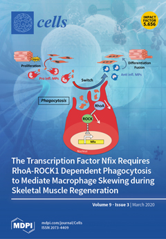
Macrophages (MPs) are crucial for tissue regeneration/repair. In skeletal muscle, MPs adopt a pro-inflammatory phenotype that stimulates a proliferation of myogenic cells and then switches to an anti-inflammatory status that induces myogenic differentiation. The phagocytosis drives this phenotypical switch. We show that the transcription factor Nfix is expressed by MPs and required for the pro-inflammatory to anti-inflammatory switch. Upon acute injury, a delay of muscle regeneration was evident in mice where Nfix has been deleted in the myeloid line. Moreover, phagocytosis induced by the inhibition of the RhoA-ROCK1 pathway leads to Nfix expression and the acquisition of the anti-inflammatory phenotype. We identified Nfix as a link between RhoA-ROCK1-dependent phagocytosis and the MP phenotypical switch, thus establishing a new role for Nfix in macrophage biology for tissue recovery. View this paper.
  • Issues are regarded as officially published after their release is announced to the table of contents alert mailing list.
  • You may sign up for e-mail alerts to receive table of contents of newly released issues.
  • PDF is the official format for papers published in both, html and pdf forms. To view the papers in pdf format, click on the "PDF Full-text" link, and use the free Adobe Reader to open them.
Order results
Result details
Section
Select all
Export citation of selected articles as:
21 pages, 6371 KiB  
Article
The Switch from NF-YAl to NF-YAs Isoform Impairs Myotubes Formation
by Debora Libetti, Andrea Bernardini, Sarah Sertic, Graziella Messina, Diletta Dolfini and Roberto Mantovani
Cells 2020, 9(3), 789; https://doi.org/10.3390/cells9030789 - 24 Mar 2020
Cited by 9 | Viewed by 3431
Abstract
NF-YA, the regulatory subunit of the trimeric transcription factor (TF) NF-Y, is regulated by alternative splicing (AS) generating two major isoforms, “long” (NF-YAl) and “short” (NF-YAs). Muscle cells express NF-YAl. We ablated exon 3 in mouse C2C12 cells by a four-guide CRISPR/Cas9n strategy, [...] Read more.
NF-YA, the regulatory subunit of the trimeric transcription factor (TF) NF-Y, is regulated by alternative splicing (AS) generating two major isoforms, “long” (NF-YAl) and “short” (NF-YAs). Muscle cells express NF-YAl. We ablated exon 3 in mouse C2C12 cells by a four-guide CRISPR/Cas9n strategy, obtaining clones expressing exclusively NF-YAs (C2-YAl-KO). C2-YAl-KO cells grow normally, but are unable to differentiate. Myogenin and—to a lesser extent, MyoD— levels are substantially lower in C2-YAl-KO, before and after differentiation. Expression of the fusogenic Myomaker and Myomixer genes, crucial for the early phases of the process, is not induced. Myomaker and Myomixer promoters are bound by MyoD and Myogenin, and Myogenin overexpression induces their expression in C2-YAl-KO. NF-Y inactivation reduces MyoD and Myogenin, but not directly: the Myogenin promoter is CCAAT-less, and the canonical CCAAT of the MyoD promoter is not bound by NF-Y in vivo. We propose that NF-YAl, but not NF-YAs, maintains muscle commitment by indirectly regulating Myogenin and MyoD expression in C2C12 cells. These experiments are the first genetic evidence that the two NF-YA isoforms have functionally distinct roles. Full article
Show Figures

Figure 1

14 pages, 2604 KiB  
Article
Phenol-Soluble Modulin-Mediated Aggregation of Community-Associated Methicillin-Resistant Staphylococcus Aureus in Human Cerebrospinal Fluid
by Deok-ryeong Kim, Yeonhee Lee, Hyeon-kyeong Kim, Wooseong Kim, Yun-Gon Kim, Yung-Hun Yang, Jae-Seok Kim and Hwang-Soo Joo
Cells 2020, 9(3), 788; https://doi.org/10.3390/cells9030788 - 24 Mar 2020
Cited by 9 | Viewed by 2734
Abstract
Phenol-soluble modulins (PSMs) are major determinants of Staphylococcus aureus virulence and their increased production in community-associated methicillin-resistant S. aureus (CA-MRSA) likely contributes to the enhanced virulence of MRSA strains. Here, we analyzed the differences in bacterial cell aggregation according to PSM presence in [...] Read more.
Phenol-soluble modulins (PSMs) are major determinants of Staphylococcus aureus virulence and their increased production in community-associated methicillin-resistant S. aureus (CA-MRSA) likely contributes to the enhanced virulence of MRSA strains. Here, we analyzed the differences in bacterial cell aggregation according to PSM presence in the specific human cerebrospinal fluid (CSF) environment. CSF samples from the intraventricular or lumbar intrathecal area of each patient and tryptic soy broth media were mixed at a 1:1 ratio, inoculated with WT and PSM-deleted mutants (Δpsm) of the CA-MRSA strain, USA300 LAC, and incubated overnight. Cell aggregation images were acquired after culture and image analysis was performed. The cell aggregation ratio in WT samples differed significantly between the two sampling sites (intraventricular: 0.2% vs. lumbar intrathecal: 6.7%, p < 0.001). The cell aggregation ratio in Δpsm samples also differed significantly between the two sampling sites (intraventricular: 0.0% vs. lumbar intrathecal: 1.2%, p < 0.001). Division of the study cases into two groups according to the aggregated area ratio (WT/Δpsm; group A: ratio of ≥ 2, group B: ratio of < 2) showed that the median aggregation ratio value differed significantly between groups A and B (5.5 and 0, respectively, p < 0.001). The differences in CSF distribution and PSM presence within the specific CSF environment are significant factors affecting bacterial cell aggregation. Full article
(This article belongs to the Section Cellular Pathology)
Show Figures

Figure 1

20 pages, 3242 KiB  
Review
The FOXO’s Advantages of Being a Family: Considerations on Function and Evolution
by Michel Schmitt-Ney
Cells 2020, 9(3), 787; https://doi.org/10.3390/cells9030787 - 24 Mar 2020
Cited by 35 | Viewed by 4695
Abstract
The nematode Caenorhabditis elegans possesses a unique (with various isoforms) FOXO transcription factor DAF-16, which is notorious for its role in aging and its regulation by the insulin-PI3K-AKT pathway. In humans, five genes (including a protein-coding pseudogene) encode for FOXO transcription factors that [...] Read more.
The nematode Caenorhabditis elegans possesses a unique (with various isoforms) FOXO transcription factor DAF-16, which is notorious for its role in aging and its regulation by the insulin-PI3K-AKT pathway. In humans, five genes (including a protein-coding pseudogene) encode for FOXO transcription factors that are targeted by the PI3K-AKT axis, such as in C. elegans. This common regulation and highly conserved DNA-binding domain are the pillars of this family. In this review, I will discuss the possible meaning of possessing a group of very similar proteins and how it can generate additional functionality to more complex organisms. I frame this discussion in relation to the much larger super family of Forkhead proteins to which they belong. FOXO members are very often co-expressed in the same cell type. The overlap of function and expression creates a certain redundancy that might be a safeguard against the accidental loss of FOXO function, which could otherwise lead to disease, particularly, cancer. This is one of the points that will be examined in this “family affair” report. Full article
(This article belongs to the Special Issue The FoxO Transcription Factors and Metabolic Regulation)
Show Figures

Figure 1

17 pages, 2499 KiB  
Article
Systematic Identification of Housekeeping Genes Possibly Used as References in Caenorhabditis elegans by Large-Scale Data Integration
by Jingxin Tao, Youjin Hao, Xudong Li, Huachun Yin, Xiner Nie, Jie Zhang, Boying Xu, Qiao Chen and Bo Li
Cells 2020, 9(3), 786; https://doi.org/10.3390/cells9030786 - 24 Mar 2020
Cited by 12 | Viewed by 4810
Abstract
For accurate gene expression quantification, normalization of gene expression data against reliable reference genes is required. It is known that the expression levels of commonly used reference genes vary considerably under different experimental conditions, and therefore, their use for data normalization is limited. [...] Read more.
For accurate gene expression quantification, normalization of gene expression data against reliable reference genes is required. It is known that the expression levels of commonly used reference genes vary considerably under different experimental conditions, and therefore, their use for data normalization is limited. In this study, an unbiased identification of reference genes in Caenorhabditis elegans was performed based on 145 microarray datasets (2296 gene array samples) covering different developmental stages, different tissues, drug treatments, lifestyle, and various stresses. As a result, thirteen housekeeping genes (rps-23, rps-26, rps-27, rps-16, rps-2, rps-4, rps-17, rpl-24.1, rpl-27, rpl-33, rpl-36, rpl-35, and rpl-15) with enhanced stability were comprehensively identified by using six popular normalization algorithms and RankAggreg method. Functional enrichment analysis revealed that these genes were significantly overrepresented in GO terms or KEGG pathways related to ribosomes. Validation analysis using recently published datasets revealed that the expressions of newly identified candidate reference genes were more stable than the commonly used reference genes. Based on the results, we recommended using rpl-33 and rps-26 as the optimal reference genes for microarray and rps-2 and rps-4 for RNA-sequencing data validation. More importantly, the most stable rps-23 should be a promising reference gene for both data types. This study, for the first time, successfully displays a large-scale microarray data driven genome-wide identification of stable reference genes for normalizing gene expression data and provides a potential guideline on the selection of universal internal reference genes in C. elegans, for quantitative gene expression analysis. Full article
(This article belongs to the Special Issue Biocomputing and Synthetic Biology in Cells)
Show Figures

Figure 1

36 pages, 1549 KiB  
Review
Focusing on Adenosine Receptors as a Potential Targeted Therapy in Human Diseases
by Wiwin Is Effendi, Tatsuya Nagano, Kazuyuki Kobayashi and Yoshihiro Nishimura
Cells 2020, 9(3), 785; https://doi.org/10.3390/cells9030785 - 24 Mar 2020
Cited by 88 | Viewed by 10609
Abstract
Adenosine is involved in a range of physiological and pathological effects through membrane-bound receptors linked to G proteins. There are four subtypes of adenosine receptors, described as A1AR, A2AAR, A2BAR, and A3AR, which are the [...] Read more.
Adenosine is involved in a range of physiological and pathological effects through membrane-bound receptors linked to G proteins. There are four subtypes of adenosine receptors, described as A1AR, A2AAR, A2BAR, and A3AR, which are the center of cAMP signal pathway-based drug development. Several types of agonists, partial agonists or antagonists, and allosteric substances have been synthesized from these receptors as new therapeutic drug candidates. Research efforts surrounding A1AR and A2AAR are perhaps the most enticing because of their concentration and affinity; however, as a consequence of distressing conditions, both A2BAR and A3AR levels might accumulate. This review focuses on the biological features of each adenosine receptor as the basis of ligand production and describes clinical studies of adenosine receptor-associated pharmaceuticals in human diseases. Full article
(This article belongs to the Special Issue Adenosine Receptors: From Cell Biology to Human Diseases)
Show Figures

Figure 1

25 pages, 3673 KiB  
Review
The Interplay between Peripherin 2 Complex Formation and Degenerative Retinal Diseases
by Lars Tebbe, Mashal Kakakhel, Mustafa S. Makia, Muayyad R. Al-Ubaidi and Muna I. Naash
Cells 2020, 9(3), 784; https://doi.org/10.3390/cells9030784 - 24 Mar 2020
Cited by 12 | Viewed by 3852
Abstract
Peripherin 2 (Prph2) is a photoreceptor-specific tetraspanin protein present in the outer segment (OS) rims of rod and cone photoreceptors. It shares many common features with other tetraspanins, including a large intradiscal loop which contains several cysteines. This loop enables Prph2 to associate [...] Read more.
Peripherin 2 (Prph2) is a photoreceptor-specific tetraspanin protein present in the outer segment (OS) rims of rod and cone photoreceptors. It shares many common features with other tetraspanins, including a large intradiscal loop which contains several cysteines. This loop enables Prph2 to associate with itself to form homo-oligomers or with its homologue, rod outer segment membrane protein 1 (Rom1) to form hetero-tetramers and hetero-octamers. Mutations in PRPH2 cause a multitude of retinal diseases including autosomal dominant retinitis pigmentosa (RP) or cone dominant macular dystrophies. The importance of Prph2 for photoreceptor development, maintenance and function is underscored by the fact that its absence results in a failure to initialize OS formation in rods and formation of severely disorganized OS membranous structures in cones. Although the exact role of Rom1 has not been well studied, it has been concluded that it is not necessary for disc morphogenesis but is required for fine tuning OS disc size and structure. Pathogenic mutations in PRPH2 often result in complex and multifactorial phenotypes, involving not just photoreceptors, as has historically been reasoned, but also secondary effects on the retinal pigment epithelium (RPE) and retinal/choroidal vasculature. The ability of Prph2 to form complexes was identified as a key requirement for the development and maintenance of OS structure and function. Studies using mouse models of pathogenic Prph2 mutations established a connection between changes in complex formation and disease phenotypes. Although progress has been made in the development of therapeutic approaches for retinal diseases in general, the highly complex interplay of functions mediated by Prph2 and the precise regulation of these complexes made it difficult, thus far, to develop a suitable Prph2-specific therapy. Here we describe the latest results obtained in Prph2-associated research and how mouse models provided new insights into the pathogenesis of its related diseases. Furthermore, we give an overview on the current status of the development of therapeutic solutions. Full article
(This article belongs to the Special Issue The Molecular and Cellular Basis of Retinal Diseases)
Show Figures

Figure 1

18 pages, 8198 KiB  
Article
A Wnt-BMP4 Signaling Axis Induces MSX and NOTCH Proteins and Promotes Growth Suppression and Differentiation in Neuroblastoma
by Marianna Szemes, Zsombor Melegh, Jacob Bellamy, Alexander Greenhough, Madhu Kollareddy, Daniel Catchpoole and Karim Malik
Cells 2020, 9(3), 783; https://doi.org/10.3390/cells9030783 - 23 Mar 2020
Cited by 8 | Viewed by 6361
Abstract
The Wnt and bone morphogenetic protein (BMP) signaling pathways are known to be crucial in the development of neural crest lineages, including the sympathetic nervous system. Surprisingly, their role in paediatric neuroblastoma, the prototypic tumor arising from this lineage, remains relatively uncharacterised. We [...] Read more.
The Wnt and bone morphogenetic protein (BMP) signaling pathways are known to be crucial in the development of neural crest lineages, including the sympathetic nervous system. Surprisingly, their role in paediatric neuroblastoma, the prototypic tumor arising from this lineage, remains relatively uncharacterised. We previously demonstrated that Wnt/β-catenin signaling can have cell-type-specific effects on neuroblastoma phenotypes, including growth inhibition and differentiation, and that BMP4 mRNA and protein were induced by Wnt3a/Rspo2. In this study, we characterised the phenotypic effects of BMP4 on neuroblastoma cells, demonstrating convergent induction of MSX homeobox transcription factors by Wnt and BMP4 signaling and BMP4-induced growth suppression and differentiation. An immunohistochemical analysis of BMP4 expression in primary neuroblastomas confirms a striking absence of BMP4 in poorly differentiated tumors, in contrast to a high expression in ganglion cells. These results are consistent with a tumor suppressive role for BMP4 in neuroblastoma. RNA sequencing following BMP4 treatment revealed induction of Notch signaling, verified by increases of Notch3 and Hes1 proteins. Together, our data demonstrate, for the first time, Wnt-BMP-Notch signaling crosstalk associated with growth suppression of neuroblastoma. Full article
(This article belongs to the Special Issue Wnt Signaling in Health and Diseases 2020)
Show Figures

Graphical abstract

18 pages, 12559 KiB  
Article
Cell-Substrate Patterns Driven by Curvature-Sensitive Actin Polymerization: Waves and Podosomes
by Moshe Naoz and Nir S. Gov
Cells 2020, 9(3), 782; https://doi.org/10.3390/cells9030782 - 23 Mar 2020
Cited by 5 | Viewed by 3504
Abstract
Cells adhered to an external solid substrate are observed to exhibit rich dynamics of actin structures on the basal membrane, which are distinct from those observed on the dorsal (free) membrane. Here we explore the dynamics of curved membrane proteins, or protein complexes, [...] Read more.
Cells adhered to an external solid substrate are observed to exhibit rich dynamics of actin structures on the basal membrane, which are distinct from those observed on the dorsal (free) membrane. Here we explore the dynamics of curved membrane proteins, or protein complexes, that recruit actin polymerization when the membrane is confined by the solid substrate. Such curved proteins can induce the spontaneous formation of membrane protrusions on the dorsal side of cells. However, on the basal side of the cells, such protrusions can only extend as far as the solid substrate and this constraint can convert such protrusions into propagating wave-like structures. We also demonstrate that adhesion molecules can stabilize localized protrusions that resemble some features of podosomes. This coupling of curvature and actin forces may underlie the differences in the observed actin-membrane dynamics between the basal and dorsal sides of adhered cells. Full article
(This article belongs to the Special Issue Symmetry Breaking in Cells and Tissues)
Show Figures

Figure 1

18 pages, 3121 KiB  
Article
Electrical Stimulation Induces Retinal Müller Cell Proliferation and Their Progenitor Cell Potential
by Sam Enayati, Karen Chang, Hamida Achour, Kin-Sang Cho, Fuyi Xu, Shuai Guo, Katarina Z. Enayati, Jia Xie, Eric Zhao, Tytteli Turunen, Amer Sehic, Lu Lu, Tor Paaske Utheim and Dong Feng Chen
Cells 2020, 9(3), 781; https://doi.org/10.3390/cells9030781 - 23 Mar 2020
Cited by 25 | Viewed by 4718
Abstract
Non-invasive electrical stimulation (ES) is increasingly applied to improve vision in untreatable eye conditions, such as retinitis pigmentosa and age-related macular degeneration. Our previous study suggested that ES promoted retinal function and the proliferation of progenitor-like glial cells in mice with inherited photoreceptor [...] Read more.
Non-invasive electrical stimulation (ES) is increasingly applied to improve vision in untreatable eye conditions, such as retinitis pigmentosa and age-related macular degeneration. Our previous study suggested that ES promoted retinal function and the proliferation of progenitor-like glial cells in mice with inherited photoreceptor degeneration; however, the underlying mechanism remains obscure. Müller cells (MCs) are thought to be dormant residential progenitor cells that possess a high potential for retinal neuron repair and functional plasticity. Here, we showed that ES with a ramp waveform of 20 Hz and 300 µA of current was effective at inducing mouse MC proliferation and enhancing their expression of progenitor cell markers, such as Crx (cone–rod homeobox) and Wnt7, as well as their production of trophic factors, including ciliary neurotrophic factor. RNA sequencing revealed that calcium signaling pathway activation was a key event, with a false discovery rate of 5.33 × 10⁻⁸ (p = 1.78 × 10⁻¹⁰) in ES-mediated gene profiling changes. Moreover, the calcium channel blocker, nifedipine, abolished the observed effects of ES on MC proliferation and progenitor cell gene induction, supporting a central role of ES-induced Ca2+ signaling in the MC changes. Our results suggest that low-current ES may present a convenient tool for manipulating MC behavior toward neuroregeneration and repair. Full article
Show Figures

Graphical abstract

14 pages, 2092 KiB  
Review
Role of Histone Deacetylases in Carcinogenesis: Potential Role in Cholangiocarcinoma
by Kishor Pant, Estanislao Peixoto, Seth Richard and Sergio A. Gradilone
Cells 2020, 9(3), 780; https://doi.org/10.3390/cells9030780 - 23 Mar 2020
Cited by 50 | Viewed by 5310
Abstract
Cholangiocarcinoma (CCA) is a highly invasive and metastatic form of carcinoma with bleak prognosis due to limited therapies, frequent relapse, and chemotherapy resistance. There is an urgent need to identify the molecular regulators of CCA in order to develop novel therapeutics and advance [...] Read more.
Cholangiocarcinoma (CCA) is a highly invasive and metastatic form of carcinoma with bleak prognosis due to limited therapies, frequent relapse, and chemotherapy resistance. There is an urgent need to identify the molecular regulators of CCA in order to develop novel therapeutics and advance diseases diagnosis. Many cellular proteins including histones may undergo a series of enzyme-mediated post-translational modifications including acetylation, methylation, phosphorylation, sumoylation, and crotonylation. Histone deacetylases (HDACs) play an important role in regulating epigenetic maintenance and modifications of their targets, which in turn exert critical impacts on chromatin structure, gene expression, and stability of proteins. As such, HDACs constitute a group of potential therapeutic targets for CCA. The aim of this review was to summarize the role that HDACs perform in regulating epigenetic changes, tumor development, and their potential as therapeutic targets for CCA. Full article
Show Figures

Figure 1

32 pages, 24751 KiB  
Article
Transcriptomic Insights into Mechanisms of Early Seed Maturation in the Garden Pea (Pisum sativum L.)
by Yury V. Malovichko, Oksana Y. Shtark, Ekaterina N. Vasileva, Anton A. Nizhnikov and Kirill S. Antonets
Cells 2020, 9(3), 779; https://doi.org/10.3390/cells9030779 - 23 Mar 2020
Cited by 13 | Viewed by 5163
Abstract
The garden pea (Pisum sativum L.) is a legume crop of immense economic value. Extensive breeding has led to the emergence of numerous pea varieties, of which some are distinguished by accelerated development in various stages of ontogenesis. One such trait is [...] Read more.
The garden pea (Pisum sativum L.) is a legume crop of immense economic value. Extensive breeding has led to the emergence of numerous pea varieties, of which some are distinguished by accelerated development in various stages of ontogenesis. One such trait is rapid seed maturation, which, despite novel insights into the genetic control of seed development in legumes, remains poorly studied. This article presents an attempt to dissect mechanisms of early maturation in the pea line Sprint-2 by means of whole transcriptome RNA sequencing in two developmental stages. By using a de novo assembly approach, we have obtained a reference transcriptome of 25,756 non-redundant entries expressed in pea seeds at either 10 or 20 days after pollination. Differential expression in Sprint-2 seeds has affected 13,056 transcripts. A comparison of the two pea lines with a common maturation rate demonstrates that while at 10 days after pollination, Sprint-2 seeds show development retardation linked to intensive photosynthesis, morphogenesis, and cell division, and those at 20 days show a rapid onset of desiccation marked by the cessation of translation and cell anabolism and accumulation of dehydration-protective and -storage moieties. Further inspection of certain transcript functional categories, including the chromatin constituent, transcription regulation, protein turnover, and hormonal regulation, has revealed transcriptomic trends unique to specific stages and cultivars. Among other remarkable features, Sprint-2 demonstrated an enhanced expression of transposable element-associated open reading frames and an altered expression of major maturation regulators and DNA methyltransferase genes. To the best of our knowledge, this is the first comparative transcriptomic study in which the issue of the seed maturation rate is addressed. Full article
(This article belongs to the Special Issue Bioinformatics and Computational Biology 2019)
Show Figures

Figure 1

29 pages, 6438 KiB  
Article
Fibronectin Adsorption on Electrospun Synthetic Vascular Grafts Attracts Endothelial Progenitor Cells and Promotes Endothelialization in Dynamic In Vitro Culture
by Ruben Daum, Dmitri Visser, Constanze Wild, Larysa Kutuzova, Maria Schneider, Günter Lorenz, Martin Weiss, Svenja Hinderer, Ulrich A. Stock, Martina Seifert and Katja Schenke-Layland
Cells 2020, 9(3), 778; https://doi.org/10.3390/cells9030778 - 23 Mar 2020
Cited by 37 | Viewed by 5628
Abstract
Appropriate mechanical properties and fast endothelialization of synthetic grafts are key to ensure long-term functionality of implants. We used a newly developed biostable polyurethane elastomer (TPCU) to engineer electrospun vascular scaffolds with promising mechanical properties (E-modulus: 4.8 ± 0.6 MPa, burst pressure: 3326 [...] Read more.
Appropriate mechanical properties and fast endothelialization of synthetic grafts are key to ensure long-term functionality of implants. We used a newly developed biostable polyurethane elastomer (TPCU) to engineer electrospun vascular scaffolds with promising mechanical properties (E-modulus: 4.8 ± 0.6 MPa, burst pressure: 3326 ± 78 mmHg), which were biofunctionalized with fibronectin (FN) and decorin (DCN). Neither uncoated nor biofunctionalized TPCU scaffolds induced major adverse immune responses except for minor signs of polymorph nuclear cell activation. The in vivo endothelial progenitor cell homing potential of the biofunctionalized scaffolds was simulated in vitro by attracting endothelial colony-forming cells (ECFCs). Although DCN coating did attract ECFCs in combination with FN (FN + DCN), DCN-coated TPCU scaffolds showed a cell-repellent effect in the absence of FN. In a tissue-engineering approach, the electrospun and biofunctionalized tubular grafts were cultured with primary-isolated vascular endothelial cells in a custom-made bioreactor under dynamic conditions with the aim to engineer an advanced therapy medicinal product. Both FN and FN + DCN functionalization supported the formation of a confluent and functional endothelial layer. Full article
(This article belongs to the Special Issue Stem Cell Research on Cardiology)
Show Figures

Figure 1

24 pages, 8823 KiB  
Article
The Proteasomal Deubiquitinating Enzyme PSMD14 Regulates Macroautophagy by Controlling Golgi-to-ER Retrograde Transport
by Hianara A Bustamante, Karina Cereceda, Alexis E González, Guillermo E Valenzuela, Yorka Cheuquemilla, Sergio Hernández, Eloisa Arias-Muñoz, Cristóbal Cerda-Troncoso, Susanne Bandau, Andrea Soza, Gudrun Kausel, Bredford Kerr, Gonzalo A Mardones, Jorge Cancino, Ronald T Hay, Alejandro Rojas-Fernandez and Patricia V Burgos
Cells 2020, 9(3), 777; https://doi.org/10.3390/cells9030777 - 23 Mar 2020
Cited by 12 | Viewed by 4975
Abstract
Ubiquitination regulates several biological processes, however the role of specific members of the ubiquitinome on intracellular membrane trafficking is not yet fully understood. Here, we search for ubiquitin-related genes implicated in protein membrane trafficking performing a High-Content siRNA Screening including 1187 genes of [...] Read more.
Ubiquitination regulates several biological processes, however the role of specific members of the ubiquitinome on intracellular membrane trafficking is not yet fully understood. Here, we search for ubiquitin-related genes implicated in protein membrane trafficking performing a High-Content siRNA Screening including 1187 genes of the human “ubiquitinome” using amyloid precursor protein (APP) as a reporter. We identified the deubiquitinating enzyme PSMD14, a subunit of the 19S regulatory particle of the proteasome, specific for K63-Ub chains in cells, as a novel regulator of Golgi-to-endoplasmic reticulum (ER) retrograde transport. Silencing or pharmacological inhibition of PSMD14 with Capzimin (CZM) caused a robust increase in APP levels at the Golgi apparatus and the swelling of this organelle. We showed that this phenotype is the result of rapid inhibition of Golgi-to-ER retrograde transport, a pathway implicated in the early steps of the autophagosomal formation. Indeed, we observed that inhibition of PSMD14 with CZM acts as a potent blocker of macroautophagy by a mechanism related to the retention of Atg9A and Rab1A at the Golgi apparatus. As pharmacological inhibition of the proteolytic core of the 20S proteasome did not recapitulate these effects, we concluded that PSMD14, and the K63-Ub chains, act as a crucial regulatory factor for macroautophagy by controlling Golgi-to-ER retrograde transport. Full article
(This article belongs to the Special Issue Ubiquitin and Autophagy)
Show Figures

Figure 1

18 pages, 4719 KiB  
Article
Graphene Oxide Nanosheets for Localized Hyperthermia—Physicochemical Characterization, Biocompatibility, and Induction of Tumor Cell Death
by Malgorzata J. Podolska, Alexandre Barras, Christoph Alexiou, Benjamin Frey, Udo Gaipl, Rabah Boukherroub, Sabine Szunerits, Christina Janko and Luis E. Muñoz
Cells 2020, 9(3), 776; https://doi.org/10.3390/cells9030776 - 23 Mar 2020
Cited by 19 | Viewed by 3466
Abstract
Background: The main goals of cancer treatment are not only to eradicate the tumor itself but also to elicit a specific immune response that overcomes the resistance of tumor cells against chemo- and radiotherapies. Hyperthermia was demonstrated to chemo- and radio-sensitize cancerous cells. [...] Read more.
Background: The main goals of cancer treatment are not only to eradicate the tumor itself but also to elicit a specific immune response that overcomes the resistance of tumor cells against chemo- and radiotherapies. Hyperthermia was demonstrated to chemo- and radio-sensitize cancerous cells. Many reports have confirmed the immunostimulatory effect of such multi-modal routines. Methods: We evaluated the interaction of graphene oxide (GO) nanosheets; its derivatives reduced GO and PEGylated rGO, with components of peripheral blood and evaluated its thermal conductivity to induce cell death by localized hyperthermia. Results: We confirmed the sterility and biocompatibility of the graphene nanomaterials and demonstrated that hyperthermia applied alone or in the combination with radiotherapy induced much more cell death in tumor cells than irradiation alone. Cell death was confirmed by the release of lactate dehydrogenase from dead and dying tumor cells. Conclusion: Biocompatible GO and its derivatives can be successfully used in graphene-induced hyperthermia to elicit tumor cell death. Full article
(This article belongs to the Special Issue The Interaction of Biomedical Nanoparticles with the Immune System)
Show Figures

Figure 1

16 pages, 3107 KiB  
Article
Pirfenidone Inhibits Cell Proliferation and Collagen I Production of Primary Human Intestinal Fibroblasts
by Yingying Cui, Mengfan Zhang, Changsen Leng, Tjasso Blokzijl, Bernadien H. Jansen, Gerard Dijkstra and Klaas Nico Faber
Cells 2020, 9(3), 775; https://doi.org/10.3390/cells9030775 - 22 Mar 2020
Cited by 32 | Viewed by 4405
Abstract
Intestinal fibrosis is a common complication of inflammatory bowel disease. So far, there is no safe and effective drug for intestinal fibrosis. Pirfenidone is an anti-fibrotic compound available for the treatment of idiopathic pulmonary fibrosis. Here, we explored the anti-proliferative and anti-fibrotic properties [...] Read more.
Intestinal fibrosis is a common complication of inflammatory bowel disease. So far, there is no safe and effective drug for intestinal fibrosis. Pirfenidone is an anti-fibrotic compound available for the treatment of idiopathic pulmonary fibrosis. Here, we explored the anti-proliferative and anti-fibrotic properties of pirfenidone on primary human intestinal fibroblasts (p-hIFs). p-hIFs were cultured in the absence and presence of pirfenidone. Cell proliferation was measured by a real-time cell analyzer (xCELLigence) and BrdU incorporation. Cell motility was monitored by live cell imaging. Cytotoxicity and cell viability were analyzed by Sytox green, Caspase-3 and Water Soluble Tetrazolium Salt-1 (WST-1) assays. Gene expression of fibrosis markers was determined by quantitative reverse transcription PCR (RT-qPCR). The mammalian target of rapamycin (mTOR) signaling was analyzed by Western blotting and type I collagen protein expression additionally by immunofluorescence microscopy. Pirfenidone dose-dependently inhibited p-hIF proliferation and motility, without inducing cell death. Pirfenidone suppressed mRNA levels of genes that contribute to extracellular matrix production, as well as basal and TGF-β1-induced collagen I protein production, which was associated with inhibition of the rapamycin-sensitive mTOR/p70S6K pathway in p-hIFs. Thus, pirfenidone inhibits the proliferation of intestinal fibroblasts and suppresses collagen I production through the TGF-β1/mTOR/p70S6K signaling pathway, which might be a novel and safe anti-fibrotic strategy to treat intestinal fibrosis. Full article
Show Figures

Figure 1

15 pages, 3407 KiB  
Article
Lamin A and Prelamin A Counteract Migration of Osteosarcoma Cells
by Camilla Evangelisti, Francesca Paganelli, Gaia Giuntini, Elisabetta Mattioli, Alessandra Cappellini, Giulia Ramazzotti, Irene Faenza, Maria Cristina Maltarello, Alberto M. Martelli, Katia Scotlandi, Francesca Chiarini and Giovanna Lattanzi
Cells 2020, 9(3), 774; https://doi.org/10.3390/cells9030774 - 22 Mar 2020
Cited by 14 | Viewed by 3723
Abstract
A type lamins are fundamental components of the nuclear lamina. Changes in lamin A expression correlate with malignant transformation in several cancers. However, the role of lamin A has not been explored in osteosarcoma (OS). Here, we wanted to investigate the role of [...] Read more.
A type lamins are fundamental components of the nuclear lamina. Changes in lamin A expression correlate with malignant transformation in several cancers. However, the role of lamin A has not been explored in osteosarcoma (OS). Here, we wanted to investigate the role of lamin A in normal osteoblasts (OBs) and OS cells. Thus, we studied the expression of lamin A/C in OS cells compared to OBs and evaluated the effects of lamin A overexpression in OS cell lines. We show that, while lamin A expression increases during osteoblast differentiation, all examined OS cell lines express lower lamin A levels relative to differentiated OBs. The condition of low LMNA expression confers to OS cells a significant increase in migration potential, while overexpression of lamin A reduces migration ability of OS cells. Moreover, overexpression of unprocessable prelamin A also reduces cell migration. In agreement with the latter finding, OS cells which accumulate the highest prelamin A levels upon inhibition of lamin A maturation by statins, had significantly reduced migration ability. Importantly, OS cells subjected to statin treatment underwent apoptotic cell death in a RAS-independent, lamin A-dependent manner. Our results show that pro-apoptotic effects of statins and statin inhibitory effect on OS cell migration are comparable to those obtained by prelamin A accumulation and further suggest that modulation of lamin A expression and post-translational processing can be a tool to decrease migration potential in OS cells. Full article
(This article belongs to the Collection Lamins and Laminopathies)
Show Figures

Figure 1

24 pages, 3331 KiB  
Article
Heme, A Metabolic Sensor, Directly Regulates the Activity of the KDM4 Histone Demethylase Family and Their Interactions with Partner Proteins
by Purna Chaitanya Konduri, Tianyuan Wang, Narges Salamat and Li Zhang
Cells 2020, 9(3), 773; https://doi.org/10.3390/cells9030773 - 22 Mar 2020
Cited by 2 | Viewed by 4229
Abstract
The KDM4 histone demethylase subfamily is constituted of yeast JmjC domain-containing proteins, such as Gis1, and human Gis1 orthologues, such as KDM4A/B/C. KDM4 proteins have important functions in regulating chromatin structure and gene expression in response to metabolic and nutritional stimuli. Heme acts [...] Read more.
The KDM4 histone demethylase subfamily is constituted of yeast JmjC domain-containing proteins, such as Gis1, and human Gis1 orthologues, such as KDM4A/B/C. KDM4 proteins have important functions in regulating chromatin structure and gene expression in response to metabolic and nutritional stimuli. Heme acts as a versatile signaling molecule to regulate important cellular functions in diverse organisms ranging from bacteria to humans. Here, using purified KDM4 proteins containing the JmjN/C domain, we showed that heme stimulates the histone demethylase activity of the JmjN/C domains of KDM4A and Cas well as full-length Gis1. Furthermore, we found that the C-terminal regions of KDM4 proteins, like that of Gis1, can confer heme regulation when fused to an unrelated transcriptional activator. Interestingly, biochemical pull-down of Gis1-interacting proteins followed by mass spectrometry identified 147 unique proteins associated with Gis1 under heme-sufficient and/or heme-deficient conditions. These 147 proteins included a significant number of heterocyclic compound-binding proteins, Ubl-conjugated proteins, metabolic enzymes/proteins, and acetylated proteins. These results suggested that KDM4s interact with diverse cellular proteins to form a complex network to sense metabolic and nutritional conditions like heme levels and respond by altering their interactions with other proteins and functional activities, such as histone demethylation. Full article
(This article belongs to the Section Cell Signaling)
Show Figures

Figure 1

14 pages, 546 KiB  
Review
Functional Phenotypes of Human Vγ9Vδ2 T Cells in Lymphoid Stress Surveillance
by Oliver Nussbaumer and Martin Thurnher
Cells 2020, 9(3), 772; https://doi.org/10.3390/cells9030772 - 22 Mar 2020
Cited by 12 | Viewed by 3777
Abstract
Butyrophilin and butyrophilin-like proteins select γδ T cells and direct the migration of γδ T cell subsets to distinct anatomical sites. γδ T cells expressing Vδ2 paired with Vγ9 (Vγ9Vδ2 T cells) are the predominant γδ T cell type in human peripheral blood. [...] Read more.
Butyrophilin and butyrophilin-like proteins select γδ T cells and direct the migration of γδ T cell subsets to distinct anatomical sites. γδ T cells expressing Vδ2 paired with Vγ9 (Vγ9Vδ2 T cells) are the predominant γδ T cell type in human peripheral blood. Vγ9Vδ2 T cells, which cannot be studied easily in vivo because they do not exist in rodents, are often referred to as innate-like T cells. The genetically recombined γδ T cell receptor (TCR) that responds to isoprenoid-derived pyrophosphates (phosphoantigens) produced by infected and malignant cells in a butyrophilin-dependent manner qualifies them as therapeutically relevant components of the adaptive immune system. On the other hand, cell-surface proteins such as the C-type lectin CD161 mark a functional phenotype of Vγ9Vδ2 T cells that mediates TCR-independent innate-like responses. Moreover, CD56 (neural cell adhesion molecule, NCAM) and the G protein-coupled receptor GPR56 define Vγ9Vδ2 T cells with increased cytolytic potential and, like CD161, may also be expressed by dendritic cells, principally facilitating the generation of an innate-like immunological synapse. In this review, we summarise current knowledge of Vγ9Vδ2 T cell functional phenotypes that are critical to lymphoid stress surveillance. Full article
Show Figures

Figure 1

13 pages, 4270 KiB  
Article
Selective Ablation of Dehydrodolichyl Diphosphate Synthase in Murine Retinal Pigment Epithelium (RPE) Causes RPE Atrophy and Retinal Degeneration
by Marci L. DeRamus, Stephanie J. Davis, Sriganesh Ramachandra Rao, Cyril Nyankerh, Delores Stacks, Timothy W. Kraft, Steven J. Fliesler and Steven J. Pittler
Cells 2020, 9(3), 771; https://doi.org/10.3390/cells9030771 - 21 Mar 2020
Cited by 10 | Viewed by 5438
Abstract
Patients with certain defects in the dehydrodolichyl diphosphate synthase (DHDDS) gene (RP59; OMIM #613861) exhibit classic symptoms of retinitis pigmentosa, as well as macular changes, suggestive of retinal pigment epithelium (RPE) involvement. The DHDDS enzyme is ubiquitously required for several pathways of protein [...] Read more.
Patients with certain defects in the dehydrodolichyl diphosphate synthase (DHDDS) gene (RP59; OMIM #613861) exhibit classic symptoms of retinitis pigmentosa, as well as macular changes, suggestive of retinal pigment epithelium (RPE) involvement. The DHDDS enzyme is ubiquitously required for several pathways of protein glycosylation. We wish to understand the basis for selective ocular pathology associated with certain DHDDS mutations and the contribution of specific ocular cell types to the pathology of mutant Dhdds-mediated retinal degeneration. To circumvent embryonic lethality associated with Dhdds knockout, we generated a Cre-dependent knockout allele of murine Dhdds (Dhddsflx/flx). We used targeted Cre expression to study the importance of the enzyme in the RPE. Structural alterations of the RPE and retina including reduction in outer retinal thickness, cell layer disruption, and increased RPE hyper-reflectivity were apparent at one postnatal month. At three months, RPE and photoreceptor disruption was observed non-uniformly across the retina as well as RPE transmigration into the photoreceptor layer, external limiting membrane descent towards the RPE, and patchy loss of photoreceptors. Functional loss measured by electroretinography was consistent with structural loss showing scotopic a- and b-wave reductions of 83% and 77%, respectively, at three months. These results indicate that RPE dysfunction contributes to DHDDS mutation-mediated pathology and suggests a more complicated disease mechanism than simply disruption of glycosylation. Full article
(This article belongs to the Special Issue The Molecular and Cellular Basis of Retinal Diseases)
Show Figures

Figure 1

20 pages, 2679 KiB  
Article
Validation of Reference Genes for Gene Expression Studies by RT-qPCR in HepaRG Cells during Toxicity Testing and Disease Modelling
by Joanna Brzeszczyńska, Filip Brzeszczyński, Kay Samuel, Katie Morgan, Steven D. Morley, John N. Plevris and Peter C. Hayes
Cells 2020, 9(3), 770; https://doi.org/10.3390/cells9030770 - 21 Mar 2020
Cited by 5 | Viewed by 4147
Abstract
Gene expression analysis by quantitative real-time polymerase chain reaction (RT-qPCR) is routinely used in biomedical studies. The reproducibility and reliability of the data fundamentally depends on experimental design and data interpretation. Despite the wide application of this assay, there is significant variation in [...] Read more.
Gene expression analysis by quantitative real-time polymerase chain reaction (RT-qPCR) is routinely used in biomedical studies. The reproducibility and reliability of the data fundamentally depends on experimental design and data interpretation. Despite the wide application of this assay, there is significant variation in the validation process of gene expression data from research laboratories. Since the validity of results depends on appropriate normalisation, it is crucial to select appropriate reference gene(s), where transcription of the selected gene is unaffected by experimental setting. In this study we have applied geNorm technology to investigate the transcription of 12 ‘housekeeping’ genes for use in the normalisation of RT-qPCR data acquired using a widely accepted HepaRG hepatic cell line in studies examining models of pre-clinical drug testing. geNorm data identified a number of genes unaffected by specific drug treatments and showed that different genes remained invariant in response to different drug treatments, whereas the transcription of ‘classical’ reference genes such as GAPDH (glyceralde- hyde-3-phosphate dehydrogenase) was altered by drug treatment. Comparing data normalised using the reference genes identified by geNorm with normalisation using classical housekeeping genes demonstrated substantial differences in the final results. In light of cell therapy application, RT-qPCR analyses has to be carefully evaluated to accurately interpret data obtained from dynamic cellular models undergoing sequential stages of phenotypic change. Full article
Show Figures

Figure 1

19 pages, 4529 KiB  
Article
Evolution of Epileptiform Activity in Zebrafish by Statistical-Based Integration of Electrophysiology and 2-Photon Ca2+ Imaging
by Olga Cozzolino, Federico Sicca, Emanuele Paoli, Francesco Trovato, Filippo M. Santorelli, Gian Michele Ratto and Maria Marchese
Cells 2020, 9(3), 769; https://doi.org/10.3390/cells9030769 - 21 Mar 2020
Cited by 13 | Viewed by 4132
Abstract
The study of sources and spatiotemporal evolution of ictal bursts is critical for the mechanistic understanding of epilepsy and for the validation of anti-epileptic drugs. Zebrafish is a powerful vertebrate model representing an excellent compromise between system complexity and experimental accessibility. We performed [...] Read more.
The study of sources and spatiotemporal evolution of ictal bursts is critical for the mechanistic understanding of epilepsy and for the validation of anti-epileptic drugs. Zebrafish is a powerful vertebrate model representing an excellent compromise between system complexity and experimental accessibility. We performed the quantitative evaluation of the spatial recruitment of neuronal populations during physiological and pathological activity by combining local field potential (LFP) recordings with simultaneous 2-photon Ca2+ imaging. We developed a method to extract and quantify electrophysiological transients coupled with Ca2+ events and we applied this tool to analyze two different epilepsy models and to assess the efficacy of the anti-epileptic drug valproate. Finally, by cross correlating the imaging data with the LFP, we demonstrated that the cerebellum is the main source of epileptiform transients. We have also shown that each transient was preceded by the activation of a sparse subset of neurons mostly located in the optic tectum. Full article
(This article belongs to the Special Issue Signaling Pathway Analysis and Disease Modeling in Zebrafish)
Show Figures

Figure 1

12 pages, 3894 KiB  
Review
NK cells and CD38: Implication for (Immuno)Therapy in Plasma Cell Dyscrasias
by Renato Zambello, Gregorio Barilà, Sabrina Manni, Francesco Piazza and Gianpietro Semenzato
Cells 2020, 9(3), 768; https://doi.org/10.3390/cells9030768 - 21 Mar 2020
Cited by 24 | Viewed by 7219
Abstract
Immunotherapy represents a promising new avenue for the treatment of multiple myeloma (MM) patients, particularly with the availability of Monoclonal Antibodies (mAbs) as anti-CD38 Daratumumab and Isatuximab and anti-SLAM-F7 Elotuzumab. Although a clear NK activation has been demonstrated for Elotuzumab, the effect of [...] Read more.
Immunotherapy represents a promising new avenue for the treatment of multiple myeloma (MM) patients, particularly with the availability of Monoclonal Antibodies (mAbs) as anti-CD38 Daratumumab and Isatuximab and anti-SLAM-F7 Elotuzumab. Although a clear NK activation has been demonstrated for Elotuzumab, the effect of anti-CD38 mAbs on NK system is controversial. As a matter of fact, an initial reduction of NK cells number characterizes Daratumumab therapy, limiting the potential role of this subset on myeloma immunotherapy. In this paper we discuss the role of NK cells along with anti-CD38 therapy and their implication in plasma cell dyscrasias, showing that mechanisms triggered by anti-CD38 mAbs ultimately lead to the activation of the immune system against myeloma cell growth. Full article
Show Figures

Figure 1

5 pages, 195 KiB  
Editorial
Roles and Functions of ROS and RNS in Cellular Physiology and Pathology
by Neven Zarkovic
Cells 2020, 9(3), 767; https://doi.org/10.3390/cells9030767 - 21 Mar 2020
Cited by 67 | Viewed by 5935
Abstract
Our common knowledge on oxidative stress has evolved substantially over the years, being focused mostly on the fundamental chemical reactions and the most relevant chemical species involved in human pathophysiology of oxidative stress-associated diseases. Thus, reactive oxygen species and reactive nitrogen species (ROS [...] Read more.
Our common knowledge on oxidative stress has evolved substantially over the years, being focused mostly on the fundamental chemical reactions and the most relevant chemical species involved in human pathophysiology of oxidative stress-associated diseases. Thus, reactive oxygen species and reactive nitrogen species (ROS and RNS) were identified as key players in initiating, mediating, and regulating the cellular and biochemical complexity of oxidative stress either as physiological (acting pro-hormetic) or as pathogenic (causing destructive vicious circles) processes. The papers published in this particular Special Issue of Cells show an impressive range on the pathophysiological relevance of ROS and RNS, including the relevance of second messengers of free radicals like 4-hydroxynonenal, allowing us to assume that the future will reveal even more detailed mechanisms of their positive and negative effects that might improve the monitoring of major modern diseases, and aid the development of advanced integrative biomedical treatments. Full article
18 pages, 1470 KiB  
Review
The Immune Response Against Human Cytomegalovirus Links Cellular to Systemic Senescence
by John J. Heath and Michael D. Grant
Cells 2020, 9(3), 766; https://doi.org/10.3390/cells9030766 - 20 Mar 2020
Cited by 30 | Viewed by 6223
Abstract
Aging reflects long-term decline in physiological function and integrity. Changes arise at a variable pace governed by time-dependent and -independent mechanisms that are themselves complex, interdependent and variable. Molecular decay produces inferior cells that eventually dominate over healthy counterparts in tissues they comprise. [...] Read more.
Aging reflects long-term decline in physiological function and integrity. Changes arise at a variable pace governed by time-dependent and -independent mechanisms that are themselves complex, interdependent and variable. Molecular decay produces inferior cells that eventually dominate over healthy counterparts in tissues they comprise. In a form of biological entropy, progression from molecular through cellular to tissue level degeneration culminates in organ disease or dysfunction, affecting systemic health. To better understand time-independent contributors and their potential modulation, common biophysical bases for key molecular and cellular changes underlying age-related physiological deterioration must be delineated. This review addresses the potential contribution of cytomegalovirus (CMV)-driven T cell proliferation to cellular senescence and immunosenescence. We first describe molecular processes imposing cell cycle arrest, the foundation of cellular senescence, then focus on the unique distribution, phenotype and function of CMV-specific CD8+ T cells in the context of cellular senescence and “inflammaging”. Their features position CMV infection as a pathogenic accelerant of immune cell proliferation underlying immune senescence. In human immunodeficiency virus (HIV) infection, where increased inflammation and exaggerated anti-CMV immune responses accelerate immune senescence, CMV infection has emerged as a major factor in unhealthy aging. Thus, we speculate on mechanistic links between CMV-specific CD8+ T-cell expansion, immune senescence and prevalence of age-related disorders in HIV infection. Full article
Show Figures

Figure 1

14 pages, 2802 KiB  
Article
Looking at New Unexpected Disease Targets in LMNA-Linked Lipodystrophies in the Light of Complex Cardiovascular Phenotypes: Implications for Clinical Practice
by Héléna Mosbah, Camille Vatier, Franck Boccara, Isabelle Jéru, Olivier Lascols, Marie-Christine Vantyghem, Bruno Fève, Bruno Donadille, Elisabeth Sarrazin, Sophie Benabbou, Jocelyn Inamo, Stéphane Ederhy, Ariel Cohen, Barbara Neraud, Pascale Richard, Fabien Picard, Sophie Christin-Maitre, Alban Redheuil, Karim Wahbi and Corinne Vigouroux
Cells 2020, 9(3), 765; https://doi.org/10.3390/cells9030765 - 20 Mar 2020
Cited by 9 | Viewed by 2720
Abstract
Variants in LMNA, encoding A-type lamins, are responsible for laminopathies including muscular dystrophies, lipodystrophies, and progeroid syndromes. Cardiovascular laminopathic involvement is classically described as cardiomyopathy in striated muscle laminopathies, and arterial wall dysfunction and/or valvulopathy in lipodystrophic and/or progeroid laminopathies. We report [...] Read more.
Variants in LMNA, encoding A-type lamins, are responsible for laminopathies including muscular dystrophies, lipodystrophies, and progeroid syndromes. Cardiovascular laminopathic involvement is classically described as cardiomyopathy in striated muscle laminopathies, and arterial wall dysfunction and/or valvulopathy in lipodystrophic and/or progeroid laminopathies. We report unexpected cardiovascular phenotypes in patients with LMNA-associated lipodystrophies, illustrating the complex multitissular pathophysiology of the disease and the need for specific cardiovascular investigations in affected patients. A 33-year-old woman was diagnosed with generalized lipodystrophy and atypical progeroid syndrome due to the newly identified heterozygous LMNA p.(Asp136Val) variant. Her complex cardiovascular phenotype was associated with atherosclerosis, aortic valvular disease and left ventricular hypertrophy with rhythm and conduction defects. A 29-year-old woman presented with a partial lipodystrophy syndrome and a severe coronary atherosclerosis which required a triple coronary artery bypass grafting. She carried the novel heterozygous p.(Arg60Pro) LMNA variant inherited from her mother, affected with partial lipodystrophy and dilated cardiomyopathy. Different lipodystrophy-associated LMNA pathogenic variants could target cardiac vasculature and/or muscle, leading to complex overlapping phenotypes. Unifying pathophysiological hypotheses should be explored in several cell models including adipocytes, cardiomyocytes and vascular cells. Patients with LMNA-associated lipodystrophy should be systematically investigated with 24-h ECG monitoring, echocardiography and non-invasive coronary function testing. Full article
Show Figures

Figure 1

12 pages, 2961 KiB  
Review
On the TRAIL of Better Therapies: Understanding TNFRSF Structure-Function
by Éva S. Vanamee and Denise L. Faustman
Cells 2020, 9(3), 764; https://doi.org/10.3390/cells9030764 - 20 Mar 2020
Cited by 22 | Viewed by 3708
Abstract
Tumor necrosis factor (TNF) superfamily ligands show diverse biological functions, such as the induction of apoptotic cell death or cell survival and proliferation, making them excellent therapeutic targets for cancer and autoimmunity. We review the latest literature on TNF receptor superfamily signaling with [...] Read more.
Tumor necrosis factor (TNF) superfamily ligands show diverse biological functions, such as the induction of apoptotic cell death or cell survival and proliferation, making them excellent therapeutic targets for cancer and autoimmunity. We review the latest literature on TNF receptor superfamily signaling with a focus on structure-function. Using combinatorics, we argue that receptors that cluster on the cell surface and are activated by membrane-bound ligands need to arrange in a highly ordered manner, as the probability of random ligand and receptor arrangements matching up for receptor activation is very low. A growing body of evidence indicates that antiparallel receptor dimers that sequester the ligand binding site cluster on the cell surface, forming a hexagonal lattice. Upon ligand binding, this arrangement puts the activated receptors at the right distance to accommodate the downstream signaling partners. The data also suggest that the same geometry is utilized regardless of receptor type. The unified model provides important clues about TNF receptor signaling and should aid the design of better therapies for cancer and various immune mediated diseases. Full article
(This article belongs to the Special Issue TRAIL Receptors in Health and Diseases)
Show Figures

Figure 1

20 pages, 602 KiB  
Review
The Missing Lnc: The Potential of Targeting Triple-Negative Breast Cancer and Cancer Stem Cells by Inhibiting Long Non-Coding RNAs
by Justin M Brown, Marie-Claire D Wasson and Paola Marcato
Cells 2020, 9(3), 763; https://doi.org/10.3390/cells9030763 - 20 Mar 2020
Cited by 27 | Viewed by 5049
Abstract
Treatment decisions for breast cancer are based on staging and hormone receptor expression and include chemotherapies and endocrine therapy. While effective in many cases, some breast cancers are resistant to therapy, metastasize and recur, leading to eventual death. Higher percentages of tumor-initiating cancer [...] Read more.
Treatment decisions for breast cancer are based on staging and hormone receptor expression and include chemotherapies and endocrine therapy. While effective in many cases, some breast cancers are resistant to therapy, metastasize and recur, leading to eventual death. Higher percentages of tumor-initiating cancer stem cells (CSCs) may contribute to the increased aggressiveness, chemoresistance, and worse outcomes among breast cancer. This may be particularly true in triple-negative breast cancers (TNBCs) which have higher percentages of CSCs and are associated with worse outcomes. In recent years, increasing numbers of long non-coding RNAs (lncRNAs) have been identified as playing an important role in breast cancer progression and some of these have been specifically associated within the CSC populations of breast cancers. LncRNAs are non-protein-coding transcripts greater than 200 nucleotides which can have critical functions in gene expression regulation. The preclinical evidence regarding lncRNA antagonists for the treatment of cancer is promising and therefore, presents a potential novel approach for treating breast cancer and targeting therapy-resistant CSCs within these tumors. Herein, we summarize the lncRNAs that have been identified as functionally relevant in breast CSCs. Furthermore, our review of the literature and analysis of patient datasets has revealed that many of these breast CSC-associated lncRNAs are also enriched in TNBC. Together, this suggests that these lncRNAs may be playing a particularly important role in TNBC. Thus, certain breast cancer-promoting/CSC-associated lncRNAs could be targeted in the treatment of TNBCs and the CSCs within these tumors should be susceptible to anti-lncRNA therapy. Full article
(This article belongs to the Special Issue lncRNA and Cancer)
Show Figures

Figure 1

12 pages, 944 KiB  
Review
Parallels of Resistance between Angiogenesis and Lymphangiogenesis Inhibition in Cancer Therapy
by Dennis Jones
Cells 2020, 9(3), 762; https://doi.org/10.3390/cells9030762 - 20 Mar 2020
Cited by 27 | Viewed by 3892
Abstract
Metastasis is the primary cause of cancer-related mortality. Cancer cells primarily metastasize via blood and lymphatic vessels to colonize lymph nodes and distant organs, leading to worse prognosis. Thus, strategies to limit blood and lymphatic spread of cancer have been a focal point [...] Read more.
Metastasis is the primary cause of cancer-related mortality. Cancer cells primarily metastasize via blood and lymphatic vessels to colonize lymph nodes and distant organs, leading to worse prognosis. Thus, strategies to limit blood and lymphatic spread of cancer have been a focal point of cancer research for several decades. Resistance to FDA-approved anti-angiogenic therapies designed to limit blood vessel growth has emerged as a significant clinical challenge. However, there are no FDA-approved drugs that target tumor lymphangiogenesis, despite the consequences of metastasis through the lymphatic system. This review highlights several of the key resistance mechanisms to anti-angiogenic therapy and potential challenges facing anti-lymphangiogenic therapy. Blood and lymphatic vessels are more than just conduits for nutrient, fluid, and cancer cell transport. Recent studies have elucidated how these vasculatures often regulate immune responses. Vessels that are abnormal or compromised by tumor cells can lead to immunosuppression. Therapies designed to improve lymphatic vessel function while limiting metastasis may represent a viable approach to enhance immunotherapy and limit cancer progression. Full article
(This article belongs to the Special Issue Angiogenesis in Cancer)
Show Figures

Figure 1

18 pages, 5471 KiB  
Article
Hybrid Label-Free Molecular Microscopies for Simultaneous Visualization of Changes in Cell Wall Polysaccharides of Peach at Single- and Multiple-Cell Levels during Postharvest Storage
by Weinan Huang, Yating Nie, Nan Zhu, Yifan Yang, Changqing Zhu, Minbiao Ji, Di Wu and Kunsong Chen
Cells 2020, 9(3), 761; https://doi.org/10.3390/cells9030761 - 20 Mar 2020
Cited by 13 | Viewed by 2900
Abstract
Softening of fruit during the postharvest storage, which is mainly associated with both compositional and spatial changes of polysaccharides within cell wall, affects the texture and quality of fruit. Current research on the fruit softening mechanism lacks an understanding of the overall softening [...] Read more.
Softening of fruit during the postharvest storage, which is mainly associated with both compositional and spatial changes of polysaccharides within cell wall, affects the texture and quality of fruit. Current research on the fruit softening mechanism lacks an understanding of the overall softening at the cell level. The objective of this work was to investigate the change in the spatial distribution of cell wall polysaccharides in peach flesh cells at both single- and multiple-cell levels in a label-free way during the postharvest storage. Nonmelting peaches (Prunus persica L. Batsch cv.”Zhonghuashoutao”) at commercial maturity were stored at 0 °C and 20 °C. Firmness measurement and chemical analysis were performed at each storage time. In addition, three molecular imaging techniques, namely confocal Raman microspectroscopy (CRM), Fourier transform infrared microspectroscopy (FTIRM), and stimulated Raman scattering microscopy (SRS) were used to visualize changes in the spatial distribution of cell wall polysaccharides of peach fruit in a label-free way during the postharvest storage. The combination of CRM and FTIRM provided complementary spectral information to visualize the spatial changes of cellulose, hemicellulose, and pectin in the cell wall of peach flesh during softening at the single-cell level, and found that the cell wall polysaccharides tended to be concentrated in the cell corner of parenchymal cells at the late stage. Furthermore, SRS, which is an ultrafast Raman imaging technique (approximately three or four orders of magnitude faster than CRM), was used for high-throughput cell wall phenotypes measurement. Different degradation degrees of parenchymal cells during fruit softening were found based on the gray-scale statistical analysis of SRS data. In general, cell wall polysaccharides decreased during softening and tended to be concentrated in the cell corner for most parenchymal cells at the late stage, but there were also some cells not in line with the whole softening trends. The results show that there were differences in the content and spatial changes of cell wall polysaccharides among parenchymal cells of peach fruit during the softening process, and the hybrid use of CRM, FTIRM, and SRS is a promising method for simultaneous visualization of changes in cell wall polysaccharides of peach. Full article
(This article belongs to the Section Plant, Algae and Fungi Cell Biology)
Show Figures

Figure 1

28 pages, 2627 KiB  
Review
Transcription Factors in Cancer: When Alternative Splicing Determines Opposite Cell Fates
by Silvia Belluti, Giovanna Rigillo and Carol Imbriano
Cells 2020, 9(3), 760; https://doi.org/10.3390/cells9030760 - 20 Mar 2020
Cited by 45 | Viewed by 6894
Abstract
Alternative splicing (AS) is a finely regulated mechanism for transcriptome and proteome diversification in eukaryotic cells. Correct balance between AS isoforms takes part in molecular mechanisms that properly define spatiotemporal and tissue specific transcriptional programs in physiological conditions. However, several diseases are associated [...] Read more.
Alternative splicing (AS) is a finely regulated mechanism for transcriptome and proteome diversification in eukaryotic cells. Correct balance between AS isoforms takes part in molecular mechanisms that properly define spatiotemporal and tissue specific transcriptional programs in physiological conditions. However, several diseases are associated to or even caused by AS alterations. In particular, multiple AS changes occur in cancer cells and sustain the oncogenic transcriptional program. Transcription factors (TFs) represent a key class of proteins that control gene expression by direct binding to DNA regulatory elements. AS events can generate cancer-associated TF isoforms with altered activity, leading to sustained proliferative signaling, differentiation block and apoptosis resistance, all well-known hallmarks of cancer. In this review, we focus on how AS can produce TFs isoforms with opposite transcriptional activities or antagonistic functions that severely impact on cancer biology. This summary points the attention to the relevance of the analysis of TFs splice variants in cancer, which can allow patients stratification despite the presence of interindividual genetic heterogeneity. Recurrent TFs variants that give advantage to specific cancer types not only open the opportunity to use AS transcripts as clinical biomarkers but also guide the development of new anti-cancer strategies in personalized medicine. Full article
Show Figures

Figure 1

Previous Issue
Next Issue
Back to TopTop