Next Issue
Volume 8, October
Previous Issue
Volume 8, August


Cells, Volume 8, Issue 9 (September 2019) – 170 articles

Cover Story (view full-size image): 
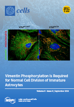
Intermediate filaments (also known as nanofilaments) of astrocytes are dynamic structures involved in cell signaling and migration and determining cell responses in health, disease, and regeneration. Upregulation of the intermediate filament proteins GFAP, vimentin (VIM), and nestin is a hallmark of reactive gliosis, which is protective in ischemic stroke or neurotrauma, but inhibits some regenerative responses. Immature astrocytes from mice with VIM mutations of serine sites phosphorylated during mitosis (VIMSA/SA) exhibit cytokinetic failure and contain VIM accumulations that colocalize with mitochondria (green: VIM-containing intermediate filaments and VIM accumulations; red: mitochondria). This phenotype is transient and disappears with VIMSA/SA astrocyte maturation and can be alleviated by the inhibition of cell proliferation. View this paper.
  • Issues are regarded as officially published after their release is announced to the table of contents alert mailing list.
  • You may sign up for e-mail alerts to receive table of contents of newly released issues.
  • PDF is the official format for papers published in both, html and pdf forms. To view the papers in pdf format, click on the "PDF Full-text" link, and use the free Adobe Reader to open them.
Order results
Result details
Section
Select all
Export citation of selected articles as:
21 pages, 3551 KiB  
Article
Proteomic Analysis of miR-195 and miR-497 Replacement Reveals Potential Candidates that Increase Sensitivity to Oxaliplatin in MSI/P53wt Colorectal Cancer Cells
by Dennis Poel, Lenka N.C. Boyd, Robin Beekhof, Tim Schelfhorst, Thang V. Pham, Sander R. Piersma, Jaco C. Knol, Connie R. Jimenez, Henk M.W. Verheul and Tineke E. Buffart
Cells 2019, 8(9), 1111; https://doi.org/10.3390/cells8091111 - 19 Sep 2019
Cited by 23 | Viewed by 3964
Abstract
Most patients with advanced colorectal cancer (CRC) eventually develop resistance to systemic combination therapy. miR-195-5p and miR-497-5p are downregulated in CRC tissues and associated with drug resistance. Sensitization to 5-FU, oxaliplatin, and irinotecan by transfection with miR-195-5p and miR-497-5p mimics was studied using [...] Read more.
Most patients with advanced colorectal cancer (CRC) eventually develop resistance to systemic combination therapy. miR-195-5p and miR-497-5p are downregulated in CRC tissues and associated with drug resistance. Sensitization to 5-FU, oxaliplatin, and irinotecan by transfection with miR-195-5p and miR-497-5p mimics was studied using cell viability and clonogenic assays in cell lines HCT116, RKO, DLD-1, and SW480. In addition, proteomic analysis of transfected cells was implemented to identify potential targets. Significantly altered proteins were subjected to STRING (protein-protein interaction networks) database analysis to study the potential mechanisms of drug resistance. Cell viability analysis of transfected cells revealed increased sensitivity to oxaliplatin in microsatellite instable (MSI)/P53 wild-type HCT116 and RKO cells. HCT116 transfected cells formed significantly fewer colonies when treated with oxaliplatin. In sensitized cells, proteomic analysis showed 158 and 202 proteins with significantly altered expression after transfection with miR-195-5p and miR-497-5p mimics respectively, of which CHUK and LUZP1 proved to be coinciding downregulated proteins. Resistance mechanisms of these proteins may be associated with nuclear factor kappa-B signaling and G1 cell-cycle arrest. In conclusion, miR-195-5p and miR-497-5p replacement enhanced sensitivity to oxaliplatin in treatment naïve MSI/P53 wild-type CRC cells. Proteomic analysis revealed potential miRNA targets associated with the cell-cycle which possibly bare a relation with chemotherapy sensitivity. Full article
Show Figures

Figure 1

28 pages, 1031 KiB  
Review
FOXO3a from the Nucleus to the Mitochondria: A Round Trip in Cellular Stress Response
by Candida Fasano, Vittoria Disciglio, Stefania Bertora, Martina Lepore Signorile and Cristiano Simone
Cells 2019, 8(9), 1110; https://doi.org/10.3390/cells8091110 - 19 Sep 2019
Cited by 124 | Viewed by 14382
Abstract
Cellular stress response is a universal mechanism that ensures the survival or negative selection of cells in challenging conditions. The transcription factor Forkhead box protein O3 (FOXO3a) is a core regulator of cellular homeostasis, stress response, and longevity since it can modulate a [...] Read more.
Cellular stress response is a universal mechanism that ensures the survival or negative selection of cells in challenging conditions. The transcription factor Forkhead box protein O3 (FOXO3a) is a core regulator of cellular homeostasis, stress response, and longevity since it can modulate a variety of stress responses upon nutrient shortage, oxidative stress, hypoxia, heat shock, and DNA damage. FOXO3a activity is regulated by post-translational modifications that drive its shuttling between different cellular compartments, thereby determining its inactivation (cytoplasm) or activation (nucleus and mitochondria). Depending on the stress stimulus and subcellular context, activated FOXO3a can induce specific sets of nuclear genes, including cell cycle inhibitors, pro-apoptotic genes, reactive oxygen species (ROS) scavengers, autophagy effectors, gluconeogenic enzymes, and others. On the other hand, upon glucose restriction, 5′-AMP-activated protein kinase (AMPK) and mitogen activated protein kinase kinase (MEK)/extracellular signal-regulated kinase (ERK) -dependent FOXO3a mitochondrial translocation allows the transcription of oxidative phosphorylation (OXPHOS) genes, restoring cellular ATP levels, while in cancer cells, mitochondrial FOXO3a mediates survival upon genotoxic stress induced by chemotherapy. Interestingly, these target genes and their related pathways are diverse and sometimes antagonistic, suggesting that FOXO3a is an adaptable player in the dynamic homeostasis of normal and stressed cells. In this review, we describe the multiple roles of FOXO3a in cellular stress response, with a focus on both its nuclear and mitochondrial functions. Full article
(This article belongs to the Special Issue Molecular and Cellular Mechanisms of Stress Responses)
Show Figures

Graphical abstract

13 pages, 6923 KiB  
Article
Dynamic Interplay between Pericytes and Endothelial Cells during Sprouting Angiogenesis
by Giulia Chiaverina, Laura di Blasio, Valentina Monica, Massimo Accardo, Miriam Palmiero, Barbara Peracino, Marianela Vara-Messler, Alberto Puliafito and Luca Primo
Cells 2019, 8(9), 1109; https://doi.org/10.3390/cells8091109 - 19 Sep 2019
Cited by 45 | Viewed by 5154
Abstract
Vascular physiology relies on the concerted dynamics of several cell types, including pericytes, endothelial, and vascular smooth muscle cells. The interactions between such cell types are inherently dynamic and are not easily described with static, fixed, experimental approaches. Pericytes are mural cells that [...] Read more.
Vascular physiology relies on the concerted dynamics of several cell types, including pericytes, endothelial, and vascular smooth muscle cells. The interactions between such cell types are inherently dynamic and are not easily described with static, fixed, experimental approaches. Pericytes are mural cells that support vascular development, remodeling, and homeostasis, and are involved in a number of pathological situations including cancer. The dynamic interplay between pericytes and endothelial cells is at the basis of vascular physiology and few experimental tools exist to properly describe and study it. Here we employ a previously developed ex vivo murine aortic explant to study the formation of new blood capillary-like structures close to physiological situation. We develop several mouse models to culture, identify, characterize, and follow simultaneously single endothelial cells and pericytes during angiogenesis. We employ microscopy and image analysis to dissect the interactions between cell types and the process of cellular recruitment on the newly forming vessel. We find that pericytes are recruited on the developing sprout by proliferation, migrate independently from endothelial cells, and can proliferate on the growing capillary. Our results help elucidating several relevant mechanisms of interactions between endothelial cells and pericytes. Full article
(This article belongs to the Special Issue Angiogenesis in Cancer)
Show Figures

Figure 1

22 pages, 1839 KiB  
Review
Environmentally-Induced Transgenerational Epigenetic Inheritance: Implication of PIWI Interacting RNAs
by Karine Casier, Antoine Boivin, Clément Carré and Laure Teysset
Cells 2019, 8(9), 1108; https://doi.org/10.3390/cells8091108 - 19 Sep 2019
Cited by 18 | Viewed by 6836
Abstract
Environmentally-induced transgenerational epigenetic inheritance is an emerging field. The understanding of associated epigenetic mechanisms is currently in progress with open questions still remaining. In this review, we present an overview of the knowledge of environmentally-induced transgenerational inheritance and associated epigenetic mechanisms, mainly in [...] Read more.
Environmentally-induced transgenerational epigenetic inheritance is an emerging field. The understanding of associated epigenetic mechanisms is currently in progress with open questions still remaining. In this review, we present an overview of the knowledge of environmentally-induced transgenerational inheritance and associated epigenetic mechanisms, mainly in animals. The second part focuses on the role of PIWI-interacting RNAs (piRNAs), a class of small RNAs involved in the maintenance of the germline genome, in epigenetic memory to put into perspective cases of environmentally-induced transgenerational inheritance involving piRNA production. Finally, the last part addresses how genomes are facing production of new piRNAs, and from a broader perspective, how this process might have consequences on evolution and on sporadic disease development. Full article
(This article belongs to the Special Issue Evolution of Epigenetic Mechanisms and Signatures)
Show Figures

Figure 1

19 pages, 917 KiB  
Review
Functions and Regulatory Mechanisms of lncRNAs in Skeletal Myogenesis, Muscle Disease and Meat Production
by Shanshan Wang, Jianjun Jin, Zaiyan Xu and Bo Zuo
Cells 2019, 8(9), 1107; https://doi.org/10.3390/cells8091107 - 19 Sep 2019
Cited by 61 | Viewed by 5466
Abstract
Myogenesis is a complex biological process, and understanding the regulatory network of skeletal myogenesis will contribute to the treatment of human muscle related diseases and improvement of agricultural animal meat production. Long noncoding RNAs (lncRNAs) serve as regulators in gene expression networks, and [...] Read more.
Myogenesis is a complex biological process, and understanding the regulatory network of skeletal myogenesis will contribute to the treatment of human muscle related diseases and improvement of agricultural animal meat production. Long noncoding RNAs (lncRNAs) serve as regulators in gene expression networks, and participate in various biological processes. Recent studies have identified functional lncRNAs involved in skeletal muscle development and disease. These lncRNAs regulate the proliferation, differentiation, and fusion of myoblasts through multiple mechanisms, such as chromatin modification, transcription regulation, and microRNA sponge activity. In this review, we presented the latest advances regarding the functions and regulatory activities of lncRNAs involved in muscle development, muscle disease, and meat production. Moreover, challenges and future perspectives related to the identification of functional lncRNAs were also discussed. Full article
Show Figures

Figure 1

18 pages, 2389 KiB  
Article
Regulation of Ketogenic Enzyme HMGCS2 by Wnt/β-catenin/PPARγ Pathway in Intestinal Cells
by Ji Tae Kim, Chang Li, Heidi L. Weiss, Yuning Zhou, Chunming Liu, Qingding Wang and B. Mark Evers
Cells 2019, 8(9), 1106; https://doi.org/10.3390/cells8091106 - 19 Sep 2019
Cited by 42 | Viewed by 7007
Abstract
The Wnt/β-catenin pathway plays a crucial role in development and renewal of the intestinal epithelium. Mitochondrial 3-hydroxy-3-methylglutaryl-CoA synthase 2 (HMGCS2), a rate-limiting ketogenic enzyme in the synthesis of ketone body β-hydroxybutyrate (βHB), contributes to the regulation of intestinal cell differentiation. Here, we have [...] Read more.
The Wnt/β-catenin pathway plays a crucial role in development and renewal of the intestinal epithelium. Mitochondrial 3-hydroxy-3-methylglutaryl-CoA synthase 2 (HMGCS2), a rate-limiting ketogenic enzyme in the synthesis of ketone body β-hydroxybutyrate (βHB), contributes to the regulation of intestinal cell differentiation. Here, we have shown that HMGCS2 is a novel target of Wnt/β-catenin/PPARγ signaling in intestinal epithelial cancer cell lines and normal intestinal organoids. Inhibition of the Wnt/β-catenin pathway resulted in increased protein and mRNA expression of HMGCS2 and βHB production in human colon cancer cell lines LS174T and Caco2. In addition, Wnt inhibition increased expression of PPARγ and its target genes, FABP2 and PLIN2, in these cells. Conversely, activation of Wnt/β-catenin signaling decreased protein and mRNA levels of HMGCS2, βHB production, and expression of PPARγ and its target genes in LS174T and Caco2 cells and mouse intestinal organoids. Moreover, inhibition of PPARγ reduced HMGCS2 expression and βHB production, while activation of PPARγ increased HMGCS2 expression and βHB synthesis. Furthermore, PPARγ bound the promoter of HMGCS2 and this binding was enhanced by β-catenin knockdown. Finally, we showed that HMGCS2 inhibited, while Wnt/β-catenin stimulated, glycolysis, which contributed to regulation of intestinal cell differentiation. Our results identified HMGCS2 as a downstream target of Wnt/β-catenin/PPARγ signaling in intestinal epithelial cells. Moreover, our findings suggest that Wnt/β-catenin/PPARγ signaling regulates intestinal cell differentiation, at least in part, through regulation of ketogenesis. Full article
(This article belongs to the Section Cell Signaling)
Show Figures

Figure 1

18 pages, 2784 KiB  
Review
Cellular Stress Responses in Radiotherapy
by Wanyeon Kim, Sungmin Lee, Danbi Seo, Dain Kim, Kyeongmin Kim, EunGi Kim, JiHoon Kang, Ki Moon Seong, HyeSook Youn and BuHyun Youn
Cells 2019, 8(9), 1105; https://doi.org/10.3390/cells8091105 - 18 Sep 2019
Cited by 174 | Viewed by 15415
Abstract
Radiotherapy is one of the major cancer treatment strategies. Exposure to penetrating radiation causes cellular stress, directly or indirectly, due to the generation of reactive oxygen species, DNA damage, and subcellular organelle damage and autophagy. These radiation-induced damage responses cooperatively contribute to cancer [...] Read more.
Radiotherapy is one of the major cancer treatment strategies. Exposure to penetrating radiation causes cellular stress, directly or indirectly, due to the generation of reactive oxygen species, DNA damage, and subcellular organelle damage and autophagy. These radiation-induced damage responses cooperatively contribute to cancer cell death, but paradoxically, radiotherapy also causes the activation of damage-repair and survival signaling to alleviate radiation-induced cytotoxic effects in a small percentage of cancer cells, and these activations are responsible for tumor radio-resistance. The present study describes the molecular mechanisms responsible for radiation-induced cellular stress response and radioresistance, and the therapeutic approaches used to overcome radioresistance. Full article
(This article belongs to the Special Issue Molecular and Cellular Mechanisms of Stress Responses)
Show Figures

Figure 1

18 pages, 4489 KiB  
Article
Interleukin 21 Receptor/Ligand Interaction Is Linked to Disease Progression in Pancreatic Cancer
by Alica Linnebacher, Philipp Mayer, Nicole Marnet, Frank Bergmann, Esther Herpel, Steffie Revia, Libo Yin, Li Liu, Thilo Hackert, Thomas Giese, Ingrid Herr and Matthias M. Gaida
Cells 2019, 8(9), 1104; https://doi.org/10.3390/cells8091104 - 18 Sep 2019
Cited by 11 | Viewed by 4292
Abstract
Pancreatic ductal adenocarcinoma (PDAC) displays a marked fibro-inflammatory microenvironment in which infiltrated immune cells fail to eliminate the tumor cells and often—rather paradoxically—promote tumor progression. Of special interest are tumor-promoting T cells that assume a Th17-like phenotype because their presence in PDAC tissue [...] Read more.
Pancreatic ductal adenocarcinoma (PDAC) displays a marked fibro-inflammatory microenvironment in which infiltrated immune cells fail to eliminate the tumor cells and often—rather paradoxically—promote tumor progression. Of special interest are tumor-promoting T cells that assume a Th17-like phenotype because their presence in PDAC tissue is associated with a poor prognosis. In that context, the role of IL-21, a major cytokine released by Th17-like cells, was assessed. In all tissue samples (n = 264) IL-21+ immune cells were detected by immunohistochemistry and high density of those cells was associated with poor prognosis. In the majority of patients (221/264), tumor cells expressed the receptor for IL-21 (IL-21R) and also a downstream target of IL-21, Blimp-1 (199/264). Blimp-1 expression closely correlated with IL-21R expression and multivariate analysis revealed that expression of both IL-21R and Blimp-1 was associated with shorter survival time of the patients. In vitro data using pancreatic tumor cells lines provided a possible explanation: IL-21 activated ERK and STAT3 pathways and upregulated Blimp-1. Moreover, IL-21 increased invasion of tumor cell lines in a Blimp-1-dependent manner. As an in vivo correlate, an avian xenograft model was used. Here again Blimp-1 expression was significantly upregulated in IL-21 stimulated tumor cells. In summary, our data showed an association of IL-21+ immune cell infiltration and IL-21 receptor expression in PDAC with poor survival, most likely due to an IL-21-mediated promotion of tumor cell invasion and enhanced colony formation, supporting the notion of the tumor-promoting abilities of the tumor microenvironment. Full article
(This article belongs to the Special Issue Molecular and Cellular Mechanisms of Cancers: Pancreatic Cancer)
Show Figures

Figure 1

18 pages, 3800 KiB  
Article
Understanding the Modus Operandi of MicroRNA Regulatory Clusters
by Arthur C. Oliveira, Luiz A. Bovolenta, Lucas Alves, Lucas Figueiredo, Amanda O. Ribeiro, Vinicius F. Campos, Ney Lemke and Danillo Pinhal
Cells 2019, 8(9), 1103; https://doi.org/10.3390/cells8091103 - 18 Sep 2019
Cited by 9 | Viewed by 3947
Abstract
MicroRNAs (miRNAs) are non-coding RNAs that regulate a wide range of biological pathways by post-transcriptionally modulating gene expression levels. Given that even a single miRNA may simultaneously control several genes enrolled in multiple biological functions, one would expect that these tiny RNAs have [...] Read more.
MicroRNAs (miRNAs) are non-coding RNAs that regulate a wide range of biological pathways by post-transcriptionally modulating gene expression levels. Given that even a single miRNA may simultaneously control several genes enrolled in multiple biological functions, one would expect that these tiny RNAs have the ability to properly sort among distinctive cellular processes to drive protein production. To test this hypothesis, we scrutinized previously published microarray datasets and clustered protein-coding gene expression profiles according to the intensity of fold-change levels caused by the exogenous transfection of 10 miRNAs (miR-1, miR-7, miR-9, miR-124, miR-128a, miR-132, miR-133a, miR-142, miR-148b, miR-181a) in a human cell line. Through an in silico functional enrichment analysis, we discovered non-randomic regulatory patterns, proper of each cluster identified. We demonstrated that miRNAs are capable of equivalently modulate the expression signatures of target genes in regulatory clusters according to the biological function they are assigned to. Moreover, target prediction analysis applied to ten vertebrate species, suggest that such miRNA regulatory modus operandi is evolutionarily conserved within vertebrates. Overall, we discovered a complex regulatory cluster-module strategy driven by miRNAs, which relies on the controlled intensity of the repression over distinct targets under specific biological contexts. Our discovery helps to clarify the mechanisms underlying the functional activity of miRNAs and makes it easier to take the fastest and most accurate path in the search for the functions of miRNAs in any distinct biological process of interest. Full article
(This article belongs to the Collection Regulatory Functions of microRNAs)
Show Figures

Figure 1

35 pages, 1887 KiB  
Review
Trends and Challenges in Tumor Anti-Angiogenic Therapies
by József Jászai and Mirko H.H. Schmidt
Cells 2019, 8(9), 1102; https://doi.org/10.3390/cells8091102 - 18 Sep 2019
Cited by 148 | Viewed by 14155
Abstract
Excessive abnormal angiogenesis plays a pivotal role in tumor progression and is a hallmark of solid tumors. This process is driven by an imbalance between pro- and anti-angiogenic factors dominated by the tissue hypoxia-triggered overproduction of vascular endothelial growth factor (VEGF). VEGF-mediated signaling [...] Read more.
Excessive abnormal angiogenesis plays a pivotal role in tumor progression and is a hallmark of solid tumors. This process is driven by an imbalance between pro- and anti-angiogenic factors dominated by the tissue hypoxia-triggered overproduction of vascular endothelial growth factor (VEGF). VEGF-mediated signaling has quickly become one of the most promising anti-angiogenic therapeutic targets in oncology. Nevertheless, the clinical efficacy of this approach is severely limited in certain tumor types or shows only transient efficacy in patients. Acquired or intrinsic therapy resistance associated with anti-VEGF monotherapeutic approaches indicates the necessity of a paradigm change when targeting neoangiogenesis in solid tumors. In this context, the elaboration of the conceptual framework of “vessel normalization” might be a promising approach to increase the efficacy of anti-angiogenic therapies and the survival rates of patients. Indeed, the promotion of vessel maturation instead of regressing tumors by vaso-obliteration could result in reduced tumor hypoxia and improved drug delivery. The implementation of such anti-angiogenic strategies, however, faces several pitfalls due to the potential involvement of multiple pro-angiogenic factors and modulatory effects of the innate and adaptive immune system. Thus, effective treatments bypassing relapses associated with anti-VEGF monotherapies or breaking the intrinsic therapy resistance of solid tumors might use combination therapies or agents with a multimodal mode of action. This review enumerates some of the current approaches and possible future directions of treating solid tumors by targeting neovascularization. Full article
(This article belongs to the Special Issue Angiogenesis in Cancer)
Show Figures

Figure 1

14 pages, 1904 KiB  
Article
Increased Glycolysis and Higher Lactate Production in Hyperglycemic Myotubes
by Jenny Lund, D. Margriet Ouwens, Marianne Wettergreen, Siril S. Bakke, G. Hege Thoresen and Vigdis Aas
Cells 2019, 8(9), 1101; https://doi.org/10.3390/cells8091101 - 18 Sep 2019
Cited by 27 | Viewed by 6382
Abstract
Previous studies have shown that chronic hyperglycemia impairs glucose and fatty acid oxidation in cultured human myotubes. To further study the hyperglycemia-induced suppression of oxidation, lactate oxidation, mitochondrial function and glycolytic rate were evaluated. Further, we examined the intracellular content of reactive oxygen [...] Read more.
Previous studies have shown that chronic hyperglycemia impairs glucose and fatty acid oxidation in cultured human myotubes. To further study the hyperglycemia-induced suppression of oxidation, lactate oxidation, mitochondrial function and glycolytic rate were evaluated. Further, we examined the intracellular content of reactive oxygen species (ROS), production of lactate and conducted pathway-ANOVA analysis on microarray data. In addition, the roles of the pentose phosphate pathway (PPP) and the hexosamine pathway were evaluated. Lactic acid oxidation was suppressed in hyperglycemic versus normoglycaemic myotubes. No changes in mitochondrial function or ROS concentration were observed. Pathway-ANOVA analysis indicated several upregulated pathways in hyperglycemic cells, including glycolysis and PPP. Functional studies showed that glycolysis and lactate production were higher in hyperglycemic than normoglycaemic cells. However, there were no indications of involvement of PPP or the hexosamine pathway. In conclusion, hyperglycemia reduced substrate oxidation while increasing glycolysis and lactate production in cultured human myotubes. Full article
Show Figures

Figure 1

15 pages, 1545 KiB  
Review
New Insights into the Liver–Visceral Adipose Axis During Hepatic Resection and Liver Transplantation
by María Eugenia Cornide-Petronio, Mónica B. Jiménez-Castro, Jordi Gracia-Sancho and Carmen Peralta
Cells 2019, 8(9), 1100; https://doi.org/10.3390/cells8091100 - 18 Sep 2019
Cited by 7 | Viewed by 3607
Abstract
In the last decade, adipose tissue has emerged as an endocrine organ with a key role in energy homeostasis. In addition, there is close crosstalk between the adipose tissue and the liver, since pro- and anti-inflammatory substances produced at the visceral adipose tissue [...] Read more.
In the last decade, adipose tissue has emerged as an endocrine organ with a key role in energy homeostasis. In addition, there is close crosstalk between the adipose tissue and the liver, since pro- and anti-inflammatory substances produced at the visceral adipose tissue level directly target the liver through the portal vein. During surgical procedures, including hepatic resection and liver transplantation, ischemia–reperfusion injury induces damage and regenerative failure. It has been suggested that adipose tissue is associated with both pathological or, on the contrary, with protective effects on damage and regenerative response after liver surgery. The present review aims to summarize the current knowledge on the crosstalk between the adipose tissue and the liver during liver surgery. Therapeutic strategies as well as the clinical and scientific controversies in this field are discussed. The different experimental models, such as lipectomy, to evaluate the role of adipose tissue in both steatotic and nonsteatotic livers undergoing surgery, are described. Such information may be useful for the establishment of protective strategies aimed at regulating the liver–visceral adipose tissue axis and improving the postoperative outcomes in clinical liver surgery. Full article
(This article belongs to the Special Issue Adipose Tissue Inflammation 2022)
Show Figures

Figure 1

15 pages, 3748 KiB  
Article
CX3CR1 Mediates the Development of Monocyte-Derived Dendritic Cells during Hepatic Inflammation
by Salvatore Sutti, Stefania Bruzzì, Felix Heymann, Anke Liepelt, Oliver Krenkel, Alberto Toscani, Naresh Naik Ramavath, Diego Cotella, Emanuele Albano and Frank Tacke
Cells 2019, 8(9), 1099; https://doi.org/10.3390/cells8091099 - 18 Sep 2019
Cited by 23 | Viewed by 5039
Abstract
Recent evidence suggests that hepatic dendritic cells (HDCs) contribute to the evolution of chronic liver diseases. However, the HDC subsets involved and the mechanisms driving these responses are still poorly understood. In this study, we have investigated the role of the fractalkine receptor [...] Read more.
Recent evidence suggests that hepatic dendritic cells (HDCs) contribute to the evolution of chronic liver diseases. However, the HDC subsets involved and the mechanisms driving these responses are still poorly understood. In this study, we have investigated the role of the fractalkine receptor CX3CR1 in modulating monocyte-derived dendritic cell (moDC) differentiation during liver inflammation. The phenotype of HDC and functional relevance of CX3CR1 was assessed in mice following necro-inflammatory liver injury induced by the hepatotoxic agent carbon tetrachloride (CCl4) and in steatohepatitis caused by a methionine/choline-deficient (MCD) diet. In both the experimental models, hepatic inflammation was associated with a massive expansion of CD11c+/MHCIIhigh/CD11b+ myeloid HDCs. These cells also expressed the monocyte markers Ly6C, chemokine (C-C Motif) receptor 2 (CCR2), F4/80 and CD88, along with CX3CR1, allowing their tentative identification as moDCs. Mice defective in CX3CR1 showed a reduction in liver-moDC recruitment following CCl4 poisoning in parallel with a defective maturation of monocytes into moDCs. The lack of CX3CR1 also affected moDC differentiation from bone marrow myeloid cells induced by granulocyte-macrophage colony stimulating factor (GM-CSF) and interleukin-4 (IL-4) in vitro. In wild-type mice, treatment with the CX3CR1 antagonist CX3-AT (150 µg, i.p.) 24 h after CCl4 administration reduced liver moDCS and significantly ameliorated hepatic injury and inflammation. Altogether, these results highlight the possible involvement of moDCs in promoting hepatic inflammation following liver injury and indicated a novel role of CX3CL1/CX3CR1 dyad in driving the differentiation of hepatic moDCs. Full article
(This article belongs to the Special Issue Recent Advances in Liver Repair Strategies)
Show Figures

Figure 1

16 pages, 1886 KiB  
Review
How Cancer Exploits Ribosomal RNA Biogenesis: A Journey beyond the Boundaries of rRNA Transcription
by Marco Gaviraghi, Claudia Vivori and Giovanni Tonon
Cells 2019, 8(9), 1098; https://doi.org/10.3390/cells8091098 - 17 Sep 2019
Cited by 23 | Viewed by 5366
Abstract
The generation of new ribosomes is a coordinated process essential to sustain cell growth. As such, it is tightly regulated according to cell needs. As cancer cells require intense protein translation to ensure their enhanced growth rate, they exploit various mechanisms to boost [...] Read more.
The generation of new ribosomes is a coordinated process essential to sustain cell growth. As such, it is tightly regulated according to cell needs. As cancer cells require intense protein translation to ensure their enhanced growth rate, they exploit various mechanisms to boost ribosome biogenesis. In this review, we will summarize how oncogenes and tumor suppressors modulate the biosynthesis of the RNA component of ribosomes, starting from the description of well-characterized pathways that converge on ribosomal RNA transcription while including novel insights that reveal unexpected regulatory networks hacked by cancer cells to unleash ribosome production. Full article
(This article belongs to the Section Intracellular and Plasma Membranes)
Show Figures

Figure 1

22 pages, 8302 KiB  
Article
DNA Damage Changes Distribution Pattern and Levels of HP1 Protein Isoforms in the Nucleolus and Increases Phosphorylation of HP1β-Ser88
by Soňa Legartová, Gabriela Lochmanová, Zbyněk Zdráhal, Stanislav Kozubek, Jiří Šponer, Miroslav Krepl, Pavlína Pokorná and Eva Bártová
Cells 2019, 8(9), 1097; https://doi.org/10.3390/cells8091097 - 17 Sep 2019
Cited by 8 | Viewed by 5174
Abstract
The family of heterochromatin protein 1 (HP1) isoforms is essential for chromatin packaging, regulation of gene expression, and repair of damaged DNA. Here we document that γ-radiation reduced the number of HP1α-positive foci, but not HP1β and HP1γ foci, located in the vicinity [...] Read more.
The family of heterochromatin protein 1 (HP1) isoforms is essential for chromatin packaging, regulation of gene expression, and repair of damaged DNA. Here we document that γ-radiation reduced the number of HP1α-positive foci, but not HP1β and HP1γ foci, located in the vicinity of the fibrillarin-positive region of the nucleolus. The additional analysis confirmed that γ-radiation has the ability to significantly decrease the level of HP1α in rDNA promoter and rDNA encoding 28S rRNA. By mass spectrometry, we showed that treatment by γ-rays enhanced the HP1β serine 88 phosphorylation (S88ph), but other analyzed modifications of HP1β, including S161ph/Y163ph, S171ph, and S174ph, were not changed in cells exposed to γ-rays or treated by the HDAC inhibitor (HDACi). Interestingly, a combination of HDACi and γ-radiation increased the level of HP1α and HP1γ. The level of HP1β remained identical before and after the HDACi/γ-rays treatment, but HDACi strengthened HP1β interaction with the KRAB-associated protein 1 (KAP1) protein. Conversely, HP1γ did not interact with KAP1, although approximately 40% of HP1γ foci co-localized with accumulated KAP1. Especially HP1γ foci at the periphery of nucleoli were mostly absent of KAP1. Together, DNA damage changed the morphology, levels, and interaction properties of HP1 isoforms. Also, γ-irradiation-induced hyperphosphorylation of the HP1β protein; thus, HP1β-S88ph could be considered as an important marker of DNA damage. Full article
(This article belongs to the Special Issue Nucleolar Organization and Functions in Health and Disease)
Show Figures

Figure 1

23 pages, 6196 KiB  
Article
Reappraisal of Human HOG and MO3.13 Cell Lines as a Model to Study Oligodendrocyte Functioning
by Kim M. A. De Kleijn, Wieteke A. Zuure, Jolien Peijnenborg, Josje M. Heuvelmans and Gerard J. M. Martens
Cells 2019, 8(9), 1096; https://doi.org/10.3390/cells8091096 - 17 Sep 2019
Cited by 18 | Viewed by 7074
Abstract
Myelination of neuronal axons is essential for proper brain functioning and requires mature myelinating oligodendrocytes (myOLs). The human OL cell lines HOG and MO3.13 have been widely used as in vitro models to study OL (dys) functioning. Here we applied a number of [...] Read more.
Myelination of neuronal axons is essential for proper brain functioning and requires mature myelinating oligodendrocytes (myOLs). The human OL cell lines HOG and MO3.13 have been widely used as in vitro models to study OL (dys) functioning. Here we applied a number of protocols aimed at differentiating HOG and MO3.13 cells into myOLs. However, none of the differentiation protocols led to increased expression of terminal OL differentiation or myelin-sheath formation markers. Surprisingly, the applied protocols did cause changes in the expression of markers for early OLs, neurons, astrocytes and Schwann cells. Furthermore, we noticed that mRNA expression levels in HOG and MO3.13 cells may be affected by the density of the cultured cells. Finally, HOG and MO3.13 co-cultured with human neuronal SH-SY5Y cells did not show myelin formation under several pro-OL-differentiation and pro-myelinating conditions. Together, our results illustrate the difficulty of inducing maturation of HOG and MO3.13 cells into myOLs, implying that these oligodendrocytic cell lines may not represent an appropriate model to study the (dys)functioning of human (my)OLs and OL-linked disease mechanisms. Full article
(This article belongs to the Special Issue The Molecular and Cellular Basis for Parkinson's Disease)
Show Figures

Figure 1

21 pages, 2515 KiB  
Article
Fatty Acid-Treated Induced Pluripotent Stem Cell-Derived Human Cardiomyocytes Exhibit Adult Cardiomyocyte-Like Energy Metabolism Phenotypes
by Yuichi Horikoshi, Yasheng Yan, Maia Terashvili, Clive Wells, Hisako Horikoshi, Satoshi Fujita, Zeljko J. Bosnjak and Xiaowen Bai
Cells 2019, 8(9), 1095; https://doi.org/10.3390/cells8091095 - 17 Sep 2019
Cited by 95 | Viewed by 11808
Abstract
Human induced pluripotent stem cell (iPSC)-derived cardiomyocytes (CMs) (iPSC-CMs) are a promising cell source for myocardial regeneration, disease modeling and drug assessment. However, iPSC-CMs exhibit immature fetal CM-like characteristics that are different from adult CMs in several aspects, including cellular structure and metabolism. [...] Read more.
Human induced pluripotent stem cell (iPSC)-derived cardiomyocytes (CMs) (iPSC-CMs) are a promising cell source for myocardial regeneration, disease modeling and drug assessment. However, iPSC-CMs exhibit immature fetal CM-like characteristics that are different from adult CMs in several aspects, including cellular structure and metabolism. As an example, glycolysis is a major energy source for immature CMs. As CMs mature, the mitochondrial oxidative capacity increases, with fatty acid β-oxidation becoming a key energy source to meet the heart’s high energy demand. The immaturity of iPSC-CMs thereby limits their applications. The aim of this study was to investigate whether the energy substrate fatty acid-treated iPSC-CMs exhibit adult CM-like metabolic properties. After 20 days of differentiation from human iPSCs, iPSC-CMs were sequentially cultured with CM purification medium (lactate+/glucose-) for 7 days and maturation medium (fatty acids+/glucose-) for 3–7 days by mimicking the adult CM’s preference of utilizing fatty acids as a major metabolic substrate. The purity and maturity of iPSC-CMs were characterized via the analysis of: (1) Expression of CM-specific markers (e.g., troponin T, and sodium and potassium channels) using RT-qPCR, Western blot or immunofluorescence staining and electron microscopy imaging; and (2) cell energy metabolic profiles using the XF96 Extracellular Flux Analyzer. iPSCs-CMs (98% purity) cultured in maturation medium exhibited enhanced elongation, increased mitochondrial numbers with more aligned Z-lines, and increased expression of matured CM-related genes, suggesting that fatty acid-contained medium promotes iPSC-CMs to undergo maturation. In addition, the oxygen consumption rate (OCR) linked to basal respiration, ATP production, and maximal respiration and spare respiratory capacity (representing mitochondrial function) was increased in matured iPSC-CMs. Mature iPSC-CMs also displayed a larger change in basal and maximum respirations due to the utilization of exogenous fatty acids (palmitate) compared with non-matured control iPSC-CMs. Etomoxir (a carnitine palmitoyltransferase 1 inhibitor) but not 2-deoxyglucose (an inhibitor of glycolysis) abolished the palmitate pretreatment-mediated OCR increases in mature iPSC-CMs. Collectively, our data demonstrate for the first time that fatty acid treatment promotes metabolic maturation of iPSC-CMs (as evidenced by enhanced mitochondrial oxidative function and strong capacity of utilizing fatty acids as energy source). These matured iPSC-CMs might be a promising human CM source for broad biomedical application. Full article
(This article belongs to the Special Issue Stem Cell-based Therapy and Disease Modeling)
Show Figures

Graphical abstract

18 pages, 3844 KiB  
Article
Maternal Protein Restriction Modulates Angiogenesis and AQP9 Expression Leading to a Delay in Postnatal Epididymal Development in Rat
by Talita de Mello Santos, Marilia Martins Cavariani, Dhrielly Natália Pereira, Bruno César Schimming, Luiz Gustavo de Almeida Chuffa and Raquel Fantin Domeniconi
Cells 2019, 8(9), 1094; https://doi.org/10.3390/cells8091094 - 17 Sep 2019
Cited by 5 | Viewed by 3319
Abstract
The maternal nutritional status is essential to the health and well-being of the fetus. Maternal protein restriction during the perinatal stage causes sperm alterations in the offspring that are associated with epididymal dysfunctions. Vascular endothelial growth factor (VEGF) and its receptor, VEGFr-2, as [...] Read more.
The maternal nutritional status is essential to the health and well-being of the fetus. Maternal protein restriction during the perinatal stage causes sperm alterations in the offspring that are associated with epididymal dysfunctions. Vascular endothelial growth factor (VEGF) and its receptor, VEGFr-2, as well as aquaporins (AQPs) are important regulators of angiogenesis and the epididymal microenvironment and are associated with male fertility. We investigated the effects of maternal protein restriction on epididymal angiogenesis and AQP expression in the early stages of postnatal epididymal development. Pregnant rats were divided into two experimental groups that received either a normoprotein (17% protein) or low-protein diet (6% protein) during gestation and lactation. At postnatal day (PND)7 and PND14, male offspring were euthanized, the epididymides were subjected to morphometric and microvascular density analyses and to VEGF-A, VEGF-r2, AQP1 and AQP9 expression analyses. The maternal low-protein diet decreased AQP9 and VEGFr-2 expression, decreased epididymal microvascularity and altered the morphometric features of the epididymal epithelium; no changes in AQP1 expression were observed at the beginning of postnatal epididymal development. Maternal protein restriction alters microvascularization and affects molecules involved in the epidydimal microenvironment, resulting in morphometric alterations related to a delay in the beginning of epididymis postnatal development. Full article
(This article belongs to the Special Issue Aquaporins 2019)
Show Figures

Graphical abstract

19 pages, 3987 KiB  
Article
Single Cell Mass Cytometry of Non-Small Cell Lung Cancer Cells Reveals Complexity of In Vivo and Three-Dimensional Models over the Petri-Dish
by Róbert Alföldi, József Á. Balog, Nóra Faragó, Miklós Halmai, Edit Kotogány, Patrícia Neuperger, Lajos I. Nagy, Liliána Z. Fehér, Gábor J. Szebeni and László G. Puskás
Cells 2019, 8(9), 1093; https://doi.org/10.3390/cells8091093 - 16 Sep 2019
Cited by 18 | Viewed by 6593
Abstract
Single cell genomics and proteomics with the combination of innovative three-dimensional (3D) cell culture techniques can open new avenues toward the understanding of intra-tumor heterogeneity. Here, we characterize lung cancer markers using single cell mass cytometry to compare different in vitro cell culturing [...] Read more.
Single cell genomics and proteomics with the combination of innovative three-dimensional (3D) cell culture techniques can open new avenues toward the understanding of intra-tumor heterogeneity. Here, we characterize lung cancer markers using single cell mass cytometry to compare different in vitro cell culturing methods: two-dimensional (2D), carrier-free, or bead-based 3D culturing with in vivo xenografts. Proliferation, viability, and cell cycle phase distribution has been investigated. Gene expression analysis enabled the selection of markers that were overexpressed: TMEM45A, SLC16A3, CD66, SLC2A1, CA9, CD24, or repressed: EGFR either in vivo or in long-term 3D cultures. Additionally, TRA-1-60, pan-keratins, CD326, Galectin-3, and CD274, markers with known clinical significance have been investigated at single cell resolution. The described twelve markers convincingly highlighted a unique pattern reflecting intra-tumor heterogeneity of 3D samples and in vivo A549 lung cancer cells. In 3D systems CA9, CD24, and EGFR showed higher expression than in vivo. Multidimensional single cell proteome profiling revealed that 3D cultures represent a transition from 2D to in vivo conditions by intermediate marker expression of TRA-1-60, TMEM45A, pan-keratin, CD326, MCT4, Gal-3, CD66, GLUT1, and CD274. Therefore, 3D cultures of NSCLC cells bearing more putative cancer targets should be used in drug screening as the preferred technique rather than the Petri-dish. Full article
(This article belongs to the Special Issue Single Cell Analysis)
Show Figures

Figure 1

13 pages, 6062 KiB  
Article
Expression of FGF8, FGF18, and FGFR4 in Gastroesophageal Adenocarcinomas
by Gerd Jomrich, Xenia Hudec, Felix Harpain, Daniel Winkler, Gerald Timelthaler, Thomas Mohr, Brigitte Marian and Sebastian F. Schoppmann
Cells 2019, 8(9), 1092; https://doi.org/10.3390/cells8091092 - 16 Sep 2019
Cited by 14 | Viewed by 3521
Abstract
Even though distinctive advances in the field of esophageal cancer therapy have occurred over the last few years, patients’ survival rates remain poor. FGF8, FGF18, and FGFR4 have been identified as promising biomarkers in a number of cancers; however no data exist on [...] Read more.
Even though distinctive advances in the field of esophageal cancer therapy have occurred over the last few years, patients’ survival rates remain poor. FGF8, FGF18, and FGFR4 have been identified as promising biomarkers in a number of cancers; however no data exist on expression of FGF8, FGF18, and FGFR4 in adenocarcinomas of the esophago-gastric junction (AEG). A preliminary analysis of the Cancer Genome Atlas (TCGA) database on FGF8, FGF18, and FGFR4 mRNA expression data of patients with AEG was performed. Furthermore, protein levels of FGF8, FGF18, and FGFR4 in diagnostic biopsies and post-operative specimens in neoadjuvantly treated and primarily resected patients using immunohistochemistry were investigated. A total of 242 patients was analyzed in this study: 87 patients were investigated in the TCGA data set analysis and 155 patients in the analysis of protein expression using immunohistochemistry. High protein levels of FGF8, FGF18, and FGFR4 were detected in 94 (60.7%), 49 (31.6%) and 84 (54.2%) patients, respectively. Multivariable Cox proportional hazard regression models revealed that high expression of FGF8 was an independent prognostic factor for diminished overall survival for all patients and for neoadjuvantly treated patients. By contrast, FGF18 overexpression was significantly associated with longer survival rates in neoadjuvantly treated patients. In addition, FGF8 protein level correlated with Mandard regression due to neoadjuvant therapy, indicating potential as a predictive marker. In summary, FGF8 and FGF18 are promising candidates for prognostic factors in adenocarcinomas of the esophago-gastric junction and new potential targets for new anti-cancer therapies. Full article
(This article belongs to the Special Issue Fibroblast Growth Factor Receptor (FGFR) Signaling Pathway in Tumor)
Show Figures

Figure 1

14 pages, 1781 KiB  
Article
Expression of FGFR1–4 in Malignant Pleural Mesothelioma Tissue and Corresponding Cell Lines and its Relationship to Patient Survival and FGFR Inhibitor Sensitivity
by Gregor Vlacic, Mir A. Hoda, Thomas Klikovits, Katharina Sinn, Elisabeth Gschwandtner, Katja Mohorcic, Karin Schelch, Christine Pirker, Barbara Peter-Vörösmarty, Jelena Brankovic, Balazs Dome, Viktoria Laszlo, Tanja Cufer, Ales Rozman, Walter Klepetko, Bettina Grasl-Kraupp, Balazs Hegedus, Walter Berger, Izidor Kern and Michael Grusch
Cells 2019, 8(9), 1091; https://doi.org/10.3390/cells8091091 - 16 Sep 2019
Cited by 10 | Viewed by 3534
Abstract
Malignant pleural mesothelioma (MPM) is a devastating malignancy with limited therapeutic options. Fibroblast growth factor receptors (FGFR) and their ligands were shown to contribute to MPM aggressiveness and it was suggested that subgroups of MPM patients could benefit from FGFR-targeted inhibitors. In the [...] Read more.
Malignant pleural mesothelioma (MPM) is a devastating malignancy with limited therapeutic options. Fibroblast growth factor receptors (FGFR) and their ligands were shown to contribute to MPM aggressiveness and it was suggested that subgroups of MPM patients could benefit from FGFR-targeted inhibitors. In the current investigation, we determined the expression of all four FGFRs (FGFR1–FGFR4) by immunohistochemistry in tissue samples from 94 MPM patients. From 13 of these patients, we were able to establish stable cell lines, which were subjected to FGFR1–4 staining, transcript analysis by quantitative RT-PCR, and treatment with the FGFR inhibitor infigratinib. While FGFR1 and FGFR2 were widely expressed in MPM tissue and cell lines, FGFR3 and FGFR4 showed more restricted expression. FGFR1 and FGFR2 showed no correlation with clinicopathologic data or patient survival, but presence of FGFR3 in 42% and of FGFR4 in 7% of patients correlated with shorter overall survival. Immunostaining in cell lines was more homogenous than in the corresponding tissue samples. Neither transcript nor protein expression of FGFR1–4 correlated with response to infigratinib treatment in MPM cell lines. We conclude that FGFR3 and FGFR4, but not FGFR1 or FGFR2, have prognostic significance in MPM and that FGFR expression is not sufficient to predict FGFR inhibitor response in MPM cell lines. Full article
(This article belongs to the Special Issue Fibroblast Growth Factor Receptor (FGFR) Signaling Pathway in Tumor)
Show Figures

Figure 1

20 pages, 886 KiB  
Review
Therapeutic Approaches Targeting Nucleolus in Cancer
by Pietro Carotenuto, Annalisa Pecoraro, Gaetano Palma, Giulia Russo and Annapina Russo
Cells 2019, 8(9), 1090; https://doi.org/10.3390/cells8091090 - 16 Sep 2019
Cited by 54 | Viewed by 5173
Abstract
The nucleolus is a distinct sub-cellular compartment structure in the nucleus. First observed more than 200 years ago, the nucleolus is detectable by microscopy in eukaryotic cells and visible during the interphase as a sub-nuclear structure immersed in the nucleoplasm, from which it [...] Read more.
The nucleolus is a distinct sub-cellular compartment structure in the nucleus. First observed more than 200 years ago, the nucleolus is detectable by microscopy in eukaryotic cells and visible during the interphase as a sub-nuclear structure immersed in the nucleoplasm, from which it is not separated from any membrane. A huge number of studies, spanning over a century, have identified ribosome biogenesis as the main function of the nucleolus. Recently, novel functions, independent from ribosome biogenesis, have been proposed by several proteomic, genomic, and functional studies. Several works have confirmed the non-canonical role for nucleoli in regulating important cellular processes including genome stability, cell-cycle control, the cellular senescence, stress responses, and biogenesis of ribonucleoprotein particles (RNPs). Many authors have shown that both canonical and non-canonical functions of the nucleolus are associated with several cancer-related processes. The association between the nucleolus and cancer, first proposed by cytological and histopathological studies showing that the number and shape of nucleoli are commonly altered in almost any type of cancer, has been confirmed at the molecular level by several authors who demonstrated that numerous mechanisms occurring in the nucleolus are altered in tumors. Recently, therapeutic approaches targeting the nucleolus in cancer have started to be considered as an emerging “hallmark” of cancer and several therapeutic interventions have been developed. This review proposes an up-to-date overview of available strategies targeting the nucleolus, focusing on novel targeted therapeutic approaches. Finally, a target-based classification of currently available treatment will be proposed. Full article
(This article belongs to the Special Issue Nucleolar Organization and Functions in Health and Disease)
Show Figures

Figure 1

27 pages, 5139 KiB  
Article
Fisetin, a 3,7,3′,4′-Tetrahydroxyflavone Inhibits the PI3K/Akt/mTOR and MAPK Pathways and Ameliorates Psoriasis Pathology in 2D and 3D Organotypic Human Inflammatory Skin Models
by Jean Christopher Chamcheu, Stephane Esnault, Vaqar M. Adhami, Andrea L. Noll, Sergette Banang-Mbeumi, Tithi Roy, Sitanshu S. Singh, Shile Huang, Konstantin G. Kousoulas and Hasan Mukhtar
Cells 2019, 8(9), 1089; https://doi.org/10.3390/cells8091089 - 15 Sep 2019
Cited by 52 | Viewed by 8322
Abstract
Psoriasis is a chronic immune-mediated skin disease that involves the interaction of immune and skin cells, and is characterized by cytokine-driven epidermal hyperplasia, deviant differentiation, inflammation, and angiogenesis. Because the available treatments for psoriasis have significant limitations, dietary products are potential natural sources [...] Read more.
Psoriasis is a chronic immune-mediated skin disease that involves the interaction of immune and skin cells, and is characterized by cytokine-driven epidermal hyperplasia, deviant differentiation, inflammation, and angiogenesis. Because the available treatments for psoriasis have significant limitations, dietary products are potential natural sources of therapeutic molecules, which can repair the molecular defects associated with psoriasis and could possibly be developed for its management. Fisetin (3,7,3′,4′-tetrahydroxyflavone), a phytochemical naturally found in pigmented fruits and vegetables, has demonstrated proapoptotic and antioxidant effects in several malignancies. This study utilized biochemical, cellular, pharmacological, and tissue engineering tools to characterize the effects of fisetin on normal human epidermal keratinocytes (NHEKs), peripheral blood mononuclear cells (PBMC), and CD4+ T lymphocytes in 2D and 3D psoriasis-like disease models. Fisetin treatment of NHEKs dose- and time-dependently induced differentiation and inhibited interleukin-22-induced proliferation, as well as activation of the PI3K/Akt/mTOR pathway. Fisetin treatment of TNF-α stimulated NHEKs also significantly inhibited the activation of p38 and JNK, but had enhanced effect on ERK1/2 (MAPK). In addition, fisetin treatment significantly decreased the secretion of Th1/Th-17 pro-inflammatory cytokines, particularly IFN-γ and IL-17A by 12-O-tetradecanolylphorbol 13-acetate (TPA)-stimulated NHEKs and anti-CD3/CD28-activated human PBMCs. Furthermore, we established the in vivo relevance of fisetin functions, using a 3D full-thickness human skin model of psoriasis (FTRHSP) that closely mimics in vivo human psoriatic skin lesions. Herein, fisetin significantly ameliorated psoriasis-like disease features, and decreased the production of IL-17 by CD4+ T lymphocytes co-cultured with FTRHSP. Collectively, our data identify the prodifferentiative, antiproliferative, and anti-inflammatory effects of fisetin, via modulation of the PI3K-Akt-mTOR and p38/JNK pathways and the production of cytokines in 2D and 3D human skin models of psoriasis. These results suggest that fisetin has a great potential to be developed as an effective and inexpensive agent for the treatment of psoriasis and other related inflammatory skin disorders. Full article
Show Figures

Graphical abstract

21 pages, 1518 KiB  
Review
ER-Mitochondria Communication in Cells of the Innate Immune System
by Dmitry Namgaladze, Vera Khodzhaeva and Bernhard Brüne
Cells 2019, 8(9), 1088; https://doi.org/10.3390/cells8091088 - 15 Sep 2019
Cited by 39 | Viewed by 10750
Abstract
In cells the interorganelle communication comprises vesicular and non-vesicular mechanisms. Non-vesicular material transfer predominantly takes place at regions of close organelle apposition termed membrane contact sites and is facilitated by a growing number of specialized proteins. Contacts of the endoplasmic reticulum (ER) and [...] Read more.
In cells the interorganelle communication comprises vesicular and non-vesicular mechanisms. Non-vesicular material transfer predominantly takes place at regions of close organelle apposition termed membrane contact sites and is facilitated by a growing number of specialized proteins. Contacts of the endoplasmic reticulum (ER) and mitochondria are now recognized to be essential for diverse biological processes such as calcium homeostasis, phospholipid biosynthesis, apoptosis, and autophagy. In addition to these universal roles, ER-mitochondria communication serves also cell type-specific functions. In this review, we summarize the current knowledge on ER-mitochondria contacts in cells of the innate immune system, especially in macrophages. We discuss ER- mitochondria communication in the context of macrophage fatty acid metabolism linked to inflammatory and ER stress responses, its roles in apoptotic cell engulfment, activation of the inflammasome, and antiviral defense. Full article
(This article belongs to the Section Mitochondria)
Show Figures

Figure 1

20 pages, 3922 KiB  
Article
The Anti-Apoptotic Effect of ASC-Exosomes in an In Vitro ALS Model and Their Proteomic Analysis
by Roberta Bonafede, Jessica Brandi, Marcello Manfredi, Ilaria Scambi, Lorenzo Schiaffino, Flavia Merigo, Ermanna Turano, Bruno Bonetti, Emilio Marengo, Daniela Cecconi and Raffaella Mariotti
Cells 2019, 8(9), 1087; https://doi.org/10.3390/cells8091087 - 14 Sep 2019
Cited by 56 | Viewed by 5752
Abstract
Stem cell therapy represents a promising approach in the treatment of several neurodegenerative disorders, including amyotrophic lateral sclerosis (ALS). The beneficial effect of stem cells is exerted by paracrine mediators, as exosomes, suggesting a possible potential use of these extracellular vesicles as non-cell [...] Read more.
Stem cell therapy represents a promising approach in the treatment of several neurodegenerative disorders, including amyotrophic lateral sclerosis (ALS). The beneficial effect of stem cells is exerted by paracrine mediators, as exosomes, suggesting a possible potential use of these extracellular vesicles as non-cell based therapy. We demonstrated that exosomes isolated from adipose stem cells (ASC) display a neuroprotective role in an in vitro model of ALS. Moreover, the internalization of ASC-exosomes by the cells was shown and the molecules and the mechanisms by which exosomes could exert their beneficial effect were addressed. We performed for the first time a comprehensive proteomic analysis of exosomes derived from murine ASC. We identified a total of 189 proteins and the shotgun proteomics analysis revealed that the exosomal proteins are mainly involved in cell adhesion and negative regulation of the apoptotic process. We correlated the protein content to the anti-apoptotic effect of exosomes observing a downregulation of pro-apoptotic proteins Bax and cleaved caspase-3 and upregulation of anti-apoptotic protein Bcl-2 α, in an in vitro model of ALS after cell treatment with exosomes. Overall, this study shows the neuroprotective effect of ASC-exosomes after their internalization and their global protein profile, that could be useful to understand how exosomes act, demonstrating that they can be employed as therapy in neurodegenerative diseases. Full article
(This article belongs to the Section Intracellular and Plasma Membranes)
Show Figures

Figure 1

15 pages, 1410 KiB  
Article
Regulation and Function of C-Type Natriuretic Peptide (CNP) in Gonadotrope-Derived Cell Lines
by Samantha M Mirczuk, Andrew J Lessey, Alice R Catterick, Rebecca M Perrett, Christopher J Scudder, Jordan E Read, Victoria J Lipscomb, Stijn J Niessen, Andrew J Childs, Craig A McArdle, Imelda M McGonnell and Robert C Fowkes
Cells 2019, 8(9), 1086; https://doi.org/10.3390/cells8091086 - 14 Sep 2019
Cited by 8 | Viewed by 3685
Abstract
C-type natriuretic peptide (CNP) is the most conserved member of the mammalian natriuretic peptide family, and is implicated in the endocrine regulation of growth, metabolism and reproduction. CNP is expressed throughout the body, but is particularly abundant in the central nervous system and [...] Read more.
C-type natriuretic peptide (CNP) is the most conserved member of the mammalian natriuretic peptide family, and is implicated in the endocrine regulation of growth, metabolism and reproduction. CNP is expressed throughout the body, but is particularly abundant in the central nervous system and anterior pituitary gland. Pituitary gonadotropes are regulated by pulsatile release of gonadotropin releasing hormone (GnRH) from the hypothalamus, to control reproductive function. GnRH and CNP reciprocally regulate their respective signalling pathways in αT3-1 gonadotrope cells, but effects of pulsatile GnRH stimulation on CNP expression has not been explored. Here, we examine the sensitivity of the natriuretic peptide system in LβT2 and αT3-1 gonadotrope cell lines to continuous and pulsatile GnRH stimulation, and investigate putative CNP target genes in gonadotropes. Multiplex RT-qPCR assays confirmed that primary mouse pituitary tissue express Nppc, Npr2 (encoding CNP and guanylyl cyclase B (GC-B), respectively) and Furin (a CNP processing enzyme), but failed to express transcripts for Nppa or Nppb (encoding ANP and BNP, respectively). Pulsatile, but not continuous, GnRH stimulation of LβT2 cells caused significant increases in Nppc and Npr2 expression within 4 h, but failed to alter natriuretic peptide gene expression in αT3-1 cells. CNP enhanced expression of cJun, Egr1, Nr5a1 and Nr0b1, within 8 h in LβT2 cells, but inhibited Nr5a1 expression in αT3-1 cells. Collectively, these data show the gonadotrope natriuretic peptide system is sensitive to pulsatile GnRH signalling, and gonadotrope transcription factors are putative CNP-target genes. Such findings represent additional mechanisms by which CNP may regulate reproductive function. Full article
Show Figures

Figure 1

12 pages, 1741 KiB  
Article
Empagliflozin Protects HK-2 Cells from High Glucose-Mediated Injuries via a Mitochondrial Mechanism
by Wen-Chin Lee, You-Ying Chau, Hwee-Yeong Ng, Chiu-Hua Chen, Pei-Wen Wang, Chia-Wei Liou, Tsu-Kung Lin and Jin-Bor Chen
Cells 2019, 8(9), 1085; https://doi.org/10.3390/cells8091085 - 14 Sep 2019
Cited by 50 | Viewed by 9787
Abstract
Empagliflozin is known to retard the progression of kidney disease in diabetic patients. However, the underlying mechanism is incompletely understood. High glucose induces oxidative stress in renal tubules, eventually leading to mitochondrial damage. Here, we investigated whether empagliflozin exhibits protective functions in renal [...] Read more.
Empagliflozin is known to retard the progression of kidney disease in diabetic patients. However, the underlying mechanism is incompletely understood. High glucose induces oxidative stress in renal tubules, eventually leading to mitochondrial damage. Here, we investigated whether empagliflozin exhibits protective functions in renal tubules via a mitochondrial mechanism. We used human proximal tubular cell (PTC) line HK-2 and employed western blotting, terminal deoxynucleotidyl transferase dUTP nick end labelling assay, fluorescence staining, flow cytometry, and enzyme-linked immunosorbent assay to investigate the impact of high glucose and empagliflozin on cellular apoptosis, mitochondrial morphology, and functions including mitochondrial membrane potential (MMP), reactive oxygen species (ROS) production, and adenosine triphosphate (ATP) generation. We found that PTCs were susceptible to high glucose-induced mitochondrial fragmentation, and empagliflozin ameliorated this effect via the regulation of mitochondrial fission (FIS1 and DRP1) and fusion (MFN1 and MFN2) proteins. Empagliflozin reduced the high glucose-induced cellular apoptosis and improved mitochondrial functions by restoring mitochondrial ROS production, MMP, and ATP generation. Our results suggest that empagliflozin may protect renal PTCs from high glucose-mediated injuries through a mitochondrial mechanism. This could be one of the novel mechanisms explaining the benefits demonstrated in EMPA-REG OUTCOME trial. Full article
Show Figures

Figure 1

12 pages, 2074 KiB  
Article
Effect of Adenomatous Polyposis Coli Loss on Tumorigenic Potential in Pancreatic Ductal Adenocarcinoma
by Jennifer M. Cole, Kaitlyn Simmons and Jenifer R. Prosperi
Cells 2019, 8(9), 1084; https://doi.org/10.3390/cells8091084 - 14 Sep 2019
Cited by 7 | Viewed by 3465
Abstract
Loss of the Adenomatous Polyposis Coli (APC) tumor suppressor in colorectal cancer elicits rapid signaling through the Wnt/β-catenin signaling pathway. In contrast to this well-established role of APC, recent studies from our laboratory demonstrated that APC functions through Wnt-independent pathways to [...] Read more.
Loss of the Adenomatous Polyposis Coli (APC) tumor suppressor in colorectal cancer elicits rapid signaling through the Wnt/β-catenin signaling pathway. In contrast to this well-established role of APC, recent studies from our laboratory demonstrated that APC functions through Wnt-independent pathways to mediate in vitro and in vivo models of breast tumorigenesis. Pancreatic ductal adenocarcinoma (PDAC) has an overall median survival of less than one year with a 5-year survival rate of 7.2%. APC is lost in a subset of pancreatic cancers, but the impact on Wnt signaling or tumor development is unclear. Given the lack of effective treatment strategies for pancreatic cancer, it is important to understand the functional implications of APC loss in pancreatic cancer cell lines. Therefore, the goal of this project is to study how APC loss affects Wnt pathway activation and in vitro tumor phenotypes. Using lentiviral shRNA, we successfully knocked down APC expression in six pancreatic cancer cell lines (AsPC-1, BxPC3, L3.6pl, HPAF-II, Hs 766T, MIA PaCa-2). No changes were observed in localization of β-catenin or reporter assays to assess β-catenin/TCF interaction. Despite this lack of Wnt/β-catenin pathway activation, the majority of APC knockdown cell lines exhibit an increase in cell proliferation. Cell migration assays showed that the BxPC-3 and L3.6pl cells were impacted by APC knockdown, showing faster wound healing in scratch wound assays. Interestingly, APC knockdown had no effect on gemcitabine treatment, which is the standard care for pancreatic cancer. It is important to understand the functional implications of APC loss in pancreatic cancer cells lines, which could be used as a target for therapeutics. Full article
Show Figures

Figure 1

13 pages, 902 KiB  
Review
Improving Cancer Immunotherapy by Targeting the Hypoxic Tumor Microenvironment: New Opportunities and Challenges
by Muhammad Zaeem Noman, Meriem Hasmim, Audrey Lequeux, Malina Xiao, Caroline Duhem, Salem Chouaib, Guy Berchem and Bassam Janji
Cells 2019, 8(9), 1083; https://doi.org/10.3390/cells8091083 - 14 Sep 2019
Cited by 145 | Viewed by 9094
Abstract
Initially believed to be a disease of deregulated cellular and genetic expression, cancer is now also considered a disease of the tumor microenvironment. Over the past two decades, significant and rapid progress has been made to understand the complexity of the tumor microenvironment [...] Read more.
Initially believed to be a disease of deregulated cellular and genetic expression, cancer is now also considered a disease of the tumor microenvironment. Over the past two decades, significant and rapid progress has been made to understand the complexity of the tumor microenvironment and its contribution to shaping the response to various anti-cancer therapies, including immunotherapy. Nevertheless, it has become clear that the tumor microenvironment is one of the main hallmarks of cancer. Therefore, a major challenge is to identify key druggable factors and pathways in the tumor microenvironment that can be manipulated to improve the efficacy of current cancer therapies. Among the different tumor microenvironmental factors, this review will focus on hypoxia as a key process that evolved in the tumor microenvironment. We will briefly describe our current understanding of the molecular mechanisms by which hypoxia negatively affects tumor immunity and shapes the anti-tumor immune response. We believe that such understanding will provide insight into the therapeutic value of targeting hypoxia and assist in the design of innovative combination approaches to improve the efficacy of current cancer therapies, including immunotherapy. Full article
(This article belongs to the Special Issue Tumor Microenvironment: Interaction and Metabolism)
Show Figures

Figure 1

20 pages, 4450 KiB  
Article
Endoglin Protein Interactome Profiling Identifies TRIM21 and Galectin-3 as New Binding Partners
by Eunate Gallardo-Vara, Lidia Ruiz-Llorente, Juan Casado-Vela, María J. Ruiz-Rodríguez, Natalia López-Andrés, Asit K. Pattnaik, Miguel Quintanilla and Carmelo Bernabeu
Cells 2019, 8(9), 1082; https://doi.org/10.3390/cells8091082 - 13 Sep 2019
Cited by 22 | Viewed by 5319
Abstract
Endoglin is a 180-kDa glycoprotein receptor primarily expressed by the vascular endothelium and involved in cardiovascular disease and cancer. Heterozygous mutations in the endoglin gene (ENG) cause hereditary hemorrhagic telangiectasia type 1, a vascular disease that presents with nasal and gastrointestinal bleeding, skin [...] Read more.
Endoglin is a 180-kDa glycoprotein receptor primarily expressed by the vascular endothelium and involved in cardiovascular disease and cancer. Heterozygous mutations in the endoglin gene (ENG) cause hereditary hemorrhagic telangiectasia type 1, a vascular disease that presents with nasal and gastrointestinal bleeding, skin and mucosa telangiectases, and arteriovenous malformations in internal organs. A circulating form of endoglin (alias soluble endoglin, sEng), proteolytically released from the membrane-bound protein, has been observed in several inflammation-related pathological conditions and appears to contribute to endothelial dysfunction and cancer development through unknown mechanisms. Membrane-bound endoglin is an auxiliary component of the TGF-β receptor complex and the extracellular region of endoglin has been shown to interact with types I and II TGF-β receptors, as well as with BMP9 and BMP10 ligands, both members of the TGF-β family. To search for novel protein interactors, we screened a microarray containing over 9000 unique human proteins using recombinant sEng as bait. We find that sEng binds with high affinity, at least, to 22 new proteins. Among these, we validated the interaction of endoglin with galectin-3, a secreted member of the lectin family with capacity to bind membrane glycoproteins, and with tripartite motif-containing protein 21 (TRIM21), an E3 ubiquitin-protein ligase. Using human endothelial cells and Chinese hamster ovary cells, we showed that endoglin co-immunoprecipitates and co-localizes with galectin-3 or TRIM21. These results open new research avenues on endoglin function and regulation. Full article
(This article belongs to the Special Issue TGF-beta/BMP Signaling Pathway)
Show Figures

Figure 1

Previous Issue
Next Issue
Back to TopTop