Next Issue
Volume 8, September
Previous Issue
Volume 8, July


Cells, Volume 8, Issue 8 (August 2019) – 174 articles

Cover Story (view full-size image): 
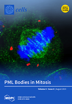
The figure shows promyelocytic leukemia (PML) bodies at different stages of the cell cycle. In interphase cells, nuclear PML forms spherical nuclear structures which recruit and release various nuclear proteins. In mitosis, these structures aggregate to form multispherical complexes. The mitotic PML bodies end up in the cytoplasm of newly divided G1 cells, where they immediately become covered with nuclear import factors. In this volume of Cells, Lång et al. review biochemical and biophysical transitions of PML bodies during cell cycle progression, and they discuss this behavior in light of putative PML body cellular functions.  The confocal images in the cover illustration show fixed HaCaT cells with immunofluorescently labeled PML (red), importin β (green), and DAPI (blue). View this paper
  • Issues are regarded as officially published after their release is announced to the table of contents alert mailing list.
  • You may sign up for e-mail alerts to receive table of contents of newly released issues.
  • PDF is the official format for papers published in both, html and pdf forms. To view the papers in pdf format, click on the "PDF Full-text" link, and use the free Adobe Reader to open them.
Order results
Result details
Section
Select all
Export citation of selected articles as:
16 pages, 2290 KiB  
Article
Neutral Lipids Are Not a Source of Arachidonic Acid for Lipid Mediator Signaling in Human Foamy Monocytes
by Carlos Guijas, Miguel A. Bermúdez, Clara Meana, Alma M. Astudillo, Laura Pereira, Lidia Fernández-Caballero, María A. Balboa and Jesús Balsinde
Cells 2019, 8(8), 941; https://doi.org/10.3390/cells8080941 - 20 Aug 2019
Cited by 13 | Viewed by 3850
Abstract
Human monocytes exposed to free arachidonic acid (AA), a secretory product of endothelial cells, acquire a foamy phenotype which is due to the accumulation of cytoplasmic lipid droplets with high AA content. Recruitment of foamy monocytes to the inflamed endothelium contributes to the [...] Read more.
Human monocytes exposed to free arachidonic acid (AA), a secretory product of endothelial cells, acquire a foamy phenotype which is due to the accumulation of cytoplasmic lipid droplets with high AA content. Recruitment of foamy monocytes to the inflamed endothelium contributes to the development of atherosclerotic lesions. In this work, we investigated the potential role of AA stored in the neutral lipids of foamy monocytes to be cleaved by lipases and contribute to lipid mediator signaling. To this end, we used mass spectrometry-based lipidomic approaches combined with strategies to generate monocytes with different concentrations of AA. Results from our experiments indicate that the phospholipid AA pool in monocytes is stable and does not change upon exposure of the cells to the external AA. On the contrary, the AA pool in triacylglycerol is expandable and can accommodate relatively large amounts of fatty acid. Stimulation of the cells with opsonized zymosan results in the expected decreases of cellular AA. Under all conditions examined, all of the AA decreases observed in stimulated cells were accounted for by decreases in the phospholipid pool; we failed to detect any contribution of the triacylglycerol pool to the response. Experiments utilizing selective inhibitors of phospholipid or triacylglyerol hydrolysis confirmed that the phospholipid pool is the sole contributor of the AA liberated by stimulated cells. Thus, the AA in the triacylglycerol is not a source of free AA for the lipid mediator signaling during stimulation of human foamy monocytes and may be used for other cellular functions. Full article
(This article belongs to the Special Issue Phospholipids: Dynamic Lipid Signaling in Health and Diseases)
Show Figures

Figure 1

16 pages, 2900 KiB  
Article
Loss of Protein Kinase Csnk2b/CK2β at Neuromuscular Junctions Affects Morphology and Dynamics of Aggregated Nicotinic Acetylcholine Receptors, Neuromuscular Transmission, and Synaptic Gene Expression
by Nane Eiber, Michael Rehman, Bojana Kravic, Rüdiger Rudolf, Marco Sandri and Said Hashemolhosseini
Cells 2019, 8(8), 940; https://doi.org/10.3390/cells8080940 - 20 Aug 2019
Cited by 12 | Viewed by 3511
Abstract
The protein kinase Csnk2/CK2 is important for cell proliferation, differentiation, and survival. Previously, we showed that CK2 binds distinctive proteins at neuromuscular junctions (NMJs) of mice and phosphorylates some of them. CK2 likely stabilizes clustered nicotinic acetylcholine receptors (AChRs). In the absence of [...] Read more.
The protein kinase Csnk2/CK2 is important for cell proliferation, differentiation, and survival. Previously, we showed that CK2 binds distinctive proteins at neuromuscular junctions (NMJs) of mice and phosphorylates some of them. CK2 likely stabilizes clustered nicotinic acetylcholine receptors (AChRs). In the absence of the β-subunit of CK2 in skeletal muscle fibers, mice develop an age-dependent decrease of grip strength accompanied by NMJ fragmentation and impairments of neuromuscular transmission. However, the precise role of CK2β regarding the clustering of AChRs and downstream signaling at NMJs is unknown. Here, we compared conditional CK2β-deficient mice with controls and found in the mutants (1) a lower decrement of endplate potentials after repetitive stimulation and decrements of nerve-evoked compound muscle action potentials decayed more rapidly after synaptic transmission was partially blocked, (2) that their muscle weakness was partially rescued by administration of an acetylcholine esterase inhibitor, (3) fragmented NMJs and impaired AChR clustering was detected in muscles and cultured muscle cells, (4) enlarged myonuclei, (5) impaired synaptic gene expression, and (6) a high turnover rate of their AChR clusters in vivo. Altogether, our data demonstrate a role for CK2 at the NMJ by maintaining a high density of AChRs and ensuring physiological synaptic gene expression. Full article
Show Figures

Figure 1

17 pages, 742 KiB  
Review
Transcriptional Regulation of Differentiation and Functions of Effector T Regulatory Cells
by Shin-ichi Koizumi and Hiroki Ishikawa
Cells 2019, 8(8), 939; https://doi.org/10.3390/cells8080939 - 20 Aug 2019
Cited by 42 | Viewed by 7234
Abstract
Foxp3-expressing regulatory T (Treg) cells can suppress the activity of various types of immune cells and play key roles in the maintenance of self-tolerance and in the regulation of immune responses against pathogens and tumor cells. Treg cells consist of heterogeneous subsets that [...] Read more.
Foxp3-expressing regulatory T (Treg) cells can suppress the activity of various types of immune cells and play key roles in the maintenance of self-tolerance and in the regulation of immune responses against pathogens and tumor cells. Treg cells consist of heterogeneous subsets that have distinct phenotypes and functions. Upon antigen stimulation, naïve-like thymus-derived Treg cells, which circulate in secondary lymphoid organs, can differentiate into effector Treg (eTreg) cells and migrate to and control immune homeostasis of peripheral tissues. eTreg cells are heterogeneous in terms of their ability to localize to specific tissues and suppress particular types of immune responses. Differentiation and function of diverse eTreg subsets are regulated by a variety of transcription factors that are activated by antigens and cytokines. In this article, we review the current understanding of the transcriptional regulation of differentiation and function of eTreg cells. Full article
(This article belongs to the Special Issue Regulatory T (Treg) Cells in Health and Diseases)
Show Figures

Figure 1

18 pages, 4005 KiB  
Article
DROSHA-Dependent AIM2 Inflammasome Activation Contributes to Lung Inflammation during Idiopathic Pulmonary Fibrosis
by Soo Jung Cho, Kyoung Sook Hong, Ji Hun Jeong, Mihye Lee, Augustine M. K. Choi, Heather W. Stout-Delgado and Jong-Seok Moon
Cells 2019, 8(8), 938; https://doi.org/10.3390/cells8080938 - 20 Aug 2019
Cited by 20 | Viewed by 4884
Abstract
Idiopathic pulmonary fibrosis (IPF) has been linked to chronic lung inflammation. Drosha ribonuclease III (DROSHA), a class 2 ribonuclease III enzyme, plays a key role in microRNA (miRNA) biogenesis. However, the mechanisms by which DROSHA affects the lung inflammation during idiopathic pulmonary fibrosis [...] Read more.
Idiopathic pulmonary fibrosis (IPF) has been linked to chronic lung inflammation. Drosha ribonuclease III (DROSHA), a class 2 ribonuclease III enzyme, plays a key role in microRNA (miRNA) biogenesis. However, the mechanisms by which DROSHA affects the lung inflammation during idiopathic pulmonary fibrosis (IPF) remain unclear. Here, we demonstrate that DROSHA regulates the absent in melanoma 2 (AIM2) inflammasome activation during idiopathic pulmonary fibrosis (IPF). Both DROSHA and AIM2 protein expression were elevated in alveolar macrophages of patients with IPF. We also found that DROSHA and AIM2 protein expression were increased in alveolar macrophages of lung tissues in a mouse model of bleomycin-induced pulmonary fibrosis. DROSHA deficiency suppressed AIM2 inflammasome-dependent caspase-1 activation and interleukin (IL)-1β and IL-18 secretion in primary mouse alveolar macrophages and bone marrow-derived macrophages (BMDMs). Transduction of microRNA (miRNA) increased the formation of the adaptor apoptosis-associated speck-like protein containing a caspase recruitment domain (ASC) specks, which is required for AIM2 inflammasome activation in BMDMs. Our results suggest that DROSHA promotes AIM2 inflammasome activation-dependent lung inflammation during IPF. Full article
(This article belongs to the Special Issue Mechanisms of Inflammasome Activation)
Show Figures

Graphical abstract

16 pages, 2227 KiB  
Article
Leukocyte-Derived Extracellular Vesicles in Blood with and without EpCAM Enrichment
by Afroditi Nanou, Leonie L. Zeune and Leon W.M.M. Terstappen
Cells 2019, 8(8), 937; https://doi.org/10.3390/cells8080937 - 20 Aug 2019
Cited by 11 | Viewed by 4848
Abstract
Large tumor-derived Extracellular Vesicles (tdEVs) detected in blood of metastatic prostate, breast, colorectal, and non-small cell lung cancer patients after enrichment for Epithelial Cell Adhesion Molecule (EpCAM) expression and labeling with 4′,6-diamidino-2-phenylindole (DAPI), phycoerythrin-conjugated antibodies against Cytokeratins (CK-PE), and allophycocyanin-conjugated antibody against the [...] Read more.
Large tumor-derived Extracellular Vesicles (tdEVs) detected in blood of metastatic prostate, breast, colorectal, and non-small cell lung cancer patients after enrichment for Epithelial Cell Adhesion Molecule (EpCAM) expression and labeling with 4′,6-diamidino-2-phenylindole (DAPI), phycoerythrin-conjugated antibodies against Cytokeratins (CK-PE), and allophycocyanin-conjugated antibody against the cluster of differentiation 45 (CD45-APC), are negatively associated with the overall survival of patients. Here, we investigated whether, similarly to tdEVs, leukocyte-derived EVs (ldEVs) could also be detected in EpCAM-enriched blood. Presence of ldEVs and leukocytes in image data sets of EpCAM-enriched samples of 25 healthy individuals and 75 metastatic cancer patients was evaluated using the ACCEPT software. Large ldEVs could indeed be detected, but in contrast to the 20-fold higher frequency of tdEVs as compared to Circulating Tumor Cells (CTCs), ldEVs were present in a 5-fold lower frequency as compared to leukocytes. To evaluate whether these ldEVs pre-exist in the blood or are formed during the CellSearch procedure, the blood of healthy individuals without EpCAM enrichment was labelled with the nuclear dye Hoechst and fluorescently tagged monoclonal antibodies recognizing the leukocyte-specific CD45, platelet-specific CD61, and red blood cell-specific CD235a. Fluorescence microscopy imaging using a similar setup as the CellSearch was performed and demonstrated the presence of a similar population of ldEVs present at a 3-fold lower frequency as compared to leukocytes. Full article
Show Figures

Figure 1

17 pages, 4562 KiB  
Article
Physioxia Has a Beneficial Effect on Cartilage Matrix Production in Interleukin-1 Beta-Inhibited Mesenchymal Stem Cell Chondrogenesis
by Girish Pattappa, Ruth Schewior, Isabelle Hofmeister, Jennifer Seja, Johannes Zellner, Brian Johnstone, Denitsa Docheva and Peter Angele
Cells 2019, 8(8), 936; https://doi.org/10.3390/cells8080936 - 20 Aug 2019
Cited by 30 | Viewed by 4104
Abstract
Osteoarthritis (OA) is a degenerative condition that involves the production of inflammatory cytokines (e.g., interleukin-1β (IL-1β), tumour necrosis factor-alpha (TNF-α) and interleukin-6 (IL-6)) that stimulate degradative enzymes, matrix metalloproteinases (MMPs) and aggrecanases (ADAMTS) resulting in articular cartilage breakdown. The presence of interleukin-1β (IL-1β) [...] Read more.
Osteoarthritis (OA) is a degenerative condition that involves the production of inflammatory cytokines (e.g., interleukin-1β (IL-1β), tumour necrosis factor-alpha (TNF-α) and interleukin-6 (IL-6)) that stimulate degradative enzymes, matrix metalloproteinases (MMPs) and aggrecanases (ADAMTS) resulting in articular cartilage breakdown. The presence of interleukin-1β (IL-1β) is one reason for poor clinical outcomes in current cell-based tissue engineering strategies for treating focal early osteoarthritic defects. Mesenchymal stem cells (MSCs) are a potential cell source for articular cartilage regeneration, although IL-1β has been shown to inhibit in vitro chondrogenesis. In vivo, articular chondrocytes reside under a low oxygen environment between 2–5% oxygen (physioxia) and have been shown to enhance in vitro MSC chondrogenic matrix content with reduced hypertrophic marker expression under these conditions. The present investigation sought to understand the effect of physioxia on IL-1β inhibited MSC chondrogenesis. MSCs expanded under physioxic (2% oxygen) and hyperoxic (20%) conditions, then chondrogenically differentiated as pellets in the presence of TGF-β1 and either 0.1 or 0.5 ng/mL IL-1β. Results showed that there were donor variations in response to physioxic culture based on intrinsic GAG content under hyperoxia. In physioxia responsive donors, MSC chondrogenesis significantly increased GAG and collagen II content, whilst hypertrophic markers were reduced compared with hyperoxia. In the presence of IL-1β, these donors showed a significant increase in cartilage matrix gene expression and GAG content relative to hyperoxic conditions. In contrast, a set of MSC donors were unresponsive to physioxia and showed no significant increase in matrix production independent of IL-1β presence. Thus, physioxia has a beneficial effect on MSC cartilage matrix production in responsive donors with or without IL-1β application. The mechanisms controlling the MSC chondrogenic response in both physioxia responsive and unresponsive donors are to be elucidated in future investigations. Full article
(This article belongs to the Special Issue Pathways Contributing to Cartilage and Bone Destruction in Arthritis)
Show Figures

Figure 1

17 pages, 8750 KiB  
Article
MicroRNA-21 Mediates the Protective Effect of Cardiomyocyte-Derived Conditioned Medium on Ameliorating Myocardial Infarction in Rats
by Chih-Hung Chen, Shu-Yuan Hsu, Chien-Chih Chiu and Steve Leu
Cells 2019, 8(8), 935; https://doi.org/10.3390/cells8080935 - 19 Aug 2019
Cited by 34 | Viewed by 4263
Abstract
Conditioned medium derived from ischemic myocardium improves rodent cardiac function after myocardial infarction. Exosomal miRNA-mediated intercellular communication is considered to mediate the protective effect of conditioned medium against ischemic injury. Oxygen–glucose-deprivation (OGD)-treated cardiac cells and a rat model with acute myocardial infarction (AMI) [...] Read more.
Conditioned medium derived from ischemic myocardium improves rodent cardiac function after myocardial infarction. Exosomal miRNA-mediated intercellular communication is considered to mediate the protective effect of conditioned medium against ischemic injury. Oxygen–glucose-deprivation (OGD)-treated cardiac cells and a rat model with acute myocardial infarction (AMI) were applied. The expression profiles of myocardial-disease-associated miRNAs in cardiomyocytes, cardiac fibroblasts, ventricular myocardium, and conditioned medium derived from cardiomyocytes under ischemic stresses were analyzed. Primary cultured cell model and a rat model with myocardial infarction were applied to examine the role of miRNA in regulating cardiomyocyte apoptosis, fibroblast activation, immune cell infiltration, and myocardial infarction. Results showed that expression levels of miR-21 in cardiomyocytes, cardiac fibroblasts, and conditioned medium (CM) derived from cardiomyocytes were up-regulated with OGD treatment. With the depletion of miR-21, the protective effect of CM on cardiomyocytes against oxidative stress, enhanced fibroblast activation, and promotion of angiogenesis in endothelial cells were reduced. Administration of CM reduced the infarcted size and immune cell infiltration in myocardium of rats with AMI, while depletion of miR-21 reduced the effect of CM. In conclusion, miR-21 plays a role in intercellular communication among ischemic cardiac cells. The expression of miR-21 is important for the protective effect of conditioned medium against myocardial infarction. Full article
Show Figures

Figure 1

15 pages, 4260 KiB  
Article
Tissue Specific Differentiation of Human Chondrocytes Depends on Cell Microenvironment and Serum Selection
by Annemarie Ecke, Anne-Helen Lutter, Jenny Scholka, Anna Hansch, Roland Becker and Ursula Anderer
Cells 2019, 8(8), 934; https://doi.org/10.3390/cells8080934 - 19 Aug 2019
Cited by 17 | Viewed by 4741
Abstract
Therapeutic options to cure osteoarthritis (OA) are not yet available, although cell-based therapies for the treatment of traumatic defects of cartilage have already been developed using, e.g., articular chondrocytes. In order to adapt cell-based therapies to treat OA, appropriate cell culture conditions are [...] Read more.
Therapeutic options to cure osteoarthritis (OA) are not yet available, although cell-based therapies for the treatment of traumatic defects of cartilage have already been developed using, e.g., articular chondrocytes. In order to adapt cell-based therapies to treat OA, appropriate cell culture conditions are necessary. Chondrocytes require a 3-dimensional (3D) environment for redifferentiation after 2-dimensional (2D) expansion. Fetal bovine serum (FBS) is commonly used as a medium supplement, although the usage of a xenogeneic serum could mask the intrinsic behavior of human cells in vitro. The aim of this study was to compare human articular chondrocytes cultivated as monolayers (2D) and the development of microtissues (3D) in the presence of FBS with those cultivated with human serum (HS). Evaluation of the expression of various markers via immunocytochemistry on monolayer cells revealed a higher dedifferentiation degree of chondrocytes cultivated with HS. Scaffold-free microtissues were generated using the agar overlay technique, and their differentiation level was evaluated via histochemistry and immunohistochemistry. Microtissues cultivated in the medium with FBS showed a higher redifferentiation level. This was evidenced by bigger microtissues and a more cartilage-like composition of the matrix with not any/less positivity for cartilage-specific markers in HS versus moderate-to-high positivity in FBS-cultured microtissues. The present study showed that the differentiation degree of chondrocytes depends both on the microenvironment of the cells and the serum type with FBS achieving the best results. However, HS should be preferred for the engineering of cartilage-like microtissues, as it rather enables a "human-based" situation in vitro. Hence, cultivation conditions might be further optimized to gain an even more adequate and donor-independent redifferentiation of chondrocytes in microtissues, e.g., designing a suitable chemically-defined serum supplement. Full article
Show Figures

Figure 1

14 pages, 7868 KiB  
Article
Estrogen and EGFR Pathways Regulate Notch Signaling in Opposing Directions for Multi-Ciliogenesis in the Fallopian Tube
by Maobi Zhu, Tomohiko Iwano and Sen Takeda
Cells 2019, 8(8), 933; https://doi.org/10.3390/cells8080933 - 19 Aug 2019
Cited by 19 | Viewed by 4456
Abstract
The lumen of the fallopian tube (FT) is lined with columnar epithelium composed of secretory and ciliated cells, both of which are important for reproduction. However, the molecular mechanism regulating cell fate remains controversial. In this study, we established a primary culture system [...] Read more.
The lumen of the fallopian tube (FT) is lined with columnar epithelium composed of secretory and ciliated cells, both of which are important for reproduction. However, the molecular mechanism regulating cell fate remains controversial. In this study, we established a primary culture system using porcine fallopian tube epithelial cells (FTECs) to study the differentiation mechanism. We found that estrogen promoted the differentiation of multi-ciliated cells (MCCs) through estrogen receptor β, following the reduction of DLL1, a ligand of Notch. Meanwhile, epidermal growth factor (EGF), a regulator of epithelial homeostasis and differentiation, suppressed ciliogenesis by the activation of Notch signaling. However, the estrogen pathway did not affect the activation of the EGF pathway. Taken together, the differentiation of MMCs in FT depends on the balance of EGF and estrogen signaling, either of which inhibits or stimulates the Notch signaling pathway respectively. Full article
(This article belongs to the Collection Cilia and Flagella: Structure, Function and Beyond)
Show Figures

Figure 1

17 pages, 2432 KiB  
Article
Extracellular Juxtamembrane Motif Critical for TrkB Preformed Dimer and Activation
by Jianying Shen, Dang Sun, Jingyu Shao, Yanbo Chen, Keliang Pang, Wei Guo and Bai Lu
Cells 2019, 8(8), 932; https://doi.org/10.3390/cells8080932 - 19 Aug 2019
Cited by 10 | Viewed by 4553
Abstract
Receptor tyrosine kinases are believed to be activated through ligand-induced dimerization. We now demonstrate that in cultured neurons, a substantial amount of endogenous TrkB, the receptor for brain-derived neurotrophic factor (BDNF), exists as an inactive preformed dimer, and the application of BDNF activates [...] Read more.
Receptor tyrosine kinases are believed to be activated through ligand-induced dimerization. We now demonstrate that in cultured neurons, a substantial amount of endogenous TrkB, the receptor for brain-derived neurotrophic factor (BDNF), exists as an inactive preformed dimer, and the application of BDNF activates the pre-existing dimer. Deletion of the extracellular juxtamembrane motif (EJM) of TrkB increased the amount of preformed dimer, suggesting an inhibitory role of EJM on dimer formation. Further, binding of an agonistic antibody (MM12) specific to human TrkB-EJM activated the full-length TrkB and unexpectedly also truncated TrkB lacking ECD (TrkBdelECD365), suggesting that TrkB is activated by attenuating the inhibitory effect of EJM through MM12 binding-induced conformational changes. Finally, in cells co-expressing rat and human TrkB, MM12 could only activate TrkB human-human dimer but not TrkB human-rat TrkB dimer, indicating that MM12 binding to two TrkB monomers is required for activation. Our results support a model that TrkB preforms as an inactive dimer and BDNF induces TrkB conformation changes leading to its activation. Full article
(This article belongs to the Section Cell Signaling)
Show Figures

Figure 1

22 pages, 7309 KiB  
Article
Internalization Characterization of Si Nanorod with Camouflaged Cell Membrane Proteins Reveals ATXN2 as a Negative Regulator
by Yi Lu, Jing Dai, Na Kong, Jianghuai Liu, Jinkang Gong and Yuan Yao
Cells 2019, 8(8), 931; https://doi.org/10.3390/cells8080931 - 19 Aug 2019
Cited by 6 | Viewed by 4261
Abstract
The fabrication of shape-controlled nanocarriers is critical for efficient delivery of biomolecules across the cell membrane. Surface coating of the nanocarrier can improve internalization efficiency. Here, we developed a facile method of silicon nanorod fabrication leading to a controlled size and shape. We [...] Read more.
The fabrication of shape-controlled nanocarriers is critical for efficient delivery of biomolecules across the cell membrane. Surface coating of the nanocarrier can improve internalization efficiency. Here, we developed a facile method of silicon nanorod fabrication leading to a controlled size and shape. We then systematically evaluated five surface modifications with membrane proteins from different cancer cell lines including MCF7, MD231, Hela, Panc-PDX, and Panc-1. We demonstrated that silicon nanorods coated with either a homolytic or heterolytic membrane protein coating have significantly improved internalization efficiency as compared with uncoated Si nanorods. To elucidate the molecular mechanism of the improved efficiency associated with a modified coating, we analyzed the coating membrane proteins derived from five cell lines with proteomics and identified 601 proteins shared by different cell sources. These proteins may function as cell-substrate adhesion molecules that contribute to the enhanced internalization. We also tested the internalization efficiency of nanorods with different coatings in each of the five cell lines to determine the influencing factors from target cells. We found that the internalization efficiency varied among different target cells, and the ranking of the average efficiency was as follows: Hela > Panc-PDX > MD231 > MCF7 > Panc-1. The bioinformatics analysis suggested that the low internalization efficiency in Panc-1 cells might be associated with the upregulation of ATXN2, which is a negative regulator of endocytosis. We further demonstrated that ATXN2 knockdown with specific siRNA significantly improved nanorod internalization efficiency in Panc-1 cells suggesting that ATXN2 can be a reference for efficiency prediction of nanoparticle delivery to tumor cells. Thus, we studied the effect of different cancer cell membrane proteins on nanorod uptake efficiencies. These results can improve nanorod internalization to cancer cells, including a fundamental understanding of the internalization efficiency of cancer cells. Full article
Show Figures

Graphical abstract

18 pages, 3009 KiB  
Article
EGFR–c-Src-Mediated HDAC3 Phosphorylation Exacerbates Invasion of Breast Cancer Cells
by Sung-Min Kwak, Jaesung Seo, Jin-Taek Hwang, Gi-Jun Sung, Ji-Hye Song, Ji-Hoon Jeong, Seung-Hyun Lee, Ho-Geun Yoon, Hyo-Kyoung Choi and Kyung-Chul Choi
Cells 2019, 8(8), 930; https://doi.org/10.3390/cells8080930 - 19 Aug 2019
Cited by 17 | Viewed by 4758
Abstract
Breast cancer is one of the leading causes of morbidity and mortality among women. Epidermal growth factor receptor (EGFR) and proto-oncogene tyrosine-protein kinase Src (c-Src) are critical components of the signaling pathways that are associated with breast cancer. However, the regulatory mechanism of [...] Read more.
Breast cancer is one of the leading causes of morbidity and mortality among women. Epidermal growth factor receptor (EGFR) and proto-oncogene tyrosine-protein kinase Src (c-Src) are critical components of the signaling pathways that are associated with breast cancer. However, the regulatory mechanism of histone deacetylase 3 (HDAC3) in these pathways remains unclear. Using the Net Phos 3.1 program for the analysis of kinase consensus motifs, we found two c-Src-mediated putative phosphorylation sites, tyrosine (Tyr, Y)-328 and Y331 on HDAC3, and generated a phospho-specific HDAC3 antibody against these sites. c-Src-mediated phosphorylation was observed in the cells expressing wild-type HDAC3 (HDAC3WT), but not in cells overexpressing phosphorylation-defective HDAC3 (HDAC3Y328/331A). Phosphorylated HDAC3 showed relatively higher deacetylase activity, and PP2, which is a c-Src inhibitor, blocked HDAC3 phosphorylation and reduced its enzymatic activity. EGF treatment resulted in HDAC3 phosphorylation in both MDA-MB-231 and EGFR-overexpressing MCF7 (MCF7-EGFR) cells, but not in MCF7 cells. Total internal reflection fluorescence analysis showed that HDAC3 was recruited to the plasma membrane following EGF stimulation. HDAC3 inhibition with either c-Src knockdown or PP2 treatment significantly ameliorated the invasiveness of breast cancer cells. Altogether, our findings reveal an EGF signaling cascade involving EGFR, c-Src, and HDAC3 in breast cancer cells. Full article
Show Figures

Graphical abstract

23 pages, 1554 KiB  
Review
The Intricate Interplay between Epigenetic Events, Alternative Splicing and Noncoding RNA Deregulation in Colorectal Cancer
by Raheleh Amirkhah, Hojjat Naderi-Meshkin, Jaynish S. Shah, Philip D. Dunne and Ulf Schmitz
Cells 2019, 8(8), 929; https://doi.org/10.3390/cells8080929 - 19 Aug 2019
Cited by 32 | Viewed by 7831
Abstract
Colorectal cancer (CRC) results from a transformation of colonic epithelial cells into adenocarcinoma cells due to genetic and epigenetic instabilities, alongside remodelling of the surrounding stromal tumour microenvironment. Epithelial-specific epigenetic variations escorting this process include chromatin remodelling, histone modifications and aberrant DNA methylation, [...] Read more.
Colorectal cancer (CRC) results from a transformation of colonic epithelial cells into adenocarcinoma cells due to genetic and epigenetic instabilities, alongside remodelling of the surrounding stromal tumour microenvironment. Epithelial-specific epigenetic variations escorting this process include chromatin remodelling, histone modifications and aberrant DNA methylation, which influence gene expression, alternative splicing and function of non-coding RNA. In this review, we first highlight epigenetic modulators, modifiers and mediators in CRC, then we elaborate on causes and consequences of epigenetic alterations in CRC pathogenesis alongside an appraisal of the complex feedback mechanisms realized through alternative splicing and non-coding RNA regulation. An emphasis in our review is put on how this intricate network of epigenetic and post-transcriptional gene regulation evolves during the initiation, progression and metastasis formation in CRC. Full article
Show Figures

Figure 1

20 pages, 5076 KiB  
Article
17β-Estradiol Modulates SIRT1 and Halts Oxidative Stress-Mediated Cognitive Impairment in a Male Aging Mouse Model
by Mehtab Khan, Rahat Ullah, Shafiq Ur Rehman, Shahid Ali Shah, Kamran Saeed, Tahir Muhammad, Hyun Young Park, Myeung Hoon Jo, Kyonghwan Choe, Bart P.F. Rutten and Myeong Ok Kim
Cells 2019, 8(8), 928; https://doi.org/10.3390/cells8080928 - 19 Aug 2019
Cited by 65 | Viewed by 6127
Abstract
Oxidative stress has been considered the main mediator in neurodegenerative disease and in normal aging processes. Several studies have reported that the accumulation of reactive oxygen species (ROS), elevated oxidative stress, and neuroinflammation result in cellular malfunction. These conditions lead to neuronal cell [...] Read more.
Oxidative stress has been considered the main mediator in neurodegenerative disease and in normal aging processes. Several studies have reported that the accumulation of reactive oxygen species (ROS), elevated oxidative stress, and neuroinflammation result in cellular malfunction. These conditions lead to neuronal cell death in aging-related neurodegenerative disorders such as Alzheimer’s disease (AD) and Parkinson’s disease. Chronic administration of d-galactose (d-gal) for a period of 10 weeks causes ROS generation and neuroinflammation, ultimately leading to cognitive impairment. In this study, we evaluated the estrogen receptor α (ERα)/silent mating type information regulation 2 homolog 1 (SIRT1)-dependent antioxidant efficacy of 17β-estradiol against d-gal-induced oxidative damage-mediated cognitive dysfunction in a male mouse model. The results indicate that 17β-estradiol, by stimulating ERα/SIRT1, halts d-gal-induced oxidative stress–mediated JNK/NF-ҡB overexpression, neuroinflammation and neuronal apoptosis. Moreover, 17β-estradiol ameliorated d-gal-induced AD-like pathophysiology, synaptic dysfunction and memory impairment in adult mouse brains. Interestingly, inhibition of SIRT1 with Ex527 (a potent and selective SIRT1 inhibitor) further enhanced d-gal-induced toxicity and abolished the beneficial effect of 17β-estradiol. Most importantly, for the first time, our molecular docking study reveals that 17β-estradiol allosterically increases the expression of SIRT1 and abolishes the inhibitory potential of d-ga. In summary, we can conclude that 17β-estradiol, in an ERα/SIRT1-dependent manner, abrogates d-gal-induced oxidative stress–mediated memory impairment, neuroinflammation, and neurodegeneration in adult mice. Full article
(This article belongs to the Special Issue Estrogen Receptor Expression and Function in Health and Disease)
Show Figures

Graphical abstract

18 pages, 655 KiB  
Review
Targeting the CD40-CD154 Signaling Pathway for Treatment of Autoimmune Arthritis
by Jenn-Haung Lai, Shue-Fen Luo and Ling-Jun Ho
Cells 2019, 8(8), 927; https://doi.org/10.3390/cells8080927 - 18 Aug 2019
Cited by 27 | Viewed by 10581
Abstract
Full activation of T lymphocytes requires signals from both T cell receptors and costimulatory molecules. In addition to CD28, several T cell molecules could deliver costimulatory signals, including CD154, which primarily interacts with CD40 on B-cells. CD40 is a critical molecule regulating several [...] Read more.
Full activation of T lymphocytes requires signals from both T cell receptors and costimulatory molecules. In addition to CD28, several T cell molecules could deliver costimulatory signals, including CD154, which primarily interacts with CD40 on B-cells. CD40 is a critical molecule regulating several B-cell functions, such as antibody production, germinal center formation and cellular proliferation. Upregulated expression of CD40 and CD154 occurs in immune effector cells and non-immune cells in different autoimmune diseases. In addition, therapeutic benefits have been observed by blocking the CD40-CD154 interaction in animals with collagen-induced arthritis. Given the therapeutic success of the biologics abatacept, which blocks CD28 costimulation, and rituximab, which deletes B cells in the treatment of autoimmune arthritis, the inhibition of the CD40-CD154 axis has two advantages, namely, attenuating CD154-mediated T cell costimulation and suppressing CD40-mediated B-cell stimulation. Furthermore, blockade of the CD40-CD154 interaction drives the conversion of CD4+ T cells to regulatory T cells that mediate immunosuppression. Currently, several biological products targeting the CD40-CD154 axis have been developed and are undergoing early phase clinical trials with encouraging success in several autoimmune disorders, including autoimmune arthritis. This review addresses the roles of the CD40-CD154 axis in the pathogenesis of autoimmune arthritis and its potential as a therapeutic target. Full article
(This article belongs to the Special Issue Pathways Contributing to Cartilage and Bone Destruction in Arthritis)
Show Figures

Graphical abstract

19 pages, 927 KiB  
Review
Cancer Stem Cells and Targeting Strategies
by Luisa Barbato, Marco Bocchetti, Anna Di Biase and Tarik Regad
Cells 2019, 8(8), 926; https://doi.org/10.3390/cells8080926 - 18 Aug 2019
Cited by 125 | Viewed by 7859
Abstract
Chemoresistance is a major problem in cancer therapy as cancer cells develop mechanisms that counteract the effect of chemotherapeutic compounds, leading to relapse and the development of more aggressive cancers that contribute to poor prognosis and survival rates of treated patients. Cancer stem [...] Read more.
Chemoresistance is a major problem in cancer therapy as cancer cells develop mechanisms that counteract the effect of chemotherapeutic compounds, leading to relapse and the development of more aggressive cancers that contribute to poor prognosis and survival rates of treated patients. Cancer stem cells (CSCs) play a key role in this event. Apart from their slow proliferative property, CSCs have developed a range of cellular processes that involve drug efflux, drug enzymatic inactivation and other mechanisms. In addition, the microenvironment where CSCs evolve (CSC niche), effectively contributes to their role in cancer initiation, progression and chemoresistance. In the CSC niche, immune cells, mesenchymal stem cells (MSCs), endothelial cells and cancer associated fibroblasts (CAFs) contribute to the maintenance of CSC malignancy via the secretion of factors that promote cancer progression and resistance to chemotherapy. Due to these factors that hinder successful cancer therapies, CSCs are a subject of intense research that aims at better understanding of CSC behaviour and at developing efficient targeting therapies. In this review, we provide an overview of cancer stem cells, their role in cancer initiation, progression and chemoresistance, and discuss the progress that has been made in the development of CSC targeted therapies. Full article
(This article belongs to the Special Issue CSCs Identification and Targeting Therapies: Advances and Challenges)
Show Figures

Graphical abstract

28 pages, 5805 KiB  
Review
The uPAR System as a Potential Therapeutic Target in the Diseased Eye
by Maurizio Cammalleri, Massimo Dal Monte, Vincenzo Pavone, Mario De Rosa, Dario Rusciano and Paola Bagnoli
Cells 2019, 8(8), 925; https://doi.org/10.3390/cells8080925 - 18 Aug 2019
Cited by 10 | Viewed by 5146
Abstract
Dysregulation of vascular networks is characteristic of eye diseases associated with retinal cell degeneration and visual loss. Visual impairment is also the consequence of photoreceptor degeneration in inherited eye diseases with a major inflammatory component, but without angiogenic profile. Among the pathways with [...] Read more.
Dysregulation of vascular networks is characteristic of eye diseases associated with retinal cell degeneration and visual loss. Visual impairment is also the consequence of photoreceptor degeneration in inherited eye diseases with a major inflammatory component, but without angiogenic profile. Among the pathways with high impact on vascular/degenerative diseases of the eye, a central role is played by a system formed by the ligand urokinase-type plasminogen activator (uPA) and its receptor uPAR. The uPAR system, although extensively investigated in tumors, still remains a key issue in vascular diseases of the eye and even less studied in inherited retinal pathologies such as retinitis pigmantosa (RP). Its spectrum of action has been extended far beyond a classical pro-angiogenic function and has emerged as a central actor in inflammation. Preclinical studies in more prevalent eye diseases characterized by neovascular formation, as in retinopathy of prematurity, wet macular degeneration and rubeosis iridis or vasopermeability excess as in diabetic retinopathy, suggest a critical role of increased uPAR signaling indicating the potentiality of its modulation to counteract neovessel formation and microvascular dysfunction. The additional observation that the uPAR system plays a major role in RP by limiting the inflammatory cascade triggered by rod degeneration rises further questions about its role in the diseased eye. Full article
Show Figures

Figure 1

12 pages, 3329 KiB  
Article
mTOR-Dependent Stimulation of IL20RA Orchestrates Immune Cell Trafficking through Lymphatic Endothelium in Patients with Crohn’s Disease
by Federica Ungaro, Valentina Garlatti, Luca Massimino, Antonino Spinelli, Michele Carvello, Matteo Sacchi, Salvatore Spanò, Gaia Colasante, Nicholas Valassina, Stefania Vetrano, Alberto Malesci, Laurent Peyrin-Biroulet, Silvio Danese and Silvia D’Alessio
Cells 2019, 8(8), 924; https://doi.org/10.3390/cells8080924 - 18 Aug 2019
Cited by 11 | Viewed by 4144
Abstract
Crohn’s disease (CD) is a chronic inflammatory condition that can affect different portions of the gastrointestinal tract. Lymphatic drainage was demonstrated to be dysfunctional in CD pathogenesis, ultimately causing the failure of the resolution of intestinal inflammation. To investigate the molecular mechanisms underlying [...] Read more.
Crohn’s disease (CD) is a chronic inflammatory condition that can affect different portions of the gastrointestinal tract. Lymphatic drainage was demonstrated to be dysfunctional in CD pathogenesis, ultimately causing the failure of the resolution of intestinal inflammation. To investigate the molecular mechanisms underlying these dysfunctions, we isolated human intestinal lymphatic endothelial cells (HILECs) from surgical specimens of patients undergoing resection for complicated CD (CD HILEC) and from a disease-free margin of surgical specimens of patients undergoing resection for cancer (healthy HILEC). Both cell types underwent transcriptomic profiling, and their barrier functionality was tested using a transwell-based co-culture system between HILEC and lamina propria mononuclear cells (LPMCs). Results showed CD HILEC displayed a peculiar transcriptomic signature that highlighted mTOR signaling as an orchestrator of leukocyte trafficking through the lymphatic barrier of CD patients. Moreover, we demonstrated that LPMC transmigration through the lymphatic endothelium of patients with CD depends on the capability of mTOR to trigger interleukin 20 receptor subunit α (IL20RA)-mediated intracellular signaling. Conclusively, our study suggests that leukocyte trafficking through the intestinal lymphatic microvasculature can be controlled by modulating IL20RA, thus leading to the resolution of chronic inflammation in patients with CD. Full article
Show Figures

Figure 1

19 pages, 4027 KiB  
Article
miR-263b Controls Circadian Behavior and the Structural Plasticity of Pacemaker Neurons by Regulating the LIM-Only Protein Beadex
by Xiaoge Nian, Wenfeng Chen, Weiwei Bai, Zhangwu Zhao and Yong Zhang
Cells 2019, 8(8), 923; https://doi.org/10.3390/cells8080923 - 18 Aug 2019
Cited by 13 | Viewed by 3957
Abstract
Circadian clocks drive rhythmic physiology and behavior to allow adaption to daily environmental changes. In Drosophila, the small ventral lateral neurons (sLNvs) are primary pacemakers that control circadian rhythms. Circadian changes are observed in the dorsal axonal projections of the sLNvs, but [...] Read more.
Circadian clocks drive rhythmic physiology and behavior to allow adaption to daily environmental changes. In Drosophila, the small ventral lateral neurons (sLNvs) are primary pacemakers that control circadian rhythms. Circadian changes are observed in the dorsal axonal projections of the sLNvs, but their physiological importance and the underlying mechanism are unclear. Here, we identified miR-263b as an important regulator of circadian rhythms and structural plasticity of sLNvs in Drosophila. Depletion of miR-263b (miR-263bKO) in flies dramatically impaired locomotor rhythms under constant darkness. Indeed, miR-263b is required for the structural plasticity of sLNvs. miR-263b regulates circadian rhythms through inhibition of expression of the LIM-only protein Beadex (Bx). Consistently, overexpression of Bx or loss-of-function mutation (BxhdpR26) phenocopied miR-263bKO and miR-263b overexpression in behavior and molecular characteristics. In addition, mutating the miR-263b binding sites in the Bx 3′ UTR using CRISPR/Cas9 recapitulated the circadian phenotypes of miR-263bKO flies. Together, these results establish miR-263b as an important regulator of circadian locomotor behavior and structural plasticity. Full article
(This article belongs to the Collection Regulatory Functions of microRNAs)
Show Figures

Figure 1

35 pages, 1494 KiB  
Review
Interplay between Intrinsic and Innate Immunity during HIV Infection
by Louis Bergantz, Frédéric Subra, Eric Deprez, Olivier Delelis and Clémence Richetta
Cells 2019, 8(8), 922; https://doi.org/10.3390/cells8080922 - 17 Aug 2019
Cited by 23 | Viewed by 7524
Abstract
Restriction factors are antiviral components of intrinsic immunity which constitute a first line of defense by blocking different steps of the human immunodeficiency virus (HIV) replication cycle. In immune cells, HIV infection is also sensed by several pattern recognition receptors (PRRs), leading to [...] Read more.
Restriction factors are antiviral components of intrinsic immunity which constitute a first line of defense by blocking different steps of the human immunodeficiency virus (HIV) replication cycle. In immune cells, HIV infection is also sensed by several pattern recognition receptors (PRRs), leading to type I interferon (IFN-I) and inflammatory cytokines production that upregulate antiviral interferon-stimulated genes (ISGs). Several studies suggest a link between these two types of immunity. Indeed, restriction factors, that are generally interferon-inducible, are able to modulate immune responses. This review highlights recent knowledge of the interplay between restriction factors and immunity inducing antiviral defenses. Counteraction of this intrinsic and innate immunity by HIV viral proteins will also be discussed. Full article
(This article belongs to the Special Issue HIV and Host Interaction)
Show Figures

Figure 1

15 pages, 1595 KiB  
Article
Genes Controlled by DNA Methylation Are Involved in Wilms Tumor Progression
by João Victor da Silva Guerra, Bruna Maria de Sá Pereira, Jéssica Gonçalves Vieira da Cruz, Nicole de Miranda Scherer, Carolina Furtado, Rafaela Montalvão de Azevedo, Paulo Sergio Lopes de Oliveira, Paulo Faria, Mariana Boroni, Beatriz de Camargo and Mariana Maschietto
Cells 2019, 8(8), 921; https://doi.org/10.3390/cells8080921 - 17 Aug 2019
Cited by 12 | Viewed by 5825
Abstract
To identify underlying mechanisms involved with metastasis formation in Wilms tumors (WTs), we performed comprehensive DNA methylation and gene expression analyses of matched normal kidney (NK), WT blastemal component, and metastatic tissues (MT) from patients treated under SIOP 2001 protocol. A linear Bayesian [...] Read more.
To identify underlying mechanisms involved with metastasis formation in Wilms tumors (WTs), we performed comprehensive DNA methylation and gene expression analyses of matched normal kidney (NK), WT blastemal component, and metastatic tissues (MT) from patients treated under SIOP 2001 protocol. A linear Bayesian framework model identified 497 differentially methylated positions (DMPs) between groups that discriminated NK from WT, but MT samples were divided in two groups. Accordingly, methylation variance grouped NK and three MT samples tightly together and all WT with four MT samples that showed high variability. WT were hypomethylated compared to NK, and MT had a hypermethylated pattern compared to both groups. The methylation patterns were in agreement with methylases and demethylases expression. Methylation data pointed to the existence of two groups of metastases. While hierarchical clustering analysis based on the expression of all 2569 differentially expressed genes (DEGs) discriminated WT and MT from all NK samples, the hierarchical clustering based on the expression of 44 genes with a differentially methylated region (DMR) located in their promoter region revealed two groups: one containing all NKs and three MTs and one containing all WT and four MTs. Methylation changes might be controlling expression of genes associated with WT progression. The 44 genes are candidates to be further explored as a signature for metastasis formation in WT. Full article
(This article belongs to the Special Issue Plasticity in Cancer and in Microenvironmental Cells)
Show Figures

Graphical abstract

15 pages, 1476 KiB  
Article
The Snapdragon LATE ELONGATED HYPOCOTYL Plays A Dual Role in Activating Floral Growth and Scent Emission
by Marta I. Terry, Fernando Pérez-Sanz, Pedro J. Navarro, Julia Weiss and Marcos Egea-Cortines
Cells 2019, 8(8), 920; https://doi.org/10.3390/cells8080920 - 17 Aug 2019
Cited by 12 | Viewed by 4311
Abstract
The plant circadian clock controls a large number of internal processes, including growth and metabolism. Scent emission displays a circadian pattern in many species such as the snapdragon. Here we show that knocking down LATE ELONGATED HYPOCOTYL in Antirrhinum majus affects growth and [...] Read more.
The plant circadian clock controls a large number of internal processes, including growth and metabolism. Scent emission displays a circadian pattern in many species such as the snapdragon. Here we show that knocking down LATE ELONGATED HYPOCOTYL in Antirrhinum majus affects growth and scent emission. In order to gain an understanding of the growth kinetics, we took a phenomic approach using in-house artificial vision systems, obtaining time-lapse videos. Wild type flowers showed a higher growth speed than knockdown plants. The maximal growth rate was decreased by 22% in plants with lower LHY expression. Floral volatiles were differentially affected as RNAi plants showed advanced emission of compounds synthesized from cinnamic acid and delayed emission of metabolites of benzoic acid. The monoterpenes myrcene and ocimene were delayed, whereas the sesquiterpene farnesene was advanced. Overall, transgenic lines showed an altered volatile emission pattern and displayed a modified scent profile. Our results show that AmLHY plays an important role in the quantitative and qualitative control of floral growth and scent emission. Full article
Show Figures

Figure 1

18 pages, 6296 KiB  
Article
The Combination of IFN β and TNF Induces an Antiviral and Immunoregulatory Program via Non-Canonical Pathways Involving STAT2 and IRF9
by Mélissa K. Mariani, Pouria Dasmeh, Audray Fortin, Elise Caron, Mario Kalamujic, Alexander N. Harrison, Diana I. Hotea, Dacquin M. Kasumba, Sandra L. Cervantes-Ortiz, Espérance Mukawera, Adrian W. R. Serohijos and Nathalie Grandvaux
Cells 2019, 8(8), 919; https://doi.org/10.3390/cells8080919 - 17 Aug 2019
Cited by 11 | Viewed by 5438
Abstract
Interferon (IFN) β and Tumor Necrosis Factor (TNF) are key players in immunity against viruses. Compelling evidence has shown that the antiviral and inflammatory transcriptional response induced by IFNβ is reprogrammed by crosstalk with TNF. IFNβ mainly induces interferon-stimulated genes by the Janus [...] Read more.
Interferon (IFN) β and Tumor Necrosis Factor (TNF) are key players in immunity against viruses. Compelling evidence has shown that the antiviral and inflammatory transcriptional response induced by IFNβ is reprogrammed by crosstalk with TNF. IFNβ mainly induces interferon-stimulated genes by the Janus kinase (JAK)/signal transducer and activator of transcription (STAT) pathway involving the canonical ISGF3 transcriptional complex, composed of STAT1, STAT2, and IRF9. The signaling pathways engaged downstream of the combination of IFNβ and TNF remain elusive, but previous observations suggested the existence of a response independent of STAT1. Here, using genome-wide transcriptional analysis by RNASeq, we observed a broad antiviral and immunoregulatory response initiated in the absence of STAT1 upon IFNβ and TNF costimulation. Additional stratification of this transcriptional response revealed that STAT2 and IRF9 mediate the expression of a wide spectrum of genes. While a subset of genes was regulated by the concerted action of STAT2 and IRF9, other gene sets were independently regulated by STAT2 or IRF9. Collectively, our data supports a model in which STAT2 and IRF9 act through non-canonical parallel pathways to regulate distinct pool of antiviral and immunoregulatory genes in conditions with elevated levels of both IFNβ and TNF. Full article
(This article belongs to the Special Issue Cell Biology of Viral Infections)
Show Figures

Figure 1

10 pages, 2196 KiB  
Article
The Cisplatin-Derived Increase of Mitochondrial Reactive Oxygen Species Enhances the Effectiveness of Photodynamic Therapy via Transporter Regulation
by Hiromi Kurokawa, Hiromu Ito and Hirofumi Matsui
Cells 2019, 8(8), 918; https://doi.org/10.3390/cells8080918 - 17 Aug 2019
Cited by 12 | Viewed by 3580
Abstract
Photodynamic therapy (PDT) is a cancer treatment involving the generation of reactive oxygen species (ROS) by laser irradiation of porphyrins that accumulate in cancer tissues. 5-aminolevulinic acid (ALA), a porphyrin precursor, is often used as a photosensitizer. ALA is imported into cells via [...] Read more.
Photodynamic therapy (PDT) is a cancer treatment involving the generation of reactive oxygen species (ROS) by laser irradiation of porphyrins that accumulate in cancer tissues. 5-aminolevulinic acid (ALA), a porphyrin precursor, is often used as a photosensitizer. ALA is imported into cells via peptide transporter 1 (PEPT1), and porphyrin is exported via ATP-binding cassette member 2 of subfamily G (ABCG2). Thus, cancer cell-specific porphyrin accumulation involves regulation of both transporters to enhance the ALA-PDT effect. We reported previously that mitochondrial ROS (mitROS) upregulated PEPT1 expression and downregulated ABCG2 expression. Therefore, we propose that increasing mitROS production will enhance ALA-PDT cytotoxicity. Cisplatin is a chemotherapeutic drug that induces intracellular ROS generation. In this study, we investigated whether cisplatin-increased mitROS production in gastric cancer cell lines (RGK36 and RGK45) enhanced the cytotoxicity of ALA-PDT by regulation the expression of both PEPT1 and ABCG2. The results showed that cisplatin increased intracellular mitROS production in cancer but not normal cells (RGM1). PEPT1 was upregulated and ABCG2 downregulated in cancer cells treated with cisplatin. Moreover, intracellular porphyrin accumulation and ALA-PDT cytotoxicity increased. We conclude that cisplatin treatment increases the intracellular mitROS concentration and upregulates PEPT1 and downregulates ABCG2 expression. Full article
(This article belongs to the Section Cell Signaling)
Show Figures

Figure 1

14 pages, 6569 KiB  
Article
Biomimetic In Vitro Model of Cell Infiltration into Skin Scaffolds for Pre-Screening and Testing of Biomaterial-Based Therapies
by Rafael Ballesteros-Cillero, Evan Davison-Kotler, Nupur Kohli, William S. Marshall and Elena García-Gareta
Cells 2019, 8(8), 917; https://doi.org/10.3390/cells8080917 - 17 Aug 2019
Cited by 7 | Viewed by 5466
Abstract
Due to great clinical need, research where different biomaterials are tested as 3D scaffolds for skin tissue engineering has increased. In vitro studies use a cell suspension that is simply pipetted onto the material and cultured until the cells migrate and proliferate within [...] Read more.
Due to great clinical need, research where different biomaterials are tested as 3D scaffolds for skin tissue engineering has increased. In vitro studies use a cell suspension that is simply pipetted onto the material and cultured until the cells migrate and proliferate within the 3D scaffold, which does not mimic the in vivo reality. Our aim was to engineer a novel biomimetic in vitro model that mimics the natural cell infiltration process occurring in wound healing, thus offering a realistic approach when pre-screening and testing new skin substitutes. Our model consists of porous membrane cell culture inserts coated with gelatin and seeded with human dermal fibroblasts, inside which two different commercially available dermal substitutes were placed. Several features relevant to the wound healing process (matrix contraction, cell infiltration and proliferation, integration of the biomaterial with the surrounding tissue, and secretion of exogenous cytokines and growth factors) were evaluated. Our results showed that cells spontaneously infiltrate the materials and that our engineered model is able to induce and detect subtle differences between different biomaterials. The model allows for room for improvements or “adds-on” and miniaturization and can contribute to the development of functional and efficient skin substitutes for burns and chronic wounds. Full article
Show Figures

Figure 1

16 pages, 13333 KiB  
Article
Absence of Uncoupling Protein-3 at Thermoneutrality Impacts Lipid Handling and Energy Homeostasis in Mice
by Assunta Lombardi, Rosa Anna Busiello, Rita De Matteis, Lillà Lionetti, Sabrina Savarese, Maria Moreno, Alessandra Gentile, Elena Silvestri, Rosalba Senese, Pieter de Lange, Federica Cioffi, Antonia Lanni and Fernando Goglia
Cells 2019, 8(8), 916; https://doi.org/10.3390/cells8080916 - 17 Aug 2019
Cited by 7 | Viewed by 3398
Abstract
The role of uncoupling protein-3 (UCP3) in energy and lipid metabolism was investigated. Male wild-type (WT) and UCP3-null (KO) mice that were housed at thermoneutrality (30 °C) were used as the animal model. In KO mice, the ability of skeletal muscle mitochondria to [...] Read more.
The role of uncoupling protein-3 (UCP3) in energy and lipid metabolism was investigated. Male wild-type (WT) and UCP3-null (KO) mice that were housed at thermoneutrality (30 °C) were used as the animal model. In KO mice, the ability of skeletal muscle mitochondria to oxidize fatty acids (but not pyruvate or succinate) was reduced. At whole animal level, adult KO mice presented blunted resting metabolic rates, energy expenditure, food intake, and the use of lipids as metabolic substrates. When WT and KO mice were fed with a standard/low-fat diet for 80 days, since weaning, they showed similar weight gain and body composition. Interestingly, KO mice showed lower fat accumulation in visceral adipose tissue and higher ectopic fat accumulation in liver and skeletal muscle. When fed with a high-fat diet for 80 days, since weaning, KO mice showed enhanced energy efficiency and an increased lipid gain (thus leading to a change in body composition between the two genotypes). We conclude that UCP3 plays a role in energy and lipid homeostasis and in preserving lean tissues by lipotoxicity, in mice that were housed at thermoneutrality. Full article
(This article belongs to the Section Mitochondria)
Show Figures

Figure 1

17 pages, 1660 KiB  
Review
The NAE Pathway: Autobahn to the Nucleus for Cell Surface Receptors
by Poonam Shah, Alexandre Chaumet, Stephen J. Royle and Frederic A. Bard
Cells 2019, 8(8), 915; https://doi.org/10.3390/cells8080915 - 16 Aug 2019
Cited by 22 | Viewed by 6833
Abstract
Various growth factors and full-length cell surface receptors such as EGFR are translocated from the cell surface to the nucleoplasm, baffling cell biologists to the mechanisms and functions of this process. Elevated levels of nuclear EGFR correlate with poor prognosis in various cancers. [...] Read more.
Various growth factors and full-length cell surface receptors such as EGFR are translocated from the cell surface to the nucleoplasm, baffling cell biologists to the mechanisms and functions of this process. Elevated levels of nuclear EGFR correlate with poor prognosis in various cancers. In recent years, nuclear EGFR has been implicated in regulating gene transcription, cell proliferation and DNA damage repair. Different models have been proposed to explain how the receptors are transported into the nucleus. However, a clear consensus has yet to be reached. Recently, we described the nuclear envelope associated endosomes (NAE) pathway, which delivers EGFR from the cell surface to the nucleus. This pathway involves transport, docking and fusion of NAEs with the outer membrane of the nuclear envelope. EGFR is then presumed to be transported through the nuclear pore complex, extracted from membranes and solubilised. The SUN1/2 nuclear envelope proteins, Importin-beta, nuclear pore complex proteins and the Sec61 translocon have been implicated in the process. While this framework can explain the cell surface to nucleus traffic of EGFR and other cell surface receptors, it raises several questions that we consider in this review, together with implications for health and disease. Full article
(This article belongs to the Special Issue Membrane Traffic in Health and Disease)
Show Figures

Figure 1

19 pages, 1523 KiB  
Review
Functions and the Emerging Role of the Foetal Liver into Regenerative Medicine
by Antonella Giancotti, Marco Monti, Lorenzo Nevi, Samira Safarikia, Valentina D’Ambrosio, Roberto Brunelli, Cristina Pajno, Sara Corno, Violante Di Donato, Angela Musella, Michele Francesco Chiappetta, Daniela Bosco, Pierluigi Benedetti Panici, Domenico Alvaro and Vincenzo Cardinale
Cells 2019, 8(8), 914; https://doi.org/10.3390/cells8080914 - 16 Aug 2019
Cited by 26 | Viewed by 9945
Abstract
During foetal life, the liver plays the important roles of connection and transient hematopoietic function. Foetal liver cells develop in an environment called a hematopoietic stem cell niche composed of several cell types, where stem cells can proliferate and give rise to mature [...] Read more.
During foetal life, the liver plays the important roles of connection and transient hematopoietic function. Foetal liver cells develop in an environment called a hematopoietic stem cell niche composed of several cell types, where stem cells can proliferate and give rise to mature blood cells. Embryologically, at about the third week of gestation, the liver appears, and it grows rapidly from the fifth to 10th week under WNT/β-Catenin signaling pathway stimulation, which induces hepatic progenitor cells proliferation and differentiation into hepatocytes. Development of new strategies and identification of new cell sources should represent the main aim in liver regenerative medicine and cell therapy. Cells isolated from organs with endodermal origin, like the liver, bile ducts, and pancreas, could be preferable cell sources. Furthermore, stem cells isolated from these organs could be more susceptible to differentiate into mature liver cells after transplantation with respect to stem cells isolated from organs or tissues with a different embryological origin. The foetal liver possesses unique features given the co-existence of cells having endodermal and mesenchymal origin, and it could be highly available source candidate for regenerative medicine in both the liver and pancreas. Taking into account these advantages, the foetal liver can be the highest potential and available cell source for cell therapy regarding liver diseases and diabetes. Full article
(This article belongs to the Special Issue Hepatic Stem Cells in Liver and Biliary Regeneration)
Show Figures

Figure 1

15 pages, 736 KiB  
Review
IGF2/IGF1R Signaling as a Therapeutic Target in MYB-Positive Adenoid Cystic Carcinomas and Other Fusion Gene-Driven Tumors
by Mattias K. Andersson, Pierre Åman and Göran Stenman
Cells 2019, 8(8), 913; https://doi.org/10.3390/cells8080913 - 16 Aug 2019
Cited by 31 | Viewed by 5228
Abstract
Chromosome rearrangements resulting in pathogenetically important gene fusions are a common feature of many cancers. They are often potent oncogenic drivers and have key functions in central cellular processes and pathways and encode transcription factors, transcriptional co-regulators, growth factor receptors, tyrosine kinases, and [...] Read more.
Chromosome rearrangements resulting in pathogenetically important gene fusions are a common feature of many cancers. They are often potent oncogenic drivers and have key functions in central cellular processes and pathways and encode transcription factors, transcriptional co-regulators, growth factor receptors, tyrosine kinases, and chromatin modifiers. In addition to being useful diagnostic biomarkers, they are also targets for development of new molecularly targeted therapies. Studies in recent decades have shown that several oncogenic gene fusions interact with the insulin-like growth factor (IGF) signaling pathway. For example, the MYB–NFIB fusion in adenoid cystic carcinoma is regulated by IGF1R through an autocrine loop, and IGF1R is a downstream target of the EWSR1–WT1 and PAX3–FKHR fusions in desmoplastic small round cell tumors and alveolar rhabdomyosarcoma, respectively. Here, we will discuss the mechanisms behind the interactions between oncogenic gene fusions and the IGF signaling pathway. We will also discuss the role of therapeutic inhibition of IGF1R in fusion gene driven malignancies. Full article
Show Figures

Figure 1

19 pages, 870 KiB  
Review
Keeping the Centromere under Control: A Promising Role for DNA Methylation
by Andrea Scelfo and Daniele Fachinetti
Cells 2019, 8(8), 912; https://doi.org/10.3390/cells8080912 - 16 Aug 2019
Cited by 33 | Viewed by 8027
Abstract
In order to maintain cell and organism homeostasis, the genetic material has to be faithfully and equally inherited through cell divisions while preserving its integrity. Centromeres play an essential task in this process; they are special sites on chromosomes where kinetochores form on [...] Read more.
In order to maintain cell and organism homeostasis, the genetic material has to be faithfully and equally inherited through cell divisions while preserving its integrity. Centromeres play an essential task in this process; they are special sites on chromosomes where kinetochores form on repetitive DNA sequences to enable accurate chromosome segregation. Recent evidence suggests that centromeric DNA sequences, and epigenetic regulation of centromeres, have important roles in centromere physiology. In particular, DNA methylation is abundant at the centromere, and aberrant DNA methylation, observed in certain tumors, has been correlated to aneuploidy and genomic instability. In this review, we evaluate past and current insights on the relationship between centromere function and the DNA methylation pattern of its underlying sequences. Full article
(This article belongs to the Special Issue Molecular Regulation of Mitosis and Its Role in Disease)
Show Figures

Figure 1

Previous Issue
Next Issue
Back to TopTop