Next Issue
Volume 8, December
Previous Issue
Volume 8, October


Cells, Volume 8, Issue 11 (November 2019) – 168 articles

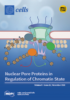
Cover Story (view full-size image): Nuclear pore complexes (NPCs) are large protein assemblies which span the nuclear envelope and regulate nucleocytoplasmic transport. Research over the last 15 years has revealed an additional key function of the NPC and its constituent nucleoporins (Nups) in the regulation of gene expression via physical interactions with the genome. Although many of the precise mechanisms behind the gene regulatory roles of Nups remain enigmatic, one emerging trend is the apparent ability of Nups to influence chromatin structure. In the accompanying review, we discuss recent findings on this topic and highlight how this influence can occur via interactions between specific Nups and both repressive and activating histone modifying machinery, and at the level of chromatin compaction, through conserved interactions of Nups with chromatin remodeling enzymes. View this paper.
  • Issues are regarded as officially published after their release is announced to the table of contents alert mailing list.
  • You may sign up for e-mail alerts to receive table of contents of newly released issues.
  • PDF is the official format for papers published in both, html and pdf forms. To view the papers in pdf format, click on the "PDF Full-text" link, and use the free Adobe Reader to open them.
Order results
Result details
Section
Select all
Export citation of selected articles as:
20 pages, 7929 KiB  
Article
A Novel 3D Scaffold for Cell Growth to Assess Electroporation Efficacy
by Monica Dettin, Elisabetta Sieni, Annj Zamuner, Ramona Marino, Paolo Sgarbossa, Maria Lucibello, Anna Lisa Tosi, Flavio Keller, Luca Giovanni Campana and Emanuela Signori
Cells 2019, 8(11), 1470; https://doi.org/10.3390/cells8111470 - 19 Nov 2019
Cited by 7 | Viewed by 4403
Abstract
Tumor electroporation (EP) refers to the permeabilization of the cell membrane by means of short electric pulses thus allowing the potentiation of chemotherapeutic drugs. Standard plate adhesion 2D cell cultures can simulate the in vivo environment only partially due to lack of cell–cell [...] Read more.
Tumor electroporation (EP) refers to the permeabilization of the cell membrane by means of short electric pulses thus allowing the potentiation of chemotherapeutic drugs. Standard plate adhesion 2D cell cultures can simulate the in vivo environment only partially due to lack of cell–cell interaction and extracellular matrix (ECM). In this study, we assessed a novel 3D scaffold for cell cultures based on hyaluronic acid and ionic-complementary self-assembling peptides (SAPs), by studying the growth patterns of two different breast carcinoma cell lines (HCC1569 and MDA-MB231). This 3D scaffold modulates cell shape and induces extracellular matrix deposit around cells. In the MDA-MB 231 cell line, it allows three-dimensional growth of structures known as spheroids, while in HCC1569 it achieves a cell organization similar to that observed in vivo. Interestingly, we were able to visualize the electroporation effect on the cells seeded in the new scaffold by means of standard propidium iodide assay and fluorescence microscopy. Thanks to the presence of cell–cell and cell–ECM interactions, the new 3D scaffold may represent a more reliable support for EP studies than 2D cancer cell cultures and may be used to test new EP-delivered drugs and novel EP protocols. Full article
Show Figures

Figure 1

14 pages, 3199 KiB  
Article
Suppression of Angiogenesis by Targeting Cyclin-Dependent Kinase 7 in Human Umbilical Vein Endothelial Cells and Renal Cell Carcinoma: An In Vitro and In Vivo Study
by Chung-Sheng Shi, Kuan-Lin Kuo, Mei-Sin Chen, Po-Ming Chow, Shing-Hwa Liu, Yu-Wei Chang, Wei-Chou Lin, Shih-Ming Liao, Chen-Hsun Hsu, Fu-Shun Hsu, Hong-Chiang Chang and Kuo-How Huang
Cells 2019, 8(11), 1469; https://doi.org/10.3390/cells8111469 - 19 Nov 2019
Cited by 9 | Viewed by 3408
Abstract
Cancer cells rely on aberrant transcription for growth and survival. Cyclin-dependent kinases (CDKs) play critical roles in regulating gene transcription by modulating the activity of RNA polymerase II (RNAPII). THZ1, a selective covalent inhibitor of CDK7, has antitumor effects in several human cancers. [...] Read more.
Cancer cells rely on aberrant transcription for growth and survival. Cyclin-dependent kinases (CDKs) play critical roles in regulating gene transcription by modulating the activity of RNA polymerase II (RNAPII). THZ1, a selective covalent inhibitor of CDK7, has antitumor effects in several human cancers. In this study, we investigated the role and therapeutic potential of CDK7 in regulating the angiogenic activity of endothelial cells and human renal cell carcinoma (RCC). Our results revealed that vascular endothelial growth factor (VEGF), a critical activator of angiogenesis, upregulated the expression of CDK7 and RNAPII, and the phosphorylation of RNAPII at serine 5 and 7 in human umbilical vein endothelial cells (HUVECs), indicating the transcriptional activity of CDK7 may be involved in VEGF-activated angiogenic activity of endothelium. Furthermore, through suppressing CDK7 activity, THZ1 suppressed VEGF-activated proliferation and migration, as well as enhanced apoptosis of HUVECs. Moreover, THZ1 inhibited VEGF-activated capillary tube formation and CDK7 knockdown consistently diminished tube formation in HUVECs. Additionally, THZ1 reduced VEGF expression in human RCC cells (786-O and Caki-2), and THZ1 treatment inhibited tumor growth, vascularity, and angiogenic marker (CD31) expression in RCC xenografts. Our results demonstrated that CDK7-mediated transcription was involved in the angiogenic activity of endothelium and human RCC. THZ1 suppressed VEGF-mediated VEGFR2 downstream activation of angiogenesis, providing a new perspective for antitumor therapy in RCC patients. Full article
(This article belongs to the Special Issue Angiogenesis in Cancer)
Show Figures

Figure 1

20 pages, 2411 KiB  
Article
Identification of Novel Adenylyl Cyclase 5 (AC5) Signaling Networks in D1 and D2 Medium Spiny Neurons using Bimolecular Fluorescence Complementation Screening
by Trevor B. Doyle, Brian S. Muntean, Karin F. Ejendal, Michael P. Hayes, Monica Soto-Velasquez, Kirill A. Martemyanov, Carmen W. Dessauer, Chang-Deng Hu and Val J. Watts
Cells 2019, 8(11), 1468; https://doi.org/10.3390/cells8111468 - 19 Nov 2019
Cited by 13 | Viewed by 4126
Abstract
Adenylyl cyclase type 5 (AC5), as the principal isoform expressed in striatal medium spiny neurons (MSNs), is essential for the integration of both stimulatory and inhibitory midbrain signals that initiate from dopaminergic G protein-coupled receptor (GPCR) activation. The spatial and temporal control of [...] Read more.
Adenylyl cyclase type 5 (AC5), as the principal isoform expressed in striatal medium spiny neurons (MSNs), is essential for the integration of both stimulatory and inhibitory midbrain signals that initiate from dopaminergic G protein-coupled receptor (GPCR) activation. The spatial and temporal control of cAMP signaling is dependent upon the composition of local regulatory protein networks. However, there is little understanding of how adenylyl cyclase protein interaction networks adapt to the multifarious pressures of integrating acute versus chronic and inhibitory vs. stimulatory receptor signaling in striatal MSNs. Here, we presented the development of a novel bimolecular fluorescence complementation (BiFC)-based protein-protein interaction screening methodology to further identify and characterize elements important for homeostatic control of dopamine-modulated AC5 signaling in a neuronal model cell line and striatal MSNs. We identified two novel AC5 modulators: the protein phosphatase 2A (PP2A) catalytic subunit (PPP2CB) and the intracellular trafficking associated protein—NSF (N-ethylmaleimide-sensitive factor) attachment protein alpha (NAPA). The effects of genetic knockdown (KD) of each gene were evaluated in several cellular models, including D1- and D2-dopamine receptor-expressing MSNs from CAMPER mice. The knockdown of PPP2CB was associated with a reduction in acute and sensitized adenylyl cyclase activity, implicating PP2A is an important and persistent regulator of adenylyl cyclase activity. In contrast, the effects of NAPA knockdown were more nuanced and appeared to involve an activity-dependent protein interaction network. Taken together, these data represent a novel screening method and workflow for the identification and validation of adenylyl cyclase protein-protein interaction networks under diverse cAMP signaling paradigms. Full article
(This article belongs to the Special Issue New Advances in Cyclic AMP Signalling)
Show Figures

Figure 1

21 pages, 4957 KiB  
Article
The G Protein-Coupled Bile Acid Receptor TGR5 (Gpbar1) Modulates Endothelin-1 Signaling in Liver
by Caroline Klindt, Maria Reich, Birte Hellwig, Jan Stindt, Jörg Rahnenführer, Jan G. Hengstler, Karl Köhrer, Kristina Schoonjans, Dieter Häussinger and Verena Keitel
Cells 2019, 8(11), 1467; https://doi.org/10.3390/cells8111467 - 19 Nov 2019
Cited by 37 | Viewed by 5416
Abstract
TGR5 (Gpbar1) is a G protein-coupled receptor responsive to bile acids (BAs), which is expressed in different non-parenchymal cells of the liver, including biliary epithelial cells, liver-resident macrophages, sinusoidal endothelial cells (LSECs), and activated hepatic stellate cells (HSCs). Mice with targeted deletion of [...] Read more.
TGR5 (Gpbar1) is a G protein-coupled receptor responsive to bile acids (BAs), which is expressed in different non-parenchymal cells of the liver, including biliary epithelial cells, liver-resident macrophages, sinusoidal endothelial cells (LSECs), and activated hepatic stellate cells (HSCs). Mice with targeted deletion of TGR5 are more susceptible towards cholestatic liver injury induced by cholic acid-feeding and bile duct ligation, resulting in a reduced proliferative response and increased liver injury. Conjugated lithocholic acid (LCA) represents the most potent TGR5 BA ligand and LCA-feeding has been used as a model to rapidly induce severe cholestatic liver injury in mice. Thus, TGR5 knockout (KO) mice and wildtype (WT) littermates were fed a diet supplemented with 1% LCA for 84 h. Liver injury and gene expression changes induced by the LCA diet revealed an enrichment of pathways associated with inflammation, proliferation, and matrix remodeling. Knockout of TGR5 in mice caused upregulation of endothelin-1 (ET-1) expression in the livers. Analysis of TGR5-dependent ET-1 signaling in isolated LSECs and HSCs demonstrated that TGR5 activation reduces ET-1 expression and secretion from LSECs and triggers internalization of the ET-1 receptor in HSCs, dampening ET-1 responsiveness. Thus, we identified two independent mechanisms by which TGR5 inhibits ET-1 signaling and modulates portal pressure. Full article
Show Figures

Figure 1

19 pages, 4263 KiB  
Article
Acetylshikonin Sensitizes Hepatocellular Carcinoma Cells to Apoptosis through ROS-Mediated Caspase Activation
by Ming Hong, Jinke Li, Siying Li and Mohammed M.Almutairi
Cells 2019, 8(11), 1466; https://doi.org/10.3390/cells8111466 - 19 Nov 2019
Cited by 21 | Viewed by 4788 | Retraction
Abstract
The tumor necrosis factor-related apoptosis-inducing ligand (TRAIL) has shown strong and explicit cancer cell-selectivity, which results in little toxicity toward normal tissues, and has been recognized as a potential, relatively safe anticancer agent. However, several cancers are resistant to the apoptosis induced by [...] Read more.
The tumor necrosis factor-related apoptosis-inducing ligand (TRAIL) has shown strong and explicit cancer cell-selectivity, which results in little toxicity toward normal tissues, and has been recognized as a potential, relatively safe anticancer agent. However, several cancers are resistant to the apoptosis induced by TRAIL. A recent study found that shikonin b (alkannin, 5,8-dihydroxy-2-[(1S)-1-hydroxy-4-methylpent-3-en-1-yl]naphthalene-1,4-dione) might induce apoptosis in TRAIL-resistant cholangiocarcinoma cells through reactive oxygen species (ROS)-mediated caspases activation. However, the strong cytotoxic activity has limited its potential as an anticancer drug. Thus, the current study intends to discover novel shikonin derivatives which can sensitize the liver cancer cell to TRAIL-induced apoptosis while exhibiting little toxicity toward the normal hepatic cell. The trypan blue exclusion assay, western blot assay, 4′,6-diamidino-2-phenylindole (DAPI) staining and the terminal deoxynucleotidyl transferase dUTP nick end labeling (TUNEL) assay as well as the ‘comet’ assay, were used to study the underlying mechanisms of cell death and to search for any mechanisms of an enhancement of TRAIL-mediated apoptosis in the presence of ASH. Herein, we demonstrated that non-cytotoxic doses of acetylshikonin (ASH), one of the shikonin derivatives, in combination with TRAIL, could promote apoptosis in HepG2 cells. Further studies showed that application of ASH in a non-cytotoxic dose (2.5 μM) could increase intracellular ROS production and induce DNA damage, which might trigger a cell intrinsic apoptosis pathway in the TRAIL-resistant HepG2 cell. Combination treatment with a non-cytotoxic dose of ASH and TRAIL activated caspase and increased the cleavage of PARP-1 in the HepG2 cell. However, when intracellular ROS production was suppressed by N-acetyl-l-cysteine (NAC), the synergistic effects of ASH and TRAIL on hepatocellular carcinoma (HCC) cell apoptosis was abolished. Furthermore, NAC could alleviate p53 and the p53 upregulated modulator of apoptosis (PUMA) expression induced by TRAIL and ASH. Small (or short) interfering RNA (siRNA) targeting PUMA or p53 significantly reversed ASH-mediated sensitization to TRAIL-induced apoptosis. In addition, Bax gene deficiency also abolished ASH-induced TRAIL sensitization. An orthotopical HCC implantation mice model further confirmed that co-treated ASH overcomes TRAIL resistance in HCC cells without exhibiting potent toxicity in vivo. In conclusion, the above data suggested that ROS could induce DNA damage and activating p53/PUMA/Bax signaling, and thus, this resulted in the permeabilization of mitochondrial outer membrane and activating caspases as well as sensitizing the HCC cell to apoptosis induced by TRAIL and ASH treatment. Full article
(This article belongs to the Special Issue Cellular and Molecular Mechanisms of Hepatocellular Carcinoma)
Show Figures

Figure 1

20 pages, 3414 KiB  
Review
The Non-Canonical Aspects of MicroRNAs: Many Roads to Gene Regulation
by Christiaan J. Stavast and Stefan J. Erkeland
Cells 2019, 8(11), 1465; https://doi.org/10.3390/cells8111465 - 19 Nov 2019
Cited by 229 | Viewed by 9676
Abstract
MicroRNAs (miRNAs) are critical regulators of gene expression. As miRNAs are frequently deregulated in many human diseases, including cancer and immunological disorders, it is important to understand their biological functions. Typically, miRNA-encoding genes are transcribed by RNA Polymerase II and generate primary transcripts [...] Read more.
MicroRNAs (miRNAs) are critical regulators of gene expression. As miRNAs are frequently deregulated in many human diseases, including cancer and immunological disorders, it is important to understand their biological functions. Typically, miRNA-encoding genes are transcribed by RNA Polymerase II and generate primary transcripts that are processed by RNase III-endonucleases DROSHA and DICER into small RNAs of approximately 21 nucleotides. All miRNAs are loaded into Argonaute proteins in the RNA-induced silencing complex (RISC) and act as post-transcriptional regulators by binding to the 3′- untranslated region (UTR) of mRNAs. This seed-dependent miRNA binding inhibits the translation and/or promotes the degradation of mRNA targets. Surprisingly, recent data presents evidence for a target-mediated decay mechanism that controls the level of specific miRNAs. In addition, several non-canonical miRNA-containing genes have been recently described and unexpected functions of miRNAs have been identified. For instance, several miRNAs are located in the nucleus, where they are involved in the transcriptional activation or silencing of target genes. These epigenetic modifiers are recruited by RISC and guided by miRNAs to specific loci in the genome. Here, we will review non-canonical aspects of miRNA biology, including novel regulators of miRNA expression and functions of miRNAs in the nucleus. Full article
(This article belongs to the Collection Regulatory Functions of microRNAs)
Show Figures

Figure 1

15 pages, 2810 KiB  
Article
Sex Differences in High Fat Diet-Induced Metabolic Alterations Correlate with Changes in the Modulation of GRK2 Levels
by Alba C. Arcones, Marta Cruces-Sande, Paula Ramos, Federico Mayor and Cristina Murga
Cells 2019, 8(11), 1464; https://doi.org/10.3390/cells8111464 - 19 Nov 2019
Cited by 16 | Viewed by 6381
Abstract
A differential sex-related sensitivity has been reported in obesity and insulin resistance-related cardio-metabolic diseases, with a lower incidence of these pathologies being observed in young females when compared to age-matched males. However, such relative protection is lost with age. The mechanisms underlying such [...] Read more.
A differential sex-related sensitivity has been reported in obesity and insulin resistance-related cardio-metabolic diseases, with a lower incidence of these pathologies being observed in young females when compared to age-matched males. However, such relative protection is lost with age. The mechanisms underlying such sex and age-related changes in the susceptibility to diabetes and obesity are not fully understood. Herein, we report that the relative protection that is displayed by young female mice, as compared to male littermates, against some of the metabolic alterations that are induced by feeding a high fat diet (HFD), correlates with a lower upregulation of the protein levels of G protein-coupled receptor kinase (GRK2), which is a key regulator of both insulin and G protein-coupled receptor signaling, in the liver and adipose tissue. Interestingly, when the HFD is initiated in middle-aged (32 weeks) female mice, these animals are no longer protected and display a more overt obese and insulin-resistant phenotype, along with a more evident increase in the GRK2 protein levels in metabolically relevant tissues in such conditions. Our data suggest that GRK2 dosage might be involved in the sex and age-biased sensitivity to insulin resistance-related pathologies. Full article
Show Figures

Figure 1

13 pages, 624 KiB  
Review
Estrogen Receptors and Melanoma: A Review
by Emi Dika, Annalisa Patrizi, Martina Lambertini, Nicholas Manuelpillai, Michelangelo Fiorentino, Annalisa Altimari, Manuela Ferracin, Mattia Lauriola, Enrica Fabbri, Elena Campione, Giulia Veronesi and Federica Scarfì
Cells 2019, 8(11), 1463; https://doi.org/10.3390/cells8111463 - 19 Nov 2019
Cited by 41 | Viewed by 6021
Abstract
In the last three decades cutaneous melanoma has been widely investigated as a steroid hormone-sensitive cancer. Following this hypothesis, many epidemiological studies have investigated the relationship between estrogens and melanoma. No evidence to date has supported this association due to the great complexity [...] Read more.
In the last three decades cutaneous melanoma has been widely investigated as a steroid hormone-sensitive cancer. Following this hypothesis, many epidemiological studies have investigated the relationship between estrogens and melanoma. No evidence to date has supported this association due to the great complexity of genetic, external and environmental factors underlying the development of this cancer. Molecular mechanisms through which estrogen and their receptor exert a role in melanoma genesis are still under investigation with new studies increasingly focusing on the discovery of new molecular targets for therapeutic treatments. Full article
(This article belongs to the Special Issue Estrogen Receptor Expression and Function in Health and Disease)
Show Figures

Figure 1

20 pages, 2040 KiB  
Review
Recent Advances in EPAC-Targeted Therapies: A Biophysical Perspective
by Alveena Ahmed, Stephen Boulton, Hongzhao Shao, Madoka Akimoto, Amarnath Natarajan, Xiaodong Cheng and Giuseppe Melacini
Cells 2019, 8(11), 1462; https://doi.org/10.3390/cells8111462 - 19 Nov 2019
Cited by 14 | Viewed by 4191
Abstract
The universal second messenger cAMP regulates diverse intracellular processes by interacting with ubiquitously expressed proteins, such as Protein Kinase A (PKA) and the Exchange Protein directly Activated by cAMP (EPAC). EPAC is implicated in multiple pathologies, thus several EPAC-specific inhibitors have been identified [...] Read more.
The universal second messenger cAMP regulates diverse intracellular processes by interacting with ubiquitously expressed proteins, such as Protein Kinase A (PKA) and the Exchange Protein directly Activated by cAMP (EPAC). EPAC is implicated in multiple pathologies, thus several EPAC-specific inhibitors have been identified in recent years. However, the mechanisms and molecular interactions underlying the EPAC inhibition elicited by such compounds are still poorly understood. Additionally, being hydrophobic low molecular weight species, EPAC-specific inhibitors are prone to forming colloidal aggregates, which result in non-specific aggregation-based inhibition (ABI) in aqueous systems. Here, we review from a biophysical perspective the molecular basis of the specific and non-specific interactions of two EPAC antagonists—CE3F4R, a non-competitive inhibitor, and ESI-09, a competitive inhibitor of EPAC. Additionally, we discuss the value of common ABI attenuators (e.g., TX and HSA) to reduce false positives at the expense of introducing false negatives when screening aggregation-prone compounds. We hope this review provides the EPAC community effective criteria to evaluate similar compounds, aiding in the optimization of existing drug leads, and informing the development of the next generation of EPAC-specific inhibitors. Full article
(This article belongs to the Special Issue New Advances in Cyclic AMP Signalling)
Show Figures

Figure 1

20 pages, 1022 KiB  
Review
The Role of MicroRNAs upon Epithelial-to-Mesenchymal Transition in Inflammatory Bowel Disease
by Éva Boros and István Nagy
Cells 2019, 8(11), 1461; https://doi.org/10.3390/cells8111461 - 19 Nov 2019
Cited by 13 | Viewed by 4379
Abstract
Increasing evidence suggest the significance of inflammation in the progression of cancer, for example the development of colorectal cancer in Inflammatory Bowel Disease (IBD) patients. Long-lasting inflammation in the gastrointestinal tract causes serious systemic complications and breaks the homeostasis of the intestine, where [...] Read more.
Increasing evidence suggest the significance of inflammation in the progression of cancer, for example the development of colorectal cancer in Inflammatory Bowel Disease (IBD) patients. Long-lasting inflammation in the gastrointestinal tract causes serious systemic complications and breaks the homeostasis of the intestine, where the altered expression of regulatory genes and miRNAs trigger malignant transformations. Several steps lead from acute inflammation to malignancies: epithelial-to-mesenchymal transition (EMT) and inhibitory microRNAs (miRNAs) are known factors during multistage carcinogenesis and IBD pathogenesis. In this review, we outline the interactions between EMT components and miRNAs that may affect cancer development during IBD. Full article
Show Figures

Figure 1

14 pages, 3008 KiB  
Article
Carbamazepine Enhances Adipogenesis by Inhibiting Wnt/β-Catenin Expression
by Dong Uk Im, Sang Chon Kim, Gia Cac Chau and Sung Hee Um
Cells 2019, 8(11), 1460; https://doi.org/10.3390/cells8111460 - 18 Nov 2019
Cited by 20 | Viewed by 4667
Abstract
Carbamazepine is a drug that is widely used in the treatment of epilepsy and bipolar disorder. The prevalence of obesity in patients treated with carbamazepine has been frequently reported. However, whether carbamazepine affects adipogenesis, one of the critical steps in the development of [...] Read more.
Carbamazepine is a drug that is widely used in the treatment of epilepsy and bipolar disorder. The prevalence of obesity in patients treated with carbamazepine has been frequently reported. However, whether carbamazepine affects adipogenesis, one of the critical steps in the development of obesity, remains unclear. Here, we show that carbamazepine increased the expression levels of peroxisome proliferator-activated receptor γ (PPARγ), CCAAT/enhancer-binding protein β (C/EBPβ), and fatty acid synthase (FASN) in 3T3-L1 cells. Notably, carbamazepine inhibited the expression levels of β-catenin, a negative regulator of adipogenesis, leading to enhanced adipogenesis. Conversely, β-catenin overexpression abolished the effect of carbamazepine on adipogenic gene expression. However, depletion of β-catenin further enhanced PPARγ expression. In addition, carbamazepine reduced β-catenin expression by lowering the levels of phospho-low density lipoprotein receptor-related protein 6 (p-LRP6) and phospho-glycogen synthase kinase 3β (p-GSK3β) in Wnt/β-catenin signaling. Moreover, carbamazepine reduced Wnt mRNA expression and decreased the promoter activities of TCF, the target of β-catenin during adipogenesis. These results suggest that carbamazepine enhances adipogenesis by suppressing Wnt/β-catenin expression, indicating its potential effects on obesity-related metabolism. Full article
(This article belongs to the Special Issue Adipocytes and Metabolic Health)
Show Figures

Figure 1

37 pages, 958 KiB  
Review
Novel Epigenetic Biomarkers in Pregnancy-Related Disorders and Cancers
by Valentina Karin-Kujundzic, Ida Marija Sola, Nina Predavec, Anamarija Potkonjak, Ema Somen, Pavao Mioc, Alan Serman, Semir Vranic and Ljiljana Serman
Cells 2019, 8(11), 1459; https://doi.org/10.3390/cells8111459 - 18 Nov 2019
Cited by 11 | Viewed by 5248
Abstract
As the majority of cancers and gestational diseases are prognostically stage- and grade-dependent, the ultimate goal of ongoing studies in precision medicine is to provide early and timely diagnosis of such disorders. These studies have enabled the development of various new diagnostic biomarkers, [...] Read more.
As the majority of cancers and gestational diseases are prognostically stage- and grade-dependent, the ultimate goal of ongoing studies in precision medicine is to provide early and timely diagnosis of such disorders. These studies have enabled the development of various new diagnostic biomarkers, such as free circulating nucleic acids, and detection of their epigenetic changes. Recently, extracellular vesicles including exosomes, microvesicles, oncosomes, and apoptotic bodies have been recognized as powerful diagnostic tools. Extracellular vesicles carry specific proteins, lipids, DNAs, mRNAs, and miRNAs of the cells that produced them, thus reflecting the function of these cells. It is believed that exosomes, in particular, may be the optimal biomarkers of pathological pregnancies and cancers, especially those that are frequently diagnosed at an advanced stage, such as ovarian cancer. In the present review, we survey and critically appraise novel epigenetic biomarkers related to free circulating nucleic acids and extracellular vesicles, focusing especially on their status in trophoblasts (pregnancy) and neoplastic cells (cancers). Full article
(This article belongs to the Special Issue Navigating Through the Epigenetic Pathways of Cancer)
Show Figures

Figure 1

23 pages, 1060 KiB  
Review
Emerging Role of Cellular Prion Protein in the Maintenance and Expansion of Glioma Stem Cells
by Stefano Thellung, Alessandro Corsaro, Alessia G. Bosio, Martina Zambito, Federica Barbieri, Michele Mazzanti and Tullio Florio
Cells 2019, 8(11), 1458; https://doi.org/10.3390/cells8111458 - 18 Nov 2019
Cited by 12 | Viewed by 3904
Abstract
Cellular prion protein (PrPC) is a membrane-anchored glycoprotein representing the physiological counterpart of PrP scrapie (PrPSc), which plays a pathogenetic role in prion diseases. Relatively little information is however available about physiological role of PrPC. Although PrP [...] Read more.
Cellular prion protein (PrPC) is a membrane-anchored glycoprotein representing the physiological counterpart of PrP scrapie (PrPSc), which plays a pathogenetic role in prion diseases. Relatively little information is however available about physiological role of PrPC. Although PrPC ablation in mice does not induce lethal phenotypes, impairment of neuronal and bone marrow plasticity was reported in embryos and adult animals. In neurons, PrPC stimulates neurite growth, prevents oxidative stress-dependent cell death, and favors antiapoptotic signaling. However, PrPC activity is not restricted to post-mitotic neurons, but promotes cell proliferation and migration during embryogenesis and tissue regeneration in adult. PrPC acts as scaffold to stabilize the binding between different membrane receptors, growth factors, and basement proteins, contributing to tumorigenesis. Indeed, ablation of PrPC expression reduces cancer cell proliferation and migration and restores cell sensitivity to chemotherapy. Conversely, PrPC overexpression in cancer stem cells (CSCs) from different tumors, including gliomas—the most malignant brain tumors—is predictive for poor prognosis, and correlates with relapses. The mechanisms of the PrPC role in tumorigenesis and its molecular partners in this activity are the topic of the present review, with a particular focus on PrPC contribution to glioma CSCs multipotency, invasiveness, and tumorigenicity. Full article
Show Figures

Figure 1

16 pages, 3508 KiB  
Article
Natural Histogel-Based Bio-Scaffolds for Sustaining Angiogenesis in Beige Adipose Tissue
by Margherita Di Somma, Wandert Schaafsma, Elisabetta Grillo, Maria Vliora, Eleni Dakou, Michela Corsini, Cosetta Ravelli, Roberto Ronca, Paraskevi Sakellariou, Jef Vanparijs, Begona Castro and Stefania Mitola
Cells 2019, 8(11), 1457; https://doi.org/10.3390/cells8111457 - 18 Nov 2019
Cited by 11 | Viewed by 3871
Abstract
In the treatment of obesity and its related disorders, one of the measures adopted is weight reduction by controlling nutrition and increasing physical activity. A valid alternative to restore the physiological function of the human body could be the increase of energy consumption [...] Read more.
In the treatment of obesity and its related disorders, one of the measures adopted is weight reduction by controlling nutrition and increasing physical activity. A valid alternative to restore the physiological function of the human body could be the increase of energy consumption by inducing the browning of adipose tissue. To this purpose, we tested the ability of Histogel, a natural mixture of glycosaminoglycans isolated from animal Wharton jelly, to sustain the differentiation of adipose derived mesenchymal cells (ADSCs) into brown-like cells expressing UCP-1. Differentiated cells show a higher energy metabolism compared to undifferentiated mesenchymal cells. Furthermore, Histogel acts as a pro-angiogenic matrix, induces endothelial cell proliferation and sprouting in a three-dimensional gel in vitro, and stimulates neovascularization when applied in vivo on top of the chicken embryo chorioallantoic membrane or injected subcutaneously in mice. In addition to the pro-angiogenic activity of Histogel, also the ADSC derived beige cells contribute to activating endothelial cells. These data led us to propose Histogel as a promising scaffold for the modulation of the thermogenic behavior of adipose tissue. Indeed, Histogel simultaneously supports the acquisition of brown tissue markers and activates the vasculature process necessary for the correct function of the thermogenic tissue. Thus, Histogel represents a valid candidate for the development of bioscaffolds to increase the amount of brown adipose tissue in patients with metabolic disorders. Full article
(This article belongs to the Special Issue Adipose-Derived Stromal/Stem Cells)
Show Figures

Figure 1

21 pages, 4144 KiB  
Article
Differential Roles of Lipin1 and Lipin2 in the Hepatitis C Virus Replication Cycle
by Victoria Castro, Gema Calvo, Ginés Ávila-Pérez, Marlène Dreux and Pablo Gastaminza
Cells 2019, 8(11), 1456; https://doi.org/10.3390/cells8111456 - 18 Nov 2019
Cited by 4 | Viewed by 3601
Abstract
Although their origin, nature and structure are not identical, a common feature of positive-strand RNA viruses is their ability to subvert host lipids and intracellular membranes to generate replication and assembly complexes. Recently, lipin1, a cellular enzyme that converts phosphatidic acid into diacylglycerol, [...] Read more.
Although their origin, nature and structure are not identical, a common feature of positive-strand RNA viruses is their ability to subvert host lipids and intracellular membranes to generate replication and assembly complexes. Recently, lipin1, a cellular enzyme that converts phosphatidic acid into diacylglycerol, has been implicated in the formation of the membranous web that hosts hepatitis C virus (HCV) replicase. In the liver, lipin1 cooperates with lipin2 to maintain glycerolipid homeostasis. We extended our previous study of the lipin family on HCV infection, by determining the impact of the lipin2 silencing on viral replication. Our data reveal that lipin2 silencing interferes with HCV virion secretion at late stages of the infection, without significantly affecting viral replication or assembly. Moreover, uninfected lipin2-, but not lipin1-deficient cells display alterations in mitochondrial and Golgi apparatus morphology, suggesting that lipin2 contributes to the maintenance of the overall organelle architecture. Finally, our data suggest a broader function of lipin2 for replication of HCV and other RNA viruses, in contrast with the specific impact of lipin1 silencing on HCV replication. Overall, this study reveals distinctive functions of lipin1 and lipin2 in cells of hepatic origin, a context in which they are often considered functionally redundant. Full article
(This article belongs to the Special Issue Hepatitis C Virus and Host Interactions)
Show Figures

Figure 1

11 pages, 2601 KiB  
Article
miRNA-29b Inhibits Prostate Tumor Growth and Induces Apoptosis by Increasing Bim Expression
by Subhayan Sur, Robert Steele, Xingyi Shi and Ratna B. Ray
Cells 2019, 8(11), 1455; https://doi.org/10.3390/cells8111455 - 18 Nov 2019
Cited by 66 | Viewed by 5159
Abstract
Prostate cancer is one of the most common cancers among men. Currently available therapies improve patient survival against local prostate cancer but have shown severe side effects. Advanced prostate cancer is still incurable. Studies have suggested the involvement of non-coding RNAs, especially micro-RNAs [...] Read more.
Prostate cancer is one of the most common cancers among men. Currently available therapies improve patient survival against local prostate cancer but have shown severe side effects. Advanced prostate cancer is still incurable. Studies have suggested the involvement of non-coding RNAs, especially micro-RNAs (miRNAs), in the regulation of multiple cellular events in cancer and thus several clinical trials are ongoing using miRNAs mimics or inhibitors. We previously demonstrated that miRNA-29b-3p (miR-29b) was downregulated in prostate cancer and that the overexpression of miR-29b limited prostate cancer metastasis. However, the therapeutic potential of the miR-29b against prostate cancer remains unknown. Here, we evaluated the therapeutic role of miR-29b in in vivo prostate tumors in a mouse model. Intratumoral injection of mimic miR-29b significantly inhibited prostate cancer xenograft tumor growth in nude mice. Subsequent study demonstrated that the overexpression of miR-29b reduced prostate cancer cell PC3 proliferation in a time dependent manner and induced cell death. Mechanistic study using a cancer pathway specific transcriptomic array revealed a significant overexpression of the pro-apoptotic gene BCL2L11 (Bim) in the miR-29b overexpressed PC3 cells, which was further verified in PC3 cells overexpressing miR-29b. We also observed a significant induction of Bim protein in miR-29b treated xenograft tumors. The induction of cytosolic accumulation of cytochrome C and PARP cleavage in miR-29b overexpressed PC3 cells was observed. Thus, our results suggest that miR-29b can be used as a potential molecule for prostate cancer therapy. Full article
(This article belongs to the Section Cell Signaling)
Show Figures

Figure 1

14 pages, 2841 KiB  
Article
Mitochondrial Mass Assessment in a Selected Cell Line under Different Metabolic Conditions
by Anna Costanzini, Gianluca Sgarbi, Alessandra Maresca, Valentina Del Dotto, Giancarlo Solaini and Alessandra Baracca
Cells 2019, 8(11), 1454; https://doi.org/10.3390/cells8111454 - 18 Nov 2019
Cited by 8 | Viewed by 4986
Abstract
Changes of quantity and/or morphology of cell mitochondria are often associated with metabolic modulation, pathology, and apoptosis. Exogenous fluorescent probes used to investigate changes in mitochondrial content and dynamics are strongly dependent, for their internalization, on the mitochondrial membrane potential and composition, thus [...] Read more.
Changes of quantity and/or morphology of cell mitochondria are often associated with metabolic modulation, pathology, and apoptosis. Exogenous fluorescent probes used to investigate changes in mitochondrial content and dynamics are strongly dependent, for their internalization, on the mitochondrial membrane potential and composition, thus limiting the reliability of measurements. To overcome this limitation, genetically encoded recombinant fluorescent proteins, targeted to different cellular districts, were used as reporters. Here, we explored the potential use of mitochondrially targeted red fluorescent probe (mtRFP) to quantify, by flow cytometry, mitochondrial mass changes in cells exposed to different experimental conditions. We first demonstrated that the mtRFP fluorescence intensity is stable during cell culture and it is related with the citrate synthase activity, an established marker of the mitochondrial mass. Incidentally, the expression of mtRFP inside mitochondria did not alter the oxygen consumption rate under both state 3 and 4 respiration conditions. In addition, using this method, we showed for the first time that different inducers of mitochondrial mass change, such as hypoxia exposure or resveratrol treatment of cells, could be consistently detected. We suggest that transfection and selection of stable clones expressing mtRFP is a reliable method to monitor mitochondrial mass changes, particularly when pathophysiological or experimental conditions change ΔΨm, as it occurs during mitochondrial uncoupling or hypoxia/anoxia conditions. Full article
(This article belongs to the Section Intracellular and Plasma Membranes)
Show Figures

Figure 1

9 pages, 1366 KiB  
Review
Targeting the Stress-Induced Protein NUPR1 to Treat Pancreatic Adenocarcinoma
by Patricia Santofimia-Castaño, Yi Xia, Ling Peng, Adrián Velázquez-Campoy, Olga Abián, Wenjun Lan, Gwen Lomberk, Raul Urrutia, Bruno Rizzuti, Philippe Soubeyran, José Luis Neira and Juan Iovanna
Cells 2019, 8(11), 1453; https://doi.org/10.3390/cells8111453 - 17 Nov 2019
Cited by 29 | Viewed by 5006
Abstract
Cancer cells activate stress-response mechanisms to adapt themselves to a variety of stressful conditions. Among these protective mechanisms, those controlled by the stress-induced nuclear protein 1 (NUPR1) belong to the most conserved ones. NUPR1 is an 82-residue-long, monomeric, basic and intrinsically disordered protein [...] Read more.
Cancer cells activate stress-response mechanisms to adapt themselves to a variety of stressful conditions. Among these protective mechanisms, those controlled by the stress-induced nuclear protein 1 (NUPR1) belong to the most conserved ones. NUPR1 is an 82-residue-long, monomeric, basic and intrinsically disordered protein (IDP), which was found to be invariably overexpressed in some, if not all, cancer tissues. Remarkably, we and others have previously showed that genetic inactivation of the Nupr1 gene antagonizes the growth of pancreatic cancer as well as several other tumors. With the use of a multidisciplinary strategy by combining biophysical, biochemical, bioinformatic, and biological approaches, a trifluoperazine-derived compound, named ZZW-115, has been identified as an inhibitor of the NUPR1 functions. The anticancer activity of the ZZW-115 was first validated on a large panel of cancer cells. Furthermore, ZZW-115 produced a dose-dependent tumor regression of the tumor size in xenografted mice. Mechanistically, we have demonstrated that NUPR1 binds to several importins. Because ZZW-115 binds NUPR1 through the region around the amino acid Thr68, which is located into the nuclear location signal (NLS) region of the protein, we demonstrated that treatment with ZZW-115 inhibits completely the translocation of NUPR1 from the cytoplasm to the nucleus by competing with importins. Full article
(This article belongs to the Special Issue Molecular and Cellular Mechanisms of Cancers: Pancreatic Cancer)
Show Figures

Figure 1

16 pages, 3218 KiB  
Article
Let-7 miRNA’s Expression Profile and Its Potential Prognostic Role in Uterine Leiomyosarcoma
by Bruna Cristine de Almeida, Laura Gonzalez dos Anjos, Miyuki Uno, Isabela Werneck da Cunha, Fernando Augusto Soares, Glauco Baiocchi, Edmund Chada Baracat and Katia Candido Carvalho
Cells 2019, 8(11), 1452; https://doi.org/10.3390/cells8111452 - 17 Nov 2019
Cited by 16 | Viewed by 3000
Abstract
The lethal-7 (let-7) family is an important microRNA (miRNA) group that usually exerts functions as a tumor suppressor. We aimed to evaluate the expression profile of let-7a, let-7b, let-7c, let-7d, let-7e, let-7f, let-7g, and let-7i and to assess their value as prognostic [...] Read more.
The lethal-7 (let-7) family is an important microRNA (miRNA) group that usually exerts functions as a tumor suppressor. We aimed to evaluate the expression profile of let-7a, let-7b, let-7c, let-7d, let-7e, let-7f, let-7g, and let-7i and to assess their value as prognostic markers in uterine leiomyosarcoma (LMS) patients. The miRNAs expression profile was assessed in 34 LMS and 13 normal myometrium (MM) paraffin-embedded samples. All let-7 family members showed downregulation in LMS. Our findings showed that patients with let-7e downregulation had worse overall survival (OS) and is an independent prognostic factor (hazard ratio [HR] = 2.24). In addition, almost half the patients had distant metastasis. LMS patients with downregulated let-7b and let-7d had worse disease-free survival (DFS); they are not independent prognostic factors (HR = 2.65). Patients’ ages were associated with let-7d, let-7e and let-7f (p = 0.0160) downregulation. In conclusion, all the let-7 family members were downregulated in LMS patients, and the greater the loss of expression of these molecules, the greater their relationship with worse prognosis of patients. Let-7e expression might influence the OS, while let-7b and le-7d might influence the DFS. The lowest expression levels of let-7d, let-7e, and let-7f were associated with the oldest patients. Our findings indicate strong evidence of let-7’s role as a potential prognostic biomarker in LMS. Full article
(This article belongs to the Special Issue Cancer Related microRNAs)
Show Figures

Figure 1

15 pages, 6858 KiB  
Article
Functional Ultrastructure of the Excretory Gland Cell in Zoonotic Anisakids (Anisakidae, Nematoda)
by Ivona Mladineo, Jerko Hrabar, Hrvoje Smodlaka, Lauren Palmer, Kristen Sakamaki, Kleoniki Keklikoglou and Pantelis Katharios
Cells 2019, 8(11), 1451; https://doi.org/10.3390/cells8111451 - 17 Nov 2019
Cited by 4 | Viewed by 3496
Abstract
Excretory and secretory products are crucial for parasite infectivity and host immunomodulation, but the functioning and ultrastructure of the excretory gland cell (EC) that produces these products are still scarcely understood and described. In light of growing reports on anisakiasis cases in Europe, [...] Read more.
Excretory and secretory products are crucial for parasite infectivity and host immunomodulation, but the functioning and ultrastructure of the excretory gland cell (EC) that produces these products are still scarcely understood and described. In light of growing reports on anisakiasis cases in Europe, we aimed to characterise the EC of larval Anisakis pegreffii and adult Pseudoterranova azarasi. In the latter, EC starts 0.85 mm from the head tip, measuring 1.936 × 0.564 mm. Larval EC shows a long nucleus with thorn-like extravaginations toward the cytoplasm, numerous electron-dense and -lucent secretory granules spanning from the perinuclear to subplasmalemmal space, an elevated number of free ribosomes, small, spherical mitochondria with few cristae and a laminated matrix, small and few Golgi apparatuses, and few endoplasmic reticula, with wide cisternae complexes. Ultrastructure suggests that anaerobic glycolysis is the main metabolic pathway, obtained through nutrient endocytosis across the pseudocoelomic surface of the EC plasmalemma and its endocytic canaliculi. Thorn-like extravaginations of EC karyotheca likely mediate specific processes (Ca2+ signaling, gene expression, transport, nuclear lipid metabolism) into the extremely wide EC cytosol, enabling focal delivery of a signal to specific sites in a short time. These functional annotations of parasitic EC should help to clarify anisakiasis pathogenesis. Full article
(This article belongs to the Section Intracellular and Plasma Membranes)
Show Figures

Figure 1

16 pages, 4750 KiB  
Article
HIF1α-Dependent Metabolic Signals Control the Differentiation of Follicular Helper T Cells
by Lin Dong, Ying He, Shuping Zhou, Yejin Cao, Yan Li, Yujing Bi and Guangwei Liu
Cells 2019, 8(11), 1450; https://doi.org/10.3390/cells8111450 - 17 Nov 2019
Cited by 25 | Viewed by 3491
Abstract
Follicular helper T (TFH) cells are critical for germinal center (GC) formation and are responsible for effective B cell-mediated immunity; metabolic signaling is an important regulatory mechanism for the differentiation of TFH cells. However, the precise roles of hypoxia inducible [...] Read more.
Follicular helper T (TFH) cells are critical for germinal center (GC) formation and are responsible for effective B cell-mediated immunity; metabolic signaling is an important regulatory mechanism for the differentiation of TFH cells. However, the precise roles of hypoxia inducible factor (HIF) 1α-dependent glycolysis and oxidative phosphorylation (OXPHOS) metabolic signaling remain unclear in TFH cell differentiation. Herein, we investigated the effects of glycolysis and OXPHOS on TFH cell differentiation and GC responses using a pharmacological approach in mice under a steady immune status or an activated immune status, which can be caused by foreign antigen stimulation and viral infection. GC and TFH cell responses are related to signals from glycolytic metabolism in mice of different ages. Foreign, specific antigen-induced GC, and TFH cell responses and metabolic signals are essential upon PR8 infection. Glycolysis and succinate-mediated OXPHOS are required for the GC response and TFH cell differentiation. Furthermore, HIF1α is responsible for glycolysis- and OXPHOS-induced alterations in the GC response and TFH cell differentiation under steady or activated conditions in vivo. Blocking glycolysis and upregulating OXPHOS signaling significantly recovered TFH cell differentiation upon PR8 infection and ameliorated inflammatory damage in mice. Thus, our data provide a comprehensive experimental basis for fully understanding the precise roles of HIF1α-mediated glycolysis and OXPHOS metabolic signaling in regulating the GC response and TFH cell differentiation during stable physiological conditions or an antiviral immune response. Full article
(This article belongs to the Section Cellular Immunology)
Show Figures

Figure 1

18 pages, 731 KiB  
Review
Myocardial Adaptation in Pseudohypoxia: Signaling and Regulation of mPTP via Mitochondrial Connexin 43 and Cardiolipin
by Miroslav Ferko, Natália Andelová, Barbara Szeiffová Bačová and Magdaléna Jašová
Cells 2019, 8(11), 1449; https://doi.org/10.3390/cells8111449 - 17 Nov 2019
Cited by 15 | Viewed by 3950
Abstract
Therapies intended to mitigate cardiovascular complications cannot be applied in practice without detailed knowledge of molecular mechanisms. Mitochondria, as the end-effector of cardioprotection, represent one of the possible therapeutic approaches. The present review provides an overview of factors affecting the regulation processes of [...] Read more.
Therapies intended to mitigate cardiovascular complications cannot be applied in practice without detailed knowledge of molecular mechanisms. Mitochondria, as the end-effector of cardioprotection, represent one of the possible therapeutic approaches. The present review provides an overview of factors affecting the regulation processes of mitochondria at the level of mitochondrial permeability transition pores (mPTP) resulting in comprehensive myocardial protection. The regulation of mPTP seems to be an important part of the mechanisms for maintaining the energy equilibrium of the heart under pathological conditions. Mitochondrial connexin 43 is involved in the regulation process by inhibition of mPTP opening. These individual cardioprotective mechanisms can be interconnected in the process of mitochondrial oxidative phosphorylation resulting in the maintenance of adenosine triphosphate (ATP) production. In this context, the degree of mitochondrial membrane fluidity appears to be a key factor in the preservation of ATP synthase rotation required for ATP formation. Moreover, changes in the composition of the cardiolipin’s structure in the mitochondrial membrane can significantly affect the energy system under unfavorable conditions. This review aims to elucidate functional and structural changes of cardiac mitochondria subjected to preconditioning, with an emphasis on signaling pathways leading to mitochondrial energy maintenance during partial oxygen deprivation. Full article
(This article belongs to the Section Intracellular and Plasma Membranes)
Show Figures

Figure 1

34 pages, 7519 KiB  
Review
Nerve, Muscle, and Synaptogenesis
by Lauren Eric Swenarchuk
Cells 2019, 8(11), 1448; https://doi.org/10.3390/cells8111448 - 16 Nov 2019
Cited by 14 | Viewed by 7030
Abstract
The vertebrate skeletal neuromuscular junction (NMJ) has long served as a model system for studying synapse structure, function, and development. Over the last several decades, a neuron-specific isoform of agrin, a heparan sulfate proteoglycan, has been identified as playing a central role in [...] Read more.
The vertebrate skeletal neuromuscular junction (NMJ) has long served as a model system for studying synapse structure, function, and development. Over the last several decades, a neuron-specific isoform of agrin, a heparan sulfate proteoglycan, has been identified as playing a central role in synapse formation at all vertebrate skeletal neuromuscular synapses. While agrin was initially postulated to be the inductive molecule that initiates synaptogenesis, this model has been modified in response to work showing that postsynaptic differentiation can develop in the absence of innervation, and that synapses can form in transgenic mice in which the agrin gene is ablated. In place of a unitary mechanism for neuromuscular synapse formation, studies in both mice and zebrafish have led to the proposal that two mechanisms mediate synaptogenesis, with some synapses being induced by nerve contact while others involve the incorporation of prepatterned postsynaptic structures. Moreover, the current model also proposes that agrin can serve two functions, to induce synaptogenesis and to stabilize new synapses, once these are formed. This review examines the evidence for these propositions, and concludes that it remains possible that a single molecular mechanism mediates synaptogenesis at all NMJs, and that agrin acts as a stabilizer, while its role as inducer is open to question. Moreover, if agrin does not act to initiate synaptogenesis, it follows that as yet uncharacterized molecular interactions are required to play this essential inductive role. Several alternatives to agrin for this function are suggested, including focal pericellular proteolysis and integrin signaling, but all require experimental validation. Full article
Show Figures

Figure 1

28 pages, 1603 KiB  
Review
Ending Restenosis: Inhibition of Vascular Smooth Muscle Cell Proliferation by cAMP
by Sarah A. Smith, Andrew C. Newby and Mark Bond
Cells 2019, 8(11), 1447; https://doi.org/10.3390/cells8111447 - 16 Nov 2019
Cited by 37 | Viewed by 6277
Abstract
Increased vascular smooth muscle cell (VSMC) proliferation contributes towards restenosis after angioplasty, vein graft intimal thickening and atherogenesis. The second messenger 3′ 5′ cyclic adenosine monophosphate (cAMP) plays an important role in maintaining VSMC quiescence in healthy vessels and repressing VSMC proliferation during [...] Read more.
Increased vascular smooth muscle cell (VSMC) proliferation contributes towards restenosis after angioplasty, vein graft intimal thickening and atherogenesis. The second messenger 3′ 5′ cyclic adenosine monophosphate (cAMP) plays an important role in maintaining VSMC quiescence in healthy vessels and repressing VSMC proliferation during resolution of vascular injury. Although the anti-mitogenic properties of cAMP in VSMC have been recognised for many years, it is only recently that we gained a detailed understanding of the underlying signalling mechanisms. Stimuli that elevate cAMP in VSMC inhibit G1-S phase cell cycle progression by inhibiting expression of cyclins and preventing S-Phase Kinase Associated Protein-2 (Skp2-mediated degradation of cyclin-dependent kinase inhibitors. Early studies implicated inhibition of MAPK signalling, although this does not fully explain the anti-mitogenic effects of cAMP. The cAMP effectors, Protein Kinase A (PKA) and Exchange Protein Activated by cAMP (EPAC) act together to inhibit VSMC proliferation by inducing Cyclic-AMP Response Element Binding protein (CREB) activity and inhibiting members of the RhoGTPases, which results in remodelling of the actin cytoskeleton. Cyclic-AMP induced actin remodelling controls proliferation by modulating the activity of Serum Response Factor (SRF) and TEA Domain Transcription Factors (TEAD), which regulate expression of genes required for proliferation. Here we review recent research characterising these mechanisms, highlighting novel drug targets that may allow the anti-mitogenic properties of cAMP to be harnessed therapeutically to limit restenosis. Full article
(This article belongs to the Special Issue New Advances in Cyclic AMP Signalling)
Show Figures

Figure 1

28 pages, 3042 KiB  
Review
Dissecting Aging and Senescence—Current Concepts and Open Lessons
by Christian Schmeer, Alexandra Kretz, Diane Wengerodt, Milan Stojiljkovic and Otto W. Witte
Cells 2019, 8(11), 1446; https://doi.org/10.3390/cells8111446 - 15 Nov 2019
Cited by 82 | Viewed by 17270
Abstract
In contrast to the programmed nature of development, it is still a matter of debate whether aging is an adaptive and regulated process, or merely a consequence arising from a stochastic accumulation of harmful events that culminate in a global state of reduced [...] Read more.
In contrast to the programmed nature of development, it is still a matter of debate whether aging is an adaptive and regulated process, or merely a consequence arising from a stochastic accumulation of harmful events that culminate in a global state of reduced fitness, risk for disease acquisition, and death. Similarly unanswered are the questions of whether aging is reversible and can be turned into rejuvenation as well as how aging is distinguishable from and influenced by cellular senescence. With the discovery of beneficial aspects of cellular senescence and evidence of senescence being not limited to replicative cellular states, a redefinition of our comprehension of aging and senescence appears scientifically overdue. Here, we provide a factor-based comparison of current knowledge on aging and senescence, which we converge on four suggested concepts, thereby implementing the newly emerging cellular and molecular aspects of geroconversion and amitosenescence, and the signatures of a genetic state termed genosenium. We also address the possibility of an aging-associated secretory phenotype in analogy to the well-characterized senescence-associated secretory phenotype and delineate the impact of epigenetic regulation in aging and senescence. Future advances will elucidate the biological and molecular fingerprints intrinsic to either process. Full article
(This article belongs to the Special Issue Aging and Regeneration)
Show Figures

Figure 1

17 pages, 4704 KiB  
Article
CCN2 Mediates S1P-Induced Upregulation of COX2 Expression in Human Granulosa-Lutein Cells
by Liao-Liao Hu, Hsun-Ming Chang, Yuyin Yi, Yingtao Liu, Elizabeth L. Taylor, Li-Ping Zheng and Peter C.K. Leung
Cells 2019, 8(11), 1445; https://doi.org/10.3390/cells8111445 - 15 Nov 2019
Cited by 5 | Viewed by 4133
Abstract
CCN1 and CCN2 are members of the CCN family and play essential roles in the regulation of multiple female reproductive functions, including ovulation. Cyclooxygenase-2 (COX2) is a critical mediator of ovulation and can be induced by sphingosine-1-phosphate (S1P) through the S1P1/3-mediated [...] Read more.
CCN1 and CCN2 are members of the CCN family and play essential roles in the regulation of multiple female reproductive functions, including ovulation. Cyclooxygenase-2 (COX2) is a critical mediator of ovulation and can be induced by sphingosine-1-phosphate (S1P) through the S1P1/3-mediated Yes-associated protein (YAP) signaling. However, it is unclear whether CCN1 or CCN2 can mediate S1P-induced upregulation of COX2 expression and increase in prostaglandin E2 (PGE2) production in human granulosa-lutein (hGL) cells. In the present study, we investigated the effects of S1P on the expressions of CCN1 and CCN2 in hGL cells. Additionally, we used a dual inhibition approach (siRNA-mediated silencing and small molecular inhibitors) to investigate the molecular mechanisms of S1P effects. Our results showed that S1P treatment significantly upregulated the expression of CCN1 and CCN2 in a concentration-dependent manner in hGL cells. Additionally, inhibition or silencing of S1P1, but not S1P3, completely abolished the S1P-induced upregulation of CCN2 expression. Furthermore, we demonstrated that S1P-induced nuclear translocation of YAP and inhibition or silencing of YAP completely abolished the S1P-induced upregulation of CCN1 and CCN2 expression. Notably, silencing of CCN2, but not CCN1, completely reversed the S1P-induced upregulation of COX2 expression and the increase in PGE2 production. Thus, CCN2 mediates the S1P-induced upregulation of COX2 expression through the S1P1-mediated signaling pathway in hGL cells. Our findings expand our understanding of the molecular mechanism underlying the S1P-mediated cellular activities in the human ovary. Full article
(This article belongs to the Special Issue Protein Kinases: Signalling and Disease)
Show Figures

Graphical abstract

15 pages, 1633 KiB  
Article
The Envelope Residues E152/156/158 of Zika Virus Influence the Early Stages of Virus Infection in Human Cells
by Sandra Bos, Wildriss Viranaicken, Etienne Frumence, Ge Li, Philippe Desprès, Richard Y. Zhao and Gilles Gadea
Cells 2019, 8(11), 1444; https://doi.org/10.3390/cells8111444 - 15 Nov 2019
Cited by 11 | Viewed by 3388
Abstract
Emerging infections of mosquito-borne Zika virus (ZIKV) pose an increasing threat to human health, as documented over the recent years in South Pacific islands and the Americas in recent years. To better understand molecular mechanisms underlying the increase in human cases with severe [...] Read more.
Emerging infections of mosquito-borne Zika virus (ZIKV) pose an increasing threat to human health, as documented over the recent years in South Pacific islands and the Americas in recent years. To better understand molecular mechanisms underlying the increase in human cases with severe pathologies, we recently demonstrated the functional roles of structural proteins capsid (C), pre-membrane (prM), and envelop (E) of ZIKV epidemic strains with the initiation of viral infection in human cells. Specifically, we found that the C-prM region contributes to permissiveness of human host cells to ZIKV infection and ZIKV-induced cytopathic effects, whereas the E protein is associated with viral attachment and early infection. In the present study, we further characterize ZIKV E proteins by investigating the roles of residues isoleucine 152 (Ile152), threonine 156 (Thr156), and histidine 158 (His158) (i.e., the E-152/156/158 residues), which surround a unique N-glycosylation site (E-154), in permissiveness of human host cells to epidemic ZIKV infection. For comparison purpose, we generated mutant molecular clones of epidemic BeH819015 (BR15) and historical MR766-NIID (MR766) strains that carry each other’s E-152/156/158 residues, respectively. We observed that the BR15 mutant containing the E-152/156/158 residues from MR766 was less infectious in A549-Dual™ cells than parental virus. In contrast, the MR766 mutant containing E-152/156/158 residues from BR15 displayed increased infectivity. The observed differences in infectivity were, however, not correlated with changes in viral binding onto host-cells or cellular responses to viral infection. Instead, the E-152/156/158 residues from BR15 were associated with an increased efficiency of viral membrane fusion inside infected cells due to conformational changes of E protein that enhance exposure of the fusion loop. Our data highlight an important contribution of E-152/156/158 residues to the early steps of ZIKV infection in human cells. Full article
(This article belongs to the Special Issue Zika Virus and Host Interactions)
Show Figures

Figure 1

21 pages, 2417 KiB  
Article
Cholest-4,6-Dien-3-One Promote Epithelial-To-Mesenchymal Transition (EMT) in Biliary Tree Stem/Progenitor Cell Cultures In Vitro
by Lorenzo Nevi, Daniele Costantini, Samira Safarikia, Sabina Di Matteo, Fabio Melandro, Pasquale Bartolomeo Berloco and Vincenzo Cardinale
Cells 2019, 8(11), 1443; https://doi.org/10.3390/cells8111443 - 15 Nov 2019
Cited by 6 | Viewed by 2976
Abstract
Human biliary tree stem/progenitor cells (hBTSCs), reside in peribiliary glands, are mainly stimulated by primary sclerosing cholangitis (PSC) and cholangiocarcinoma. In these pathologies, hBTSCs displayed epithelial-to-mesenchymal transition (EMT), senescence characteristics, and impaired differentiation. Here, we investigated the effects of cholest-4,6-dien-3-one, an oxysterol involved [...] Read more.
Human biliary tree stem/progenitor cells (hBTSCs), reside in peribiliary glands, are mainly stimulated by primary sclerosing cholangitis (PSC) and cholangiocarcinoma. In these pathologies, hBTSCs displayed epithelial-to-mesenchymal transition (EMT), senescence characteristics, and impaired differentiation. Here, we investigated the effects of cholest-4,6-dien-3-one, an oxysterol involved in cholangiopathies, on hBTSCs biology. hBTSCs were isolated from donor organs, cultured in self-renewal control conditions, differentiated in mature cholangiocytes by specifically tailored medium, or exposed for 10 days to concentration of cholest-4,6-dien-3-one (0.14 mM). Viability, proliferation, senescence, EMT genes expression, telomerase activity, interleukin 6 (IL6) secretion, differentiation capacity, and HDAC6 gene expression were analyzed. Although the effect of cholest-4,6-dien-3-one was not detected on hBTSCs viability, we found a significant increase in cell proliferation, senescence, and IL6 secretion. Interestingly, cholest-4.6-dien-3-one impaired differentiation in mature cholangiocytes and, simultaneously, induced the EMT markers, significantly reduced the telomerase activity, and induced HDAC6 gene expression. Moreover, cholest-4,6-dien-3-one enhanced bone morphogenic protein 4 (Bmp-4) and sonic hedgehog (Shh) pathways in hBTSCs. The same pathways activated by human recombinant proteins induced the expression of EMT markers in hBTSCs. In conclusion, we demonstrated that chronic exposition of cholest-4,6-dien-3-one induced cell proliferation, EMT markers, and senescence in hBTSC, and also impaired the differentiation in mature cholangiocytes. Full article
(This article belongs to the Section Stem Cells)
Show Figures

Figure 1

12 pages, 5782 KiB  
Article
Clusterin Attenuates Hepatic Fibrosis by Inhibiting Hepatic Stellate Cell Activation and Downregulating the Smad3 Signaling Pathway
by Hye-Young Seo, So-Hee Lee, Ji-Ha Lee, Yu Na Kang, Young-Keun Choi, Jae Seok Hwang, Keun-Gyu Park, Byoung Kuk Jang and Mi Kyung Kim
Cells 2019, 8(11), 1442; https://doi.org/10.3390/cells8111442 - 14 Nov 2019
Cited by 17 | Viewed by 4147
Abstract
Clusterin is a glycoprotein that is expressed in most human tissues and found in body fluids. In our previous studies we demonstrated that clusterin has a protective effect against hepatic lipid accumulation and renal fibrosis; however, the role of clusterin in hepatic fibrosis [...] Read more.
Clusterin is a glycoprotein that is expressed in most human tissues and found in body fluids. In our previous studies we demonstrated that clusterin has a protective effect against hepatic lipid accumulation and renal fibrosis; however, the role of clusterin in hepatic fibrosis is unknown. Here, we examined whether clusterin had protective effects against hepatic fibrosis using in vitro and in vivo models. Clusterin was upregulated in the livers of human cirrhotic patients and in thioacetamide (TAA)-induced and bile duct ligation mouse models of liver fibrosis. Loss and overexpression of clusterin promoted and attenuated hepatic fibrosis after TAA injection, respectively. In addition, we found that clusterin attenuates hepatic fibrosis by inhibiting the activation of hepatic stellate cells and Smad3 signaling pathways. Thus, clusterin plays an important role in hepatic fibrosis. Full article
Show Figures

Figure 1

15 pages, 6334 KiB  
Article
Licochalcone A-Induced Apoptosis Through the Activation of p38MAPK Pathway Mediated Mitochondrial Pathways of Apoptosis in Human Osteosarcoma Cells In Vitro and In Vivo
by Renn-Chia Lin, Shun-Fa Yang, Hui-Ling Chiou, Shu-Ching Hsieh, Shiua-Hua Wen, Ko-Hsiu Lu and Yi-Hsien Hsieh
Cells 2019, 8(11), 1441; https://doi.org/10.3390/cells8111441 - 14 Nov 2019
Cited by 40 | Viewed by 8443
Abstract
Background: Licochalcone A (LicA) is isolated from the roots of Glycyrrhiza glabra and possesses antitumor and anti-invasive activities against several tumor cells. However, the antitumor effects of LicA on human osteosarcoma cells have yet to be demonstrated either in vitro or in vivo. [...] Read more.
Background: Licochalcone A (LicA) is isolated from the roots of Glycyrrhiza glabra and possesses antitumor and anti-invasive activities against several tumor cells. However, the antitumor effects of LicA on human osteosarcoma cells have yet to be demonstrated either in vitro or in vivo. Methods: Cell viability was measured by MTT assay. Apoptosis and mitochondrial dysfunction were detected with Annexin V/PI staining and JC-1 staining by flow cytometry. The expressions of caspase- or mitochondrial-related proteins were demonstrated by western blotting. Antitumor effect of LicA on 143B xenograft mice in vivo. Results: LicA could inhibit cell proliferation and induce apoptosis in human osteosarcoma cells, as evidenced by a decrease in cell viability, loss of mitochondrial membrane potentials, and activation of caspases. LicA treatment substantially reduced the expression of Bcl-2 and Mcl-1 and increased the expression of cleaved-caspase-3, cleaved-caspase-9, cleaved-PARP, and Bax in HOS and U2OS cells. Moreover, mitochondrial membrane potential and apoptosis suppression mediated by Z-VAD or tauroursodeoxycholic acid significantly reduced LicA-induced mitochondria-dependent apoptosis. The study also determined that LicA treatment induced p38MAPK phosphorylation, but siRNA-p38 or BIRB796 substantially reversed cell viability through the inhibition of mitochondria-dependent apoptosis pathways. Finally, an in vivo study revealed that LicA significantly inhibited 143B xenograft tumor growth. Conclusions: These findings demonstrate that LicA has antitumor activities against human osteosarcoma cells through p38MAPK regulation of mitochondria-mediated intrinsic apoptotic pathways in vitro and in vivo. Full article
(This article belongs to the Section Cell Signaling)
Show Figures

Graphical abstract

Previous Issue
Next Issue
Back to TopTop