Next Issue
Volume 8, November
Previous Issue
Volume 8, September


Cells, Volume 8, Issue 10 (October 2019) – 191 articles

Cover Story (view full-size image): 
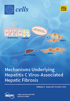
Hepatitis C virus (HCV) infection in human often causes liver fibrosis, although the virus does not replicate in hepatic stellate cells. Exosomes carry different biomolecules as cargo, including soluble and membrane-bound proteins, lipids, and nucleic acids. HCV-infected hepatocytes secrete exosomes for intercellular communication and play a role in the induction of liver fibrosis. Exosomes are easily uptaken by quiescent hepatic stellate cells. The exposure of biomolecules from internalized exosomes promotes activation of quiescent hepatic stellate cells which, in turn, leads to fibrosis. Other mechanisms also play an important role in establishing liver fibrosis. View this paper.
  • Issues are regarded as officially published after their release is announced to the table of contents alert mailing list.
  • You may sign up for e-mail alerts to receive table of contents of newly released issues.
  • PDF is the official format for papers published in both, html and pdf forms. To view the papers in pdf format, click on the "PDF Full-text" link, and use the free Adobe Reader to open them.
Order results
Result details
Section
Select all
Export citation of selected articles as:
14 pages, 3059 KiB  
Article
Dental Epithelial Stem Cells as a Source for Mammary Gland Regeneration and Milk Producing Cells In Vivo
by Lucia Jimenez-Rojo, Pierfrancesco Pagella, Hidemitsu Harada and Thimios A. Mitsiadis
Cells 2019, 8(10), 1302; https://doi.org/10.3390/cells8101302 - 22 Oct 2019
Cited by 9 | Viewed by 31085
Abstract
The continuous growth of rodent incisors is ensured by clusters of mesenchymal and epithelial stem cells that are located at the posterior part of these teeth. Genetic lineage tracing studies have shown that dental epithelial stem cells (DESCs) are able to generate all [...] Read more.
The continuous growth of rodent incisors is ensured by clusters of mesenchymal and epithelial stem cells that are located at the posterior part of these teeth. Genetic lineage tracing studies have shown that dental epithelial stem cells (DESCs) are able to generate all epithelial cell populations within incisors during homeostasis. However, it remains unclear whether these cells have the ability to adopt alternative fates in response to extrinsic factors. Here, we have studied the plasticity of DESCs in the context of mammary gland regeneration. Transplantation of DESCs together with mammary epithelial cells into the mammary stroma resulted in the formation of chimeric ductal epithelial structures in which DESCs adopted all the possible mammary fates including milk-producing alveolar cells. In addition, when transplanted without mammary epithelial cells, DESCs developed branching rudiments and cysts. These in vivo findings demonstrate that when outside their niche, DESCs redirect their fates according to their new microenvironment and thus can contribute to the regeneration of non-dental tissues. Full article
(This article belongs to the Special Issue Stem Cell Therapy in Oral and Maxillofacial Region)
Show Figures

Figure 1

19 pages, 3211 KiB  
Article
Invertebrate Retinal Progenitors as Regenerative Models in a Microfluidic System
by Caroline D. Pena, Stephanie Zhang, Robert Majeska, Tadmiri Venkatesh and Maribel Vazquez
Cells 2019, 8(10), 1301; https://doi.org/10.3390/cells8101301 - 22 Oct 2019
Cited by 10 | Viewed by 3532
Abstract
Regenerative retinal therapies have introduced progenitor cells to replace dysfunctional or injured neurons and regain visual function. While contemporary cell replacement therapies have delivered retinal progenitor cells (RPCs) within customized biomaterials to promote viability and enable transplantation, outcomes have been severely limited by [...] Read more.
Regenerative retinal therapies have introduced progenitor cells to replace dysfunctional or injured neurons and regain visual function. While contemporary cell replacement therapies have delivered retinal progenitor cells (RPCs) within customized biomaterials to promote viability and enable transplantation, outcomes have been severely limited by the misdirected and/or insufficient migration of transplanted cells. RPCs must achieve appropriate spatial and functional positioning in host retina, collectively, to restore vision, whereas movement of clustered cells differs substantially from the single cell migration studied in classical chemotaxis models. Defining how RPCs interact with each other, neighboring cell types and surrounding extracellular matrixes are critical to our understanding of retinogenesis and the development of effective, cell-based approaches to retinal replacement. The current article describes a new bio-engineering approach to investigate the migratory responses of innate collections of RPCs upon extracellular substrates by combining microfluidics with the well-established invertebrate model of Drosophila melanogaster. Experiments utilized microfluidics to investigate how the composition, size, and adhesion of RPC clusters on defined extracellular substrates affected migration to exogenous chemotactic signaling. Results demonstrated that retinal cluster size and composition influenced RPC clustering upon extracellular substrates of concanavalin (Con-A), Laminin (LM), and poly-L-lysine (PLL), and that RPC cluster size greatly altered collective migratory responses to signaling from Fibroblast Growth Factor (FGF), a primary chemotactic agent in Drosophila. These results highlight the significance of examining collective cell-biomaterial interactions on bio-substrates of emerging biomaterials to aid directional migration of transplanted cells. Our approach further introduces the benefits of pairing genetically controlled models with experimentally controlled microenvironments to advance cell replacement therapies. Full article
(This article belongs to the Special Issue Cell Biological Techniques and Cell-Biomaterial Interactions)
Show Figures

Figure 1

10 pages, 1441 KiB  
Article
Association of the Rheumatoid Arthritis Severity Variant rs26232 with the Invasive Activity of Synovial Fibroblasts
by Emma R Dorris, Eimear Linehan, Michelle Trenkmann, Douglas J Veale, Ursula Fearon and Anthony G. Wilson
Cells 2019, 8(10), 1300; https://doi.org/10.3390/cells8101300 - 22 Oct 2019
Cited by 4 | Viewed by 2958
Abstract
rs26232, located in intron one of C5orf30, is associated with the susceptibility to and severity of rheumatoid arthritis (RA). Here, we investigate the relationship between this variant and the biological activities of rheumatoid arthritis synovial fibroblasts (RASFs). RASFs were isolated from the [...] Read more.
rs26232, located in intron one of C5orf30, is associated with the susceptibility to and severity of rheumatoid arthritis (RA). Here, we investigate the relationship between this variant and the biological activities of rheumatoid arthritis synovial fibroblasts (RASFs). RASFs were isolated from the knee joints of 33 RA patients. The rs26232 genotype was determined and cellular migration, invasion, and apoptosis were compared using in vitro techniques. The production of adhesion molecules, chemokines, and proteases was measured by ELISA or flow cytometry. Cohort genotypes were CC n = 16; CT n = 14; TT n = 3. In comparison with the RASFs of the CT genotype, the CC genotype showed a 1.48-fold greater invasiveness in vitro (p = 0.02), 1.6-fold higher expression intracellular adhesion molecule (ICAM)-1 (p = 0.001), and 5-fold IFN-γ inducible protein-10 (IP-10) (p = 0.01). There was no association of the rs26232 genotype with the expression levels of either total C5orf30 mRNA or any of the three transcript variants. The rs26232 C allele, which has previously been associated with both the risk and severity of RA, is associated with greater invasive activity of RASFs in vitro, and with higher expression of ICAM-1 and IP-10. In resting RASFs, rs26232 is not a quantitative trait locus for C5orf30 mRNA, indicating a more complex mechanism underlying the genotype‒phenotype relationship. Full article
(This article belongs to the Special Issue The Molecular and Cellular Basis for Rheumatoid Arthritis)
Show Figures

Figure 1

17 pages, 2820 KiB  
Article
Opposite Effects of Moderate and Extreme Cx43 Deficiency in Conditional Cx43-Deficient Mice on Angiotensin II-Induced Cardiac Fibrosis
by Laura Valls-Lacalle, Corall Negre-Pujol, Cristina Rodríguez, Saray Varona, Antoni Valera-Cañellas, Marta Consegal, Jose Martínez-González and Antonio Rodríguez-Sinovas
Cells 2019, 8(10), 1299; https://doi.org/10.3390/cells8101299 - 22 Oct 2019
Cited by 12 | Viewed by 3998
Abstract
Connexin 43 (Cx43) is essential for cardiac electrical coupling, but its effects on myocardial fibrosis is controversial. Here, we analyzed the role of Cx43 in myocardial fibrosis caused by angiotensin II (AngII) using Cx43fl/fl and Cx43Cre-ER(T)/fl inducible knock-out (Cx43 content: 50%) [...] Read more.
Connexin 43 (Cx43) is essential for cardiac electrical coupling, but its effects on myocardial fibrosis is controversial. Here, we analyzed the role of Cx43 in myocardial fibrosis caused by angiotensin II (AngII) using Cx43fl/fl and Cx43Cre-ER(T)/fl inducible knock-out (Cx43 content: 50%) mice treated with vehicle or 4-hydroxytamoxifen (4-OHT) to induce a Cre-ER(T)-mediated global deletion of the Cx43 floxed allele. Myocardial collagen content was enhanced by AngII in all groups (n = 8–10/group, p < 0.05). However, animals with partial Cx43 deficiency (vehicle-treated Cx43Cre-ER(T)/fl) had a significantly higher AngII-induced collagen accumulation that reverted when treated with 4-OHT, which abolished Cx43 expression. The exaggerated fibrotic response to AngII in partially deficient Cx43Cre-ER(T)/fl mice was associated with enhanced p38 MAPK activation and was not evident in Cx43 heterozygous (Cx43+/-) mice. In contrast, normalization of interstitial collagen in 4-OHT-treated Cx43Cre-ER(T)/fl animals correlated with enhanced MMP-9 activity, IL-6 and NOX2 mRNA expression, and macrophage content, and with reduced α-SMA and SM22α in isolated fibroblasts. In conclusion, our data demonstrates an exaggerated, p38 MAPK-dependent, fibrotic response to AngII in partially deficient Cx43Cre-ER(T)/fl mice, and a paradoxical normalization of collagen deposition in animals with an almost complete Cx43 ablation, an effect associated with increased MMP-9 activity and inflammatory response and reduced fibroblasts differentiation. Full article
(This article belongs to the Special Issue Cells in Cardiovascular Disease)
Show Figures

Figure 1

17 pages, 5279 KiB  
Article
MicroRNA-29a Suppresses CD36 to Ameliorate High Fat Diet-Induced Steatohepatitis and Liver Fibrosis in Mice
by Hung-Yu Lin, Feng-Sheng Wang, Ya-Ling Yang and Ying-Hsien Huang
Cells 2019, 8(10), 1298; https://doi.org/10.3390/cells8101298 - 22 Oct 2019
Cited by 67 | Viewed by 7296
Abstract
MicroRNA-29 (miR-29) has been shown to play a critical role in reducing inflammation and fibrosis following liver injury. Non-alcoholic fatty liver disease (NAFLD) occurs when fat is deposited (steatosis) in the liver due to causes other than excessive alcohol use and is associated [...] Read more.
MicroRNA-29 (miR-29) has been shown to play a critical role in reducing inflammation and fibrosis following liver injury. Non-alcoholic fatty liver disease (NAFLD) occurs when fat is deposited (steatosis) in the liver due to causes other than excessive alcohol use and is associated with liver fibrosis. In this study, we asked whether miR-29a could reduce experimental high fat diet (HFD)-induced obesity and liver fibrosis in mice. We performed systematical expression analyses of miR-29a transgenic mice (miR-29aTg mice) and wild-type littermates subjected to HFD-induced NAFLD. The results demonstrated that increased miR-29a not only alleviated HFD-induced body weight gain but also subcutaneous, visceral, and intestinal fat accumulation and hepatocellular steatosis in mice. Furthermore, hepatic tissue in the miR-29aTg mice displayed a weak fibrotic matrix concomitant with low fibrotic collagen1α1 expression within the affected tissues compared to the wild-type (WT) mice fed the HFD diet. Increased miR-29a signaling also resulted in the downregulation of expression of the epithelial mesenchymal transition-executing transcription factor snail, mesenchymal markers vimentin, and such pro-inflammation markers as il6 and mcp1 within the liver tissue. Meanwhile, miR-29aTg-HFD mice exhibited significantly lower levels of peroxisome proliferator-activated receptor γ (PPARγ), mitochondrial transcription factor A TFAM, and mitochondria DNA content in the liver than the WT-HFD mice. An in vitro luciferase reporter assay further confirmed that miR-29a mimic transfection reduced fatty acid translocase CD36 expression in HepG2 cells. Conclusion: Our data provide new insights that miR-29a can improve HDF-induced obesity, hepatocellular steatosis, and fibrosis, as well as highlight the role of miR-29a in regulation of NAFLD. Full article
Show Figures

Graphical abstract

26 pages, 7021 KiB  
Article
Inhibition of Non-Small Cell Lung Cancer Cells by Oxy210, an Oxysterol-Derivative that Antagonizes TGFβ and Hedgehog Signaling
by Frank Stappenbeck, Feng Wang, Liu-Ya Tang, Ying E. Zhang and Farhad Parhami
Cells 2019, 8(10), 1297; https://doi.org/10.3390/cells8101297 - 22 Oct 2019
Cited by 13 | Viewed by 4677
Abstract
Non-Small Cell Lung Cancer (NSCLC) is a common malignancy and leading cause of death by cancer. Metastasis and drug resistance are serious clinical problems encountered in NSCLC therapy. Aberrant activation of the Transforming Growth Factor beta (TGFβ) and Hedgehog (Hh) signal transduction cascades [...] Read more.
Non-Small Cell Lung Cancer (NSCLC) is a common malignancy and leading cause of death by cancer. Metastasis and drug resistance are serious clinical problems encountered in NSCLC therapy. Aberrant activation of the Transforming Growth Factor beta (TGFβ) and Hedgehog (Hh) signal transduction cascades often associate with poor prognosis and aggressive disease progression in NSCLC, as these signals can drive cell proliferation, angiogenesis, metastasis, immune evasion and emergence of drug resistance. Therefore, simultaneous inhibition of TGFβ and Hh signaling, by a single agent, or in combination with other drugs, could yield therapeutic benefits in NSCLC and other cancers. In the current study, we report on the biological and pharmacological evaluation of Oxy210, an oxysterol-based dual inhibitor of TGFβ and Hh signaling. In NSCLC cells, Oxy210 inhibits proliferation, epithelial-mesenchymal transition (EMT) and invasive activity. Combining Oxy210 with Carboplatin (CP) increases the anti-proliferative response to CP and inhibits TGFβ-induced resistance to CP in A549 NSCLC cells. In addition, Oxy210 displays encouraging drug-like properties, including chemical scalability, metabolic stability and oral bioavailability in mice. Unlike other known inhibitors, Oxy210 antagonizes TGFβ and Hh signaling independently of TGFβ receptor kinase inhibition and downstream of Smoothened, respectively. Full article
(This article belongs to the Special Issue Hedgehog Signaling in Development and Cancer)
Show Figures

Figure 1

19 pages, 3017 KiB  
Article
Hepatitis C Virus Improves Human Tregs Suppressive Function and Promotes Their Recruitment to the Liver
by Laurissa Ouaguia, Olivier Moralès, Lynda Aoudjehane, Czeslaw Wychowski, Abhishek Kumar, Jean Dubuisson, Yvon Calmus, Filomena Conti and Nadira Delhem
Cells 2019, 8(10), 1296; https://doi.org/10.3390/cells8101296 - 22 Oct 2019
Cited by 6 | Viewed by 2876
Abstract
Background: The role of regulatory T cells (Tregs) is now well established in the progression of hepatocellular carcinoma (HCC) linked to Hepatitis C virus (HCV) infection. However, nothing is known about the potential interplay between Tregs and HCV. In this pilot study, we [...] Read more.
Background: The role of regulatory T cells (Tregs) is now well established in the progression of hepatocellular carcinoma (HCC) linked to Hepatitis C virus (HCV) infection. However, nothing is known about the potential interplay between Tregs and HCV. In this pilot study, we have investigated the ability of Tregs to hang HCV on and the subsequent effect on their suppressive function and phenotype. Moreover, we have evaluated how HCV could promote the recruitment of Tregs by infected primary human hepatocytes. Methods: Tregs of healthy donors were incubated with JFH-1/HCVcc. Viral inoculation was assessed using adapted assays (RT-qPCR, Flow Citometry (FACS) and Western Blot (WB). Expression of Tregs phenotypic (CD4, CD25, CD127 and Foxp3) and functional (IL-10, GZMB, TGF-β1 and IL-2) markers was monitored by RT-qPCR, FACS and ELISA. Suppressive activity was validated by suppressive assays. Tregs recruitment by infected primary hepatic cells was evaluated using Boyden Chamber. Results: Tregs express the classical HCV receptors (CD81, CLDN1 and LDLR) and some co-receptors (CD5). HCV inoculation significantly increases the suppressive phenotype and activity of Tregs, and raises their anergy by inducing an unexpected IL-2 production. Moreover, HCV infection induces the expression of chemokines (CCL17, CXCL16, and CCL20) by primary hepatic human hepatocytes and chemokine receptors (CCR4, CXCR6 and CCR6) by Tregs. Finally, infected hepatocytes have a significantly higher potential to recruit Tregs in a seemingly CCL20-dependent manner. Conclusions: Direct interaction between HCV and Tregs represents a newly defined mechanism that could potentiate HCV immune evasion and favor intratumoral recruitment contributing to HCC progression. Full article
(This article belongs to the Special Issue Regulatory T (Treg) Cells in Health and Diseases)
Show Figures

Figure 1

17 pages, 1806 KiB  
Article
Tumor Cells Develop Defined Cellular Phenotypes After 3D-Bioprinting in Different Bioinks
by Sonja K. Schmidt, Rafael Schmid, Andreas Arkudas, Annika Kengelbach-Weigand and Anja K. Bosserhoff
Cells 2019, 8(10), 1295; https://doi.org/10.3390/cells8101295 - 22 Oct 2019
Cited by 34 | Viewed by 5017
Abstract
Malignant melanoma is often used as a model tumor for the establishment of novel therapies. It is known that two-dimensional (2D) culture methods are not sufficient to elucidate the various processes during cancer development and progression. Therefore, it is of major interest to [...] Read more.
Malignant melanoma is often used as a model tumor for the establishment of novel therapies. It is known that two-dimensional (2D) culture methods are not sufficient to elucidate the various processes during cancer development and progression. Therefore, it is of major interest to establish defined biofabricated three-dimensional (3D) models, which help to decipher complex cellular interactions. To get an impression of their printability and subsequent behavior, we printed fluorescently labeled melanoma cell lines with Matrigel and two different types of commercially available bioinks, without or with modification (RGD (Arginine-Glycine-Aspartate)-sequence/laminin-mixture) for increased cell-matrix communication. In general, we demonstrated the printability of melanoma cells in all tested biomaterials and survival of the printed cells throughout 14 days of cultivation. Melanoma cell lines revealed specific differential behavior in the respective inks. Whereas in Matrigel, the cells were able to spread, proliferate and form dense networks throughout the construct, the cells showed no proliferation at all in alginate-based bioink. In gelatin methacrylate-based bioink, the cells proliferated in clusters. Surprisingly, the modifications of the bioinks with RGD or the laminin blend did not affect the analyzed cellular behavior. Our results underline the importance of precisely adapting extracellular matrices to individual requirements of specific 3D bioprinting applications. Full article
Show Figures

Figure 1

7 pages, 238 KiB  
Editorial
Tubulin: Structure, Functions and Roles in Disease
by Pavla Binarová and Jack Tuszynski
Cells 2019, 8(10), 1294; https://doi.org/10.3390/cells8101294 - 22 Oct 2019
Cited by 69 | Viewed by 7378
Abstract
Highly conserved α- and β-tubulin heterodimers assemble into dynamic microtubules and perform multiple important cellular functions such as structural support, pathway for transport and force generation in cell division. Tubulin exists in different forms of isotypes expressed by specific genes with spatially- and [...] Read more.
Highly conserved α- and β-tubulin heterodimers assemble into dynamic microtubules and perform multiple important cellular functions such as structural support, pathway for transport and force generation in cell division. Tubulin exists in different forms of isotypes expressed by specific genes with spatially- and temporally-regulated expression levels. Some tubulin isotypes are differentially expressed in normal and neoplastic cells, providing a basis for cancer chemotherapy drug development. Moreover, specific tubulin isotypes are overexpressed and localized in the nuclei of cancer cells and/or show bioenergetic functions through the regulation of the permeability of mitochondrial ion channels. It has also become clear that tubulin isotypes are involved in multiple cellular functions without being incorporated into microtubule structures. Understanding the mutations of tubulin isotypes specifically expressed in tumors and their post-translational modifications might help to identify precise molecular targets for the design of novel anti-microtubular drugs. Knowledge of tubulin mutations present in tubulinopathies brings into focus cellular functions of tubulin in brain pathologies such as Alzheimer’s disease. Uncovering signaling pathways which affect tubulin functions during antigen-mediated activation of mast cells presents a major challenge in developing new strategies for the treatment of inflammatory and allergic diseases. γ-tubulin, a conserved member of the eukaryotic tubulin superfamily specialized for microtubule nucleation is a target of cell cycle and stress signaling. Besides its microtubule nucleation role, γ-tubulin functions in nuclear and cell cycle related processes. This special issue “Tubulin: Structure, Functions and Roles in Disease” contains eight articles, five of which are original research papers and three are review papers that cover diverse areas of tubulin biology and functions under normal and pathological conditions. Full article
(This article belongs to the Special Issue Tubulin: Structure, Functions and Roles in Disease)
17 pages, 695 KiB  
Review
What Do Microglia Really Do in Healthy Adult Brain?
by Marcus Augusto-Oliveira, Gabriela P. Arrifano, Amanda Lopes-Araújo, Leticia Santos-Sacramento, Priscila Y. Takeda, Daniel C. Anthony, João O. Malva and Maria Elena Crespo-Lopez
Cells 2019, 8(10), 1293; https://doi.org/10.3390/cells8101293 - 22 Oct 2019
Cited by 82 | Viewed by 14831
Abstract
Microglia originate from yolk sac-primitive macrophages and auto-proliferate into adulthood without replacement by bone marrow-derived circulating cells. In inflammation, stroke, aging, or infection, microglia have been shown to contribute to brain pathology in both deleterious and beneficial ways, which have been studied extensively. [...] Read more.
Microglia originate from yolk sac-primitive macrophages and auto-proliferate into adulthood without replacement by bone marrow-derived circulating cells. In inflammation, stroke, aging, or infection, microglia have been shown to contribute to brain pathology in both deleterious and beneficial ways, which have been studied extensively. However, less is known about their role in the healthy adult brain. Astrocytes and oligodendrocytes are widely accepted to strongly contribute to the maintenance of brain homeostasis and to modulate neuronal function. On the other hand, contribution of microglia to cognition and behavior is only beginning to be understood. The ability to probe their function has become possible using microglial depletion assays and conditional mutants. Studies have shown that the absence of microglia results in cognitive and learning deficits in rodents during development, but this effect is less pronounced in adults. However, evidence suggests that microglia play a role in cognition and learning in adulthood and, at a cellular level, may modulate adult neurogenesis. This review presents the case for repositioning microglia as key contributors to the maintenance of homeostasis and cognitive processes in the healthy adult brain, in addition to their classical role as sentinels coordinating the neuroinflammatory response to tissue damage and disease. Full article
Show Figures

Figure 1

13 pages, 3711 KiB  
Protocol
Turning the World Upside-Down in Cellulose for Improved Culturing and Imaging of Respiratory Challenges within a Human 3D Model
by Viktoria Zaderer, Martin Hermann, Cornelia Lass-Flörl, Wilfried Posch and Doris Wilflingseder
Cells 2019, 8(10), 1292; https://doi.org/10.3390/cells8101292 - 21 Oct 2019
Cited by 16 | Viewed by 9709
Abstract
Polarized growth of human-derived respiratory epithelial cells on hydrogel-coated filters offers big advantages concerning detailed experiments with respect to drug screening or host pathogen interactions. Different microscopic approaches, such as confocal analyses and high content screening, help to examine such 3D respiratory samples, [...] Read more.
Polarized growth of human-derived respiratory epithelial cells on hydrogel-coated filters offers big advantages concerning detailed experiments with respect to drug screening or host pathogen interactions. Different microscopic approaches, such as confocal analyses and high content screening, help to examine such 3D respiratory samples, resulting in high-resolution pictures and enabling quantitative analyses of high cell numbers. A major problem employing these techniques relates to single-use instead of multiple-use of Transwell filters and difficulties in the digestion of collagen if subsequent analyses are needed. Up to date, cells are seeded in collagen-based matrices to the inner field of Transwell inserts, which makes it impossible to image due to the design of the inserts and hard to perform other analyses since digestion of the collagen matrix also affects Transwell grown cells. To overcome these problems, we optimized culturing conditions for monitoring cell differentiation or repeated dose experiments over a long time period. For this, cells are seeded upside-down to the bottom side of filters within an animal-free cellulose hydrogel. These cells were then grown inverted under static conditions and were differentiated in air-liquid interphase (ALI). Full differentiation of goblet (Normal Human Bronchial Epithelial (NHBE))/Club (small airway epithelia (SAE)) cells and ciliated cells was detected after 12 days in ALI. Inverted cell cultures could then be used for ‘follow-up’ live cell imaging experiments, as well as, flow-cytometric analyses due to easy digestion of the cellulose compared to classical collagen matrices. Additionally, this culture technique also enables easy addition of immune cells, such as dendritic cells (DCs), macrophages, neutrophils, T or B cells alone or in combination, to the inner field of the Transwell to monitor immune cell behavior after repeated respiratory challenge. Our detailed protocol offers the possibility of culturing human primary polarized cells into a fully differentiated, thick epithelium without any animal components over >700 days. Furthermore, this animal-free, inverted system allows investigation of the same inserts, because the complete Transwell can be readily transferred to glass-bottom dishes for live cell imaging analyses and then returned to its original plate for further cultivation. Full article
Show Figures

Graphical abstract

25 pages, 1143 KiB  
Review
Current Paradigms of Tolerogenic Dendritic Cells and Clinical Implications for Systemic Lupus Erythematosus
by Patcharee Ritprajak, Chamraj Kaewraemruaen and Nattiya Hirankarn
Cells 2019, 8(10), 1291; https://doi.org/10.3390/cells8101291 - 21 Oct 2019
Cited by 24 | Viewed by 5085
Abstract
Tolerogenic dendritic cells (tolDCs) are central players in the initiation and maintenance of immune tolerance and subsequent prevention of autoimmunity. Recent advances in treatment of autoimmune diseases including systemic lupus erythematosus (SLE) have focused on inducing specific tolerance to avoid long-term use of [...] Read more.
Tolerogenic dendritic cells (tolDCs) are central players in the initiation and maintenance of immune tolerance and subsequent prevention of autoimmunity. Recent advances in treatment of autoimmune diseases including systemic lupus erythematosus (SLE) have focused on inducing specific tolerance to avoid long-term use of immunosuppressive drugs. Therefore, DC-targeted therapies to either suppress DC immunogenicity or to promote DC tolerogenicity are of high interest. This review describes details of the typical characteristics of in vivo and ex vivo tolDC, which will help to select a protocol that can generate tolDC with high functional quality for clinical treatment of autoimmune disease in individual patients. In addition, we discuss the recent studies uncovering metabolic pathways and their interrelation intertwined with DC tolerogenicity. This review also highlights the clinical implications of tolDC-based therapy for SLE treatment, examines the current clinical therapeutics in patients with SLE, which can generate tolDC in vivo, and further discusses on possibility and limitation on each strategy. This synthesis provides new perspectives on development of novel therapeutic approaches for SLE and other autoimmune diseases. Full article
(This article belongs to the Special Issue The Molecular and Cellular Basis for Lupus)
Show Figures

Figure 1

13 pages, 2838 KiB  
Article
Helicobacter pylori Induces IL-33 Production and Recruits ST-2 to Lipid Rafts to Exacerbate Inflammation
by Chia-Jung Kuo, Chun-Ya Chen, Horng-Ren Lo, Chun-Lung Feng, Hui-Yu Wu, Mei-Zi Huang, Tung-Nan Liao, Yu-An Chen and Chih-Ho Lai
Cells 2019, 8(10), 1290; https://doi.org/10.3390/cells8101290 - 21 Oct 2019
Cited by 16 | Viewed by 4658
Abstract
Helicobacter pylori colonizes human gastric epithelial cells and contributes to the development of several gastrointestinal disorders. Interleukin (IL)-33 is involved in various immune responses, with reported proinflammatory and anti-inflammatory effects, which may be associated with colitis and colitis-associated cancer. IL-33 induces the inflammatory [...] Read more.
Helicobacter pylori colonizes human gastric epithelial cells and contributes to the development of several gastrointestinal disorders. Interleukin (IL)-33 is involved in various immune responses, with reported proinflammatory and anti-inflammatory effects, which may be associated with colitis and colitis-associated cancer. IL-33 induces the inflammatory cascade through its receptor, suppression of tumorigenicity-2 (ST-2). Binding of IL-33 to membrane-bound ST-2 (mST-2) recruits the IL-1 receptor accessory protein (IL-1RAcP) and activates intracellular signaling pathways. However, whether IL-33/ST-2 is triggered by H. pylori infection and whether this interaction occurs in lipid rafts remain unclear. Our study showed that both IL-33 and ST-2 expression levels were significantly elevated in H. pylori-infected cells. Confocal microscopy showed that ST-2 mobilized into the membrane lipid rafts during infection. Depletion of membrane cholesterol dampened H. pylori-induced IL-33 and IL-8 production. Furthermore, in vivo studies revealed IL-33/ST-2 upregulation, and severe leukocyte infiltration was observed in gastric tissues infected with H. pylori. Together, these results demonstrate that ST-2 recruitment into the lipid rafts serves as a platform for IL-33-dependent H. pylori infection, which aggravates inflammation in the stomach. Full article
(This article belongs to the Special Issue Immunomodulatory Factors in Host Defense)
Show Figures

Graphical abstract

19 pages, 2969 KiB  
Article
The Charcot–Marie Tooth Disease Mutation R94Q in MFN2 Decreases ATP Production but Increases Mitochondrial Respiration under Conditions of Mild Oxidative Stress
by Christina Wolf, Rahel Zimmermann, Osamah Thaher, Diones Bueno, Verena Wüllner, Michael K.E. Schäfer, Philipp Albrecht and Axel Methner
Cells 2019, 8(10), 1289; https://doi.org/10.3390/cells8101289 - 21 Oct 2019
Cited by 15 | Viewed by 4282
Abstract
Charcot–Marie tooth disease is a hereditary polyneuropathy caused by mutations in Mitofusin-2 (MFN2), a GTPase in the outer mitochondrial membrane involved in the regulation of mitochondrial fusion and bioenergetics. Autosomal-dominant inheritance of a R94Q mutation in MFN2 causes the axonal subtype 2A2A which [...] Read more.
Charcot–Marie tooth disease is a hereditary polyneuropathy caused by mutations in Mitofusin-2 (MFN2), a GTPase in the outer mitochondrial membrane involved in the regulation of mitochondrial fusion and bioenergetics. Autosomal-dominant inheritance of a R94Q mutation in MFN2 causes the axonal subtype 2A2A which is characterized by early onset and progressive atrophy of distal muscles caused by motoneuronal degeneration. Here, we studied mitochondrial shape, respiration, cytosolic, and mitochondrial ATP content as well as mitochondrial quality control in MFN2-deficient fibroblasts stably expressing wildtype or R94Q MFN2. Under normal culture conditions, R94Q cells had slightly more fragmented mitochondria but a similar mitochondrial oxygen consumption, membrane potential, and ATP production as wildtype cells. However, when inducing mild oxidative stress 24 h before analysis using 100 µM hydrogen peroxide, R94Q cells exhibited significantly increased respiration but decreased mitochondrial ATP production. This was accompanied by increased glucose uptake and an up-regulation of hexokinase 1 and pyruvate kinase M2, suggesting increased pyruvate shuttling into mitochondria. Interestingly, these changes coincided with decreased levels of PINK1/Parkin-mediated mitophagy in R94Q cells. We conclude that mitochondria harboring the disease-causing R94Q mutation in MFN2 are more susceptible to oxidative stress, which causes uncoupling of respiration and ATP production possibly by a less efficient mitochondrial quality control. Full article
Show Figures

Figure 1

23 pages, 9489 KiB  
Article
Subcutaneous and Visceral Adipose-Derived Mesenchymal Stem Cells: Commonality and Diversity
by Andreas Ritter, Alexandra Friemel, Susanne Roth, Nina-Naomi Kreis, Samira Catharina Hoock, Babek Khan Safdar, Kyra Fischer, Charlotte Möllmann, Christine Solbach, Frank Louwen and Juping Yuan
Cells 2019, 8(10), 1288; https://doi.org/10.3390/cells8101288 - 21 Oct 2019
Cited by 34 | Viewed by 3736
Abstract
Adipose-derived mesenchymal stem cells (ASCs) are considered to be a useful tool for regenerative medicine, owing to their capabilities in differentiation, self-renewal, and immunomodulation. These cells have become a focus in the clinical setting due to their abundance and easy isolation. However, ASCs [...] Read more.
Adipose-derived mesenchymal stem cells (ASCs) are considered to be a useful tool for regenerative medicine, owing to their capabilities in differentiation, self-renewal, and immunomodulation. These cells have become a focus in the clinical setting due to their abundance and easy isolation. However, ASCs from different depots are not well characterized. Here, we analyzed the functional similarities and differences of subcutaneous and visceral ASCs. Subcutaneous ASCs have an extraordinarily directed mode of motility and a highly dynamic focal adhesion turnover, even though they share similar surface markers, whereas visceral ASCs move in an undirected random pattern with more stable focal adhesions. Visceral ASCs have a higher potential to differentiate into adipogenic and osteogenic cells when compared to subcutaneous ASCs. In line with these observations, visceral ASCs demonstrate a more active sonic hedgehog pathway that is linked to a high expression of cilia/differentiation related genes. Moreover, visceral ASCs secrete higher levels of inflammatory cytokines interleukin-6, interleukin-8, and tumor necrosis factor α relative to subcutaneous ASCs. These findings highlight, that both ASC subpopulations share multiple cellular features, but significantly differ in their functions. The functional diversity of ASCs depends on their origin, cellular context and surrounding microenvironment within adipose tissues. The data provide important insight into the biology of ASCs, which might be useful in choosing the adequate ASC subpopulation for regenerative therapies. Full article
(This article belongs to the Special Issue Adipose-Derived Stromal/Stem Cells)
Show Figures

Figure 1

17 pages, 8243 KiB  
Article
Estimating Dynamic Cellular Morphological Properties via the Combination of the RTCA System and a Hough-Transform-Based Algorithm
by Lejun Zhang, Yang Ye, Rana Dhar, Jinsong Deng and Huifang Tang
Cells 2019, 8(10), 1287; https://doi.org/10.3390/cells8101287 - 21 Oct 2019
Cited by 9 | Viewed by 2954
Abstract
The xCELLigence real-time cell analysis (RTCA) system has the potential to detect cellular proliferation, migration, cytotoxicity, adherence, and remodeling. Although the RTCA system is widely recognized as a noninvasive and efficient tool for real-time monitoring of cellular fate, it cannot describe detailed cell [...] Read more.
The xCELLigence real-time cell analysis (RTCA) system has the potential to detect cellular proliferation, migration, cytotoxicity, adherence, and remodeling. Although the RTCA system is widely recognized as a noninvasive and efficient tool for real-time monitoring of cellular fate, it cannot describe detailed cell morphological parameters, such as length and intensity. Transforming growth factor beta(TGF-β) induced the epithelial–mesenchymal transition (EMT), which produces significant changes in cellular morphology, so we used TGF-β to treat A549 epithelial cells in this study. We compared it with lipopolysaccharide (LPS) and cigarette smoke extract (CSE) as stimulators. We developed an efficient algorithm to quantify the morphological cell changes. This algorithm is comprised of three major parts: image preprocessing, Hough transform (HT), and post-processing. We used the RTCA system to record the A549 cell index. Western blot was used to confirm the EMT. The RTCA system showed that different stimulators produce different cell index curves. The algorithm determined the lengths of the detected lines of cells, and the results were similar to the RTCA system in the TGF-β group. The Western blot results show that TGF-β changed the EMT markers, but the other stimulator remained unchanged. Optics-based computer vision techniques can supply the requisite information for the RTCA system based on good correspondence between the results. Full article
Show Figures

Figure 1

17 pages, 2101 KiB  
Article
A Machine Learning-Based Prediction Platform for P-Glycoprotein Modulators and Its Validation by Molecular Docking
by Onat Kadioglu and Thomas Efferth
Cells 2019, 8(10), 1286; https://doi.org/10.3390/cells8101286 - 21 Oct 2019
Cited by 21 | Viewed by 4355
Abstract
P-glycoprotein (P-gp) is an important determinant of multidrug resistance (MDR) because its overexpression is associated with increased efflux of various established chemotherapy drugs in many clinically resistant and refractory tumors. This leads to insufficient therapeutic targeting of tumor populations, representing a major drawback [...] Read more.
P-glycoprotein (P-gp) is an important determinant of multidrug resistance (MDR) because its overexpression is associated with increased efflux of various established chemotherapy drugs in many clinically resistant and refractory tumors. This leads to insufficient therapeutic targeting of tumor populations, representing a major drawback of cancer chemotherapy. Therefore, P-gp is a target for pharmacological inhibitors to overcome MDR. In the present study, we utilized machine learning strategies to establish a model for P-gp modulators to predict whether a given compound would behave as substrate or inhibitor of P-gp. Random forest feature selection algorithm-based leave-one-out random sampling was used. Testing the model with an external validation set revealed high performance scores. A P-gp modulator list of compounds from the ChEMBL database was used to test the performance, and predictions from both substrate and inhibitor classes were selected for the last step of validation with molecular docking. Predicted substrates revealed similar docking poses than that of doxorubicin, and predicted inhibitors revealed similar docking poses than that of the known P-gp inhibitor elacridar, implying the validity of the predictions. We conclude that the machine-learning approach introduced in this investigation may serve as a tool for the rapid detection of P-gp substrates and inhibitors in large chemical libraries. Full article
(This article belongs to the Special Issue ABC Transporters: From Basic Functions to Diseases)
Show Figures

Figure 1

14 pages, 1399 KiB  
Review
Nuclear Mechanics in the Fission Yeast
by Paola Gallardo, Ramón R. Barrales, Rafael R. Daga and Silvia Salas-Pino
Cells 2019, 8(10), 1285; https://doi.org/10.3390/cells8101285 - 20 Oct 2019
Cited by 6 | Viewed by 4990
Abstract
In eukaryotic cells, the organization of the genome within the nucleus requires the nuclear envelope (NE) and its associated proteins. The nucleus is subjected to mechanical forces produced by the cytoskeleton. The physical properties of the NE and the linkage of chromatin in [...] Read more.
In eukaryotic cells, the organization of the genome within the nucleus requires the nuclear envelope (NE) and its associated proteins. The nucleus is subjected to mechanical forces produced by the cytoskeleton. The physical properties of the NE and the linkage of chromatin in compacted conformation at sites of cytoskeleton contacts seem to be key for withstanding nuclear mechanical stress. Mechanical perturbations of the nucleus normally occur during nuclear positioning and migration. In addition, cell contraction or expansion occurring for instance during cell migration or upon changes in osmotic conditions also result innuclear mechanical stress. Recent studies in Schizosaccharomyces pombe (fission yeast) have revealed unexpected functions of cytoplasmic microtubules in nuclear architecture and chromosome behavior, and have pointed to NE-chromatin tethers as protective elements during nuclear mechanics. Here, we review and discuss how fission yeast cells can be used to understand principles underlying the dynamic interplay between genome organization and function and the effect of forces applied to the nucleus by the microtubule cytoskeleton. Full article
(This article belongs to the Special Issue Nuclear Organisation)
Show Figures

Figure 1

18 pages, 4480 KiB  
Article
Transcriptional Regulator TonEBP Mediates Oxidative Damages in Ischemic Kidney Injury
by Eun Jin Yoo, Sun Woo Lim, Hyun Je Kang, Hyun Park, Sora Yoon, Dougu Nam, Satoru Sanada, Mi Jin Kwon, Whaseon Lee-Kwon, Soo Youn Choi and Hyug Moo Kwon
Cells 2019, 8(10), 1284; https://doi.org/10.3390/cells8101284 - 20 Oct 2019
Cited by 2 | Viewed by 3279
Abstract
TonEBP (tonicity-responsive enhancer binding protein) is a transcriptional regulator whose expression is elevated in response to various forms of stress including hyperglycemia, inflammation, and hypoxia. Here we investigated the role of TonEBP in acute kidney injury (AKI) using a line of TonEBP haplo-deficient [...] Read more.
TonEBP (tonicity-responsive enhancer binding protein) is a transcriptional regulator whose expression is elevated in response to various forms of stress including hyperglycemia, inflammation, and hypoxia. Here we investigated the role of TonEBP in acute kidney injury (AKI) using a line of TonEBP haplo-deficient mice subjected to bilateral renal ischemia followed by reperfusion (I/R). In the TonEBP haplo-deficient animals, induction of TonEBP, oxidative stress, inflammation, cell death, and functional injury in the kidney in response to I/R were all reduced. Analyses of renal transcriptome revealed that genes in several cellular pathways including peroxisome and mitochondrial inner membrane were suppressed in response to I/R, and the suppression was relieved in the TonEBP deficiency. Production of reactive oxygen species (ROS) and the cellular injury was reproduced in a renal epithelial cell line in response to hypoxia, ATP depletion, or hydrogen peroxide. The knockdown of TonEBP reduced ROS production and cellular injury in correlation with increased expression of the suppressed genes. The cellular injury was also blocked by inhibitors of necrosis. These results demonstrate that ischemic insult suppresses many genes involved in cellular metabolism leading to local oxidative stress by way of TonEBP induction. Thus, TonEBP is a promising target to prevent AKI. Full article
Show Figures

Graphical abstract

15 pages, 1676 KiB  
Article
Increased Expression of Meteorin-Like Hormone in Type 2 Diabetes and Obesity and Its Association with Irisin
by Irina AlKhairi, Preethi Cherian, Mohamed Abu-Farha, Ashraf Al Madhoun, Rasheeba Nizam, Motasem Melhem, Mohamed Jamal, Suleiman Al-Sabah, Hamad Ali, Jaakko Tuomilehto, Fahd Al-Mulla and Jehad Abubaker
Cells 2019, 8(10), 1283; https://doi.org/10.3390/cells8101283 - 19 Oct 2019
Cited by 52 | Viewed by 5067
Abstract
Type 2 diabetes (T2D) is a growing pandemic associated with metabolic dysregulation and chronic inflammation. Meteorin-like hormone (METRNL) is an adipomyokine that is linked to T2D. Our objective was to evaluate the changes in METRNL levels in T2D and obesity and assess the [...] Read more.
Type 2 diabetes (T2D) is a growing pandemic associated with metabolic dysregulation and chronic inflammation. Meteorin-like hormone (METRNL) is an adipomyokine that is linked to T2D. Our objective was to evaluate the changes in METRNL levels in T2D and obesity and assess the association of METRNL levels with irisin. Overall, 228 Arab individuals were enrolled. Plasma levels of METRNL and irisin were assessed using immunoassay. Plasma levels of METRNL and irisin were significantly higher in T2D patients than in non-diabetic patients (p < 0.05). When the population was stratified based on obesity, METRNL and irisin levels were significantly higher in obese than in non-obese individuals (p < 0.05). We found a significant positive correlation between METRNL and irisin (r = 0.233 and p = 0.001). Additionally, METRNL and irisin showed significant correlation with various metabolic biomarkers associated with T2D and Obesity. Our data shows elevated METRNL plasma levels in individuals with T2D, further exacerbated with obesity. Additionally, a strong positive association was observed between METRNL and irisin. Further studies are necessary to examine the role of these proteins in T2D and obesity, against their ethnic background and to understand the mechanistic significance of their possible interplay. Full article
(This article belongs to the Special Issue Adipocytes and Metabolic Health)
Show Figures

Figure 1

15 pages, 3727 KiB  
Article
Differentiation of Baboon (Papio anubis) Induced-Pluripotent Stem Cells into Enucleated Red Blood Cells
by Emmanuel N. Olivier, Kai Wang, Joshua Grossman, Nadim Mahmud and Eric E. Bouhassira
Cells 2019, 8(10), 1282; https://doi.org/10.3390/cells8101282 - 19 Oct 2019
Cited by 3 | Viewed by 4451
Abstract
As cell culture methods and stem cell biology have progressed, the in vitro production of cultured RBCs (cRBCs) has emerged as a viable option to produce cells for transfusion or to carry therapeutic cargoes. RBCs produced in culture can be quality-tested either by [...] Read more.
As cell culture methods and stem cell biology have progressed, the in vitro production of cultured RBCs (cRBCs) has emerged as a viable option to produce cells for transfusion or to carry therapeutic cargoes. RBCs produced in culture can be quality-tested either by xeno-transfusion of human cells into immuno-deficient animals, or by transfusion of autologous cells in immuno-competent models. Although murine xeno-transfusion methods have improved, they must be complemented by studies in immuno-competent models. Non-human primates (NHPs) are important pre-clinical, large animal models due to their high biological and developmental similarities with humans, including their comparable hematopoietic and immune systems. Among NHPs, baboons are particularly attractive to validate cRBCs because of the wealth of data available on the characteristics of RBCs in this species that have been generated by past blood transfusion studies. We report here that we have developed a method to produce enucleated cRBCs by differentiation of baboon induced pluripotent stem cells (iPSCs). This method will enable the use of baboons to evaluate therapeutic cRBCs and generate essential pre-clinical data in an immuno-competent, large animal model. Production of the enucleated baboon cRBCs was achieved by adapting the PSC-RED protocol that we previously developed for human cells. Baboon-PSC-RED is an efficient chemically-defined method to differentiate iPSCs into cRBCs that are about 40% to 50% enucleated. PSC-RED is relatively low cost because it requires no albumin and only small amounts of recombinant transferrin. Full article
(This article belongs to the Special Issue iPS Cells for Disease Modeling)
Show Figures

Figure 1

19 pages, 1934 KiB  
Review
Enhancer Dysfunction in 3D Genome and Disease
by Ji-Han Xia and Gong-Hong Wei
Cells 2019, 8(10), 1281; https://doi.org/10.3390/cells8101281 - 19 Oct 2019
Cited by 13 | Viewed by 8277
Abstract
Spatiotemporal patterns of gene expression depend on enhancer elements and other factors during individual development and disease progression. The rapid progress of high-throughput techniques has led to well-defined enhancer chromatin properties. Various genome-wide methods have revealed a large number of enhancers and the [...] Read more.
Spatiotemporal patterns of gene expression depend on enhancer elements and other factors during individual development and disease progression. The rapid progress of high-throughput techniques has led to well-defined enhancer chromatin properties. Various genome-wide methods have revealed a large number of enhancers and the discovery of three-dimensional (3D) genome architecture showing the distant interacting mechanisms of enhancers that loop to target gene promoters. Whole genome sequencing projects directed at cancer have led to the discovery of substantial enhancer dysfunction in misregulating gene expression and in tumor initiation and progression. Results from genome-wide association studies (GWAS) combined with functional genomics analyses have elucidated the functional impacts of many cancer risk-associated variants that are enriched within the enhancer regions of chromatin. Risk variants dysregulate the expression of enhancer variant-associated genes via 3D genomic interactions. Moreover, these enhancer variants often alter the chromatin binding affinity for cancer-relevant transcription factors, which in turn leads to aberrant expression of the genes associated with cancer susceptibility. In this review, we investigate the extent to which these genetic regulatory circuits affect cancer predisposition and how the recent development of genome-editing methods have enabled the determination of the impacts of genomic variation and alteration on cancer phenotype, which will eventually lead to better management plans and treatment responses to human cancer in the clinic. Full article
(This article belongs to the Special Issue Nuclear Organisation)
Show Figures

Figure 1

21 pages, 2338 KiB  
Review
The Adaptive Immune System in Multiple Sclerosis: An Estrogen-Mediated Point of View
by Alessandro Maglione, Simona Rolla, Stefania Federica De Mercanti, Santina Cutrupi and Marinella Clerico
Cells 2019, 8(10), 1280; https://doi.org/10.3390/cells8101280 - 19 Oct 2019
Cited by 26 | Viewed by 4539
Abstract
Multiple sclerosis (MS) is a chronic central nervous system inflammatory disease that leads to demyelination and neurodegeneration. The third trimester of pregnancy, which is characterized by high levels of estrogens, has been shown to be associated with reduced relapse rates compared with the [...] Read more.
Multiple sclerosis (MS) is a chronic central nervous system inflammatory disease that leads to demyelination and neurodegeneration. The third trimester of pregnancy, which is characterized by high levels of estrogens, has been shown to be associated with reduced relapse rates compared with the rates before pregnancy. These effects could be related to the anti-inflammatory properties of estrogens, which orchestrate the reshuffling of the immune system toward immunotolerance to allow for fetal growth. The action of these hormones is mediated by the transcriptional regulation activity of estrogen receptors (ERs). Estrogen levels and ER expression define a specific balance of immune cell types. In this review, we explore the role of estradiol (E2) and ERs in the adaptive immune system, with a focus on estrogen-mediated cellular, molecular, and epigenetic mechanisms related to immune tolerance and neuroprotection in MS. The epigenome dynamics of immune systems are described as key molecular mechanisms that act on the regulation of immune cell identity. This is a completely unexplored field, suggesting a future path for more extensive research on estrogen-induced coregulatory complexes and molecular circuitry as targets for therapeutics in MS. Full article
(This article belongs to the Special Issue The Molecular and Cellular Basis for Multiple Sclerosis)
Show Figures

Figure 1

26 pages, 1395 KiB  
Review
Magnetic-Assisted Treatment of Liver Fibrosis
by Kateryna Levada, Alexander Omelyanchik, Valeria Rodionova, Ralf Weiskirchen and Matthias Bartneck
Cells 2019, 8(10), 1279; https://doi.org/10.3390/cells8101279 - 19 Oct 2019
Cited by 24 | Viewed by 6029
Abstract
Chronic liver injury can be induced by viruses, toxins, cellular activation, and metabolic dysregulation and can lead to liver fibrosis. Hepatic fibrosis still remains a major burden on the global health systems. Nonalcoholic fatty liver disease (NAFLD) and nonalcoholic steatohepatitis (NASH) are considered [...] Read more.
Chronic liver injury can be induced by viruses, toxins, cellular activation, and metabolic dysregulation and can lead to liver fibrosis. Hepatic fibrosis still remains a major burden on the global health systems. Nonalcoholic fatty liver disease (NAFLD) and nonalcoholic steatohepatitis (NASH) are considered the main cause of liver fibrosis. Hepatic stellate cells are key targets in antifibrotic treatment, but selective engagement of these cells is an unresolved issue. Current strategies for antifibrotic drugs, which are at the critical stage 3 clinical trials, target metabolic regulation, immune cell activation, and cell death. Here, we report on the critical factors for liver fibrosis, and on prospective novel drugs, which might soon enter the market. Apart from the current clinical trials, novel perspectives for anti-fibrotic treatment may arise from magnetic particles and controlled magnetic forces in various different fields. Magnetic-assisted techniques can, for instance, enable cell engineering and cell therapy to fight cancer, might enable to control the shape or orientation of single cells or tissues mechanically. Furthermore, magnetic forces may improve localized drug delivery mediated by magnetism-induced conformational changes, and they may also enhance non-invasive imaging applications. Full article
Show Figures

Figure 1

21 pages, 3076 KiB  
Article
Intrinsically Disordered SRC-3/AIB1 Protein Undergoes Homeostatic Nuclear Extrusion by Nuclear Budding While Ectopic Expression Induces Nucleophagy
by Miguel A. Cabrita, L. Isabel Renart, Rosanna Lau and M. A. Christine Pratt
Cells 2019, 8(10), 1278; https://doi.org/10.3390/cells8101278 - 19 Oct 2019
Cited by 2 | Viewed by 3800
Abstract
SRC-3/AIB1 (Amplified in Breast Cancer-1) is a nuclear receptor coactivator for the estrogen receptor in breast cancer cells. It is also an intrinsically disordered protein when not engaged with transcriptional binding partners and degraded upon transcriptional coactivation. Given the amplified expression of SRC-3 [...] Read more.
SRC-3/AIB1 (Amplified in Breast Cancer-1) is a nuclear receptor coactivator for the estrogen receptor in breast cancer cells. It is also an intrinsically disordered protein when not engaged with transcriptional binding partners and degraded upon transcriptional coactivation. Given the amplified expression of SRC-3 in breast cancers, the objective of this study was to determine how increasing SRC-3 protein levels are regulated in MCF-7 breast cancer cells. We found that endogenous SRC-3 was expelled from the nucleus in vesicle-like spheres under normal growth conditions suggesting that this form of nuclear exclusion of SRC-3 is a homeostatic mechanism for regulating nuclear SRC-3 protein. Only SRC-3 not associated with CREB-binding protein (CBP) was extruded from the nucleus. We found that overexpression in MCF-7 cells results in aneuploid senescence and cell death with frequent formation of nuclear aggregates which were consistently juxtaposed to perinuclear microtubules. Transfected SRC-3 was SUMOylated and caused redistribution of nuclear promyelocytic leukemia (PML) bodies and perturbation of the nuclear membrane lamin B1, hallmarks of nucleophagy. Increased SRC-3 protein-induced autophagy and resulted in SUMO-1 localization to the nuclear membrane and formation of protrusions variously containing SRC-3 and chromatin. Aspects of SRC-3 overexpression and toxicity were recapitulated following treatment with clinically relevant agents that stabilize SRC-3 in breast cancer cells. We conclude that amplified SRC-3 levels have major impacts on nuclear protein quality control pathways and may mark cancer cells for sensitivity to protein stabilizing therapeutics. Full article
(This article belongs to the Section Cell Nuclei: Function, Transport and Receptors)
Show Figures

Graphical abstract

14 pages, 1738 KiB  
Article
Increased Expression of RUNX1 in Liver Correlates with NASH Activity Score in Patients with Non-Alcoholic Steatohepatitis (NASH)
by Savneet Kaur, Preety Rawal, Hamda Siddiqui, Sumati Rohilla, Shvetank Sharma, Dinesh M Tripathi, Sukriti Baweja, Mohsin Hassan, Sebastian Vlaic, Reinhard Guthke, Maria Thomas, Rania Dayoub, Chaggan Bihari, Shiv K. Sarin and Thomas S. Weiss
Cells 2019, 8(10), 1277; https://doi.org/10.3390/cells8101277 - 18 Oct 2019
Cited by 25 | Viewed by 5327
Abstract
Given the important role of angiogenesis in liver pathology, the current study investigated the role of Runt-related transcription factor 1 (RUNX1), a regulator of developmental angiogenesis, in the pathogenesis of non-alcoholic steatohepatitis (NASH). Quantitative RT-PCRs and a transcription factor analysis of angiogenesis-associated differentially [...] Read more.
Given the important role of angiogenesis in liver pathology, the current study investigated the role of Runt-related transcription factor 1 (RUNX1), a regulator of developmental angiogenesis, in the pathogenesis of non-alcoholic steatohepatitis (NASH). Quantitative RT-PCRs and a transcription factor analysis of angiogenesis-associated differentially expressed genes in liver tissues of healthy controls, patients with steatosis and NASH, indicated a potential role of RUNX1 in NASH. The gene expression of RUNX1 was correlated with histopathological attributes of patients. The protein expression of RUNX1 in liver was studied by immunohistochemistry. To explore the underlying mechanisms, in vitro studies using RUNX1 siRNA and overexpression plasmids were performed in endothelial cells (ECs). RUNX1 expression was significantly correlated with inflammation, fibrosis and NASH activity score in NASH patients. Its expression was conspicuous in liver non-parenchymal cells. In vitro, factors from steatotic hepatocytes and/or VEGF or TGF-β significantly induced the expression of RUNX1 in ECs. RUNX1 regulated the expression of angiogenic and adhesion molecules in ECs, including CCL2, PECAM1 and VCAM1, which was shown by silencing or over-expression of RUNX1. Furthermore, RUNX1 increased the angiogenic activity of ECs. This study reports that steatosis-induced RUNX1 augmented the expression of adhesion and angiogenic molecules and properties in ECs and may be involved in enhancing inflammation and disease severity in NASH. Full article
Show Figures

Graphical abstract

20 pages, 3118 KiB  
Article
Inhibition of JAK-STAT Signaling with Baricitinib Reduces Inflammation and Improves Cellular Homeostasis in Progeria Cells
by Chang Liu, Rouven Arnold, Gonçalo Henriques and Karima Djabali
Cells 2019, 8(10), 1276; https://doi.org/10.3390/cells8101276 - 18 Oct 2019
Cited by 50 | Viewed by 5218
Abstract
Hutchinson-Gilford progeria syndrome (HGPS), a rare premature aging disorder that leads to death at an average age of 14.7 years due to myocardial infarction or stroke, is caused by mutations in the LMNA gene. Nearly 90% of HGPS cases carry the G608G mutation [...] Read more.
Hutchinson-Gilford progeria syndrome (HGPS), a rare premature aging disorder that leads to death at an average age of 14.7 years due to myocardial infarction or stroke, is caused by mutations in the LMNA gene. Nearly 90% of HGPS cases carry the G608G mutation within exon 11 that generates a truncated prelamin A protein “progerin”. Progerin accumulates in HGPS cells and induces premature senescence at the cellular and organismal levels. Children suffering from HGPS develop numerous clinical features that overlap with normal aging, including atherosclerosis, arthritis, hair loss and lipodystrophy. To determine whether an aberrant signaling pathway might underlie the development of these four diseases (atherosclerosis, arthritis, hair loss and lipodystrophy), we performed a text mining analysis of scientific literature and databases. We found a total of 17 genes associated with all four pathologies, 14 of which were linked to the JAK1/2-STAT1/3 signaling pathway. We report that the inhibition of the JAK-STAT pathway with baricitinib, a Food and Drug Administration-approved JAK1/2 inhibitor, restored cellular homeostasis, delayed senescence and decreased proinflammatory markers in HGPS cells. Our ex vivo data using human cell models indicate that the overactivation of JAK-STAT signaling mediates premature senescence and that the inhibition of this pathway could show promise for the treatment of HGPS and age-related pathologies. Full article
Show Figures

Graphical abstract

12 pages, 2351 KiB  
Article
Capsaicin Targets tNOX (ENOX2) to Inhibit G1 Cyclin/CDK Complex, as Assessed by the Cellular Thermal Shift Assay (CETSA)
by Atikul Islam, Ally J. Su, Zih-Ming Zeng, Pin Ju Chueh and Ming-Hung Lin
Cells 2019, 8(10), 1275; https://doi.org/10.3390/cells8101275 - 18 Oct 2019
Cited by 20 | Viewed by 5999
Abstract
Capsaicin (8-methyl-N-vanillyl-6-noneamide), which is an active component in red chili peppers, is used as a chemopreventive agent that shows favorable cytotoxicity against cancer cells. Accumulating evidence indicates that capsaicin preferentially inhibits a tumor-associated NADH oxidase (tNOX, ENOX2) that is ubiquitously expressed [...] Read more.
Capsaicin (8-methyl-N-vanillyl-6-noneamide), which is an active component in red chili peppers, is used as a chemopreventive agent that shows favorable cytotoxicity against cancer cells. Accumulating evidence indicates that capsaicin preferentially inhibits a tumor-associated NADH oxidase (tNOX, ENOX2) that is ubiquitously expressed in cancer but not in non-transformed cells. This attenuates cancer cell growth by inducing apoptosis. The capsaicin-mediated inhibition of tNOX was recently shown to prolong the cell cycle. However, the molecular events underlying this regulation have not yet been investigated. In the present study, we used a cellular thermal shift assay (CETSA) to detect “target engagement” of capsaicin and its consequent impact on cell cycle progression. Our results indicated that capsaicin engaged with tNOX and triggered the proteasomal degradation of tNOX, which leads to the inhibition of NAD+-dependent SIRT1 deacetylase. Ultimately, the acetylation levels of c-Myc and p53 were enhanced, which suppressed the activation of G1 cyclin/Cyclin-dependent kinase complexes and triggered cell cycle arrest in cancer cells. The results obtained when tNOX was overexpressed in non-cancer cells validated its importance in cell cycle progression. These findings provide the first molecular insights into the regulatory role of tNOX and the anti-proliferative property of capsaicin in regulating the cell cycle of bladder cancer cells. Full article
(This article belongs to the Special Issue Molecular and Cellular Mechanisms of Stress Responses)
Show Figures

Figure 1

18 pages, 2070 KiB  
Article
The Oncogenic Signaling Pathways in BRAF-Mutant Melanoma Cells Are Modulated by Naphthalene Diimide-Like G-Quadruplex Ligands
by Marta Recagni, Martina Tassinari, Filippo Doria, Graziella Cimino-Reale, Nadia Zaffaroni, Mauro Freccero, Marco Folini and Sara N. Richter
Cells 2019, 8(10), 1274; https://doi.org/10.3390/cells8101274 - 18 Oct 2019
Cited by 11 | Viewed by 3253
Abstract
Melanoma is the most aggressive and deadly type of skin cancer. Despite the advent of targeted therapies directed against specific oncogene mutations, melanoma remains a tumor that is very difficult to treat, and ultimately remains incurable. In the past two decades, stabilization of [...] Read more.
Melanoma is the most aggressive and deadly type of skin cancer. Despite the advent of targeted therapies directed against specific oncogene mutations, melanoma remains a tumor that is very difficult to treat, and ultimately remains incurable. In the past two decades, stabilization of the non-canonical nucleic acid G-quadruplex structures within oncogene promoters has stood out as a promising approach to interfere with oncogenic signaling pathways in cancer cells, paving the way toward the development of G-quadruplex ligands as antitumor drugs. Here, we present the synthesis and screening of a library of differently functionalized core-extended naphthalene diimides for their activity against the BRAFV600E-mutant melanoma cell line. The most promising compound was able to stabilize G-quadruplexes that formed in the promoter regions of two target genes relevant to melanoma, KIT and BCL-2. This activity led to the suppression of protein expression and thus to interference with oncogenic signaling pathways involved in BRAF-mutant melanoma cell survival, apoptosis, and resistance to drugs. This G-quadruplex ligand thus represents a suitable candidate for the development of melanoma treatment options based on a new mechanism of action and could reveal particular significance in the context of resistance to targeted therapies of BRAF-mutant melanoma cells. Full article
Show Figures

Graphical abstract

21 pages, 5570 KiB  
Article
Is NO the Answer? The Nitric Oxide Pathway Can Support Bone Morphogenetic Protein 2 Mediated Signaling
by Christopher Differ, Franka Klatte-Schulz, Nicole Bormann, Susann Minkwitz, Petra Knaus and Britt Wildemann
Cells 2019, 8(10), 1273; https://doi.org/10.3390/cells8101273 - 18 Oct 2019
Cited by 8 | Viewed by 4907
Abstract
The growth factor bone morphogenetic protein 2 (BMP2) plays an important role in bone development and repair. Despite the positive effects of BMP2 in fracture healing, its use is associated with negative side effects and poor cost effectiveness, partly due to the large [...] Read more.
The growth factor bone morphogenetic protein 2 (BMP2) plays an important role in bone development and repair. Despite the positive effects of BMP2 in fracture healing, its use is associated with negative side effects and poor cost effectiveness, partly due to the large amounts of BMP2 applied. Therefore, reduction of BMP2 amounts while maintaining efficacy is of clinical importance. As nitric oxide (NO) signaling plays a role in bone fracture healing and an association with the BMP2 pathway has been indicated, this study aimed to investigate the relationship of BMP2 and NO pathways and whether NO can enhance BMP2-induced signaling and osteogenic abilities in vitro. To achieve this, the stable BMP reporter cell line C2C12BRELuc was used to quantify BMP signaling, and alkaline phosphatase (ALP) activity and gene expression were used to quantify osteogenic potency. C2C12BRELuc cells were treated with recombinant BMP2 in combination with NO donors and substrate (Deta NONOate, SNAP & L-Arginine), NOS inhibitor (LNAME), soluble guanylyl cyclase (sGC) inhibitor (LY83583) and activator (YC-1), BMP type-I receptor inhibitor (LDN-193189), or protein kinase A (PKA) inhibitor (H89). It was found that the NOS enzyme, direct NO application, and sGC enhanced BMP2 signaling and improved BMP2 induced osteogenic activity. The application of a PKA inhibitor demonstrated that BMP2 signaling is enhanced by the NO pathway via PKA, underlining the capability of BMP2 in activating the NO pathway. Collectively, this study proves the ability of the NO pathway to enhance BMP2 signaling. Full article
(This article belongs to the Special Issue TGF-beta/BMP Signaling Pathway)
Show Figures

Graphical abstract

Previous Issue
Next Issue
Back to TopTop