Next Issue
Volume 7, February
Previous Issue
Volume 6, December


Cells, Volume 7, Issue 1 (January 2018) – 8 articles

Cover Story (view full-size image): 
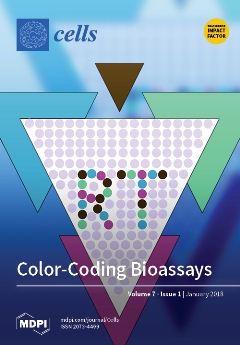
Bioassays of various types require that multiple cell types and reagents are pitapatted, often in complex patterns. As the cells and reagents are suspended/dissolved in cell culture media of one color, one has no visual feedback while pipetting, and there are no audit trails left to verify the accuracy and precision of the assay’s implementation. Here, using biologically neutral colored media, we introduce a reagent tracking platform that fills this gap. View this paper
  • Issues are regarded as officially published after their release is announced to the table of contents alert mailing list.
  • You may sign up for e-mail alerts to receive table of contents of newly released issues.
  • PDF is the official format for papers published in both, html and pdf forms. To view the papers in pdf format, click on the "PDF Full-text" link, and use the free Adobe Reader to open them.
Order results
Result details
Section
Select all
Export citation of selected articles as:
17 pages, 2604 KiB  
Article
Fine Regulation of Neutrophil Oxidative Status and Apoptosis by Ceruloplasmin and Its Derivatives
by Ekaterina A. Golenkina, Galina M. Viryasova, Svetlana I. Galkina, Tatjana V. Gaponova, Galina F. Sud’ina and Alexey V. Sokolov
Cells 2018, 7(1), 8; https://doi.org/10.3390/cells7010008 - 12 Jan 2018
Cited by 17 | Viewed by 5006
Abstract
Timely neutrophil apoptosis is an essential part of the resolution phase of acute inflammation. Ceruloplasmin, an acute-phase protein, which is the predominant copper-carrying protein in the blood, has been suggested to have a marked effect on neutrophil life span. The present work is [...] Read more.
Timely neutrophil apoptosis is an essential part of the resolution phase of acute inflammation. Ceruloplasmin, an acute-phase protein, which is the predominant copper-carrying protein in the blood, has been suggested to have a marked effect on neutrophil life span. The present work is a comparative study on the effects of intact holo-ceruloplasmin, its copper-free (apo-) and partially proteolyzed forms, and synthetic free peptides RPYLKVFNPR (883–892) and RRPYLKVFNPRR (882–893) on polymorphonuclear leukocyte (PMNL, neutrophil) oxidant status and apoptosis. The most pronounced effect on both investigated parameters was found with copper-containing samples, namely, intact and proteolyzed proteins. Both effectively reduced spontaneous and tumor necrosis factor-α (TNF-α)-induced extracellular and intracellular accumulation of superoxide radicals, but induced a sharp increase in the oxidation of intracellular 2′,7′-dichlorofluorescein upon short exposure. Therefore, intact and proteolyzed ceruloplasmin have both anti- and pro-oxidant effects on PMNLs wherein the latter effect is diminished by TNF-α and lactoferrin. Additionally, all compounds investigated were determined to be inhibitors of delayed spontaneous apoptosis. Intact enzyme retained its pro-survival activity, whereas proteolytic degradation converts ceruloplasmin from a mild inhibitor to a potent activator of TNF-α-induced neutrophil apoptosis. Full article
(This article belongs to the Section Cell Signaling)
Show Figures

Figure 1

13 pages, 2845 KiB  
Article
Effect of shRNA Mediated Silencing of YB-1 Protein on the Expression of Matrix Collagenases in Malignant Melanoma Cell In Vitro
by Wisam Nabeel Ibrahim, Abd Almonem Doolaanea and Mohammad Syaiful Bahari Bin Abdull Rasad
Cells 2018, 7(1), 7; https://doi.org/10.3390/cells7010007 - 10 Jan 2018
Cited by 10 | Viewed by 5567
Abstract
Background and Objective: YB-1 is a transcription and oncogenic factor capable of binding to DNA and RNA performing versatile functions within normal and cancer cells. Some studies reported the binding of YB-1 with a collagenases gene promoter and influencing their expression. In addition, [...] Read more.
Background and Objective: YB-1 is a transcription and oncogenic factor capable of binding to DNA and RNA performing versatile functions within normal and cancer cells. Some studies reported the binding of YB-1 with a collagenases gene promoter and influencing their expression. In addition, the role of YB-1 in malignant melanoma was not elucidated. Thus, in this study, the aim was to knock down the expression of YB-1 in A375 malignant melanoma cancer cell using the shRNA approach and study its effect on cancer cell proliferation, migration, and expression of collagenases. Methods: A375 malignant melanoma cell lines were grown in standard conditions and were transfected with three plasmids containing a retroviral pGFP-V-RS vector, two of them containing targeting sequences for YB-1 mRNA. The third plasmid contained a scrambled mRNA sequence as a negative control. Expression of YB-1 was validated using immune-fluorescence staining, RT-PCR and western blotting. The cancer cell proliferation was determined using MTT assay, serial trypan blue cell counting and cell cycle flow-cytometry analysis. Expression of collagenases (MMP1, MMP8, and MMP13) was evaluated using RT-PCR and western blotting analysis. In addition, a wound-healing assay was used to assess cell migration potential. Statistical analysis was performed using one-way ANOVA test with Bonferroni post hoc analysis to compare the quantitative results among samples. Results: The established silenced cell strains (P1 and P2) had nearly 70% knockdown in the expression of YB-1. These YB-1 silenced strains had a significant cell cycle-specific reduction in cell proliferation (p < 0.05 in serial cell counting and cell cycle flow cytometry analysis, p < 0.001 in MTT assay). In addition, YB-1 silenced strains had a remarkable reduction in cell migration potential. Expression of MMP13 was significantly reduced in YB-1 silenced strains. Conclusion: YB-1 oncoprotein is a promising target in the treatment of malignant melanoma. Silencing of this protein is associated with significant anti-proliferative, anti-invasive and MMP13 insulating properties in A375 malignant melanoma cancer cell lines. Full article
Show Figures

Figure 1

2 pages, 160 KiB  
Editorial
Acknowledgement to Reviewers of Cells in 2017
by Cells Editorial Office
Cells 2018, 7(1), 6; https://doi.org/10.3390/cells7010006 - 10 Jan 2018
Viewed by 2787
Abstract
Peer review is an essential part in the publication process, ensuring that Cells maintains high quality standards for its published papers[...]
Full article
17 pages, 3120 KiB  
Review
Multiscale and Multimodal Approaches to Study Autophagy in Model Plants
by Jessica Marion, Romain Le Bars, Laetitia Besse, Henri Batoko and Béatrice Satiat-Jeunemaitre
Cells 2018, 7(1), 5; https://doi.org/10.3390/cells7010005 - 09 Jan 2018
Cited by 15 | Viewed by 7407
Abstract
Autophagy is a catabolic process used by eukaryotic cells to maintain or restore cellular and organismal homeostasis. A better understanding of autophagy in plant biology could lead to an improvement of the recycling processes of plant cells and thus contribute, for example, towards [...] Read more.
Autophagy is a catabolic process used by eukaryotic cells to maintain or restore cellular and organismal homeostasis. A better understanding of autophagy in plant biology could lead to an improvement of the recycling processes of plant cells and thus contribute, for example, towards reducing the negative ecological consequences of nitrogen-based fertilizers in agriculture. It may also help to optimize plant adaptation to adverse biotic and abiotic conditions through appropriate plant breeding or genetic engineering to incorporate useful traits in relation to this catabolic pathway. In this review, we describe useful protocols for studying autophagy in the plant cell, taking into account some specificities of the plant model. Full article
(This article belongs to the Special Issue Assays to Monitor Autophagy in Model Systems)
Show Figures

Figure 1

9 pages, 472 KiB  
Review
Targeting FLT3 Mutations in Acute Myeloid Leukemia
by Riad El Fakih, Walid Rasheed, Yousef Hawsawi, Maamoun Alsermani and Mona Hassanein
Cells 2018, 7(1), 4; https://doi.org/10.3390/cells7010004 - 08 Jan 2018
Cited by 29 | Viewed by 9165
Abstract
The FMS-like tyrosine kinase 3 (FLT3) pathway has an important role in cellular proliferation, survival, and differentiation. Acute myeloid leukemia (AML) patients with mutated FLT3 have a large disease burden at presentation and a dismal prognosis. A number of FLT3 inhibitors have been [...] Read more.
The FMS-like tyrosine kinase 3 (FLT3) pathway has an important role in cellular proliferation, survival, and differentiation. Acute myeloid leukemia (AML) patients with mutated FLT3 have a large disease burden at presentation and a dismal prognosis. A number of FLT3 inhibitors have been developed over the years. The first-generation inhibitors are largely non-specific, while the second-generation inhibitors are more specific and more potent. These inhibitors are used to treat patients with FLT3-mutated AML in virtually all disease settings including induction, consolidation, maintenance, relapse, and after hematopoietic cell transplantation (HCT). In this article, we will review the use of FLT3 inhibitors in AML. Full article
(This article belongs to the Special Issue Receptor Tyrosine Kinases in Health and Disease)
Show Figures

Figure 1

10 pages, 1714 KiB  
Article
Reagent Tracker Dyes Permit Quality Control for Verifying Plating Accuracy in ELISPOT Tests
by Alexander Lehmann, Zoltan Megyesi, Anna Przybyla and Paul V. Lehmann
Cells 2018, 7(1), 3; https://doi.org/10.3390/cells7010003 - 03 Jan 2018
Cited by 4 | Viewed by 5547
Abstract
ELISPOT assays enable the detection of the frequency of antigen-specific T cells in the blood by measuring the secretion of cytokines, or combinations of cytokines, in response to antigenic challenges of a defined population of PBMC. As such, these assays are suited to [...] Read more.
ELISPOT assays enable the detection of the frequency of antigen-specific T cells in the blood by measuring the secretion of cytokines, or combinations of cytokines, in response to antigenic challenges of a defined population of PBMC. As such, these assays are suited to establish the magnitude and quality of T cell immunity in infectious, allergic, autoimmune and transplant settings, as well as for measurements of anti-tumor immunity. The simplicity, robustness, cost-effectiveness and scalability of ELISPOT renders it suitable for regulated immune monitoring. In response to the regulatory requirements of clinical and pre-clinical immune monitoring trials, tamper-proof audit trails have been introduced to all steps of ELISPOT analysis: from capturing the raw images of assay wells and counting of spots, to all subsequent quality control steps involved in count verification. A major shortcoming of ELISPOT and other related cellular assays is presently the lack of audit trails for the wet laboratory part of the assay, in particular, the assurance that no pipetting errors have occurred during the plating of antigens and cells. Here, we introduce a dye-based reagent tracking platform that fills this gap, thereby increasing the transparency and documentation of ELISPOT test results. Full article
Show Figures

Figure 1

646 KiB  
Review
Induced Pluripotent Stem Cell-Derived Red Blood Cells and Platelet Concentrates: From Bench to Bedside
by Daniele Focosi and Giovanni Amabile
Cells 2018, 7(1), 2; https://doi.org/10.3390/cells7010002 - 27 Dec 2017
Cited by 22 | Viewed by 6511
Abstract
Red blood cells and platelets are anucleate blood components indispensable for oxygen delivery and hemostasis, respectively. Derivation of these blood elements from induced pluripotent stem (iPS) cells has the potential to develop blood donor-independent and genetic manipulation-prone products to complement or replace current [...] Read more.
Red blood cells and platelets are anucleate blood components indispensable for oxygen delivery and hemostasis, respectively. Derivation of these blood elements from induced pluripotent stem (iPS) cells has the potential to develop blood donor-independent and genetic manipulation-prone products to complement or replace current transfusion banking, also minimizing the risk of alloimmunization. While the production of erythrocytes from iPS cells has challenges to overcome, such as differentiation into adult-type phenotype that functions properly after transfusion, platelet products are qualitatively and quantitatively approaching a clinically-applicable level owing to advances in expandable megakaryocyte (MK) lines, platelet-producing bioreactors, and novel reagents. Guidelines that assure the quality of iPS cells-derived blood products for clinical application represent a novel challenge for regulatory agencies. Considering the minimal risk of tumorigenicity and the expected significant demand of such products, ex vivo production of iPS-derived blood components can pave the way for iPS translation into the clinic. Full article
(This article belongs to the Special Issue Ten Years of iPSCs: Current Status and Future Perspectives)
Show Figures

Figure 1

1342 KiB  
Review
Intra- and Intercellular Quality Control Mechanisms of Mitochondria
by Yoshimitsu Kiriyama and Hiromi Nochi
Cells 2018, 7(1), 1; https://doi.org/10.3390/cells7010001 - 26 Dec 2017
Cited by 63 | Viewed by 11008
Abstract
Mitochondria function to generate ATP and also play important roles in cellular homeostasis, signaling, apoptosis, autophagy, and metabolism. The loss of mitochondrial function results in cell death and various types of diseases. Therefore, quality control of mitochondria via intra- and intercellular pathways is [...] Read more.
Mitochondria function to generate ATP and also play important roles in cellular homeostasis, signaling, apoptosis, autophagy, and metabolism. The loss of mitochondrial function results in cell death and various types of diseases. Therefore, quality control of mitochondria via intra- and intercellular pathways is crucial. Intracellular quality control consists of biogenesis, fusion and fission, and degradation of mitochondria in the cell, whereas intercellular quality control involves tunneling nanotubes and extracellular vesicles. In this review, we outline the current knowledge on the intra- and intercellular quality control mechanisms of mitochondria. Full article
Show Figures

Figure 1

Previous Issue
Next Issue
Back to TopTop